A Comprehensive Analysis of the Phylogeny,
Genomic Organization and Expression of
Immunoglobulin Light Chain Genes in
Alligator sinensis
, an Endangered Reptile
Species
Xifeng Wang1☯, Gang Cheng1☯, Yan Lu2, Chenglin Zhang2, Xiaobing Wu3, Haitang Han1,
Yaofeng Zhao1, Liming Ren1*
1State Key Laboratory of Agrobiotechnology, College of Biological Sciences, National Engineering Laboratory for Animal Breeding, China Agricultural University, Beijing, People’s Republic of China,2Beijing Zoo, Beijing 100044, People’s Republic of China,3College of Life Sciences, Anhui Normal University, Anhui Provincial Key Laboratory of the Conservation and Exploitation of Biological Resources, Wuhu 241000, People’s Republic of China
☯These authors contributed equally to this work. *[email protected]
Abstract
Crocodilians are evolutionarily distinct reptiles that are distantly related to lizards and are thought to be the closest relatives of birds. Compared with birds and mammals, few studies have investigated the Ig light chain of crocodilians. Here, employing anAlligator sinensis genomic bacterial artificial chromosome (BAC) library and available genome data, we char-acterized the genomic organization of theAlligator sinensisIgL gene loci. TheAlligator sinensishas two IgL isotypes,λandκ, the same asAnolis carolinensis. The Igλlocus
con-tains 6 Cλgenes, each preceded by a Jλgene, and 86 potentially functional Vλgenes
upstream of (Jλ-Cλ)n. The Igκlocus contains a single Cκgene, 6 Jκs and 62 functional Vκs.
All VLgenes are classified into a total of 31 families: 19 Vλfamilies and 12 Vκfamilies.
Based on an analysis of the chromosomal location of the light chain genes among mam-mals, birds, lizards and frogs, the data further confirm that there are two IgL isotypes in the Alligator sinensis: Igλand Igκ. By analyzing the cloned Igλ/κcDNA, we identified a biased
usage pattern of V families in the expressed Vλand Vκ. An analysis of the junctions of the
recombined VJ revealed the presence of N and P nucleotides in both expressedλandκ
sequences. Phylogenetic analysis of the V genes revealed V families shared by mammals, birds, reptiles andXenopus, suggesting that these conserved V families are orthologous and have been retained during the evolution of IgL. Our data suggest that theAlligator sinensisIgL gene repertoire is highly diverse and complex and provide insight into immuno-globulin gene evolution in vertebrates.
OPEN ACCESS
Citation:Wang X, Cheng G, Lu Y, Zhang C, Wu X, Han H, et al. (2016) A Comprehensive Analysis of the Phylogeny, Genomic Organization and Expression of Immunoglobulin Light Chain Genes inAlligator sinensis, an Endangered Reptile Species. PLoS ONE 11(2): e0147704. doi:10.1371/journal.pone.0147704
Editor:Sebastian D. Fugmann, Chang Gung University, TAIWAN
Received:September 15, 2015
Accepted:January 7, 2016
Published:February 22, 2016
Copyright:© 2016 Wang et al. This is an open access article distributed under the terms of the
Creative Commons Attribution License, which permits unrestricted use, distribution, and reproduction in any medium, provided the original author and source are credited.
Introduction
Immunoglobulin (Ig) is one of the most important primary effector molecules in the adaptive immune system of jawed vertebrates [1]. Each immunoglobulin is composed of a heavy (H) chain and one of two light (L) chain types:λorκin mammals. Each of these L chains typically covalently links to H by disulfide bonds formed by positionally conserved cysteine residues [2]. As exceptions, shark IgNAR and camelid IgGs are only composed of heavy chains [3,4]. The Ig light chain is encoded byλandκloci, which differ significantly in their genomic organiza-tion. At theλlocus, multiple Vλsegments are followed by Jλ-Cλrepeats. In contrast, the cluster of Vκgene segments is followed by a cluster of Jκgene segments and then by a single Cκgene
[5–7]. Lymphocytes can generate specific immunoglobulins against diverse antigens by a somatic recombination process, known as V (D) J recombination [8–10]. A pair of recombina-tion signal sequences (RSSs) are composed of conserved heptamer and nonamer sequences and are separated by a relatively non-conserved spacer of either 12 or 23 bp, which is recog-nized by RAG1 and RAG2. Then, RAG introduces a double-strand break (DSB) between the RSS and the coding segments [11,12]. Each of the L chains is the result of the imprecise and random combinatorial assembly of several gene fragments by a non-homologous end joining (NHEJ) pathway with the removal or addition of a random number of nucleotides [10,13]. This imprecision in the coding joint arises from short additions of self-complementary (P) or random (N) nucleotides [9], small deletions, or a combination of these and contributes to the antigen receptor diversity generated by V (D) J joining [14].
IgL genes in cartilaginous fishes belong to four major groups:κ,λ,σandσ-cart [13]. Among cartilaginous fish, theGinglymostoma cirratumL chain genes have been studied most comprehensively. In a previous study, four L chain isotypes were identified inGinglymostoma cirratum: type I (NS5), type II (NS3), type III (NS4) and type IV. The type III L chain is clearly κ, the type II light chain is somewhat moreλ-like, the type I gene is closely related to but dis-tinct from theσgene [15–17] and is referred to asσ-cart, and type IV is homologous with the L
chain isotypeσ, found first inXenopusand later in bony fish [13,17]. The IgL isotypes
cur-rently found in teleost belong toκ(L1/G and L3/F),λandσ(L2). These have been found in a cluster assemblage and, depending on the species, the number of IgL isotypes is different [17–
26].
Three types of light chains have been identified in amphibians as well, based on studies of
Xenopus laevis:ρ,σand type III [17,27–30]. Qin and colleagues completely characterized all
three gene loci inXenopus tropicalis[31] and supported the classification of amphibians in which theρgene belongs to theκgene family and type III appearsλ-like [17,29].
Evolution-arily, mammals express two types of Ig light chain,λandκ, which are expressed in varying ratios in different species [5,32–36]. InMus musculusserum, 95% of the light chains areκand 5% areλ[5], whereasBos taurusexhibit a biased usage pattern ofλchain [32]. LikeHomo
sapi-ens,Sus scrofado not show any preference for the usage of the light chain [36]. Surprisingly, unlike reptiles and mammals, birds possess only one light chain, which is orthologous to the
Homo sapiens/Mus musculusλchain [37–41]. The genomic organization of theλchain is simi-lar to the heavy chain in birds: only one functional Vλand Jλare 1.8 kb apart and are located
upstream from the Cλgene in theGallus gallus[42]. The light chain has evolved an exceptional
mechanism of generating diversity due to multiple Vλpseudogenes that modify the functional
Vλgene and can act as donors to form intrachromosomal gene conversion [43]. These results
suggested that the typical birds IgL was likely already present in the common ancestor and remained unchanged over a long period of evolution [40].
Reptilia can be divided into two main evolutionary lineages: one gave rise to Squamata, while the other gave rise to Testudines, Crocodylia, and birds [44]. Some studies have been KE698096.1, KE695928.1. The Genbank accession
numbers of the sequence are as follows: KU535866 for Alligator sinensis IgK and KU535867 for Alligator sinensis IgL.
Funding:This work was supported by the National Natural Science Foundation of China (URL:http:// www.nsfc.gov.cn/, grant number:31472085 and 31272433). The author Yaofeng Zhao received the funding. The funders had no role in study design, data collection and analysis, decision to publish, or preparation of the manuscript.
conducted to investigate Ig gene isotypes and their genomic organization in reptilia. Until now, IgM, IgD and IgY encoding genes have been identified in all Squamata species studied to date [45–47]. While it was shown that theAnolis carolinensisexpress two types of light chains:λ andκ[7,39,48], snakes lack the Igκlight chain isotype [45]. In the Testudines, IgM, IgD, IgY and IgD2 encoding genes were described, and two immunoglobulin domains of IgD2 are shown to be homologous to bird IgA domains, suggesting that they may originate from a com-mon ancestral gene [49–51]. Crocodilians appeared during the Middle Triassic, approximately 240 million years ago (MYA). Although similar in appearance, crocodilians, as reptiles, are only distantly related to lizards and are thought to be the closest relatives of birds and have thus occupied an important position in evolution [52,53]. According to phylogenetic studies, crocodilians provide a phylogenetic link to other reptiles and birds, and analysis of their Ig genes may provide important clues to understanding Ig evolution. In addition, despite living in poor conditions, crocodilians are rarely subject to infections caused by bacteria and viruses because of their strong immune systems [54,55]. However, there have been few studies on the crocodilian immune system. Recently, IgH genes of crocodilians were identified; the results indicated that there are multipleμgenes and that IgM subclasses can be expressed through
class-switch recombination. The crocodilianαgenes are the first IgA-encoding genes identified in reptiles and suggested that reptiles and birds share a common ancestral organization [56, 57].
Crocodilians are the closest phylogenetic group to birds, and they all come from a group known as archosaurs. However, little is known about the IgL locus of crocodilians. Although a previous study suggested that two distinct light chain types were present in alligator [48], the isotypes and the genomic organization of their encoding genes are still not known [39]. In this study, we present the phylogeny, genomic organization and expression of the Igλ/κof the
Alli-gator sinensisand provide insight into understanding the crocodilian immune system and the evolution of immunoglobulin in vertebrates.
Materials and Methods
Sample collection, DNA and RNA extract
Blood samples ofAlligator sinensiswere collected from the Beijing Zoo. Genomic DNA was extracted from the blood following the standard protocol. Total RNA was extracted from the blood using a TRIzol kit (TIANGEN BIOTECH, Beijing) following the manufacturer’s instruc-tions. Our studies were approved by the Animal Care and Use Committee of the China Agri-cultural University.
BAC library
AnAlligator sinensisgenomic BAC library was constructed using a service provided by Bioes-tablish Biotechnology Co., Ltd. (Beijing, China) and was stored in our laboratory [56].
BAC screening and sequencing
Based on sequences derived fromGallus gallusand other related species, we designed degener-ate primers for the Igκ/λ. We ascertained the identities of the PCR-generated product
The positive BAC clones were then sequenced by shotgun sequencing and assembled with the next generation sequencing platform by BGI (Beijing, China).
Cloning of expressed
Alligator sinensis
Ig
λ
and Ig
κ
light chain genes at
the cDNA level
ExpressedAlligator sinensisIgλand Igκchains were amplified using the 5’RACE System kit (Invitrogen, Beijing). The gene-specific primers for the Igλchain are as follows: IgLCL338L18,
5’-CAT TAG GGA GAT ACT ACA-3’; IgLCL303L21,5’-CAG GGA TCC CAG CTC TCT ACT-3’; IgLCL219L21,5’-AGG GTC TTC TCG ATG CTC TTC-3’; IgLCL129L21,5’-GCT GGC CAT GTA CTT GTT GTC-3’. The sequences of these primers are conserved in the Alliga-tor sinensisCλgene. Gene-specific primers for the Igκchain are as follows: IgLCκ301L18,5’
-ATA AAG AAA GCA TAA GAA-3’; IgLCκ236L21,5’-CGT ACA CTC GGT CCT CTT GAA-3’;
IgLCκ121L21,5’-CTG CTC TTG CTG TAC GTG TTG-3’, which are conserved in the Alliga-tor sinensisCκgene.
All PCR amplifications were performed using a proofreading enzyme Pyrobest DNA poly-merase (TaKaRa, Dalian). The PCR products were cloned into the pMD-19 T vector (TaKaRa, Dalian) and sequenced.
Southern blotting
Genomic DNA was digested with restriction endonuclease and was loaded into a 0.9% agarose gel, electrophoresed for 6 h, and transferred to a positively charged nylon membrane (Roche, Germany) for hybridization. The restriction endonucleasesBglII,NcoI,HindIII andSphI were used to digest genomic DNA to identify Igλ. Genomic DNA was digested with restriction endonucleasesKpnI,NdeI andXbaI to validate Igκ. The single exon of the Cλ/Cκprobe was labeled using a PCR digoxigenin probe synthesis kit (Roche, Germany). The primers used to amplify the Cλ/Cκexon probes were as follows: LC-F,5’-ACA GCC AAA GGC CTC TCC T-3’;
LC-R,5’-CGA TCT CTT CAG GGT CTT CTC-3’; KC-F,5’-AAA GGG GGA AGA GCC ACC-3’; KC-R,5’-TAC ACT CGG TCC TCT TGA-3’. The hybridization and detection were
per-formed following the manufacturer’s instructions.
Construction of phylogenetic trees
The phylogenetic trees were constructed using MrBayes3.1.2 [58] and were viewed in TREE-VIEW [59]. Furthermore, in order to validate the topologies of the phylogenetic trees, we also used MEGA6.0 and Phylip3.695 [60] to build all the phylogenetic trees [59]. Multiple amino-acid alignments for the tree construction were performed usingClustalW. Each Vλ/κsubgroup
was represented with one family per species chosen at random. The accession numbers of sequences used for variable regions are as follows:Heterodontus francisciσ(ABO64185); Het-erodontus franciscitype I (CAA33375);Heterodontus franciscitype II (AAA59379); Heterodon-tus franciscitype III (AAA59373);Ginglymostoma cirratumNS5 (AAV34678);Ginglymostoma cirratumσ(ABO64187);Danio reriotype I (AAG31721);Danio reriotype II (AAG31729); Danio reriotype III (AAG31698);X.laevistype III V1 (AAL40100);X.laevistype III V2 (AAL40101);X.laevistype III V3 (AAL40102);X.laevistype III V4 (AAL40103);X.laevistype III V5 (AAL40097);X.laevistype III V6 (AAL40093);X.laevisσ(NP_001087883);X.laevisρ
(AAH68859);Gallus gallusIGλV (BAB71862);Anas platyrhynchosIGλV (AAA03006);Anolis
carolinensisIGκV (ACB45832);Anolis carolinensisIGλV1 (XP_008115579);Anolis
(CAA75909);Mus musculusIGκV4-61 (CAB46123);Mus musculusIGκV5-45 (CAB46329);
Mus musculusIGκV6-25 (CAB46320);Mus musculusIGκV7-33 (AAC04340);Mus musculus IGκV8-30 (CAB46308);Mus musculusIGκV9-120 (CAA24186);Mus musculusIGκV10-95 (AAC14726);Mus musculusIGκV11-125 (CAB51813);Mus musculusIGκV12-38
(CAB46311);Mus musculusIGκV13-84 (CAB46176);Mus musculusIGκV14-130 (CAB46155);Mus musculusIGκV15-103 (CAB46175);Mus musculusIGκV16-104 (CAB46298);Mus musculusIGκV17-121 (CAB46168);Mus musculusIGκV18-36 (CAB46323);Mus musculusIGκV19-93 (CAB46297);Mus musculusIGλV1 (AAA39165);
Mus musculusIGλV3 (AAA39169);Mus musculusIGλV4 (AAA39434). All other sequences were derived in this study. The accession numbers of sequences used for constant regions are as follows:Gallus gallusλ(AAA48862);Anas platyrhynchosλ(AAA03009);
Ornithor-hynchusλ(AAO16062);Homo sapiensλ(AAA59107);Mus musculusλ(AAA39089);Bos
taurusλ(AAI46273);Didelphimorphiaλ(AAL37214);Oryctolagus cuniculusλ(AAA31360);
Anolis carolinensisλ(XP_008115579);Sus scrofaλ(AAA03572);Chelonia mydasλ
(XP_007055069.1);Chrysemys pictabelliiλ(XM_008167097.1);X.laevistype III (AAL40101);
X.tropicalistype III (AAI66944);Mus musculusκ(CAA24185);Homo sapiensκ(AAY24201);
Bos taurusκ(AAI51501);X.laevisρ(AAA49880);X.laevisσ(NP_001087883.1|);X.tropicalis
σ(AAI67133);X.tropicalisρ(AAI58339);Oryctolagus cuniculusκ(CAA10920);Sus scrofaκ
(AHB17990);Didelphimorphiaκ(AAL17618);Ornithorhynchusκ(AAO84649);Chelonia
mydasκ(EMP6807.1);Chrysemys pictabelliiκ(XM_008169724.1);Ginglymostoma cirratum NS5 (AAV34681);Ginglymostoma cirratumNS4 (A49633);Ginglymostoma cirratumNS3 (Ref.[61]);Ginglymostoma cirratumσ(ABO64188);Danio rerioIGIC1 (AAG31721);Danio rerioIGIC2 (XP_009298120);Heterodontus franciscitype I (CAA33376);Heterodontus fran-ciscitype II (CAA33375);Heterodontus franciscitype III (AAA59373);Heterodontus francisciσ
(ABO64185);Salmo salarIGIC1 (AAG18364);Salmo salarIGIC2 (AAG37201);Salmo salar
IGIC3 (AAK97642). All other sequences were derived in this study.
Sequence computations
DNA and protein sequence editing, alignments and comparisons were performed with the MegAlign software (DNASTAR). The EquCab2 assembly in Ensembl database (http://www. ensembl.org/index.html) was used to retrieve the genomic contig that contained theAlligator sinensisIgλ/Igκchain sequences. IgBLAST (http://www.ncbi.nlm.nih.gov/igblast/) was used to predict the Vλ/κsegments. Germline Vλand Vκgene segments were grouped into families
using the IMGT numbering system [62]. The RSSs for the V and J gene segments were ana-lyzed using the online program FUZZNUC (http://embossgui.sourceforge.net/demo/fuzznuc. html).
Results
Genomic organization of IgL chain gene loci in
Alligator sinensis
gene, spanning approximately 12 kb DNA, there are potentially 37 functionalλchain V genes, 32λchain V pseudogenes and one ORF. Furthermore, using the available genomic database of theAlligator sinensis(http://www.ncbi.nlm.nih.gov/), a genomic contig (AVPB01119656.1) was identified by BLAST; threeλchain C genes were identified in the contig: one is identical with Cλ4 in the ~331 kb genomic sequence, and one appears to be a pseudogene because it
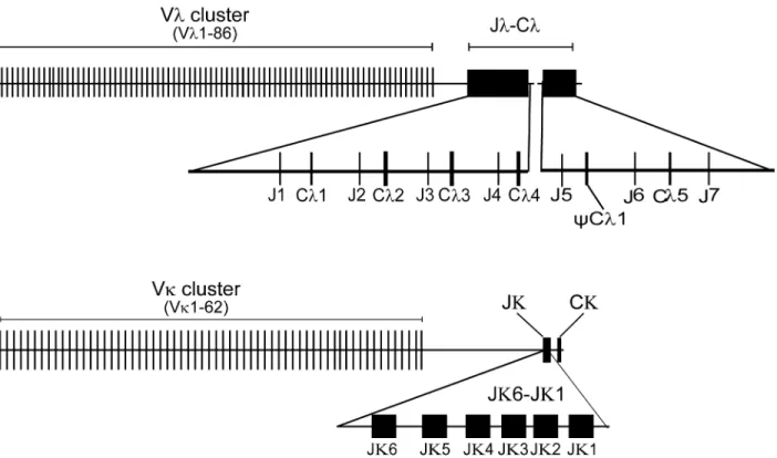
con-tains an in-frame stop codon. Furthermore, three J genes were found in the contig. There are sixλchain C genes (Cλ1, Cλ2, Cλ3, Cλ4,CCλ1 and Cλ5) and sevenλchain J genes (Jλ1, Jλ2, Jλ3, Jλ4, Jλ5, Jλ6 and Jλ7) (Fig 1andS1 Fig). All of the Cλgenes share at least 84.1% amino acid
sequence identity, of which the amino acid sequence identities between Cλ1 and Cλ2, Cλ1 and
Cλ4, Cλ2 and Cλ3, Cλ2 and Cλ4, and Cλ3 and Cλ4 are greater than 90.7%. Each Cλgene was
preceded by a single J gene segment that was 5' flanked by conserved RSS (nonamer and hepta-mer) with a 12 bp nucleotide spacer, resembling the genomic organization of theλchain gene loci in mammals (S2A Fig). However, a single J segment (Jλ7) was found downstream from
Cλ5, but no additionalλchain C genes were identified (Fig 1AandS1 Fig), which implied that
there are more Cλgenes in theAlligator sinensisIgλlocus. A protein sequence alignment of the
identified C genes with the Cλin lizards, birds and mammals uncovered an identical pattern
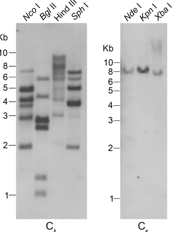
with regard to the cysteine distribution (S2B Fig). Genomic Southern blotting with the Cλexon
as a probe was conducted to verify the numbers ofλchain C genes. InBglII,NcoI andSphI digested DNA, different shades of six bands were detected, and there were more than six bands inHindIII digested DNA, which indicated that there are additional Cλgenes in the
chromo-some (Fig 2).
We performed a BLAST search against theAlligator sinensiswhole-genome shotgun sequence (WGS) assembly deposited in the Ensemble database. Seven genomic contigs (KE698600.1, AVPB01102472.1, KE698001.1, KE698031.1, KE697531.1, KE697626.1 and KE695978.1) were found to containλchain V gene segments (S2 Table). Each Vλgene, which is 3' flanked by a conserved RSS (heptamer and nonamer) with 23 bp nucleotide spacer, was identified, resembling the genomic organization of the Vλchain gene loci in mammals. In
sum-mary, a total of 86 potentially functional Vλsegments (Fig 1BandS1 Appendix), two ORFs
and 67 Vλpseudogenes were identified upstream from the (Jλ-Cλ)nsegments (S1 Fig), and 67
Vλthat either contain in-frame stop codons or lack a leading peptide appear to be pseudogenes
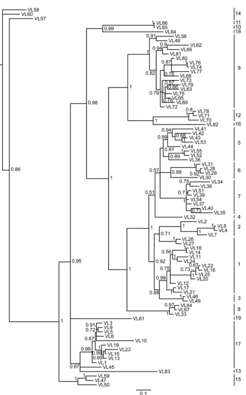
(S2 Appendix). According to the sequence identity (>75% sequence identity within a single family) and phylogenetic analysis, the potentially functional Vλgenes can be classified into at
least 19 families (Fig 3;S3andS4Figs;S3 Appendix). In addition, there may be more Vλ
seg-ments unidentified in theAlligator sinensisbased on the gaps in the contig and incomplete genomic data.
A similar approach was used to identify the Cκfrom the genome of theAlligator sinensis.
Using a PCR-based approach and sequencing, we obtained 5 Igκgene-positive BAC clones (Y146M9, Y65C14, Y77E6, Y329F14 and Y146B4) (S1 Table). An ~484 kb genomic sequence was found to contain a single copy of theAlligator sinensisCκgene, which showed homology
to several mammalian species, six Jκgene segments and 66 Vκgene segments, including 29 Vκ
pseudogenes. We performed a BLAST search against theAlligator sinensiswhole-genome shot-gun sequence (WGS) assembly deposited in the Ensemble database. Seventeen DNA contigs (AVPB01013186.1, AVPB01053098.1, AVPB01130521.1, AVPB01143799.1, KE695928.1, KE697554.1, KE697644.1, KE698008.1, KE698055.1, KE698081.1, KE698096.1, KE698098.1, KE698149.1, KE698335.1, KE698356.1, KE698428.1, and KE698585.1) comprise a leash of Vκ
genes that is variable in number from 1 to 15 (S3 Table). At least 62 potentially functional Vκ
gene segments (Fig 1BandS4 Appendix); 56 Vκpseudogenes, which either contain in-frame
stop codons or lack a leading peptide (S5 Appendix); and 4 partial Vκgenes were identified
The Cκgene as a single copy in the genome was subjected to confirmation by Southern
blot-ting. We designed a pair of degenerate primers for the Cκgene based on the conserved Cκ
sequences of theAlligator sinensis. Only a single band was observed inKpnI,NdeI andXbaI digested genomic DNA, which supported the Cκgene as a single copy present in the genome
(Fig 2). Upstream of the single copy of the Cκgene, six functional Jκs (Jκ1-Jκ6) gene segments
with RSS interrupted by a 23 bp nucleotide spacer at their 5’ends were identified (S6A Fig). An amino acid sequence alignment of the Cκgene in theAlligator sinensiswith other species
sug-gested homology to the Igκchains of several vertebrates, including theHomo sapiens,Mus
musculus,Didelphimorphia,Ornithorhynchus,Anolis carolinensis,X.laevisandX.tropicalis
(S6B Fig). The Cκprotein sequence contained three cysteines, among which the third one at
the carboxyl terminal was assumed to link heavy chains (S6B Fig).
Almost all Vκgenes were flanked on the 3’end by RSS and were separated by a 12 bp
nucleo-tide spacer to conform the 12–23 rules (S5 Appendix). All Vκgenes showed the same
transcrip-tional orientation as (Jκ)n-Cκ, with the exception of pseudogene Vκ46. The 62 potentially
functional Vκgenes can be integrated into 12 families based on the phylogenetic analysis and
the rule that Vκmembers in one family share at least 75% identity at the nucleotide level (Fig 4;
S7andS8Figs;S6 Appendix). Because gaps exist in the contigs and the genomic data are incom-plete, it is possible that more Vκgenes present in theAlligator sinensisgenome were not found.
Phylogenetic analysis of the
Alligator sinensis
Ig light chain gene segments
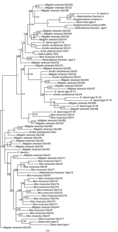
Using the amino acid sequences ofIGLV-andIGLC- encoded genes from different jawed ver-tebrates, we constructed V and C phylogenetic trees, respectively. The trees were constructed Fig 1. Schematic map of theAlligator sinensisimmunoglobulin light chain gene loci.(A) Schematic map of theAlligator sinensisimmunoglobulin light chainλgene loci. (B) Schematic map of theAlligator sinensisimmunoglobulin light chainκgene loci. V: variable gene segments; J: joining gene segments; C: constant region gene; pseudo-variable gene segments were not be shown in the figure.
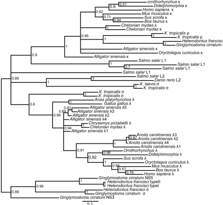
using protein sequences without CDR3. The phylogenetic trees, based on both the C domains and the V domains, support the fact that there are three major groups of IgL genes in jawed vertebrates:κ,λandσ(includingσ-cart), andAlligator sinensisκandλclearly fall into their own respective groups, suggesting that theAlligator sinensishas only two IgL isotypes:κandλ Fig 2. Southern blotting detection of theAlligator sinensisIg light chain C gene segments.Genomic DNA was digested with restriction endonucleases, which are indicated above each lane, and hybridized with probes for Cλand Cκ, respectively.
Fig 3. Phylogenetic tree analysis of the 86Alligator sinensisVλgenes.A phylogenetic tree of the
nucleotides of 86Alligator sinensisVλsegments was constructed. The 19 Vλgene families are labeled with numbers on the right. Phylogenetic trees were constructed using MrBayes3.1.2 [58] and viewed in TREEVIEW [59].
Fig 4. Phylogenetic tree analysis of the 62Alligator sinensisVκgenes.A phylogenetic tree of the
nucleotides of theAlligator sinensisVκsegments was constructed. The 12 Vκgene families are labeled with numbers on the right. Phylogenetic trees were constructed using MrBayes3.1.2 [58] and viewed in TREEVIEW [59].
(Figs5and6;S9,S10,S11andS12Figs). The results reveal that theρgene ofX.tropicalis,
tele-ost L1 and L3 and cartilaginous fish type III/NS4 is located in theκgroup, which also includes theκgenes of the Crocodilians, lizards and mammals. Teleost L2, cartilaginous fish type II/ NS3, andX.tropicalistype III all belong toλgroups, including theλgenes of the Crocodilians, lizards, birds and mammals. Theσgenes are only found in cartilaginous fish, teleost and
amphibians. Taken together with our shared synteny of theκandλlocus in theAlligator
sinen-sisand the phylogenetic analysis, these data provide convincing evidence that theAlligator sinensisexpresses two IgL isotypes:κandλ. From the phylogenetic analysis, it is not difficult to obtain the relationships between theAlligator sinensisand other species’V families.Alligator sinensisfamilies Vκ10 and Vκ11 are clustered withAnolis carolinensisVκ;Alligator sinensis
family Vκ7 is clustered withX.laevisρ; and the same phylogenetic analysis was also performed
for Vλ. As shown inFig 6,Alligator sinensisfamilies Vλ9 and Vλ19 are clustered withX.laevis
type III V5 andMus musculusfamilies 1 and 3;Alligator sinensisfamilies Vλ1-Vλ8 are related
to theAnolis carolinensisVλ1, Vλ3,Gallus gallusandAnas platyrhynchosVλandX.laevistype
III V4; andAlligator sinensisfamilies Vλ11 is clustered withX.laevistype III V6. The V genes
were orthologous in different isotypes of IgL. We found no relations between the remaining Vλ
genes and other jawed vertebrate species, suggesting that Vλgenes exhibit more abundant
diversity in theAlligator sinensis.
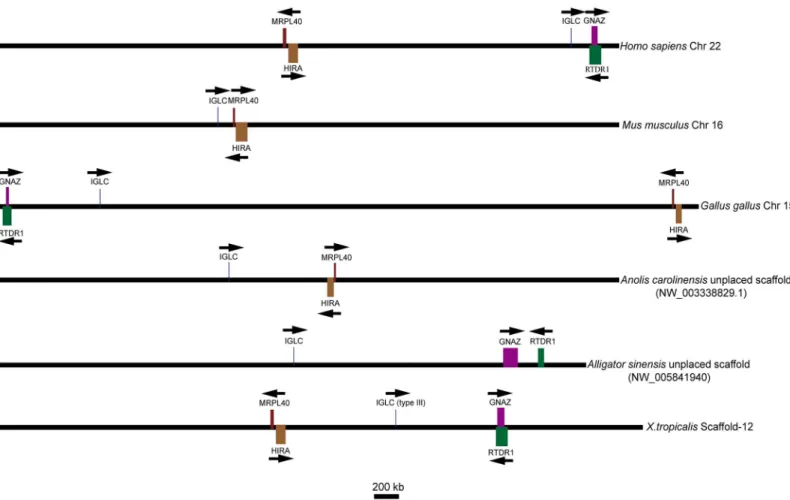
Syntenic analysis of Ig
λ
and Ig
κ
chain loci in tetrapods
To determine the identified genes belonging to theλlineage, we analyzed the chromosomal location relative to the flanking genes of the available genomic data containing the Igλloci in tetrapods.GNZA(guanine nucleotide-binding protein,αz subunit) andRTDR1(rhabdoid tumor deletion region gene 1),MRPL40(mitochondrial ribosomal protein L40) andHIRA
(histone cell cycle regulation defective homologue A) located on, respectively, the two sides of theλlocus inHomo sapienswere selected as markers to provide evidence for the gene. An available genomic contig (NW_005841940) containing the Igλlocus of theAlligator sinensis was used for analysis. The results showed three situations in which theλgenes had the same transcriptional orientation: first, the Igλlocus was flanked downstream byMRPL40andHIRA and upstream byGNZAandRTDR1, as inHomo sapiensandX.tropicalis; second, the opposite situation existed, with the Igλlocus flanked downstream byGNZAandRTDR1and upstream byMRPL40andHIRA, as inGallus gallus, which can occur via intrachromosomal gene conver-sion; and third, the Igλlocus was only flanked upstream byMRPL40andHIRA, as inMus
mus-culusandAnolis carolinensis. In the third situation,GNZAandRTDR1were identified on chromosome 10, which does not containIGLinMus musculus, and inAnolis carolinensis, the chromosomal position ofGNAZwas identified in contig (NW_003341094.1). However, no
IGLgene was found in this contig, and theRTDR1gene was not identified inAnolis carolinen-sis. InMus musculus, the chromosome was recombined, leading toGNZAandRTDR1being separated from the Igλlocus and located on another chromosome, whereas inAnolis
carolinen-sis, the position ofGNAZcould not be confirmed because of limited genomic data. The Igλ locus of theAlligator sinensiswas also flanked upstream byGNZAandRTDR1(Fig 7), whereas
MRPL40andHIRAwere located in anotherAlligator sinensisgenomic contig
musculus), and others that are not confirmed because of limited genomic data(e.g.,Anolis caro-linensis). All taxa studied showed the same flanking genes on one side or both sides of the Igλ locus. These data provide convincing evidence that the identified genes originated from the same ancestral gene as theλgene in tetrapods and originated from the same ancestral gene as the type III light chain gene inX.tropicalis. The position of the Igλlocus on chromosome inX.
tropicalismay be the oldest form in tetrapods.
Similarly, to determine the identifiedκgenes in theAlligator sinensisbelonging to theκ lineage, we performed a syntenic analysis of theκgenes using the data available for tetrapods, Fig 5. Phylogenetic analysis of the IgL chain C genes in jawed vertebrates.The phylogenetic tree was constructed using C domains. The scale shown as a bar represents the genetic distance (number of nucleotide changes at the given scale). The credibility value for each node is shown. Phylogenetic trees were constructed using MrBayes3.1.2 [58] and viewed in TREEVIEW [59].
Fig 6. Phylogenetic analysis of the IgL chain V genes in jawed vertebrates.The phylogenetic tree was constructed using V domains. The scale shown as a bar represents the genetic distance (number of nucleotide changes at the given scale). The credibility value for each node is shown. Phylogenetic trees were constructed using MrBayes3.1.2 [58] and viewed in TREEVIEW [59].
includingHomo sapiens,Mus musculus,Gallus gallus,Anolis carolinensisandX.tropicalis. We used the available long genomic contig (NW_005843366.1) containing the Igκlocus of the
Alli-gator sinensisto compare with the chromosomal location relative to the flanking genes of theκ gene in other species. The Igκloci in all analyzed species, except theGallus gallus, were flanked on the 5’side byRPIA(ribose-5-phosphate isomerase A) andEIF2AK3(eukaryotic translation initiation factor 2-αkinase 3) encoding genes (Fig 8), revealing that the Igκlocus of the
Alliga-tor sinensiswas syntenic to theHomo sapiens,Mus musculus,Anolis carolinensisandX. tropi-calis. We also searched for relevant genes upstream of the Igκlocus in the analyzed species and found some gene families that were located far from the Igκlocus, includingSCL(solute carrier family 4, sodium borate transporter) andRP(ribosomal protein). In the analyzed species, either one or two of these gene families were located in the same chromosome with the Igκ locus, except for theAlligator sinensisandX.tropicalis, which lack a complete genomic sequence. Similar to the Igλlocus, we found intrachromosomal gene conversion, as inHomo
sapiens,Anolis carolinensisandGallus gallus, and chromosome recombination leading to lost genes, as inMus musculus. The preservation of the precise order of genes near the Igκlocus on the chromosome suggested that the Igκof theHomo sapiens,Mus musculus,Anolis carolinensis andAlligator sinensisand theρofX.tropicaliswas passed down from a common ancestor.
However, we did not find any light chain gene located together with theRPIAandEIF2AK3, Fig 7. Chromosomal locations of theλgenes in different species and type III genes inX.tropicalis.Arrows indicate the transcriptional orientation of the genes. Chr: chromosome; IGLC: immunoglobulinλchain constant region gene; GNAZ: guanine nucleotide-binding protein,αz subunit; HIRA: histone cell cycle regulation defective homologue A; MRPL40: mitochondrial ribosomal protein L40; RTDR1: rhabdoid tumor deletion region gene 1. The figure was modified from Ref. [31].
butSUCLG1(succinate-CoA ligase, GDP-forming,αsubunit) was located on the 5’side of
RPIAandEIF2AK3in theGallus gallus.SUCLG1was located downstream from the same chro-mosome and far fromRPIAandEIF2AK3in theHomo sapiens(~4.0 Mb) andMus musculus
(~2.4 Mb), suggesting intrachromosomal gene conversion, such as Igλin theGallus gallus. During this process, theGallus gallusIgκlocus was lost. InAnolis carolinensis,SUCLG1is located on chromosome 5 rather than on chromosome 6, on which the Igκlocus is located. In
X.tropicalis, geneEIF2AK3was not identified with confidence. We also could not identify the geneSUCLG1in theAlligator sinensis.
IgL loci functionality and V-J junction diversity in
Alligator sinensis
Using 5’RACE, we cloned and sequenced 402 amplified cDNA fragments from the blood of
Alligator sinensiswhich was the sameAlligator sinensisto construct genomic BAC library, gen-erating 181 clones that exhibited unique V-J junctions. The sequences were somewhat different from the corresponding genome sequence of the EquCab2 assembly. Among these 181 clones, 56 clones contained a Cλ1, 44 clones contained a Cλ2, 32 clones contained a Cλ3, 3 clones
con-tained a Cλ4, and 37 clones contained another Cλchain that slightly differed from the identified
Cλ4 gene and shared at least 97.3% sequence identity with Cλ4, suggesting the existence of an
allelic variant of Cλ4. In addition, two new Cλgenes were found in clones LV6-51 and LV61,
which were distinct and shared at least 92.7% sequence identity with Cλ1, Cλ2, Cλ3, Cλ4 and
Cλ5. However, in the rest of the C region, clones exhibited chimeras: clone LV2-11 and clone
LV6-91 are Cλ2-Cλ1 chimeras; clone LV2-8 and clone LV-14 are Cλ3-Cλ2 and Cλ4-Cλ1
chime-ras, respectively; clones LV11, LV2-38 and LV56 are Cλ3-Cλ1 chimeras (S7 Appendix). All
chi-meras most likely indicated PCR artifacts. The results of the usage of Cλgenes and the genomic
organization of the Igλchain gene locus suggested the existence of additional Cλgenes in the Igλlocus ofAlligator sinensis. Furthermore, we could not amplify Jλ6-Cλ5 in theAlligator
sinensisdue to its low expression level.
Fig 8. Chromosomal locations of theκgenes in different species andρgenes inX.tropicalis.Arrows indicate the transcriptional orientation of the genes. Chr: chromosome; IGκC: immunoglobulinκchain constant region gene; RPIA: ribose-5-phosphate isomerase A; EIF2AK3: eukaryotic translation initiation factor 2-αkinase 3; SUCLG1: succinate-CoA ligase, GDP-forming,αsubunit; SLC4A11: solute carrier family 4, sodium borate transporter, member 11; SLC9A4; solute carrier family 9, sodium borate transporter, member 4; RPL31: ribosomal protein L31; RPL19-PS: ribosomal protein L19, pseudogene 8; RPL19: ribosomal protein L19. SLC4A1: solute carrier family 4, sodium borate transporter, member 1. The figure was modified from Ref. [31].
As expected, Jλ1, Jλ2, Jλ3 and Jλ4 were co-expressed with their respective Cλgenes in most
cases. However, in some cases, Jλsegments were not co-expressed with their respective Cλ
genes, such as one Jλ2-Cλ1 in clone LV25, one Jλ3-Cλ2 in clone LV109, one Jλ1-Cλ3 in clone
LV5-51 and one Jλ4-Cλ3 in clone LV6-82, which were generated by template jumping during
PCR amplification. Furthermore, two additional Cλgenes were not found in the genome and
were co-expressed with Jλ1 and Jλ2 in clones LV6-51 and LV61, respectively. By alignment, the
amino acid sequence identities of the two Cλgenes were 97.6% and 98.8% with Cλ1 and Cλ2,
respectively, suggesting that the two Cλgenes in clone LV6-51 and LV61 might be two allelic
genes with Cλ1 and Cλ2 genes. All three clones containing Cλ4 and the other 37 clones, which
contain an allelic variant of Cλ4, were co-expressed with Jλ4, indicating the existence of a Cλ4
allelic gene. Moreover, we analyzed the Jλgenes in 7 chimeras of Cλgenes; clones LV2-11 (C2
+C1) and LV6-91 (C2+C1) included Jλ2, clone LV14 (C4+C1) included Jλ4, and clones LV2-8
(C3+C2), LV11 (C3+C1), LV2-38 (C3+C1) and LV56 (C3+C1) contained Jλ3. All of these
products most likely represented PCR artifacts or were generated by template jumping during PCR amplification. We did not find Jλ5, Jλ6, Jλ7 or any other Jλin the unique 181 clones
because of their low expression. We did not find any other Cλgenes in our study, although an
isolated Jλ7 was located in the present genomic sequence. It is possible that more Cλgenes were
not found because of the incomplete genomic data for theAlligator sinensis.
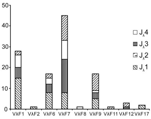
Of the 181 cDNA clones described above, 115 had an identifiable V gene, which provided 63 uniquely recombined V-J junctions (S8 Appendix), and were chosen for analysis and revealed a biased usage pattern of Vλ(Fig 9). The results showed that Vλsegments family 7 was
the most frequently used, which accounted for roughly one-third of the expressed Vλrepertoire
(45/115). Family 1, family 6 and family 9 were more frequently used segments (Fig 9). Vλ
seg-ments from families 2, 8, 11, 12 and 17 were less frequently used. The Vλsegments of other
families were not observed in the cDNA clones of theAlligator sinensis. In these 63 uniquely recombined V-J junctions, 30% of the clones (35/115) had insertion of N and P nucleotides, generally one to two nucleotides, but there were some exceptions. For example, clone LV6-73 had seven N and P nucleotides in its junction; clone LV5-13 and clone LV5-34 had six and five N and P nucleotides in their junctions, respectively; clones LV6-51 and LV6-8 had four N and P nucleotides in their junctions; and clone LV2-8 had three N and P nucleotides in its junction. On average, the length of the N + P nucleotides in these clones was 0.6 ± 1.2 nucleotides. More than 85% of the clones (98/115) had exonuclease removals at the 3’end of Vλ. Compared with
Vλ, fewer nucleotides were removed at the 5’end of Jλ(67/115) by the exonuclease activity (Vλ
3.1 ± 2.2vs. Jλ1.6 ± 1.8). The average length of the CDR3 in theseλgene clones was 10.6 ± 0.9
(S8 Appendix). The results above demonstrated the abundant diversity of the Vλgenes in the
Alligator sinensis.
We cloned and sequenced 237 cDNA fragments from theAlligator sinensisusing 5’RACE to analyze the use of Jκand Vκsegments in the expressedκchain, among which KV-4 has a
stop codon in the leading peptide. After the removal of redundant clones, 124 clones that showed unique V-J junctions were obtained for analysis. All six functional Jκsegments were
used in these clones: 51 clones contained Jκ1; 22 clones contained Jκ2; 18 and 21 clones
con-tained Jκ3 and Jκ4, respectively; 10 clones contained Jκ6; and Jκ5 was only employed in clone
KV-47. In addition, another Jκthat was not found in the genome occurred only once in clone
KV2-67, suggesting the existence of another Jκin the genome or an allelic variant of Jκ. The
results revealed a preferential Jκsegment with Jκ1 as the first preferential usage. The usage
fre-quencies of Jκ5 and Jκ6 were lower, with Jκ5 being the lowest.
We chose 91 clones from the above mentioned 124 clones that had identifiable Vκgenes for
analysis, revealing a preferential Vκusage pattern (Fig 10). The results showed that Vκ
40% of the expressed Vκrepertoire, respectively. Vκsegments from families 2, 3, 6 and 8 were
less frequently used. The Vκof other families were not observed in the cDNA clones of the
Alli-gator sinensisprobably because these families contained only one or two members and their expression levels were low. These 91 clones represented 59 uniquely recombined V-J junctions Fig 9. Usage frequency of Vλand Jλgenes in theAlligator sinensis.The number behind the Vλindicates
the number of the family.
doi:10.1371/journal.pone.0147704.g009
Fig 10. Usage frequency of Vκand Jκgenes in theAlligator sinensis.The number behind the Vκindicates
the number of families.
(S9 Appendix). More functional Vκgenes that were not found in the genome were expressed in
33 clones, suggesting more Vκgenes in theAlligator sinensisthat have not been identified
because of gaps in contigs and incomplete genomic data. The majority of V-J junctions in uniquely recombinedκchain clones lack N and P nucleotide additions. In 59 uniquely rear-ranged clones, 10 clones show putative N or P nucleotides, and the number of N and P nucleo-tides is 1 or 2, with an average of 0.16 ± 0.47 bp per clone. The exonuclease removals at the 3’
end of Vκand the 5’end of Jκwere 2.1 ± 1.5 and 1.3 ± 1.7 nucleotides. The average length of
the CDR3 was 8.8 ± 0.5 nucleotides, and 89% the expressedκV-J junctions might be formed by microhomology (S9 Appendix).
Discussion
Reptilian is comprised of Aves and non-avian reptilia (Crocodylia, Testudines and Squamata) [63,64]. Immunoglobulin genes have been studied in non-avian reptilia of Testudines species [50,51] and Squamata species [45,46,65–67]. Crocodilians are thought to be the closest rela-tives of birds, and they are believed to have strong immune systems [52–55]. Recently, the IgH gene of crocodilians was identified [56,57]. An interesting feature of the crocodilian IgH con-stant loci is the presence of a number of duplicated genes encoding five Ig classes [57]. In addi-tion, an investigation of the crocodilianαgenes suggested that reptiles and birds share a common ancestral organization [56,57]. To better understand the immune system of crocodil-ians, to provide a more complete data of crocodilians Igs, and to obtain more information about immunoglobulin evolution in mammals, birds and reptiles, we identified theAlligator sinensisIgL gene repertoire based on the genome sequence andAlligator sinensisgenomic BAC library.
Previous studies suggested that different IgL genes of jawed vertebrates were classified into four isotype groups:λ,κ,σandσ-cart. To date, all four isotypes are present only in cartilagi-nous fishes: type I (NS5), type II (NS3), type III (NS5) andσ[13]. Type III is clearlyκ, type II is
more similar toλ[15,16], type I is classified asσ-cart [13], andσis orthologous to theσisotype in amphibians [13]. Three IgL isotypes exist in amphibians, includingλ,κ, andσ[27–31], whereas most other tetrapods, including reptiles, have two IgL isotypes (λandκ) [5,7,32–36, 44]. Birds and snakes have only theλisotype [39,42,45]. The different IgL isotypes are located in different genomic regions. The genomic organizations of these regions are also different [13]. In theκlocus, multiple Jκgenes, which are present in different numbers in different spe-cies, are present in a cluster and are generally followed by a single Cκ[5]. Because theκisotype
is present in cartilaginous and bony fishes, with a clear phylogenetic relationship, and in tetra-pods, with the exception ofGallus gallus, it is believed to be the oldest and most evolutionarily conserved isotype [13]. Unlike Igκ, theλgene locus often contains several pairs of Jλ-Cλ, which are also present in different numbers in different species, located downstream from the V seg-ments [34]. Previous studies found that multiple Jλ-Cλwere duplicated after speciation [7,31].
In our recent study, two IgL lociλandκwere identified in another reptile, theAlligator
sinensis, using an available genomic database and sequencing of theAlligator sinensisgenomic BACs, which contain IgL genes. In addition, using theX.tropicalisCσas a template [31], we
performed a BLAST search against theAlligator sinensiswhole-genome shotgun sequence assembly. No similar sequence was identified (data not shown). The results are consistent with those forAnolis carolinensis, revealing onlyλandκisotypes in reptiles. We sketched the map of the genomic organization of the Igλand Igκgene loci of theAlligator sinensis(Fig 1;S1and S5Figs). As in other species, each Cλgene is preceded by a single Jλgene segment (Fig 1Aand
S1 Fig), whereas a single Cκgene follows a cluster of Jκgene segments (Fig 1BandS5 Fig). To
heptamer-12 bp spacer-nonamer and the nonamer-23 bp spacer-heptamer, which is a univer-sal rule ofIGLVandIGLJgene in all species, is demonstrated. The results reveal that the geno-mic organization of Igλin theAlligator sinensisis similar to that inX.tropicalis, lizards, birds and mammals, whereas Igκis similar to that inX.tropicalis, lizards and mammals because the κgene has been lost in birds. We found six Cλgenes and seven Jλgenes from the genomic DNA sequence, and the Cλ5 gene and Jλ5–7 were not found to be expressed, likely because of
their low expression levels. Generally, Jλ-Cλpairs are located in the genome. In our study, an
isolated Jλ7 was located on the 3’end of the Igλlocus without following a corresponding Cλ
gene. This result suggested that more Cλgenes might be located in the Igλlocus in theAlligator
sinensis, which was supported by the Southern blotting results.
Our study also found multiple germline Vλand Vκin theAlligator sinensis. A total of 155
Vλand 118 Vκgene segments were identified, which contain 69 Vλpseudogenes and 56 Vκ
pseudogenes, respectively. All Vλgenes are oriented in the same transcriptional orientation as
the Cλgene and are upstream of the (Jλ-Cλ)nor (Jκ)n. The multiple functional V genes can
increase the antibody diversity and enhance the immune response of antigen recognition and binding. The ratio of functional Vλand Vκvaries significantly in different species [5,32–36]. It
has been proposed that the number of V gene segments may be connected to the preferential use of light chain isotypes at the protein level [68]. The results of the present study indicated that Vλgermline genes are more dominant than Vκ(86 functional Vλgenesvs. 56 functional
Vκgenes) in theAlligator sinensis. It is possible that theλisotype inAlligator sinensisserum
antibodies is more abundant than theκisotype. Additionally, there is a large number of pseu-dogenes in the Vλand Vκloci. We question whether these pseudogenes are functional as those
in birds for use as donors of uniquely combined functional V genes in gene conversion [43]. These pseudogenes were likely involved in generating Ig diversity. The diversification of IgLs in theAlligator sinensisis similar to that in most tetrapods but is different from that in the Gal-lus galGal-lus. A total of 142 potentially functional Vλgenes (Vλand Vκ) are classified into 31
fami-lies in theAlligator sinensis: 19 families in Vλand 12 families in Vκ(Figs3and5;S3,S4,S7and
S8Figs,S3andS6Appendixs). For other species, 177 functional Vλgenes (Vλand Vκ) are
clas-sified into 23 families inMus musculus(http://www.imgt.org/IMGTrepertoire/), 148 functional Vλgenes (Vλand Vκ) are classified into 23 families inHomo sapiens(http://www.imgt.org/
IMGTrepertoire/), 51 functional VLgenes (Vλand Vκ) are classified into 11 families inAnolis
carolinensis[7], and only one Vλgene (or one family) is present inGallus gallus[42]. The
diversity of the IgL chain is generated by V-J recombination, somatic hypermutation, and the polymorphism of the VLgenes, including the number of VLgenes and families (classifying
family according to the similarity of sequence). Our results reveal that theAlligator sinensis
possesses at least 142 functional VLgenes (possibly more) and 31 VLgene families, although
the number of VLgenes in theAlligator sinensisis not the most plentiful in the tetrapods.
How-ever, the number of VLgene families is the greatest. The phylogenetic analyses show that many
Vλgene families in theAlligator sinensisare orthologous with other species, but the remaining
Vλgene families are characteristic of theAlligator sinensis. TheAlligator sinensisalso possesses
a large number (68) ofDHgene segments and multipleμgenes in the IgH locus, suggesting
that theDHsegments may contribute significantly to antibody diversity in crocodilians and that IgM subclasses can be expressed through class-switch recombination in the IgH gene locus [56]. These results reveal the vast diversity of Ig in theAlligator sinensis, suggesting that crocodilians have a strong immune system.
We compared IgL chains between two reptiles: theAlligator sinensisandAnolis carolinensis. We found more abundant VLgenes in theAlligator sinensisthan inAnolis carolinensis,
includ-ing functional VLgenes and pseudogenes. The analysis of the expressed Vλand Vκin the
suggesting that somatic V-J recombination can contribute to theAlligator sinensisantibody diversity, as inAnolis carolinensis[7]. Additionally, the occurrence of N or P nucleotide addi-tions at V-J juncaddi-tions is increased in theAlligator sinensiscompared to the paucity of N or P nucleotide additions in the V-J junctions inAnolis carolinensis, suggesting that crocodilians have more V-J combinatorial diversity than lizards.
We analyzed the preserved co-localization of genes on the Igλand Igκloci in different spe-cies. First, we identified a syntenic relationship between two conserved gene clusters theGNZA
andRTDR1cluster and theMRPL40andHIRAcluster with the Igλgene on the chromosome in theAlligator sinensisand other species, includingHomo sapiens,Mus musculus,Gallus gal-lus,Anolis carolinensisandX.tropicalis(Fig 7). All species retained either one or two gene clus-ters beside the Igλlocus, although two gene clusters reversed their position inGallus gallusand one gene cluster was lost inMus musculus, suggesting that the location of Igλlocus was con-served in tetrapods, includingcrocodilians. The oldest form was found inX.tropicalisand
Homo sapiensand possibly inAlligator sinensis. We also found a syntenic relationship of the Igκgene on the chromosome in different species. The results showed that conserved genes
RPIAandEIF2AK3were flanked on the 3’side of Igκin all species, except inGallus gallus(Fig 8). The two gene families,SCLandRPL, were located far upstream of the Igκlocus. The results suggested that likely intrachromosomal gene conversion occurred inGallus gallusandHomo sapiensorAnolis carolinensisduring speciation, leading toGallus gallusIgλand Igκloci changes. The flanking genes of Igλwere reversed and were lost, and the positions of SCL and RPL were reversed inHomo sapiensandAnolis carolinensis. EitherHomo sapiensorAnolis car-olinensisretained the oldest Igκlocus in the genome.
The results of the phylogenetic tree based on the C domain revealed that isotypes were grouped first, and then species were grouped (Fig 5;S9andS10Figs). The phylogenetic tree of V genes also showed the same result (Fig 6;S11andS12Figs), suggesting that IgL isotypes were individually orthologous. The phylogenetic analyses showed that theσgene was only
present incartilaginousfish, bony fish and amphibians and was absent in reptiles, birds and mammals [13,24,31,39]. Theκgene existed in all vertebrates except birds [13,39–41]. There-fore, theσgene was lost in other vertebrates after their divergence fromamphibians[13,31],
and theκgene was lost in birds [39–41]. Phylogenetic analysis of theIGLVgene, including all 19 Vλfamilies and 12 Vκfamilies in theAlligator sinensis,Alligator sinensisfamilies Vλ1-Vλ8
are related to theAnolis carolinensisVλ1, Vλ3,Gallus gallusandAnas platyrhynchosVλ, andX.
laevistype III V4(Fig 6;S11andS12Figs), which suggested that during the evolution of theλ locus, there was an ancestral locus shared by birds, reptilia and Salientia [7].Alligator sinensis
families Vλ11 is clustered withX.laevistype III V6;Alligator sinensisfamilies Vκ11 and Vκ10
are clustered withAnolis carolinensisVκ; andAlligator sinensisfamily Vκ7 is clustered withX.
laevisρ(Fig 6;S11andS12Figs), which indicated that reptilia and amphibians shared some Vλ
and Vκfamilies and originated from descendants of a common ancestor. Crocodilians possess
more VLfamilies than frogs, lizards and mammals, and there is more abundant diversity of the
V gene in crocodilians. Taken together, the results strongly suggest that we have identified two IgL loci inAlligator sinensisthat belong to theκandλlineages. We present evidence that theσ was lost in early reptilians, avian and mammalians after their divergence from amphibians [13, 31], and theκgene was absent in birds after their divergence from reptilians, similar to theδ gene [39–41].
This study investigated the genomic organization ofAlligator sinensisIgL genes. The organi-zations and structures of IgL genes are similar to those of other jawed vertebrates. The study of theAlligator sinensisλandκloci revealed a diverse and complex repertoire of IgL in
Supporting Information
S1 Appendix. Multiple sequence alignment ofAlligator sinensisVλgenes.
(DOCX)
S2 Appendix. TheAlligator sinensisVλgene DNA segment in contigs.
(DOCX)
S3 Appendix. The alignment of the deduced amino acid sequence of 86 functional Vλgenes in theAlligator sinensis.
(DOCX)
S4 Appendix. Multiple sequence alignment ofAlligator sinensisVκgenes.
(DOCX)
S5 Appendix. TheAlligator sinensisVκgene DNA segment in contigs.
(DOCX)
S6 Appendix. The alignment of the deduced amino acid sequence of 62 functional Vκgenes in theAlligator sinensis.
(DOCX)
S7 Appendix. Sequence of the C region chimeras in the cDNA clones. (DOCX)
S8 Appendix. V-J junctions of theλchain genes.The letter in the middle indicates N/P
nucleotides. The column“N+P”indicates the total nucleotide length of the N and P nucleo-tides, and the column“CDR3”indicates the codon numbers. The column“Deletions in 3’end of Vλ”indicates the number of nucleotides deleted by exonuclease activity at the 3’end of Vλ,
and the column“Deletions in 5’end of Vλ”indicates the number of nucleotides deleted by
exo-nuclease activity at the 5’end of Jλ. Germline sequences of each Vλgene segment are shown
above the cDNA clones in bold, and the CDR3 is also underlined. (DOCX)
S9 Appendix. V-J junctions of theκchain genes.The letter in the middle indicates N/P
nucleotides. The column“N+P”indicates the total nucleotide length of the N and P nucleo-tides, and the column“CDR3”indicates the codon numbers. The column“Deletions in 3’end of Vκ”indicates the number of nucleotides deleted by exonuclease activity at the 3’end of Vκ,
and the column“Deletions in 5’end of Jκ”indicates the number of nucleotides deleted by
exo-nuclease activity at the 5’end of Jκ. Germline sequences of each Vκgene segment are shown
above the cDNA clones in bold, and the CDR3 is also underlined. (DOCX)
S1 Fig. The genomic organization of theAlligator sinensisimmunoglobulinλgene locus.
V: variable gene segments;CV: pseudo-variable gene segments; ORF: variable gene segments with open reading frames but with defects in splicing sites, RSS and/or regulatory elements, and/or changing the conserved amino acids, which have been suggested to lead to incorrect folding [69]; J: joining gene segments; C: constant region gene;CC: pseudo-constant region gene. Gaps between contigs are indicated by a dotted black line, and the sequences from BAC are indicated by a bold line.
(TIF)
S2 Fig. Sequences of theAlligator sinensisJλand Cλ.(A) Nucleotide and amino acid sequences
of the sevenAlligator sinensisJλsegments. (B) Sequence comparison of the sixAlligator sinensis
platyrhynchosandAnolis carolinensis. In the alignment, dots indicate identical amino acids and A-G over the lines represent potential IgSF strands. The cysteine (C) and tryptophan (W) resi-dues are shaded.
(TIF)
S3 Fig. Phylogenetic trees based on 1000 bootstraps for theAlligator sinensisVλgene seg-ments.The phylogenetic tree was constructed using Phylip3.695 [60] and viewed in TREE-VIEW [59].
(TIF)
S4 Fig. Phylogenetic analysis of theAlligator sinensisVλgene segments.The tree is made by
Neighbor-joining P-distance and pairwise deletions using MEGA6.0. (TIF)
S5 Fig. Genomic organization of theAlligator sinensisimmunoglobulinκgene locus.V:
variable gene segments;CV: pseudo-variable gene segments; ORF: variable gene segments with open reading frames but with defects in splicing sites, RSS and/or regulatory elements, and/or changing the conserved amino acids, which have been suggested to lead to incorrect folding [69]; J: joining gene segments; C: constant region gene. Gaps between contigs are indicated by a dotted black line, and the sequences from BAC are indicated by a bold line.
(TIF)
S6 Fig. Sequences of theAlligator sinensisJκand Cκ.(A) Nucleotide and amino acid
sequences of the sixAlligator sinensisJκsegments. (B) Sequence comparison of theAlligator
sinensisCκgenes with their counterparts inHomo sapiens,Mus musculus,Didelphimorphia,
Ornithorhynchus,X.laevis,X.tropicalisandAnolis carolinensis. In the alignment, dots indicate identical amino acids and A-G over the lines represent the potential IgSF strands. The cysteine (C) and tryptophan (W) residues are shaded.
(TIF)
S7 Fig. Phylogenetic trees based on 1000 bootstraps for theAlligator sinensisVκgene seg-ments.The phylogenetic tree was constructed using Phylip3.695 [60] and viewed in TREE-VIEW [59].
(TIF)
S8 Fig. Phylogenetic analysis of theAlligator sinensisVκgene segments.The tree is made by
Neighbor-joining P-distance and pairwise deletions using MEGA6.0. (TIF)
S9 Fig. Phylogenetic trees based on 1000 bootstraps for the IgL chain C genes in jawed ver-tebrates.The phylogenetic tree was constructed using Phylip3.695 [60] and viewed in TREE-VIEW [59].
(TIF)
S10 Fig. Phylogenetic analysis of the IgL chain C genes in jawed vertebrates.The phyloge-netic tree was constructed using C domains, and by Neighbor-joining P-distance and pairwise deletions using MEGA6.0.
(TIF)
scale). The credibility value for each node is shown. The phylogenetic tree was constructed using Phylip3.695 [60] and viewed in TREEVIEW [59].
(TIF)
S12 Fig. Phylogenetic analysis of the IgL chain V genes in jawed vertebrates.The phyloge-netic tree was constructed using V domains, and by Neighbor-joining P-distance and pairwise deletions using MEGA6.0.
(TIF)
S1 Table. Primers used for screening BACs. (DOCX)
S2 Table. Summary of theAlligator sinensisgermline Vλin contigs.
(DOCX)
S3 Table. Summary of theAlligator sinensisgermline Vκin contigs.
(DOCX)
Acknowledgments
The authors are indebted to Li Ma for helping with the articles’compilation. The authors also wish to thank Drs. Xiaoxing Guan, Xueqian Cheng, Jianhui Bai and Kongpan Li for their inspiring suggestions.
Author Contributions
Conceived and designed the experiments: LR YZ HH XW GC. Performed the experiments: XW GC. Analyzed the data: LR XW GC. Contributed reagents/materials/analysis tools: YL CZ XW. Wrote the paper: LR YZ XW GC. Sample collection: YL CZ XW.
References
1. Litman GW, Anderson MK, Rast JP. Evolution of antigen binding receptors. Annual review of immunol-ogy. 1999; 17:109–47. PMID:10358755
2. Chothia C, Lesk AM, Tramontano A, Levitt M, Smith-Gill SJ, Air G, et al. Conformations of immunoglob-ulin hypervariable regions. Nature. 1989; 342(6252):877–83. PMID:2687698
3. Hamers-Casterman C, Atarhouch T, Muyldermans S, Robinson G, Hammers C, Songa EB, et al. Natu-rally occurring antibodies devoid of light chains. Nature. 1993; 363(6428):446–8. PMID:8502296 4. Greenberg AS, Avila D, Hughes M, Hughes A, McKinney EC, Flajnik MF. A new antigen receptor gene
family that undergoes rearrangement and extensive somatic diversification in sharks. Nature. 1995; 374(6518):168–73. PMID:7877689
5. Kawasaki K, Minoshima S, Nakato E, Shibuya K, Shintani A, Asakawa S, et al. Evolutionary dynamics of the human immunoglobulinκlocus and the germline repertoire of the Vκgenes. Eur J Immunol 2001; 31(4):1017–28. PMID:11298326
6. Gerdes T, Wabl M. Physical map of the mouse lambda light chain and related loci. Immunogenetics. 2002; 54(1):62–5. PMID:11976793
7. Wu Q, Wei Z, Yang Z, Wang T, Ren L, Hu X, et al. Phylogeny, genomic organization and expression of lambda and kappa immunoglobulin light chain genes in a reptile, Anolis carolinensis. Developmental and comparative immunology. 2010; 34(5):579–89. doi:10.1016/j.dci.2009.12.019PMID:20056120 8. Akira S, Okazaki K, Sakano H. Two pairs of recombination signals are sufficient to cause
immunoglobu-lin V-(D)-J joining. Science. 1987; 238(4830):1134–8. PMID:3120312
9. Gu H, Förster I, Rajewsky K. Sequence homologies, N sequence insertion and JH gene utilization in VHDJH joining: implications for the joining mechanism and the ontogenetic timing of Ly1 B cell and B-CLL progenitor generation. EMBO J. 1990; 9(7):2133. PMID:2113468
11. McBlane JF, van Gent DC, Ramsden DA, Romeo C, Cuomo CA, Gellert M, et al. Cleavage at a V (D) J recombination signal requires only RAG1 and RAG2 proteins and occurs in two steps. Cell. 1995; 83 (3):387–95. PMID:8521468
12. Fugmann SD, Lee AI, Shockett PE, Villey IJ, Schatz DG. The RAG proteins and V (D) J recombination: complexes, ends, and transposition. Annu Rev Immunol. 2000; 18(1):495–527.
13. Criscitiello MF, Flajnik MF. Four primordial immunoglobulin light chain isotypes, including lambda and kappa, identified in the most primitive living jawed vertebrates. European journal of immunology. 2007; 37(10):2683–94. PMID:17899545
14. Jung D, Giallourakis C, Mostoslavsky R, Alt FW. Mechanism and control of V(D)J recombination at the immunoglobulin heavy chain locus. Annual review of immunology. 2006; 24:541–70. PMID:16551259 15. Lee SS, Fitch D, Flajnik MF, Hsu E. Rearrangement of immunoglobulin genes in shark germ cells. J
Exp Med. 2000; 191(10):1637–48. PMID:10811858
16. Dooley H, Flajnik MF. Antibody repertoire development in cartilaginous fish. Developmental and com-parative immunology. 2006; 30(1–2):43–56. PMID:16146649
17. Edholm ES, Wilson M, Bengten E. Immunoglobulin light (IgL) chains in ectothermic vertebrates. Devel-opmental and comparative immunology. 2011; 35(9):906–15. doi:10.1016/j.dci.2011.01.012PMID: 21256861
18. Hikima J, Jung TS, Aoki T. Immunoglobulin genes and their transcriptional control in teleosts. Develop-mental and comparative immunology. 2011; 35(9):924–36. doi:10.1016/j.dci.2010.10.011PMID: 21078341
19. Daggfeldt A, Bengtén E, Pilström L. A cluster type organization of the loci of the immunoglobulin light chain in Atlantic cod (Gadus morhua L.) and rainbowttrout (Oncorhynchus mykiss Walbaum) indicated by nucleotide sequences of cDNAs and hybridization analysis. Immunogenetics. 1993; 38(3):199–209. PMID:8505063
20. Ghaffari SH, Lobb CJ. Structure and genomic organization of immunoglobulin light chain in the channel catfish. An unusual genomic organizational pattern of segmental genes. J Immunol. 1993; 151 (12):6900–12. PMID:8258698
21. Lundqvist M, Bengtén E, Strömberg S, Pilström L. Ig light chain gene in the Siberian sturgeon (Acipen-ser baeri). J Immunol. 1996; 157(5):2031–8. PMID:8757324
22. Partula S, Schwager J, Timmusk S, Pilström L, Charlemagne J. A second immunoglobulin light chain isotype in the rainbow trout. Immunogenetics. 1996; 45(1):44–51. PMID:8881036
23. Ghaffari SH, Lobb CJ. Structure and genomic organization of a second class of immunoglobulin light chain genes in the channel catfish. J Immunol. 1997; 159(1):250–8. PMID:9200461
24. Haire RN, Rast JP, Litman RT, Litman GW. Characterization of three isotypes of immunoglobulin light chains and T-cell antigen receptorαin zebrafish. Immunogenetics. 2000; 51(11):915–23. PMID: 11003385
25. Edholm ES, Wilson M, Sahoo M, Miller NW, Pilstrom L, Wermenstam NE, et al. Identification of Igsigma and Iglambda in channel catfish, Ictalurus punctatus, and Iglambda in Atlantic cod, Gadus morhua. Immunogenetics. 2009; 61(5):353–70. doi:10.1007/s00251-009-0365-zPMID:19333591
26. Bao Y, Wang T, Guo Y, Zhao Z, Li N, Zhao Y. The immunoglobulin gene loci in the teleost Gasterosteus aculeatus. Fish Shellfish Immunol. 2010; 28(1):40–8. doi:10.1016/j.fsi.2009.09.014PMID:19782140 27. Schwager J, Bürckert N, Schwager M, Wilson M. Evolution of immunoglobulin light chain genes:
analy-sis of Xenopus IgL isotypes and their contribution to antibody diversity. EMBO J. 1991; 10(3):505. PMID:1705882
28. Zezza DJ, Mikoryak C, Schwager J, Steiner L. Sequence of C region of L chains from Xenopus laevis Ig. J Immunol 1991; 146(11):4041–7. PMID:1903418
29. Zezza D, Stewart S, Steiner L. Genes encoding Xenopus laevis Ig L chains. Implications for the evolu-tion of kappa and lambda chains. J Immunol 1992; 149(12):3968–77. PMID:1460285
30. Haire R, Ota T, Rast J, Litman R, Chan F, Zon L, et al. A third Ig light chain gene isotype in Xenopus lae-vis consists of six distinct VL families and is related to mammalian lambda genes. J Immunol 1996; 157 (4):1544–50. PMID:8759737
31. Qin T, Ren L, Hu X, Guo Y, Fei J, Zhu Q, et al. Genomic organization of the immunoglobulin light chain gene loci in Xenopus tropicalis: evolutionary implications. Developmental and comparative immunol-ogy. 2008; 32(2):156–65. PMID:17624429
32. Butler J. Immunoglobulin gene organization and the mechanism of repertoire development. Scand J Immunol. 1997; 45(5):455–62. PMID:9160087