REGULAR ARTICLE
MICROBIAL LOAD AND MULTIPLE DRUG RESISTANCE OF PATHOGENIC BACTERIA ISOLATED FROM FEACES AND BODY SURFACES OF COCKROACHES IN AN URBAN AREA OF SOUTHWESTERN NIGERIA
Monsuru Adebayo Adeleke*1, Hilda Abiola Akatah1, AbdulWasiu Oladele Hassan2,
Wasiu Olalekan Adebimpe 3
1 Public Health Entomology and Parasitology Unit, Department of Biological Sciences, P.M.B
4429, Osun State University, Osogbo, Nigeria
2Department of Microbiology and Parasitology, Ladoke Akintola University Teaching
Hospital, Osogbo, Nigeria
3Department of Community Medicine, College of Health Sciences, Osun State University,
Osogbo
*Corresponding author: [email protected].
ABSTRACT
This study investigates the microbial load and antibiotic susceptibility pattern of pathogenic bacteria isolated from the faeces and body surfaces of cockroaches in Osogbo, Southwestern Nigeria. The cockroaches collected from residential areas and hospital vicinities were screened for microbial load and antibiotic susceptibility pattern using standard protocols. A total of twenty- three microorganisms namely Klebsiella aerogenes, Bacillius cereus, Proteus spp, Staphyloccocus aureus, S. saprophyticus, Enteroccocus faecalis, Staphylococus epididermis, E. coli, Listeria monoctogene, Proteus mirabilis, Citrobacter species,
Pseudomonas aeruginosa, Psuedomonas species, Seretia mensence, Candida albicans,
Candida spp., Aspergilius spp., A. flavus, A. fumigates, Mucor species and Penicilium species were isolated. The microbial load of the microorganisms was significantly higher in the isolates from hospital as compared with the residential area (p<0.05) with the exception of
Canidida species, Mucor and Penicillium which had higher or equal microbial load at the
most importantly, Ampicillin, Augumentin, Amoxicillin and Septrin (30μg). Efforts geared towards controlling the insects will be indispensable in curbing the wide spread of multi-drug resistant pathogens in the study area.
Keywords: cockroaches, microbial load, antimicrobial, multi-drug resistance, Nigeria
INTRODUCTION
Antimicrobial resistance of pathogenic microorganisms has assumed a worrisome dimension with recent trend of resistance of pathogenic bacteria to common antibiotics. This increase in antibiotic resistance was premised on the drug pressure as a result of abuse of common antibiotics by the users and uncomplimentary fake drugs in circulation (Ehinmidu 2003, Tachebe et al., 2006, Oleghe et al., 2011). The unwholesome behaviour has led to the genetic response of the microorganisms to microbial therapy which has now become an issue mitigating the control of pathogenic microorganisms in different parts of the world (Oleghe et al., 2011).
It has most often been assumed that the drug resistance in clinical isolates usually results from the contamination of resistant bacteria from the drug pressurized environment (Oleghe et al., 2011). The bacteria contaminant could be from water, food or contact with the vectors harbouring the pathogens. Cockroaches stay in filthy environments in the house, shops and even hospitals where both clinical and environmental samples coincide (Fortedor
et al., 1992). Therefore, their roles in promoting drug resistance in pathogenic
microorganisms cannot be overlooked.
Though, previous studies have implicated cockroaches as potential carriers of microorganisms and drug resistant microbes in different parts of the world (Cloarec et al., 1992; Fortedor et al., 1999; Padro et al., 2002; Tatfeng et al., 2005; Tachebe et al., 2006) there was little or no information on anti-microbial susceptibility status of the micro-organisms harboured by the cockroaches in Osogbo in particular and Nigeria in general.
MATERIAL AND METHODS
Collection of cockroaches
The study was carried out in Osogbo, Osun State Nigeria. Osogbo lies latitude of 7o49’N and a longitude of 4o37’E. The faeces and body surfaces of cockroaches collected from four randomly selected residential areas and two hospital vicinities; Ladoke Akintola University Teaching Hospital Osogbo and State Hospital Asubiaro between November 2011 and February 2012 were screened for microbial load and susceptibility pattern to antibiotics. The cockroaches were trapped with sterile hand gloves and transferred to sterile universal containers. The cockroaches were kept in the bottles until they defeacate. The cockroaches and the feaces were then transferred to separate sterile universal bottle for analysis.
Screening for pathogenic organisms
The cockroaches and the feaces were kept in the universal containers and 2ml of sterile normal saline (0.9%) was added to the universal containers and vigorously shaken for 2 minutes. 0.01ml of the sample was then taken from each container and cultured on the MacConkey , Sabouraud’s dextrose agar and chocolate agar plate and incubated overnight at 37oC.
Identification of bacteria
The colonies were identified by standard bacteriological procedures; macrosopic morphology, biochemical and gram staining in accordance with Cowan and Steel (1975) .
Gram stain
minutes, and washed off with clean water.The back of the slide was wiped clean and placed in a draining rack for the smear to air dry. The slide was examined microscopically with the oil immersion lens after the application of the oil on the slide. Gram positive bacteria gave a dark purple colour while gram negatives give a red colour.
Biochemical tests
Several biochemical tests were performed on the isolates for identification purposes as described by Baron and Finegold (1990). The catalase test was done to differentiate the bacteria that produce the enzyme catalase, such as staphylococci from the non-catalase producing bacteria such as streptococci. Citrate utilization test was done to identify
enterobacteria, the test is based on the ability to use citrate as its only source of carbon. Using Simmon’s citrate agar, slopes of the medium was prepared in bijou bottles as recommended by the manufacturer. With the aid of a sterile straight wire, the slope was streaked with a saline suspension of the test organism, and the butt was stabbed.
It was incubated at 35oC for 48hours. A bright blue colour in the medium indicates a positive citrate test (e.g Klebsiella pneumoniae), while no change in the colour of the medium gives a negative citrate test (e.g E.coli). Coagulase test was done to identify S.aureus which produces the enzyme coagulase. A drop of distilled water was placed at the end of a slide, a colony of the test organism is emulsified on it to make a suspension. A loopful of plasma was added to it and mixed gently, presence of clumping within 10 seconds indicates the presence of S. aureus, while absence indicates presence of E.coli or S.epidermidis. Oxidase test was used to identify
Pseudomonas, Neisseria, Proteus, Brucella and Pasteurella species. A piece of filter paper
enterobacteriaecae group and other group of bacilli were identified in accordance with their characteristics by comparing with standard table (Chaichanawongsaroj et al.,2004).
Fungi identification
The fungi isolates were identified by microscopic examination of the actively growing mould using morphological characters such as, the absence or presence rhizoid, colour, and micro-morphology of their sporulating structures and conida (Evans and Richrdson, 1989;
Onions, et al., 1991).
Total viable count
A ten-fold dilution was carried out on each suspension to determine the total viable count of each cockroach using the pour plate method counts were made on plates showing discrete colonies. A quantitative analysis of bacteria was calculated as described by
Salehzadeh et al., (2007). The overall load of bacteria carried by each insect was counted and expressed as colony forming unit (c.f.u).
Antibiotic sensitivities of isolated pathogenic bacteria
Statistical Analysis
The comparison of data obtained from the feaces and body surface of the cockroaches were analyzed with t-test using SPSS version 16.0.
RESULTS
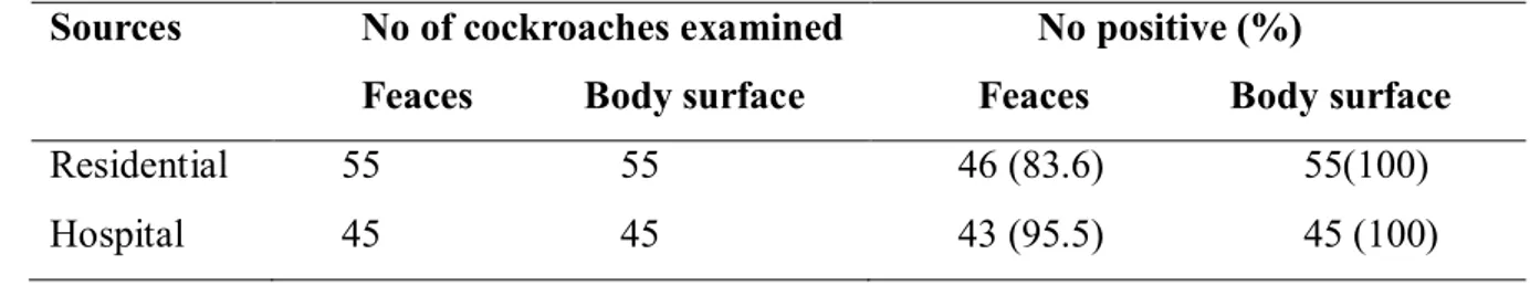
The results of the microbial analysis of the feaces and body surfaces of the cockroaches revealed that the body surface of all the cockroaches caught at both hospitals and residential areas were positive for microorganisms while 83.6% and 95.5% of the feaces from residential areas and hospitals were positive respectively (Table 1). The variation in the occurrence of the microorganisms between body surface and feaces was not significant (p>0.05). A total of twenty- three microorganisms namely Klebsiella aerogenes, Bacillius cereus, Proteus spp, Staphyloccocus aureus, S. saprophyticus, Enteroccocus faecalis, Staphylococus epididermis, E. coli, Listeria monoctogene, Proteus mirabilis, Citrobacter
species, Pseudomonas aeruginosa, Psuedomonas species, Seretia mensence, Candida
albicans, candida spp, Candida spps, Aspergilius spp, A. flavus, A. fumigates, A, Mucor
species and Penicilium species were isolated. All the microbial isolates were found in the
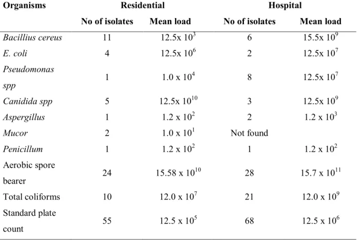
body surface of the cockroaches while ten of the twenty three isolates were found in the feaces of cockroaches (Table 2). The microbial load of the microorganisms was significantly higher in the isolates from hospital as compared with the residential area (p<0.05) with the exception of Canidida species, Mucor and Penicillium which had higher or equal microbial load at the residential areas (Table 3).
Table 1 Occurrence of the microorganisms in the faecal pellets and body surfaces of the cockroaches at the study area
Sources No of cockroaches examined
Feaces Body surface
No positive (%)
Feaces Body surface Residential 55 55 46 (83.6) 55(100) Hospital 45 45 43 (95.5) 45 (100)
Table 2 Microbial diversity in the body surfaces and feacal pellets of the cockroaches at the study area
Name of Isolates Feacal pellets Cockroach surface
Bacteria isolates
Baccillus species + +
Klebsiella aerogenes + +
Proteus species - +
Staphyloccocus aureus + +
Baccillus cereus - +
Staphyloccocus saprophyticus - +
Enteroccocus feacalis + +
Staphylococus epididermis + +
Escherichia coli + +
Listeria monoctogenes + +
Proteus mirabilis - +
Citrobacter species - +
Psuedomonas species - +
Psuedomonas aeruginosa - +
Seretia mensence - +
Fungi isolates
Candida Spcies + +
Candida albicans + +
Aspergillus fumigates - +
Aspergillus species + +
Mucor Species - +
Penicillum species - +
- Means absent
+ Means present
Table 3 Microbial load of bacteria and fungi encountered on the body surfaces and feacal pellets of the cockcroaches
Organisms Residential Hospital
No of isolates Mean load No of isolates Mean load
Bacillius cereus 11 12.5x 10³ 6 15.5x 10⁹
E. coli 4 12.5x 10⁶ 2 12.5x 10⁷
Pseudomonas
spp 1 1.0 x 10
4 8 12.5x 10⁷
Canidida spp 5 12.5x 10¹⁰ 3 12.5x 10⁹
Aspergillus 1 1.2 x 10² 2 1.2 x 10³
Mucor 2 1.0 x 101 Not found
Penicillum 1 1.2 x 10² 1 1.2 x 10²
Aerobic spore
bearer 24 15.58 x 10
10 28 15.7 x 10¹¹
Total coliforms 10 12.0 x 10⁷ 21 12.0 x 10⁹
Standard plate
count 55 12.5 x 10
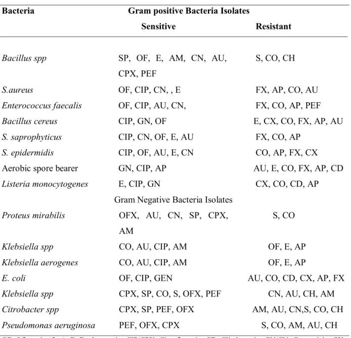
Table 4 Antibiograms of pathogenic bacteria isolated from cockroaches trapped at the residential areas
Bacteria Gram positive Bacteria Isolates
Sensitive Resistant
Bacillus spp SP, OF, E, AM, CN, AU,
CPX, PEF
S, CO, CH
S.aureus OF, CIP, CN, , E FX, AP, CO, AU
Enterococcus faecalis OF, CIP, AU, CN, FX, CO, AP, PEF
Bacillus cereus CIP, GN, OF E, CX, CO, FX, AP, AU
S. saprophyticus CIP, CN, OF, E, AU FX, CO, AP
S. epidermidis CIP, OF, AU, E, CN CO, AP, FX, CX
Aerobic spore bearer GN, CIP, AP AU, E, CO, FX, AP, CD
Listeria monocytogenes E, CIP, GN CX, CO, CD, AP
Gram Negative Bacteria Isolates
Proteus mirabilis OFX, AU, CN, SP, CPX,
AM
S, CO
Klebsiella spp CO, AU, CIP, AM OF, E, AP
Klebsiella aerogenes CO, AU, CIP, AM OF, E, AP
E. coli OF, CIP, GEN AU, CO, CD, CX, AP, FX
Klebsiella spp CPX, SP, CO, S, OFX, PEF CN, AU, CH, AM
Citrobacter spp CPX, SP, PEF, OFX AM, AU, CN,S, CO, CH
Pseudomonas aeruginosa PEF, OFX, CPX S, CO, AM, AU, CH
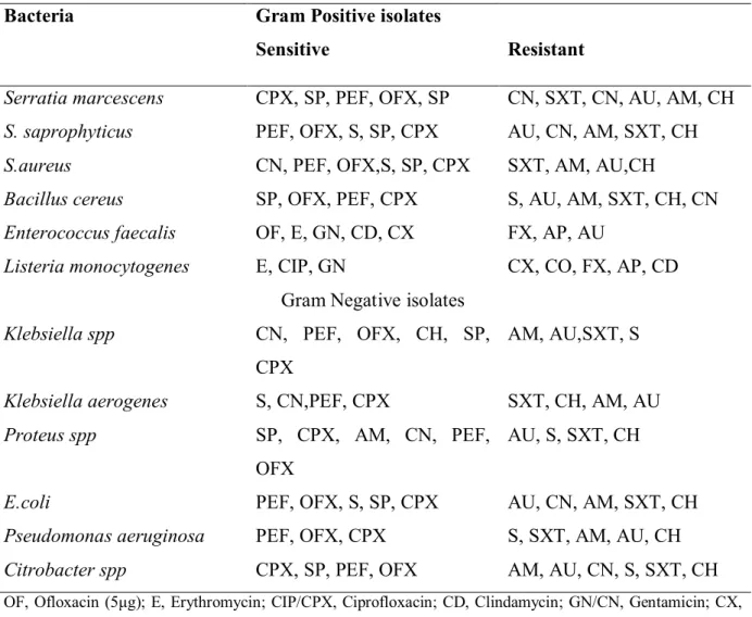
Table 5 Antibiograms of pathogenic bacteria isolated from cockroackes trapped at the hospital vicinity
Bacteria Gram Positive isolates Sensitive
Resistant
Serratia marcescens CPX, SP, PEF, OFX, SP CN, SXT, CN, AU, AM, CH
S. saprophyticus PEF, OFX, S, SP, CPX AU, CN, AM, SXT, CH
S.aureus CN, PEF, OFX,S, SP, CPX SXT, AM, AU,CH
Bacillus cereus SP, OFX, PEF, CPX S, AU, AM, SXT, CH, CN
Enterococcus faecalis OF, E, GN, CD, CX FX, AP, AU
Listeria monocytogenes E, CIP, GN CX, CO, FX, AP, CD
Gram Negative isolates
Klebsiella spp CN, PEF, OFX, CH, SP,
CPX
AM, AU,SXT, S
Klebsiella aerogenes S, CN,PEF, CPX SXT, CH, AM, AU
Proteus spp SP, CPX, AM, CN, PEF,
OFX
AU, S, SXT, CH
E.coli PEF, OFX, S, SP, CPX AU, CN, AM, SXT, CH
Pseudomonas aeruginosa PEF, OFX, CPX S, SXT, AM, AU, CH
Citrobacter spp CPX, SP, PEF, OFX AM, AU, CN, S, SXT, CH
OF, Ofloxacin (5μg); E, Erythromycin; CIP/CPX, Ciprofloxacin; CD, Clindamycin; GN/CN, Gentamicin; CX, Cephalexin; CO, Septrin (50 μg); AP, Ampicillin/Cloxacillin; FX, Flucloxacillin; AU, Augumentin; SXT Septrin (30 μg); CH, chloramphenicol; SP, sparfloxacin; AM, Amoxicillin; PEF, perfloxacin; OFX, Ofloxacin (10 μg), S, Streptomycin.
DISCUSSION
The present results clearly indicated that nearly all the cockroaches in residential areas and hospital vicinity harboured pathogenic microorganisms. This high prevalence of the microorganisms harboured in the body and feaces of the cockroaches portends public health risks to the people in residential areas and transmission of nosocomial infections in the hospitals at the study area. Most of the bacterial isolated from the cockroaches in the present study are highly pathogenic and have been implicated in many nososocomial and gastroenteric infections (Tatfeg et al., 2005, Lamiaa et al., 2010). The isolation of Candida
infections (Salehzadeh et al., 2007). The isolation of such fungi from cockroaches in hospital is alarming and could worsen the infection morbidity and mortality in the hospitals, most especially for immune compromised patients who are already overwhelmed by infections (Hong et al., 2003).
Moreover, the isolation of A. flavus from cockroaches in residential areas poses threat when considering the public health importance of this species. A. flavus has been known to produce mycotoxins which is one of the leading causes of food poisoning (Tatfeng et al., 2005; Salehzadeh et al 2007). The significant higher distribution of microorganisms and the microbial load in cockroaches from hospitals in comparison with residential areas is in consonance with the previous studies (Fortedor et al., 1999; Salehzadeh et al 2007).
Multiple drug resistance patterns were observed in all the isolates from hospitals and residential areas with the exception of Proteus species which showed double resistance in residential area. This observation contradicts the results of Salehzadeh et al (2007) but agrees with report of Fortedor et al., (1999). Though, most of the previous studies have reported the role of cockroaches as vectors of multi-drug resistant bacteria (Fortedor et al., 1999, Padro
et al., 2002), our results in the present study also showed multiple drug resistance in isolates
from residential areas. This possibly introduces new dimension to the episode of drug resistant pathogens at the study area, as wide spread and contamination of isolates earlier susceptible to antibiotics could be observed within the shortest time in the metropolis. The above speculation is possible when considering the clustering plan of the residential areas in the metropolis and the unrestricted mode of movement of cockroaches at night (Pai et al., 2003; Pai et al., 2004; Gracyk et al., 2005; Pai et al., 2005)
CONCLUSION
Our results showed high prevalence of pathogenic organisms in the body and feaces of cockroaches in Osogbo and reported the multiple resistance of the pathogenic bacteria to antibiotics. The multiple resistance of the isolates most importantly, the isolates from residential areas showed that the surveillance on pattern and origin of antimicrobial drug resistance should not be limited to only clinical isolates. It is therefore pertinent to educate the people at the resident areas on the danger inherent in harbouring the cockroaches in residential areas and hospital vicinities at the study area.
Acknowledgement:The authors acknowledge the efforts and support of the Management and Staff of Group Diagnostica, Oke-fia Osogbo to the study.
REFERENCES
BARON, E. J. – FINEGOLD, S. M. 1990. Bailey and Scott’s diagnostic microbiology, 7th ed., In St. Louis: New York 1990, p. 323 – 861.
CHAICHANAWONGSAROJ, N. – VANICHAYATANARAK, K. – PIPATKULLACHAT, T. – POLROJPANYA, M. – SOMKIATCHAROEN, S. 2004. Isolation of gram-negative bacteria from cockroaches trapped from urban environment. In Southeast Asian Journal of Tropical
Medicine and Public Health, vol. 35, 2004, no.1, p. 681-684.
CLOAREC, A. – RIVAULT, G. – FONTAINE, F. – LEGUYADER, A. 1992. Cockroaches as carriers of bacteria in multifamily dwellings. In Epidemiology and Infection, Vol.109,1992, p. 483–490.
COWAN, S. T – STEEL, J. L. 1975. Manual for the identification of Medical Bacteria, 2nd Ed., In Cambridge University Press, England 1975, p. 45-114.
EHINMIDU, J.O. 2003. Antibiotics susceptibility patterns of urine bacteria isolates in Zaria, Nigeria. In Tropical Journal of Pharmaceutical Research, vol. 2, 2003, no.2, p. 223-228
EVANS, E. G. V. – RICHRDSON, M. D. 1989. Medical mycology: a practical approach. Oxford University Press: Oxford 1989, 154p.
FORTEDOR, R. – BAREIJER, U. – VERMA, A. 1991. Cockroaches (Blattella germanica) as carriers of micro-organisms of medical importance in hospitals. In Epidemiology and
Infection, vol. 107, 1991, p. 181-187
protozoan parasites by insects. In Clinical Microbiology Reviews, vol. 35, 2005, p. 128–132.
HONG, W. – WEN, H. – LIAO, W. 2003. Fungal infection in organ transplant patients. In Chinise Medecal Journal, vol. 116, 2003, p.1421–1425.
LAMIAA, B. – ANTONIO, S. – AMIN, L. – LEBBADI, M. – AARAB, A. – GUTIERREZ, J. 2010. Antibiotic resistance patterns of bacterial strains isolated from Periplaneta americana
and Musca domestica in Tangier, Morocco. In Journal of Infection in Developing Countries,
vol. 4, 2010, no. 4, p. 194-201.
OLEGHE, P. O. – ODIMEGWU, D. C. – UDOFIA, E. – ESIMORE, C.O. 2011. Multidrug resistant bacteria isolates recovered from herbal medicinal preparations in Southeastern Setting, Nigeria. In Journal of Rural and Tropical Public Health, vol. 10, 2011, p. 70-75 ONIONS, A. H. S – ALLSOPP, A. – EGGINS, H. O. W. 1981. Introduction to industrial mycology, Edward Arnold: London 1981 , 389p.
PAI, H. H – KO, Y. C. – CHEN, E. R. 2003. Cockroaches (Periplaneta americana and Blattella germanica) as potential mechanical disseminators of Entamoeba histolytica. In Acta Tropica, vol.87, 2003, p.355–359.
PAI, H. H. – CHEN, W. C. – PENG, C. F. 2004. Cockroaches as potential vectors of nosocomial infections. In Infection Control and Hospital Epidemiology, vol. 25, 2004, p. 979– 984.
PAI, H. H. – CHEN, W. C. – PENG, C. F. 2005. Isolation of bacteria with antibiotic resistance from household cockroaches (Periplaneta americana and Blattella germanica). In Acta Tropica, vol. 93,2005,259–265.
PRADO, M. A. – PIMENTA, F .C. – HAYASHID, M. – SOUZA, P. R. – PEREIRA, M.S. – GIR, E. 2002. Enterobacteria isolated from cockroaches (Periplaneta americana) captured in a Brazilian Hospital. In Pan Americana Journal of Public Health, vol. 11,2002, p. 93–98. SALEHZADEH, A. – TAVACOL, P. – MAJHUB, H. 2007. Bacteria, fungi and Parasitic contaminants of cockroaches in Public Hospitals of Hamedah, Iran, In Journal of Vector
Borne Diseases, vol. 44, 2007, p. 105-110
TACHEBE, J. – ERKU, W. – GERBE-MICHAEL, T. – ASHENAFI, M. 2006. Cockroach-associated food-borne bacteria from hospital and restaurant in Addis Ababa, Ethopia: Distribution and antibiograms. In Journal of Rural and Tropical Public Health, vol. 5, 2006, p. 34-41