Jebmh.com
Original Article
J. Evid. Based Med. Healthc., pISSN- 2349-2562, eISSN- 2349-2570/ Vol. 3/Issue 73/Sept. 12, 2016 Page 3953
ROLE OF VACUUM ASSISTED CLOSURE (VAC) - IN WOUND HEALING
L. Lokanadha Rao1, Sravya Teja P2, V. Lakshmi Sumana3, K. Venkatesh4, M. Sidharda5, M. Prudvi6, P. Vishnu Prasad7,
M. Sai Krishna8
1Associate Professor, Department of Orthopaedics, ACSR Government Medical College, Nellore, Andhra Pradesh. 2Senior Resident, Department of Orthopaedics, Andhra Medical College, Visakhapatnam.
3Senior Resident, Department of Orthopaedics, Andhra Medical College, Visakhapatnam. 4Senior Resident, Department of Orthopaedics, Andhra Medical College, Visakhapatnam. 5Senior Resident, Department of Orthopaedics, Andhra Medical College, Visakhapatnam. 6Senior Resident, Department of Orthopaedics, Andhra Medical College, Visakhapatnam. 7Senior Resident, Department of Orthopaedics, Andhra Medical College, Visakhapatnam. 8Senior Resident, Department of Orthopaedics, Andhra Medical College, Visakhapatnam.
ABSTRACT
BACKGROUND
Large, complicated wounds pose a significant surgical problem. Negative pressure wound therapy is one of several methods enabling to obtain better treatment results in case of open infected wounds.1,2 The use of negative pressure therapy enables to obtain a reduction in the number of bacteria which significantly reduces the number of complications.3,4,5
AIMS AND OBJECTIVES:
To review the Role of VAC in wound healing in Orthopaedics.
MATERIALS AND METHODS
The cases presented in this study are those who were admitted in King George Hospital in the time period from January 2014 to August 2015. This is a prospective interventional study. In this study, 15 patients were assigned to the study group (Negative Pressure Wound Therapy- NPWT) based on their willingness for undergoing treatment.
OBSERVATIONS AND RESULTS
12 males and 3 females are involved in the study. There is decrease in the mean wound area from 64 cm2 to 38 cm2. There is decrease in the duration of hospital stay. Finally, wound is closed by SSG or secondary suturing.
DISCUSSION
NPWT is known to reduce bacterial counts, although they remain colonised with organisms. Wounds covered with NPW dressing are completely isolated from the environment, thereby reduces cross infection. In our series, we had 73.3% (11 cases) excellent results and 26.7% (4 cases) good results and no poor results. As interpretation with results, VAC therapy is effective mode of adjuvant therapy for the management of infected wounds.
CONCLUSION
VAC has been proven to be a reliable method of treating a variety of infected wounds. It greatly increases the rate of granulation tissue formation and lowers bacterial counts to accelerate wound healing. It can be used as a temporary dressing to prepare wounds optimally prior to closure or as a definitive treatment for nonsurgical and surgical wounds. VAC is now being used in a multitude of clinical settings, including the treatment of surgical wounds, infected wounds, traumatic wounds, pressure ulcers, diabetic foot ulcers and venous stasis wounds.
KEYWORDS
VAC, NPWT, Chronic osteomyelitis.
HOW TO CITE THIS ARTICLE: Rao LL, Teja SP, Sumana VL, et al.Role of vacuum assisted closure (VAC) - in wound healing. J. Evid. Based Med. Healthc. 2016; 3(73), 3953-3956. DOI: 10.18410/jebmh/2016/845
INTRODUCTION: Vacuum-assisted closure employs application of constant sub-atmospheric pressure on the wound which effectively removes excess of interstitial fluid from wound, thereby reducing bacterial proliferation and wound infection. Large, complicated wounds pose a significant surgical problem. The duration of treatment is usually long, the number of complications and therapy failures are significant, and one should not forget about high treatment costs.
Financial or Other, Competing Interest: None. Submission 18-07-2016, Peer Review 27-07-2016, Acceptance 21-08-2016, Published 10-09-2016. Corresponding Author:
Dr. L. Lokanadha Rao,
Associate Professor, Department of Orthopaedics, ACSR Government Medical College,
Jebmh.com
Original Article
J. Evid. Based Med. Healthc., pISSN- 2349-2562, eISSN- 2349-2570/ Vol. 3/Issue 73/Sept. 12, 2016 Page 3954 Negative pressure wound therapy is one of several
methods enabling to obtain better treatment results in case of open infected wounds.1,2 The use of negative pressure therapy enables to obtain a reduction in the number of bacteria which significantly reduces the number of complications.3,4,5 Vacuum-assisted closure employs application of constant sub-atmospheric pressure on the wound which effectively removes excess of interstitial fluid from wound, thereby reducing bacterial proliferation and wound infection.
AIMS AND OBJECTIVES: To review the Role of VAC in wound healing in orthopaedics. The primary efficacy end point is the complete wound closure. Secondary end points included reduction in the wound surface area over time, time to achieve complete wound closure either by surgery or secondary intention, and a reduction in complications, including amputations.
MATERIALS AND METHODS: The cases presented in this study are those who were admitted as in patients at King George Hospital in the time period from January 2014 to August 2015. This is a prospective interventional study. In this study, 15 patients were assigned to the study group (Negative Pressure Wound Therapy) based on their willingness for undergoing treatment.
Inclusion Criteria: Post-traumatic cases of infected open wounds presented to the hospital during the study period.
Post-operative infected wounds.
Non-healing wounds.
Delayed healing wounds.
Exclusion Criteria:
Chronic osteomyelitis.
Malignancy.
Raw area with exposed vessels and nerves.
Patients not willing for VAC therapy.
Imaging Studies:
X-ray of the affected limb.
MRI of affected part if needed.
Culture and sensitivity of the pus/discharge from the ulcer.
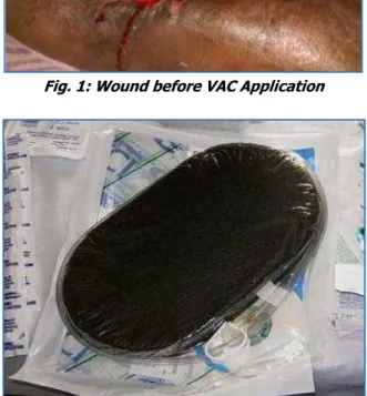
PROCEDURE: After evaluating the patient and under aseptic precautions, wound was debrided thoroughly and skin was cleaned around the wound and kept dry. Wound debridement was done after confirmation of diagnosis (Figure 1). Regular dressings were done once in 2 days basis. GranuFoam Dressing (Black Foam) was used in NPWT. It has been specifically engineered to deliver NPWT. It has pore size of 400-600 microns. It is hydrophobic. Open pore structure of VAC. GranuFoam Dressings adapt to the contours of deep or irregularly shaped wounds in order to provide equal distribution of pressure at the wound site. This is autoclavable and has requisite softness at the applied negative pressure of about 150 mmHg (Figure 2). The foam is cut to the shape of the wound (Figure 3).
Fig. 1: Wound before VAC Application
Fig. 2: GranuFoam Dressing (Black Foam)
Fig. 3: Foam is cut to the Shape of Wound
Foam is placed over the wound and transparent drape material applied to cover the foam and wound. The drape material should extend out onto the periwound about 5 cm all around. (Figure 4).
Jebmh.com
Original Article
J. Evid. Based Med. Healthc., pISSN- 2349-2562, eISSN- 2349-2570/ Vol. 3/Issue 73/Sept. 12, 2016 Page 3955 The suction tube passed under the adhesive Steridrape.
The area where the tubing enters is prone to leaks and hence care must be taken to seal that area properly. (Figure 5).
Fig. 5: After VAC Dressing
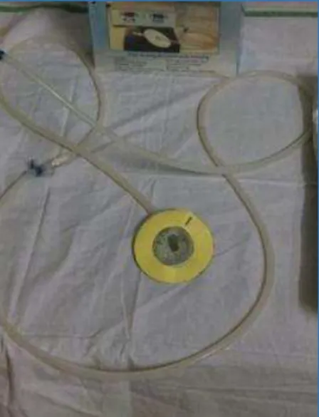
Tubing connected from dressing to tubing coming from VAC suction apparatus (Figure 6).
Fig. 6: Suction tube to connect to VAC Machine
The settings were adjusted in the VAC machine. The pressure adjusted to negative suction of 125-150 mmHg. Intermittent negative pressure was applied as machine is turned on for 5 minutes and off for 3 minutes. Upon application of the suction, the foam collapses into the wound and the negative pressure absorbs the fluid through the wound (Figure 7).
Fig. 7: VAC Machine
Dressing change was done once in 48 hrs. irrespective of the wound condition. Evidence of wound healing was confirmed by the amount of granulation tissue, culture and sensitivity, CRP and clinical findings. The initial size of the wound before placement of VAC was approximately calculated by placing a transparent sheet as close to the wound as possible. The edges of the wound were traced over the transparent sheet (Figure 8). The transparent sheet containing the wound imprint was placed over a 1 cm2 graph paper. The number of half squares, squares which were more than half and full squares were counted which determines the size of the wound (Figure 9). The same procedure was followed after every VAC dressing.
Once the wound showed evidence of healing or good granulation tissue, a split skin grafting or secondary suturing of the wound was carried out. Cases were followed 10 days after discharge.
OBSERVATIONS AND RESULTS: 15 patients aged between 16-72 years of age group with infected wounds treated with VAC therapy. 12 males and 3 females are included in the study. Large number of cases in our study comprise of infected wounds of thigh (5 cases, 33.33%), leg (6 cases, 40%), back (2 cases, 13.33%) and foot (2 cases, 13.33%). In our study, VAC dressing was applied for 6 days (6 cases, 40%), 8 days (5 cases, 33.33%), 4 days (2 cases, 13.33%), 10 days (2 cases, 13.33%). 53% (8) of the cases required less than 4 dressings and 47% (7) of the cases required more than 4 dressings for wound closure. Duration of stay in hospital was less than 1 month in 33% (5) of the cases and more than 1 month in 67% (10) of the cases. Mean wound size was 64 cm2 before the application of VAC dressing which reduced to a mean of 38 cm2 at the completion of VAC therapy. Wound closure was achieved by secondary suturing in 46.66% (7) of the cases and split skin grafting in 53.33% (8) of the cases. Wound healing was excellent in 73% (11) of the cases and good in 27% (4) of the cases.
Jebmh.com
Original Article
J. Evid. Based Med. Healthc., pISSN- 2349-2562, eISSN- 2349-2570/ Vol. 3/Issue 73/Sept. 12, 2016 Page 3956
Case 2:
Case 3:
DISCUSSION: The pathogenesis of infection in fractures fixation devices is related to micro-organisms, which grow in biofilm, and therefore its eradication is difficult.6 There are various mechanisms by which NPWT facilitates wound healing.7 It facilitates fluid removal from the wound, thereby reducing the wound size. By increasing the blood flow, NPWT helps early granulation tissue formation.
NPWT known to reduce bacterial counts, although remain colonised with organisms.8 Wounds covered with NPW dressing are completely isolated from the environment, thereby reducing cross infection. Application of negative pressure wound therapy causes changes in the tissues in the vicinity of its application. As interpretation with results, VAC therapy is effective mode of adjuvant therapy for the management of infected wounds.
CONCLUSION: VAC has been proven to be a reliable method of treating a variety of infected wounds. It greatly increases the rate of granulation tissue formation and lowers bacterial counts to accelerate wound healing. It can be used as a temporary dressing to prepare wounds optimally prior to closure or as a definitive treatment for nonsurgical and surgical wounds. The VAC device is well tolerated with few complications or contraindications and is playing an ever-expanding role in wound care. The limitation of this study is that this was a single-arm, prospective, observational study with no control group. Comparison to historical data is often difficult since different criteria are being used and no simple one-to-one comparison is possible. One more limitation is a short followup period.
REFERENCES
1. Jerome D. Advances in negative pressure wound therapy: the VAC instill. J Wound Ostomy Continence Nurs 2007;34(2):191-194.
2. Hunter JE, Teot L, Horch R, et al. Evidence based medicine: vacuum assisted closure in wound care management. Int Wound J 2007;4(3):256-269. 3. Moues CM, Vos MC, Bemd GJ, et al. Bacterial load in
relation to vacuum assisted closure wound therapy: a prospective randomised trial. Wound Repair Regen 2004;12(1):11-17.
4. Mullner T, Mrkonjic L, Kwasny O, et al. The use of negative pressure to promote the healing of tissue defects: a clinical trial using the vacuum sealing technique. Br J Plast Surg 1997;50(3):194-199. 5. Cresti S, Ouaissi M, Sielenzneff I, et al. Advantage of
Vacuum assisted closure on healing of wound associated with omentoplasty after abdominoperineal excision: a case report. World J Surg Oncol 2008;6:136.
6. Trampuz A, Zimmerli W. Diagnosis and treatment of infections associated with fracture-fixation devices. Injury 2006;37(suppl 2):S59-66.
7. Argenta LC, Morykwas MJ. Vacuum assisted closure: a new method for wound control and treatment: clinical experience. Annals of Plastic Surg 1997;38(6):563-576.