K
Channel Inhibition Differentially Regulates
Migration of Intestinal Epithelial Cells in
Inflamed vs. Non-Inflamed Conditions in a
PI3K/Akt-Mediated Manner
Sebastian Zundler1,2, Massimiliano Caioni1, Martina Müller1, Ulrike Strauch1, Claudia Kunst1, Gisela Woelfel1*
1Department of Internal Medicine I, Regensburg University Medical Center, Regensburg, Germany, 2Department of Medicine 1, University of Erlangen-Nuremberg, Kussmaul Campus for Medical Research & Translational Research Center, Erlangen, Germany
Abstract
Background
Potassium channels have been shown to determine wound healing in different tissues, but their role in intestinal epithelial restitution–the rapid closure of superficial wounds by intesti-nal epithelial cells (IEC)–remains unclear.
Methods
In this study, the regulation of IEC migration by potassium channel modulation was explored with and without additional epidermal growth factor (EGF) under baseline and interferon-γ
(IFN-γ)-pretreated conditions in scratch assays and Boyden chamber assays using the
intestinal epithelial cell lines IEC-18 and HT-29. To identify possibly involved subcellular pathways, Western Blot (WB)-analysis of ERK and Akt phosphorylation was conducted and PI3K and ERK inhibitors were used in scratch assays. Furthermore, mRNA-levels of the potassium channel KCNN4 were determined in IEC from patients suffering from inflamma-tory bowel diseases (IBD).
Results
Inhibition of Ca2+-dependent potassium channels significantly increased intestinal epithelial
restitution, which could not be further promoted by additional EGF. In contrast, inhibition of KCNN4 after pretreatment with IFN-γled to decreased or unaffected migration. This effect
was abolished by EGF. Changes in Akt, but not in ERK phosphorylation strongly correlated with these findings and PI3K but not ERK inhibition abrogated the effect of KCNN4 inhibi-tion. Levels of KCNN4 mRNA were higher in samples from IBD patients compared with controls.
OPEN ACCESS
Citation:Zundler S, Caioni M, Müller M, Strauch U, Kunst C, Woelfel G (2016) K+Channel Inhibition
Differentially Regulates Migration of Intestinal Epithelial Cells in Inflamed vs. Non-Inflamed Conditions in a PI3K/Akt-Mediated Manner. PLoS ONE 11(1): e0147736. doi:10.1371/journal. pone.0147736
Editor:Mirjam M Zegers, NCMLS, Radboud University Nijmegen Medical Center, NETHERLANDS
Received:April 8, 2015
Accepted:January 7, 2016
Published:January 29, 2016
Copyright:© 2016 Zundler et al. This is an open access article distributed under the terms of the
Creative Commons Attribution License, which permits unrestricted use, distribution, and reproduction in any medium, provided the original author and source are credited.
Data Availability Statement:All relevant data are within the paper and its Supporting Information files.
Conclusions
Taken together, we demonstrate that inhibition of KCNN4 differentially regulates IEC migra-tion in IFN-γ-pretreatedvs. non pretreated conditions. Moreover, our data propose that the
PI3K signaling cascade is responsible for this differential regulation. Therefore, we present a cellular model that contributes new aspects to epithelial barrier dysfunction in chronic intestinal inflammation, resulting in propagation of inflammation and symptoms like ulcers or diarrhea.
Introduction
A single layer of epithelial cells is lining the surface of the gastrointestinal tract. It plays an indispensable role in the transport of nutrients, ions and water, but also exerts barrier function against potentially harmful agents in the lumen, e.g. bacteria or toxic dietary products. Access of such antigens through the intestinal epithelium can result in initiation of inflammatory cas-cades. Thus, preserving the integrity of the barrier is essential.
Superficial lesions of the IEC layer–as they occur in infection or inflammation–are quickly
restored by a mechanism involving rapid restitution of the wound through dedifferentiation and migration of adjacent cells to the denuded area, followed by their proliferation and redif-ferentiation [1], [2].
It has been shown that this process of intestinal epithelial wound healing is promoted by a
number of growth factors. For example, EGF upregulates epithelial migration in intestinal [3]
and colonic cells [4] and thereby also quickly reduces damage of the epithelium as assessed by
transepithelial resistance [5]. FGF similarly influences intestinal epithelial restitution and acts
in a TGF-β-dependent manner [6]. TGF-βitself seems to selectively promote migration and
not proliferation of IEC [7] and mediates the influence of other agents regulating wound
heal-ing of IEC [8]. Important regulatory intracellular pathways involved in the effects of these
sti-muli include the NF-κB-, PI3K- and ERK-cascades: Afterin vitrowounding of IEC, NF-κB is
activated in an EGFR-dependent manner and its blockade leads to inhibition of restitution [9].
PI3K phosphorylation has been shown to be essential for wound healingin vitroby mediating
downstream GSK3βphosphorylation [10]. ERK activation occurs rapidly after mechanical
wounding of IEC [11] and is needed for chemokine receptor-dependent promotion of
restitu-tion [12].
According to findings in multiple cell lines, potassium channels also contribute to the
regu-lation of cell migration and epithelial wound healing [13]. One study reported a promotion of
IEC wound healing by blockade of potassium channels [14]. Blocking of KCNN4 in lung
den-dritic cells leads to a decreased response to chemotactic stimuli [15]. Alveolar epithelial repair
after mechanical wounding is also mediated by potassium channels, which interact with EGF
[16]. Investigations on potassium channels in rabbit corneal epithelial cell proliferation
simi-larly revealed an interaction with EGF [17]. Despite these findings, little is known about the
exact role of potassium channels and their interplay with growth factor-dependent signaling cascades in intestinal epithelial restitution.
Intestinal epithelial wound healing is of special interest as epithelial barrier dysfunction in genetically predisposed patients is believed to be one key element of the pathogenesis of IBD like Crohn's disease (CD) and ulcerative colitis (UC), although their precise etiology remains unclear. Factors contributing to barrier dysfunction include disruption of paracellular tight
Competing Interests:The authors have declared that no competing interets exist.
Abbreviations:1-EBIO,
1-ethyl-2-benzimidazolinone; BCA, bicinchoninic acid; CD, Crohn's disease; cf., confer; ChTx, charybdotoxin; Clt, clotrimazol; CRC, colorectal carcinoma; ECL, enhanced chemiluminescence; EGFR, epidermal growth factor receptor; IBD, inflammatory bowel disease; IbTx, iberiotoxin; IEC, intestinal epithelial cells; IFN-γ, interferon-gamma; PI3K,
junctions [18], reduced secretion of antimicrobial peptides [19] and loss of epithelial cells,
thereby leading to uncontrolled bacterial translocation [20].
CD is characterized by a predominant T helper 1 (Th1)-immune response, in which
proin-flammatory cytokines like IFN-γ, TNF-αand IL-12 play a central role. It has been
demon-strated that IFN-γis elevated in the mucosa and serum in CD [21] and is a key factor in
different murine models of colitis [22], [23]. It is therefore thought to be a critical cytokine
con-tributing to inflammation in CD [24].
We have recently demonstrated that IFN-γalters EGFR-downstream signaling [25].
More-over, IFN-γis able to impair other essential cellular functions such as ion transport and tight
junction organization [26], [27]. However, we have an incomplete understanding of the role of
IFN-γin intestinal epithelial wound healing.
In the present study we aimed to characterize the impact of potassium channels on
intesti-nal epithelial wound healing. We investigated a potential influence of IFN-γand sought to
identify possible downstream targets in IEC cell lines. We show that IEC migration is
differen-tially regulated by KCNN4 after pretreatment with IFN-γcompared to not pretreated
condi-tions. Moreover, we demonstrate that the PI3K-pathway could account for this finding. We thereby provide new insights into deregulation of epithelial cellular functions in chronic intes-tinal inflammation.
Materials & Methods
Culture of cell lines
The newborn rat non-transformed IEC line IEC-18 (ATCC CRL-1589; passages 25–55) was a
kind gift of Dr. Thomas Karrasch, Regensburg, who purchased the cell line from ATCC
(Manassas, VA, USA) in 2007. It is derived from ileal crypt cells [28]. IEC-18 were cultured in
DMEM with 4.5 g/L glucose and L-glutamine (PAA, Pasching, Austria) augmented with 5%
FCS (Sigma Aldrich, Steinheim, Germany), 100 U/mL penicillin, 100μg/mL streptomycin
(PAA) and 0.7 mMol bovine insulin (Sigma-Aldrich) in a humidified atmosphere at 37°C with
10% CO2[29], [30]. Subcultivation of IEC-18 was carried out twice per week in a ratio of 1:6.
The human IEC line HT-29 [31] (passages 7–12) was generously provided by Dr. Rocío
Lopez-Posadas, Erlangen, in 2015. HT-29 were cultured in DMEM with 4.5 g/L glucose and pyruvate (Life technologies, Darmstadt, Germany) with 10% FCS, 1% penicillin/streptomycin and 1% amphotericin and were subdivided 1:10 once per week. Fully supplemented cell culture medium was routinely filtered through steriflip filters (Millipore, Billerica, MA).
Scratch wound healing assays
For scratch assays of IEC-18, 400000 cells were seeded in one-well LabTek chamber slides (glass bottom; Fisher Scientific, Schwerte, Germany) 48 h prior to wounding and incubated with fully supplemented medium for the next 24 h until they had reached confluency. Cells
were then starved in serum-reduced medium (1% FCS, cf. [10]) with (“inflammatory
condi-tions”) or without (“baseline conditions”) 100 ng/mL (1000 U/mL) recombinant rat IFN-γ
(PromoCell, Heidelberg, Germany) (cf. [25]). Another 24 h later, wounding was performed
using a 10μL micropipette tip as described elsewhere [10], [14], [16], [32]. To detach cells
adhering to the wound edges the cell layer was carefully rinsed with PBS (PAA) before new serum-reduced medium with or without potassium channel modulators and/or 5 nM recombi-nant rat EGF (Peprotech, Hamburg, Germany) was added. The following potassium channel
modulators were used in the indicated concentrations: 5 mM Barium2+, 100 nM Iberiotoxin
(IbTx), 200 nM Charybdotoxin (ChTx), 10μM Clotrimazole (Clt; all from Sigma-Aldrich),
[14], [33], [34]). The latter two modulators were dissolved in DMSO (Sigma-Aldrich) leading to a final DMSO concentration of 0.25% v/v (Clt) and 1% v/v (1-EBIO), respectively, in experiments.
To determine the wound healing course, the chamber slide was placed into the prewarmed
(37°C), humidified and equilibrated (5% CO2) incubator of an Axiovert microscope and serial
images of a wound area between 120000μm² and 200000μm² were taken every 15 minutes for
the following six hours at 200-fold magnification. Imaging was controlled by AxioVision soft-ware and done with Axiocam Mr5c (all from Zeiss MicroImaging, Göttingen, Germany).
Scratch-wound assays with HT-29 cells were performed in 12 well cell culture plates (Grei-ner Bio-One, Frickenhausen, Germany). Following 24h of starvation in serum-reduced
medium with or without 100 ng/ml recombinant human IFN-γ(Peprotech), cell layers were
wounded as described above, serum-reduced medium with or without potassium channel
modulators and with or without recombinant human EGF (Peprotech) and/or 10μM of the
PI3K inhibitor Ly294002 or 10μM of the ERK inhibitor 3-(2-Aminoethyl)-5-((4-ethoxyphe-nyl)methylene)-2,4-thiazolidinedione-hydrochloride (both from Sigma-Aldrich) was added and images of the wounds were taken with inverted microscopes (Leica, Wetzlar, Germany) right after wounding (t = 0h) and after 6h.
Analyses of the wound healing experiments were performed using AxioVision and ImageJ (NIH, Bethesda, MD) software by measuring original and remaining wound areas. If necessary, contrast and/or brightness of the pictures were digitally optimized.
Migration assays in modified Boyden chambers
Lower wells of a modified Boyden Chamber (Receptor Technologies, Royal Leamington Spa,
Great Britain) were filled with 30.2μL serum-reduced medium with or without 10μM Clt and
separated from upper wells by a polycarbonate membrane with 8μm pores (Osmonics, Moers,
Germany). For adjustment of pH the chamber was transferred into an incubator for 30 min. Confluent IEC-18 were starved in serum-reduced medium for 24 h with or without 100 ng/
mL IFN-γ, washed with PBS and collected with Trypsin/EDTA (PAA). After centrifugation
cells were resuspended in serum-reduced medium at a concentration of 200000 cells/mL and 50μL of the suspension were pipetted in the upper compartment of the prepared Boyden Chamber.
After a migration period of six hours, the upper surface of the membrane was washed from attached cells with PBS and cells having migrated to the lower surface were fixed and stained with a Hemacolor kit (Merck, Darmstadt, Germany).
For cell counting, membranes mounted on a slide were placed under a microscope (Leica) and migrated cells in the center of the imprint of each well were counted at 200-fold
magnification.
Western Blot analysis
IEC-18 were seeded in cell culture dishes (Corning, Corning, NY) and starved for 24 h with or
without 100 ng/mL IFN-γafter they had reached confluency. Using a multichannel pipette
(Eppendorf, Hamburg, Germany) 16 perpendicular wounds were scratched into the cell layer
with 10μL pipette tips. Like this, an estimated number of 20% of the total cells were within a
range of 250μm from the wound margins. Afterwards, fresh serum-reduced medium with or
without 10μM Clt or 600μM 1-EBIO, each with or without 5 nM EGF was added. After 30,
were lysed in lysis buffer as described previously [25] and protein concentration was measured with a bicinchoninic acid (BCA)-protein quantification assay (Sigma-Aldrich). Volumes
con-taining 7.5μg of protein each were further diluted with aqua dest. and 6 x Laemmli buffer,
boiled for five minutes and mounted on a 4–12% polyacrylamide gel to resolve proteins by
electrophoresis. Proteins were then transferred onto a nitrocellulose membrane filter paper.
NuPAGE1
instruments and reagents were used according to the manufacturer's instructions (all from Invitrogen, Carlsbad, CA). Membranes were blocked with 5% BSA (Biomol, Ham-burg, Germany) in Tris-buffered saline with 0.1% Tween 20 (Sigma-Aldrich) (TBST) overnight.
Afterwards, membranes were incubated with the primary antibody in 5% BSA in TBST for one hour and washed six times with TBST prior to one hour of incubation with an HRP-conju-gated secondary antibody. Membranes were washed another six times and exposed to 5 mL enhanced chemiluminescence (ECL)-solution (2.5 mM luminol, 0.1 M Tris-HCl and 0.4 mM
p-coumarin acid–all from Sigma-Aldrich–in aqua dest.) mixed with 30% 1.53μL hydrogen
peroxide (Merck) for five minutes. Immunoreactive proteins were detected and densitometri-cal analyses were carried out with Image Quant software (Molecular Dynamics, Sunnyvale, CA).
The following primary antibodies were used and dissolved as indicated by the manufacturer: anti-Akt, anti-phospho-Akt (Ser473), anti-p44/42 MAPK, anti-phospho-p44/42 MAPK
(Thr202/ Tyr204) (all from Cell Signaling, Danvers, MA) and for loading control anti-β-actin
(Millipore, Billerica, MA). Secondary antibodies were anti-rabbit-IgG and anti-mouse-IgG (both from Santa Cruz, Heidelberg, Germany). For multiple use, membranes were stripped up to three times using Re-blot-plus strong solution (Chemicon, Temecula, CA).
RNA isolation and Real Time-PCR Analysis
Surgical specimens and endoscopic biopsies from IBD and control patients were collected after written informed consent. The procedure was approved by the Ethics Committee of the Uni-versity of Regensburg (record number 00/14) and performed according to the Declaration of Helsinki. IEC were isolated by accurate mechanical detachment of the mucosa from the sub-mucosal layer with a dissecting set. The mucosa was then subjected to several washing steps and manual agitation after treatment with 2 mM EDTA (Carl Roth, Karlsruhe, Germany).
Following the manufacturer's instructions, RNA was isolated and purified from cells with the RNeasy Kit (Qiagen, Hilden, Germany). By measuring absorbance at 260 nm and 280 nm RNA concentration and purity was determined. cDNA was synthesized using Affinity script reagents (Agilent, Böblingen, Germany) along the manufacturer's recommendations.
Real-Time PCR was performed in triplicates for each sample with Brilliant II Mastermix for
qPCR with high ROX (Agilent) in an AbiPrism1
Sequence Detector with SDS software (both from Applied Biosystems, Carlsbad, CA).
Primer sequences were as follows: human KCNN4 forward5'-CCC TCA TCA AAA ACA
CTC TCA CTA TG-3', reverse5'-TCC AGT CGC CTG CAC TTG-3', probe5'-FAM—TGC
TAT GGA CGA CCT CCA GCT CTC AGT T—TAMRA-3', human KCNQ1 forward5'-CGC
ATG GAG GTG CTA TGC T-3', reverse5'-GGC CTT CCG GAT GTA GAT CTT-3', probe
5'-FAM—AGA ACC CCG ACT CCT CCA CCT–TAMRA-3'(all from Eurofins MWG Operon,
Ebersberg, Germany). Human GAPDH mix was used as endogenous control (Applied Biosystems, Carlsbad, CA). PCR conditions were two minutes of incubation at 50°C, followed by ten minutes at 95°C and 40 cycles of 15 seconds at 95°C for denaturation and 60 seconds at 60°C for annealing and extension each. Analysis of the results was performed by the ddCT
Statistical analysis
Results of scratch wounding experiments and Boyden chamber assays are presented in scatter plots showing individual data points and mean with SEM. WB results are given as x-fold expression in relation to unwounded controls. Data from real-time PCR are shown as x-fold expression relative to the mean of the control group. Graphs were produced with GraphPad Prism (GraphPad Software, La Jolla, CA). Statistical comparison of groups was also performed
with GraphPad Prism using ANOVA with appropriate post hoc tests or two-tailed student’s
t-test where applicable. P-values<0.05 were considered significant. Significance levels displayed
in the graphs arep<0.05,p<0.01,p<0.001.
Results
Restitution of scratch-wounded IEC-18 monolayers is marked by
migration of adjacent cells
In order to explore the mechanisms involved in gastrointestinal epithelial wound healing and to investigate a possible contribution of potassium channels to its regulation, we examined wound healing responses of IEC lines after mechanical injury.
It is well known that migration is a cornerstone of intestinal epithelial restitution [2]. In an
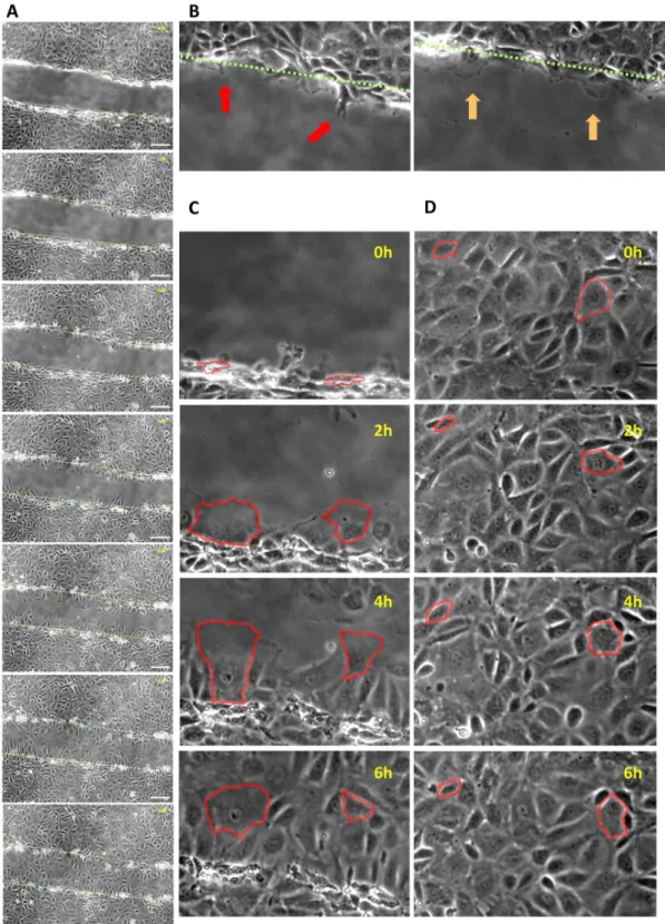
attempt to demonstrate this process, IEC-18 monolayers were wounded and serially imaged for six hours. As expected, we observed that cells in close neighborhood of the wound closed the gap by collectively migrating into the denuded area and undergoing considerable
morpho-logical changes including the protrusion of filopodia and pseudopodia (Fig 1A–1C; S1 Video).
To the contrary, distant cells did barely move or change their shape and were thus not
impli-cated in wound closure (Fig 1D;S1 Video). Moreover, cell proliferation did not relevantly
con-tribute to epithelial restitution as the absolute number of cells only marginally changed within
this timeframe (S1A Fig). This was confirmed by cell density measurements, which revealed
that the cell density at the wound margin decreased over the course of the experiments due to cell migration with accompanying increase in cell size, while the cell density at distant locations
remained approximately constant (S1B Fig). Hence, corresponding to established models [2],
proliferation seems to be dispensable for early stages of restitution.
Inhibition of Ca
2+-dependent potassium channels enhances wound
healing
To test the yet barely investigated hypothesis that potassium channels might be involved in
intestinal epithelial restitution as they are in the restitution of other tissues [13], [16] we
per-formed scratch wounding assays of IEC-18 monolayers in the presence of different potassium channel modulators. Mean wound closure was determined by the area recovered by the cell
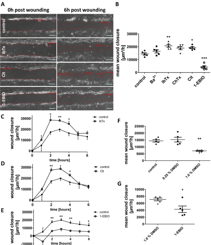
sheet within six hours (Fig 2A) and was found to be significantly higher after treatment with
IbTx (+43.8 +/- 7.8%) and Clt (+33.5 +/- 5.2%) than under control conditions. IbTx inhibits
the Ca2+-dependent large-conductance potassium channel KCNMA1, while Clt inhibits the
Ca2+-dependent intermediate-conductance potassium channel KCNN4. Addition of ChTx,
which inhibits KCNMA1 and KCNN4 as well as some voltage-gated potassium channels, resulted in a close to significant effect on wound closure (+31.4 +/- 8.6%, p = 0.06). In contrast,
1-EBIO, an activator of KCNN4 and Ca2+-dependent small-conductance potassium channels,
yielded a reduced rate of wound healing (-70.4 +/- 6.5%). Ba2+, which unspecifically inhibits
constitutively active potassium channels, had no significant effect (Fig 2B).
Fig 1. Restitution of scratch-wounded IEC-18 monolayers is marked by migration of adjacent cells.A: representative sequence of images taken every hour (from top to bottom) beginning directly after mechanical wounding of a confluent IEC-18 monolayer. The approximate location of the initial wound margin (top picture) is indicated by a green dashed line and also displayed in subsequent images. Scale bar: 100μm. B: Representative images of migrating cells
sending ahead filopodia (red arrows) and pseudopodia (orange arrows). C: Exemplary sequence of processes at the wound margin. Two adjacent cells are edged in red and shown at the indicated time points. Over time they migrate into the denuded area undergoing considerable morphological changes. D: Exemplary sequence of events within the cell sheet distant from the wound. Two representative cells are edged in red and show only bare movements or changes in shape over six hours.
Fig 2. Regulation of intestinal epithelial wound healing by potassium channel modulation.Wound healing of IEC-18 within six hours after mechanical injury. A: Representative images of wounds 0 and 6 hours after wounding in the presence of the indicated potassium channel modulators. Scale bar: 100μm.
B: Quantitative analysis of wound healing of IEC-18 incubated with different potassium channel modulators (n = 5). While IbTx and Clt cause an increase in intestinal epithelial wound healing response, wound closure is significantly reduced after administration of 1-EBIO. C-E: Time course of wound healing after administration of IbTx, Clt and 1-EBIO (n = 5). F: Comparison of different solvent concentration applied (n = 5). While 0.25% (v/v) DMSO does not affect wound closure, 1% (v/v) DMSO significantly retards wound closure. G: Direct comparison of 1-EBIO with its solvent (n = 5). Reduction in wound healing by 1-EBIO is also significant vs. 1% DMSO.
healing rates during the second, third and fourth hour after wounding. The 1-EBIO dependent
growth rates were reduced almost over the complete course of the experiment (Fig 2C–2E).
In order to find out, whether results could be distorted by the necessary use of DMSO as sol-vent for Clt (0.25%) and 1-EBIO (1%), controls were performed for 0.25% (v/v) and 1.0% (v/v)
DMSO in starvation medium (Fig 2F). While 0.25% DMSO had no impact on wound closure
(+6.1 +/- 11.9%, p = 0.90), 1% DMSO caused a significant impairment of wound healing (-50.7 +/- 2.9%). However, when compared to 1% DMSO, 1-EBIO still led to a reduced wound
clo-sure (-39.9 +/- 13.1%;Fig 2G) suggesting that 1-EBIO intrinsically downregulates wound
healing.
Preincubation with IFN-
γ
inverts effects of KCNN4 inhibition on intestinal
epithelial wound healing
We sought to explore how KCNN4-dependent intestinal epithelial wound healing is regulated
under inflamed conditions. Thus, IEC-18 cells were pretreated with 100 ng/mL IFN-γ24 h
before wounding and the impact of Clt and 1-EBIO was studied.
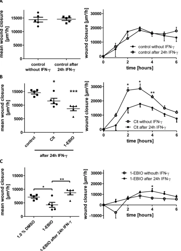
Neither overall restitution (+1.1 +/- 3.5%) nor time course of wound closure was altered by
IFN-γpretreatment under control conditions (Fig 3A). Addition of Clt exhibited a significantly
reduced wound closure (-20.9 +/- 6.9%) differing from Clt-dependent healing under baseline conditions mainly in the second, third and fourth hour of the experiments. The administration
of 1-EBIO also caused an impaired growth rate (-40.3 +/- 5.3%;Fig 3B).
Interestingly, the 1-EBIO-dependent wound healing rate was significantly higher than
with-out IFN-γpretreatment (+103.9 +/- 18.0%) and not reduced in relation to 1% DMSO alone
(+22.4 +/- 10.8%;Fig 3C) indicating that the pure action of 1-EBIO might also be reversed
under inflammatory conditions.
EGF does not further increase wound healing upon KCNN4 inhibition,
but completely abrogates the reversing effect of IFN-
γ
pretreatment on
KCNN4-dependent wound closure
As EGF and corresponding signaling pathways have been shown to influence migration and
wound healing in several types of epithelial cells [36], [37] we investigated the hypothesis that
these could also be implicated in the potassium channel-modulated wound healing response of IEC. Therefore, 5 nM EGF were added additionally to potassium channel modulators with and
without prior IFN-γincubation.
EGF alone significantly increased wound healing compared to control conditions (+61.5
+/-20.1%). Following 24h IFN-γpretreatment, rise in wound closure after EGF administration
was close to significant (+44.1 +/- 9.8%, p = 0.07). Regarding the time course of wound healing, marked differences in the second, third and fourth hour after wounding were responsible for
the effect of EGF (Fig 4A).
The combination of EGF with Clt (Fig 4B) did not further augment the Clt-dependent
growth rate observed under baseline conditions (+1.3 +/- 11.9%). After pretreatment with
IFN-γwound closure in response to Clt together with EGF was similarly raised. Thus, the
inverting effect of IFN-γpretreatment on Clt-dependent wound healing was completely
abro-gated by additional administration of EGF (+70.5 +/- 11.9%).
The addition of EGF together with 1-EBIO (Fig 4C) caused significantly higher wound
heal-ing rates than measured with 1-EBIO alone (+124.8 +/- 59.8%). Preincubation with IFN-γand
Fig 3. Differential regulation of KCNN4-dependent intestinal epithelial wound healing in response to preincubation with IFN-γ.Wound healing of IEC-18 within six hours after mechanical injury. A: Comparison of wound healing of IEC-18 under control conditions without and with 24 h-pretreatment with IFN-γ(n = 5). Both the mean wound closure over six hours (left panel) and the time course profile of wound healing (right panel) are similar. B: Wound healing
of IEC-18 after IFN-γpretreatment and incubation with Clt and 1-EBIO (n = 5). KCNN4-inhibition by Clt results in significantly reduced rates of wound closure
We therefore conclude that accelerated wound healing after inhibition of KCNN4 could be due to an activation of signaling pathways that are also activated by binding of EGF to its recep-tor EGFR, because wound closure cannot be further stimulated by additional application of EGF. Consistent with that, EGF raised growth rates reduced by 1-EBIO-dependent activation of KCNN4.
Furthermore, retardation of wound healing after KCNN4 inhibition following IFNγ
-pre-treatment could be explained by a perturbation of this linkage to EGFR-dependent pathways
by IFN-γ, as their direct activation through EGF is able to reestablish enhanced wound healing.
Of note, IbTx-dependent epithelial restitution did not differ between baseline and
inflam-matory conditions (S2 Fig) suggesting that different potassium channels play different
func-tions in regulating wound healing.
IEC migration is differentially affected by KCNN4 modulation
Although scratch assays are commonly accepted as a validin vitromodel of cell migration and
we have shown above that proliferation does not play a relevant role within six hours after wounding, we aimed at further specific assessment of IEC migration. Thus, Boyden chamber assays were performed.
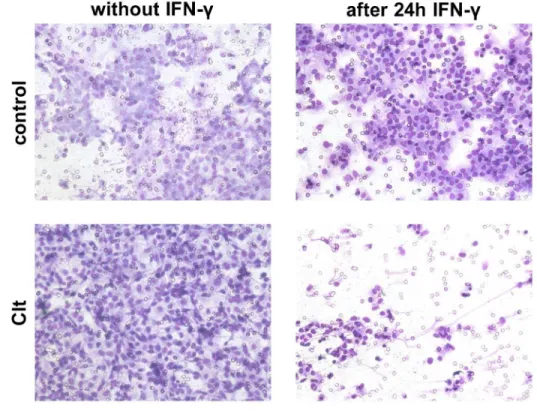
In concrete terms, cells were starved with or without 100 ng/mL IFN-γfor 24 h and allowed
to migrate through a polycarbonate membrane into compartments with starvation medium or starvation medium with Clt during six hours.
Without proinflammatory pretreatment, migration was increased in response to Clt (+34.8 +/- 8.3%) vs. control conditions.
Following preincubation with IFN-γ, migration under control conditions was not altered in
comparison to not pretreated controls (-18.0 +/- 12.9%). Clt (-66.7 +/- 34.7%) yielded a
signifi-cantly decreased number of migrating cells vs. Clt without IFN-γ-pretreatment (Fig 5).
This finding parallels scratch assay results and therefore further supports the notion, that
altered migration in response to KCNN4 modulation and IFN-γpretreatment is a major reason
for the differential regulation of wound healing observed in the scratch assays.
Changes in PI3K- but not in MAPK-signaling pathway correlate with
differential regulation of intestinal wound healing by Clt
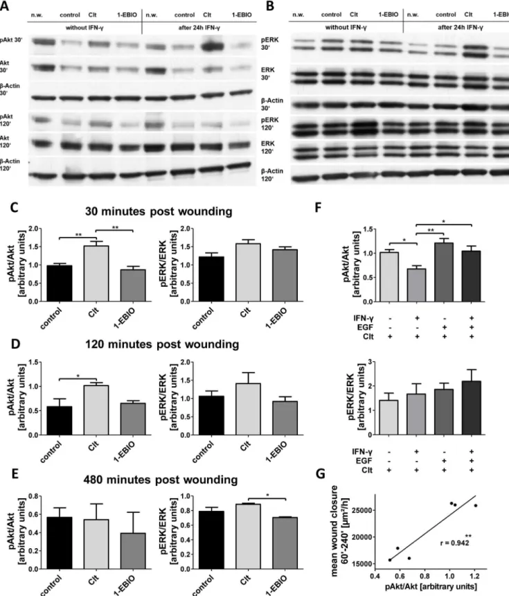
We conducted WB analysis (Fig 6A+6B) in order to explore the hypothesis that
EGFR-acti-vated pathways might be involved in the observed differential regulation of intestinal wound healing in response to Clt treatment under inflamed conditions. Therefore, phosphorylation of Akt and ERK-1/2 as key kinases of the PI3K- and MAPK-pathway was determined in the con-stellations indicated above by correlation of phosphorylated protein to total protein.
Under baseline conditions (Fig 6C–6E), Akt phosphorylation was enhanced by Clt vs.
con-trol (+55.5 +/- 12.8%) and vs. 1-EBIO (+75.7 +/- 32.4%) 30 minutes after wounding. ERK phosphorylation was not altered by these potassium channel modulators.
Two hours post wounding, increased Akt phosphorylation vs. control could once more be observed following addition of Clt (+74.3 +/- 10.4%). Again, there were no differences in ERK phosphorylation.
second, third and fourth hour (right panel). C: Comparison of dependent wound closure to 1% DMSO (left panel, n = 5) and time course of 1-EBIO-dependent restitution with and without IFN-γpreincubation (right panel, n = 5). After proinflammatory treatment, 1-EBIO-dependent wound closure is higher
than under baseline conditions.
Fig 4. Potassium channel-dependent wound healing of IEC-18 with and without 5 nm EGF under baseline or IFN-γpretreated conditions.Wound healing of IEC-18 within six hours after mechanical injury. A: Influence of EGF on intestinal epithelial wound healing (n = 5). EGF increases wound healing both under baseline and inflammatory conditions (left panel). Right panel: Time course of restitution upon EGF treatment compared to control showing marked differences in the second, third and fourth hour after wounding. B: Impact of Clt with and without additional EGF after or without IFN-γpretreatment
At 480 minutes after wounding, Akt phosphorylation was on basal levels again. Clt-depen-dent ERK phosphorylation was slightly elevated in contrast to 1-EBIO (+25.8 +/- 3.7%).
Therefore, the Clt-dependent increase of wound closure rates was paralleled by a rise of phosphorylated Akt but not ERK after 30 and 120 minutes. We subsequently particularly focused on the WB findings 120 minutes post wounding, as time course analysis of the scratch assays had shown that differences in wound healing rates were mainly due to different growth
rates in the second, third and fourth hour after wounding (Fig 2D).
In the presence of Clt (Fig 6F), IFN-γpretreated cells displayed significantly reduced Akt
phosphorylation 120min after wounding compared to not preincubated cells (-33.5 +/- 16.2%). Addition of EGF in combination with Clt under baseline conditions did not increase Akt
phos-phorylation vs. Clt alone. After preincubation with IFN-γ, additional EGF was able to
completely abolish the effect of Clt on Akt phosphorylation as levels were significantly raised (+54.7 +/- 26.7%) and comparable to those without preincubation. ERK phosphorylation was not significantly altered for either of these conditions 120 minutes after wounding.
Thus, Akt phosphorylation after 120 minutes and wound healing growth rates showed a remarkable resemblance. To investigate this in more detail, we correlated Akt-phosphorylation
and mean wound closure 60–240 minutes after wounding in controls and Clt-involving
con-stellations. With a Pearson's coefficient of 0.942 (p = 0.005) this correlation turned out to be
very strong and statistically significant (Fig 6G). Though not causally proving, this strongly
argues for an involvement of the PI3K signaling cascade in the KCNN4-mediated regulation of intestinal epithelial wound healing.
Clt-mediated increase in wound healing of HT-29 cells is abrogated by
PI3K but not ERK inhibition
In an attempt to confirm and extend our results we performed an additional series of
experi-ments with the human intestinal epithelial cell line HT-29 (Fig 7A). Again, Clt increased
wound healing vs. controls in baseline but not in inflammatory conditions, while EGF did not
further increase Clt-mediated wound healing (Fig 7B). Once more, 1-EBIO-dependent
restitu-tion was lowered (S4 Fig).
Furthermore, HT-29 were additionally treated with the PI3K inhibitor Ly294002 or the ERK inhibitor 3-(2-Aminoethyl)-5-((4-ethoxyphenyl)methylene)-2,4-thiazolidinedione-hydrochloride. Both compounds were able to reduce the increased wound healing observed
upon EGF treatment back to baseline (Fig 7C). Finally, when cells were treated with Clt and
one of the inhibitors, Clt-mediated increase in restitution was unaffected by ERK inhibition
but significantly reduced by PI3K inhibition (Fig 7D) further backing our hypothesis of a
cru-cial role of the PI3K cascade in the mediation of KCNN4-dependent effects on intestinal epi-thelial restitution.
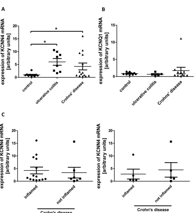
mRNA-levels of KCNN4 are augmented in IBD
Aiming to assess a possible role of KCNN4 in human IBD, we isolated IEC from surgical
speci-mens and endoscopic biopsies and quantified the expression of KCNN4 mRNA by TaqMan1
Real Time PCR.
In cells from macroscopically inflamed tissue from both UC (+531.5% +/- 137.2%; n = 8) and CD patients (+345.7% +/- 140.4%; n = 14) the expression was increased in comparison to after proinflammatory treatment. C: Impact of 1-EBIO with and without additional EGF after or without IFN-γpretreatment (n = 3–5). EGF promotes
1-EBIO-mediated wound healing both under baseline and inflammatory conditions.
Fig 5. Boyden chamber results.A: Representative images of stained cells attached to the lower surface of polycarbonate membranes after migration of IEC-18 towards medium containing Clt or not and after or without IFN-γpretreatment. B: Quantitative analysis of the number of cells migrated through a
polycarbonate membrane during six hours under the indicated conditions (n = 14–16). Clt-dependent migration is significantly higher under baseline than under inflammatory conditions.
control cells (n = 10), which were collected from macroscopically normal mucosa of patients
who underwent operation because of CRC or SD (Fig 8A). Moreover, mRNA-expression of
KCNQ1, a voltage-gated potassium channel, was determined in most of the samples to exclude an unspecific effect on potassium channels, but in no condition expression of KCNQ1 mRNA
was significantly altered (Fig 8B).
We were also able to collect some specimens from macroscopically unaffected gut tissue of CD patients (as determined by an independent pathologist) and compared KCNN4 mRNA
expression in these samples to those from inflamed tissue (Fig 8C). No significant difference
could be revealed both when comparing all samples and only those where inflamed and unin-flamed samples came from the same patient.
Discussion
The importance of intestinal epithelial wound healing for sustaining an intact intestinal barrier
in the context of inflammation has been emphasized by many authors [2], [20], [38]. Although
there is a large body of research suggesting an important role for potassium channels in wound healing and migration, their role in intestinal epithelial restitution is poorly defined. The same
applies for IFN-γ, which has been shown to impair many cellular functions [18], [25], [26],
[39], but up to now, its implications for wound healing are not completely understood.
Using the rat-derived non-transformed IEC line IEC-18 [28] in scratch assays as a
well-established model for migration-dominated IEC restitution [10], [15], [32], we showed that
inhibition of the Ca2+-dependent potassium channels KCNN4 by Clt and KCNMA1 by IbTx
caused a significant increase in wound healing response after mechanical injury. In contrast, activation of KCNN4 by 1-EBIO retarded wound closure. Modulation of constitutively active
(e.g. voltage-gated or inwardly rectifying) potassium channels by administration of Ba2+had
no effect. Further experiments with the human intestinal epithelial cell line HT-29 confirmed the effects observed with both Clt and 1-EBIO.
These modulators have been extensively used to study potassium channel effects in the
liter-ature. IbTx is a highly selective toxin with no known off-target effects [40]. Barium
unspecifi-cally inhibits a range of potassium channels through blockade of the K+selectivity filter as the
size of not hydrated Ba2+and K+ions are similar, but Ba2+binds more tightly due to its charge
[41]. Clt is a specific inhibitor of the Ca2+-dependent intermediate conductance channel
KCNN4 [33]. Although it has to be mentioned that Clt also inhibits cytochromes of the P450
family [42], this activity does not seem to play a role in the current study because cytochrome
inhibition brings along metabolic changes which would show their effects with some temporal delay. To the contrary, epithelial restitution apparently happened shortly after wounding.
Moreover, this limitation is shared by alternative KCNN4 blockers [43]. 1-EBIO has been
shown to increase KCNN4 activity. However, it is also known that it activates the small
con-ductance channels KCNN1-3 [44] and may target other molecules like adenylyl cyclase or
CFTR as well [34]. Thus, the corresponding results have to be interpreted more cautiously.
An increase in wound healing following KCNN4-inhibition by Clt was reported previously in the human colon cancer-derived cell lines T84 and CaCo2, whereas the inhibitory effect of
1-EBIO could not be demonstrated in this study [14] using a comparatively low concentration
phosphorylation is reduced by IFN-γpretreatment and reraised by additional EGF, thus correlating Clt-dependent wound closure in scratch assays. G:
Correlation of pAkt/Akt with mean wound closure in the second to fourth hour of the scratch assays in controls and Clt-involving constellations. Relative phosphorylation and wound closure rates (S1 Table) were plotted against each other and a regression line was computed. Pearson’s coefficient is 0.942 (p = 0.005).
Fig 7. Scratch-wounding of HT-29 cells confirms a differential regulation of KCNN4-dependent restitution with crucial contribution of PI3K but not ERK signaling.Confluent monolayers of HT-29 cells were wounded as explained in the Methods section and imaged right after wounding and after 6h. A: Representative images of the wounds at the indicated time points upon treatment with the indicated compounds without (left panels) or after (right panels) 24h pretreatment with IFN-γ. Wound size is denoted in red. Scale bar: 100μm. B: Quantitative analysis of wound healing under baseline conditions (left
wound closure is displayed as % difference vs. the mean of respective controls. Clt increases wound healing vs. controls in baseline but not inflammatory conditions. Clt in combination with EGF does not further increase EGF-dependent wound healing. C: Impact of PI3K (left panel) and ERK inhibition (right panel) on EGF-dependent intestinal epithelial restitution (n = 4–24). Both compounds revert the increase observed with EGF. D: Impact of PI3K (left panel) and ERK inhibition (right panel) on Clt-dependent intestinal epithelial wound healing (n = 4–24). The Clt-mediated increase in wound closure is abrogated by addition of the PI3K inhibitor but not the ERK inhibitor.
doi:10.1371/journal.pone.0147736.g007
Fig 8. KCNN4 mRNA expression in IBD.A: Relative levels of KCNN4 mRNA in IEC from controls (n = 10), UC (n = 8) and CD (n = 14). In both UC and CD the expression is increased vs. control. B: KCNQ1 mRNA levels are equal in UC (n = 5), CD (n = 11) and controls (n = 10) C: Comparison of KCNN4 expression in inflamed vs. uninflamed tissue of patients with CD. Left panel: Relative KCNN4 mRNA levels in all available samples (n = 14 inflamed, n = 7 not inflamed). Right panel: Comparison of KCNN4 levels in inflamed and uninflamed samples originating from the same patients (n = 5).
[34], [45]. Interestingly, hyperpolarization of cells by potassium-free medium and thereby
mimicking the effect of a potassium channel activator did impair wound healing [30].
Both IbTx and Clt as well as treatment with EGF exhibited significantly increased restitution rates in the second to fourth hour of the scratch assays. These effects early after wounding elic-ited the question whether the addition of potassium channel modulators might provoke the activation of intracellular signaling events to contribute to their impact on restitution. Signaling pathways like the PI3K cascade haven been identified as important mediators of intestinal epi-thelial wound healing and signaling molecule activation in response to appropriate stimuli was
demonstrated to be present early after wounding for up to five hours [10], [46], [47].
Interest-ingly, the addition of EGF, which among others activates PI3K and ERK signaling cascades, not only led to time-course profiles of wound closure comparable to those in response to IbTx Fig 9. Hypothesized impact of KCNN4 inhibition on wound-induced migration and differential regulation by IFN-γ.A: Blockade of KCNN4 by Clt (red
X) accelerates wound healing at least in part by transactivation of the PI3K pathway (red arrows). Additional EGF (blue) meets activated downstream signaling and exerts no additional effect (blue arrows). B: IFN-γpreincubation uncouples the postulated transactivation of PI3K signaling by KCNN4 inhibition
(orange line) leading to reduced migration upon KCNN4 blockade by Clt (red X). In this constellation EGF (blue) may activate downstream targets and enhance migration (blue arrows).
and Clt, but could also not further increase wound closure when added to Clt or IbTx. Thus, a transactivation of EGFR-dependent signaling pathways by potassium channel inhibition might account for these observations.
An interaction of different potassium channels with signaling molecules and especially growth factor receptors or their downstream signaling pathways has already been
demon-strated in several other cell types [48]. For example, inhibition of the channel KCNH2 in
leuke-mic cells blocked migration through a complex with VEGFR and ß1-integrin [49]. In human
T-cells, KCNA3 formed a complex with ß1-integrin, which seems to be implicated in
propaga-tion and retardapropaga-tion of migrapropaga-tion by activapropaga-tion or inhibipropaga-tion of the channel, respectively [50],
[51]. KCNMA1 has been shown to directly interact with FAK in human osteoblasts [52].
Besides these examples for conformational coupling, an interaction with signaling molecules
through changes of the membrane potential or ion flux is possible [53].
Another potential mechanism how potassium channels influence restitution is based on polar differentiation of cells during migration: In migrating MDCK-F-cells KCNN4 concentra-tion was higher in the leading edge than in the rear pole due to endocytotic recycling and
microtubular transport [54]. As Ca2+-concentration is reduced in the leading edge of migrating
cells [55], this physiologically results in a high concentration of inactive channels there.
Phar-macologic inhibition of KCNN4 could have an additive effect on this situation and thereby promote migration.
These observations are in contrast to studies demonstrating that KCNN4-inhibition
impedes migration of multiple cell lines such as dendritic lung cells [16], MDCK-F-cells [56]
and glioblastoma cells [57]. As an activation of KCNN4 in the rear pole of migrating cells is
also essential for migration [58] and goes along with increased Ca2+-concentration [55], a
cell-specific importance of these mechanisms at the front and in the back could be hypothesized. Furthermore, galvanotaxis has been proposed to account for the different effects observed in
response to KCNN4-inhibition in different tissues [13].
Though the limitations mentioned above have to be kept in mind, the observations made for 1-EBIO further support a substantial role for KCNN4 in the regulation of intestinal epithe-lial wound healing, as the activation of KCNN4 by 1-EBIO caused the opposite effect of its inhibition by Clt and additional EGF was able to reduce the impairment of wound healing exerted by 1-EBIO alone. When compared to control with 1.0% DMSO, EGF even completely abolished this impairment.
Inflammatory conditions were mimicked by 24 h pretreatment of IEC-18 with IFN-γas
described elsewhere [25]. Following KCNN4 inhibition by Clt wound healing was significantly
reduced in IEC-18 and not affected in HT-29, therefore exhibiting the opposite effect of KCNN4 blockade under baseline conditions. 1-EBIO-dependent wound healing was still
sig-nificantly reduced, but was sigsig-nificantly higher than without IFN-γpreincubation. Compared
to control with 1.0% DMSO it was even restored to normal levels, thereby endorsing the assumption of reversed action of KCNN4 on IEC migration in inflammation.
Boyden chamber assays, a widely employed method to specifically determine migration
[59], confirmed the significant difference of IEC-18 migration in response to Clt between
base-line and inflammatory conditions. In synopsis with our data on events at the wound margin and distant locations, this suggests that the scratch assay results also mainly reflect migration and proliferation does not play a substantial role during the relatively short timeframe of the experiments.
migration after IFN-γpretreatment and KCNN4 inhibition is accompanied by a re-established response of EGFR-depending pathways to EGF. Thus, it can be supposed that proinflamma-tory treatment interferes with the suggested transactivation of signaling cascades originating from EGFR.
WB analysis of Akt and ERK phosphorylation provided further evidence for this assump-tion: Akt phosphorylation two hours post wounding paralleled Clt-dependent wound healing
in the main restitution period 60–240 minutes after wounding with a very strong correlation
(r = 0.942). Regarding the deductions made from the scratch assay experiments, it is very likely that Akt as key kinase of the PI3K-pathway is transactivated by KCNN4-inhibition and thereby at least in part accounts for the observations made. This transactivation could happen either via EGFR, any other step of the cascade or directly. Indirect mechanisms of PI3K/Akt
activa-tion in IEC have been demonstrated in migraactiva-tion [10] as well as in mitogenesis [60] and
epi-thelial secretion [61].
To the contrary, ERK phosphorylation was not affected in this study, which is in line with
previous results, where we could show that IFN-γpretreatment of IEC leads to a preferential
activation of the PI3K signaling cascade compared to the ERK-MAPK-pathway [25].
IFN-γpretreatment seems to disrupt the hypothesized transactivation cascade, therefore
leading to reduced Akt phosphorylation and cell migration in response to Clt. The model
pre-sented inFig 9can be deduced from all these observations: Clt-mediated KCNN4 inhibition
enhances wound healing at least in part by transactivating PI3K signaling (Fig 9A, red)
result-ing in the inability of EGF to further increase restitution (blue). On the other hand, IFN-γ
pre-treatment prevents KCNN4-induced PI3K signaling (Fig 9B, orange and red) and EGF is able
to increase wound healing as the PI3K effector cascade is not yet activated (blue).
Reduced migration observed with 1-EBIO was not paralleled by reduced Akt-phosphoryla-tion. We believe that this is might be due to other known effects of 1-EBIO like activatory
action on other potassium channels [44] and additional impacts on other intracellular targets
like the adenylyl cyclase or CFTR [34].
When interpreting these results, it has to be considered that cells not involved in wound healing were present in WB analyses and might have affected the signal-to-noise-ratio. More-over, it has to be mentioned that these data do not prove a causal relationship underlying the demonstrated correlation of Akt phosphorylation and wound healing. However, it is a strong indication and additional scratch-wounding assays in HT-29 cells with Clt treatment in combi-nation with PI3K and ERK inhibition could demonstrate a functional relevance of PI3K for Clt-mediated increase in wound closure as PI3K but not ERK inhibition abrogated this increase. Collectively, these observations suggest a prominent role of PI3K for KCNN4-me-diated regulation of intestinal epithelial wound healing.
This is in line with former observations that the PI3K pathway essentially contributes to reg-ulation of migration in different contexts. Karrasch et al. showed that its inhibition impairs wound healing by over 50% and that it is indispensable for wound-induced phosphorylation of
GSK-3ß [10]. EGF boosts wound healing of murine colonic epithelial cells through
transloca-tion of Rac to the wound edge in a Src- and PI3K-dependent mechanism [36]. Pharmacologic
activation of the PI3K-pathway by inhibition of PTEN leads to increased wound healing in
cor-neal epithelial cells [62] and it is essential for the Gab2-mediated migration of ovarian cancer
cells [63] and melanoma cells [64]. Moreover, the PI3K-cascade is more active at the leading
edge of migrating cells, but is inhibited in the rear pole by PTEN [65].
patients with active disease is raised reactively in order to promote wound healing. Other reports favoring an important role of KCNN4 in IBD come from Ayabe et al., who showed that
inhibi-tion of KCNN4 reduces the secreinhibi-tion ofα-defensins [66], and from Simms et al., who
demon-strated an association between a single nucleotide polymorphism in KCNN4 and ileal CD [67].
Al-Hazza et al., however, report of a decrease in KCNN4 activity in IEC of patients with active UC and postulate that reduced salt and water absorption might contribute to diarrhea
[68]. However, this in in contradiction to data showing that Clt ameliorates diarrhea in
infec-tious colitis models [69].
Recently, two studies with rodent models of colitis reported a decrease in disease severity
after KCNN4 inhibition [70], [71], which seems to contradict our hypothesis of beneficial
effects of KCNN4 activation on mucosal healing under inflammatory conditions. However, in both settings, lymphocytes and/or macrophages were assumed to be the main targets of KCNN4 inhibition and the role of IEC was not directly addressed.
It can be concluded, that although available experimental data fails to provide a conclusive conception of the role of KCNN4, it appears to play an important part in the pathogenesis of IBD and emerges as a potential future therapeutic target.
Taken together, in our cellular model inhibition of the potassium channel KCNN4 in the absence of any further stimulus promotes wound healing. To the contrary, mimicking
inflam-matory conditions with IFN-γpretreatment leads to reduced or unaffected restitution rates after
inhibition of KCNN4. Activation of EGFR by EGF in inflammatory conditions reverses this finding. A transactivation of the PI3K pathway is the presumptive mediator of these effects.
Therefore, we show–to our knowledge for the first time–that intestinal epithelial restitution
is differentially regulated by specific potassium channel modulation. This reverse regulation could possibly promote or maintain chronic inflammation as it occurs in IBD.
Supporting Information
S1 Fig. Proliferation does not relevantly contribute to intestinal restitution in the observed timeframe.A: All IEC-18 cells present on hourly images of a representative scratch-wounding experiment were edged in red and counted. As displayed, the total number of cells does not substantially change, suggesting that proliferation does not relevantly contribute to intestinal restitution within the observed timeframe. B: Cell density at the wound margin and at distant locations was assessed by defining representative areas of interest at respective sites and count-ing the number of containcount-ing cells every hour from 0 to 6 hours. Upper panels show represen-tative countings at 0h (left) and 6h (right) as indicated. Lower panel: Quantirepresen-tative analysis of cell density at the wound margin and at distant locations. Density is normalized to initial cell number. While cell density in the distance does not relevantly change, cell density at the wound margin significantly decreases over the course of the experiments (n = 4). Asterisks indicate significant differences vs. initial cell density.
(TIF)
S2 Fig. Potassium channel-dependent wound healing of IEC-18 with and without 5 nm EGF under baseline or IFN-γpretreated conditions.Wound healing of IEC-18 within six hours
after mechanical injury. A: Impact of IbTx with and without additional EGF after or without
IFN-γpretreatment (n = 3–5). No significant changes in intestinal epithelial wound healing
response can be observed. B: Synopsis of intestinal epithelial restitution with or without different
potassium channel modulators and/or EGF after or without IFN-γpretreatment (n = 3–5).
Con-stellations involving the same potassium channel modulator are connected by dashed lines. For
better readability indication of significances is omitted but can be seen inFig 4B + 4C.
S3 Fig. Clt-dependent alterations in Akt but not ERK phosphorylation correlate with wound healing of IEC-18.A: Uncropped blots from which the lanes shown inFig 6A and 6B
derive. Note: For parallel processing with different antibodies membranes were cut in pieces at the dashed lines and later reassembled for developing. Exposure time was optimized for the indicated bands and quantification was only performed for these. B: Akt (left panel) and ERK phosphorylation (right panel) without potassium channel modulation under baseline and
inflammatory conditions with or without additional EGF (n = 3–5).
(TIF)
S4 Fig. Influence of DMSO on wound healing in HT-29 cells.Left panel: Impact of different
concentrations of the solvent DMSO on epithelial restitution (n = 12–24). While 0.25% DMSO
has no effect on wound closure, 1.0% DMSO leads to significantly reduced wound healing. Right panel: Direct comparison of 1-EBIO with its solvent (n = 12). Reduction in wound clo-sure by 1-EBIO is also significant vs. 1.0% DMSO.
(TIF)
S1 Table. Values used forFig 6G. (TIF)
S1 Video. Restitution of scratch-wounded IEC-18 monolayers.A confluent IEC-18 mono-layer was mechanically wounded and imaged every 15 minutes for six hours. A video sequence was composed using MAGIX Video (Berlin, Germany) and is repeated several times to demon-strate different issues. A: Representative time lapse video of epithelial restitution. B: Cells located at the wound margin migrate into the denuded area protruding filopodia (red arrows) and pseudopodia (orange arrows). C: Five representative cells directly adjacent to the wound are edged in red. They migrate into the denuded area undergoing profound morphological changes. D: Five representative cells closely but not directly neighboring the wound are edged in red. They also engage in wound closure by migrating behind the first cell line, therefore
showing properties of‘collective sheet migration’. E: Five representative cells distant from the
wound are edged in red. Over the time, they barely move nor do they considerably change shape.
(MP4)
Acknowledgments
The authors wish to thank Dr. Thomas Karrasch and Dr. Rocío Lopez-Posadas for generously providing IEC-18 and HT-29 cells, respectively. This work was supported by the German Research Foundation (DFG) within the funding programme Open Access Publishing.
Note
Parts of the results in this paper were previously presented as abstract in: Sebastian Zundler, Massimiliano Caioni, Claudia Hofmann, Gisela Paul: Potassium (K+) Channel Inhibition Impairs Epithelial Wound Healing in Inflammation Mediated by Epidermal Growth Factor
(EGF) Receptor (R) Signaling. May 2012 Volume 142, Issue 5, Supplement 1, Page S-1http://
www.gastrojournal.org/article/S0016-5085%2812%2960003-1/abstract
Author Contributions
References
1. Baumgart DC, Dignass AU. Intestinal barrier function. Curr Opin Clin Nutr Metab Care. 2002; 5: 685– 694. PMID:12394645
2. Dignass AU. Mechanisms and modulation of intestinal epithelial repair. Inflamm Bowel Dis. 2001; 7: 68–77. PMID:11233665
3. Polk DB. Epidermal growth factor receptor-stimulated intestinal epithelial cell migration requires phos-pholipase C activity. Gastroenterology. 1998; 114: 493–502. PMID:9496939
4. Wilson AJ, Gibson PR. Role of epidermal growth factor receptor in basal and stimulated colonic epithe-lial cell migration in vitro. Exp Cell Res. 1999; 250: 187–196. doi:10.1006/excr.1999.4496PMID: 10388532
5. Riegler M, Sedivy R, Sogukoglu T, Cosentini E, Bischof G, Teleky B, et al. Epidermal growth factor pro-motes rapid response to epithelial injury in rabbit duodenum in vitro. Gastroenterology. 1996; 111: 28– 36. PMID:8698221
6. Dignass AU, Tsunekawa S, Podolsky DK. Fibroblast growth factors modulate intestinal epithelial cell growth and migration. Gastroenterology. 1994; 106: 1254–1262. PMID:7513666
7. Ciacci C, Lind SE, Podolsky DK. Transforming growth factor beta regulation of migration in wounded rat intestinal epithelial monolayers. Gastroenterology. 1993; 105: 93–101. PMID:8514065
8. Felderbauer P, Bulut K, Hoeck K, Deters S, Schmidt WE, Hoffmann P. Substance P induces intestinal wound healing via fibroblasts—evidence for a TGF-beta-dependent effect. Int J Colorectal Dis. 2007; 22: 1475–1480. doi:10.1007/s00384-007-0321-zPMID:17520266
9. Egan LJ, de Lecea A, Lehrman ED, Myhre GM, Eckmann L, Kagnoff MF. Nuclear factor-kappa B acti-vation promotes restitution of wounded intestinal epithelial monolayers. Am J Physiol Cell Physiol. 2003; 285: C1028–1035. doi:10.1152/ajpcell.00167.2003PMID:12826601
10. Karrasch T, Spaeth T, Allard B, Jobin C. PI3K-dependent GSK3ß(Ser9)-phosphorylation is implicated in the intestinal epithelial cell wound-healing response. PloS One. 2011; 6: e26340. doi:10.1371/ journal.pone.0026340
11. Dieckgraefe BK, Weems DM, Santoro SA, Alpers DH. ERK and p38 MAP kinase pathways are media-tors of intestinal epithelial wound-induced signal transduction. Biochem Biophys Res Commun. 1997; 233: 389–394. doi:10.1006/bbrc.1997.6469PMID:9144545
12. Zimmerman NP, Vongsa RA, Faherty SL, Salzman NH, Dwinell MB. Targeted intestinal epithelial dele-tion of the chemokine receptor CXCR4 reveals important roles for extracellular-regulated kinase-1/2 in restitution. Lab Investig J Tech Methods Pathol. 2011; 91: 1040–1055. doi:10.1038/labinvest.2011.77 13. Schwab A, Hanley P, Fabian A, Stock C. Potassium channels keep mobile cells on the go. Physiol
Bethesda Md. 2008; 23: 212–220. doi:10.1152/physiol.00003.2008
14. Lotz MM, Wang H, Song JC, Pories SE, Matthews JB. K+ channel inhibition accelerates intestinal epi-thelial cell wound healing. Wound Repair Regen Off Publ Wound Heal Soc Eur Tissue Repair Soc. 2004; 12: 565–574. doi:10.1111/j.1067-1927.2004.012509.x
15. Shao Z, Makinde TO, Agrawal DK. Calcium-activated potassium channel KCa3.1 in lung dendritic cell migration. Am J Respir Cell Mol Biol. 2011; 45: 962–968. doi:10.1165/rcmb.2010-0514OCPMID: 21493782
16. Trinh NTN, Privé A, Kheir L, Bourret J-C, Hijazi T, Amraei MG, et al. Involvement of KATP and KvLQT1 K+ channels in EGF-stimulated alveolar epithelial cell repair processes. Am J Physiol Lung Cell Mol Physiol. 2007; 293: L870–882. doi:10.1152/ajplung.00362.2006PMID:17631610
17. Roderick C, Reinach PS, Wang L, Lu L. Modulation of rabbit corneal epithelial cell proliferation by growth factor-regulated K(+) channel activity. J Membr Biol. 2003; 196: 41–50. doi: 10.1007/s00232-003-0623-1PMID:14724755
18. Zeissig S, Bürgel N, Günzel D, Richter J, Mankertz J, Wahnschaffe U, et al. Changes in expression and distribution of claudin 2, 5 and 8 lead to discontinuous tight junctions and barrier dysfunction in active Crohn’s disease. Gut. 2007; 56: 61–72. doi:10.1136/gut.2006.094375PMID:16822808
19. Wehkamp J, Salzman NH, Porter E, Nuding S, Weichenthal M, Petras RE, et al. Reduced Paneth cell alpha-defensins in ileal Crohn’s disease. Proc Natl Acad Sci U S A. 2005; 102: 18129–18134. doi:10. 1073/pnas.0505256102PMID:16330776
20. McGuckin MA, Eri R, Simms LA, Florin THJ, Radford-Smith G. Intestinal barrier dysfunction in inflam-matory bowel diseases. Inflamm Bowel Dis. 2009; 15: 100–113. PMID:18623167
22. Ito R, Shin-Ya M, Kishida T, Urano A, Takada R, Sakagami J, et al. Interferon-gamma is causatively involved in experimental inflammatory bowel disease in mice. Clin Exp Immunol. 2006; 146: 330–338. doi:10.1111/j.1365-2249.2006.03214.xPMID:17034586
23. Obermeier F, Kojouharoff G, Hans W, Schölmerich J, Gross V, Falk W. Interferon-gamma (IFN-gamma)- and tumour necrosis factor (TNF)-induced nitric oxide as toxic effector molecule in chronic dextran sulphate sodium (DSS)-induced colitis in mice. Clin Exp Immunol. 1999; 116: 238–245. PMID: 10337013
24. Strober W, Fuss IJ. Proinflammatory cytokines in the pathogenesis of inflammatory bowel diseases. Gastroenterology. 2011; 140: 1756–1767. doi:10.1053/j.gastro.2011.02.016PMID:21530742 25. Paul G, Marchelletta RR, McCole DF, Barrett KE. Interferon-γalters downstream signaling originating
from epidermal growth factor receptor in intestinal epithelial cells: functional consequences for ion transport. J Biol Chem. 2012; 287: 2144–2155. doi:10.1074/jbc.M111.318139PMID:22069319 26. Bertelsen LS, Eckmann L, Barrett KE. Prolonged interferon-gamma exposure decreases ion transport,
NKCC1, and Na+-K+-ATPase expression in human intestinal xenografts in vivo. Am J Physiol Gastro-intest Liver Physiol. 2004; 286: G157–165. doi:10.1152/ajpgi.00227.2003PMID:12958023
27. Boivin MA, Roy PK, Bradley A, Kennedy JC, Rihani T, Ma TY. Mechanism of interferon-gamma-induced increase in T84 intestinal epithelial tight junction. J Interferon Cytokine Res Off J Int Soc Inter-feron Cytokine Res. 2009; 29: 45–54. doi:10.1089/jir.2008.0128
28. Quaroni A, Wands J, Trelstad RL, Isselbacher KJ. Epithelioid cell cultures from rat small intestine. Char-acterization by morphologic and immunologic criteria. J Cell Biol. 1979; 80: 248–265. PMID:88453 29. Basalingappa KM, Rajendran VM, Wonderlin WF. Characteristics of Kcnn4 channels in the apical
membranes of an intestinal epithelial cell line. Am J Physiol Gastrointest Liver Physiol. 2011; 301: G905–911. doi:10.1152/ajpgi.00558.2010PMID:21868633
30. Basavappa S, Vulapalli SR, Zhang H, Yule D, Coon S, Sundaram U. Chloride channels in the small intestinal cell line IEC-18. J Cell Physiol. 2005; 202: 21–31. doi:10.1002/jcp.20085PMID:15389550 31. Fogh J, Trempe G. New Human Tumor Cell Lines. In: Fogh J, editor. Human Tumor Cells in Vitro.
Springer US; 1975. pp. 115–159. Available: http://link.springer.com/chapter/10.1007/978-1-4757-1647-4_5
32. Liang C-C, Park AY, Guan J-L. In vitro scratch assay: a convenient and inexpensive method for analy-sis of cell migration in vitro. Nat Protoc. 2007; 2: 329–333. doi:10.1038/nprot.2007.30PMID:17406593 33. Barmeyer C, Rahner C, Yang Y, Sigworth FJ, Binder HJ, Rajendran VM. Cloning and identification of
tissue-specific expression of KCNN4 splice variants in rat colon. Am J Physiol Cell Physiol. 2010; 299: C251–263. doi:10.1152/ajpcell.00091.2009PMID:20445171
34. MacVinish LJ, Keogh J, Cuthbert AW. EBIO, an agent causing maintained epithelial chloride secretion by co-ordinate actions at both apical and basolateral membranes. Pflüg Arch Eur J Physiol. 2001; 443 Suppl 1: S127–131. doi:10.1007/s004240100659
35. Livak KJ, Schmittgen TD. Analysis of relative gene expression data using real-time quantitative PCR and the 2(-Delta Delta C(T)) Method. Methods San Diego Calif. 2001; 25: 402–408.
36. Dise RS, Frey MR, Whitehead RH, Polk DB. Epidermal growth factor stimulates Rac activation through Src and phosphatidylinositol 3-kinase to promote colonic epithelial cell migration. Am J Physiol Gastro-intest Liver Physiol. 2008; 294: G276–285. doi:10.1152/ajpgi.00340.2007PMID:17991704
37. Trinh NTN, Privé A, Maillé E, Noël J, Brochiero E. EGF and K+ channel activity control normal and cys-tic fibrosis bronchial epithelia repair. Am J Physiol Lung Cell Mol Physiol. 2008; 295: L866–880. doi:10. 1152/ajplung.90224.2008PMID:18757521
38. Sturm A, Dignass AU. Epithelial restitution and wound healing in inflammatory bowel disease. World J Gastroenterol. 2008; 14: 348–353. PMID:18200658
39. Wang F, Schwarz BT, Graham WV, Wang Y, Su L, Clayburgh DR, et al. IFN-gamma-induced TNFR2 expression is required for TNF-dependent intestinal epithelial barrier dysfunction. Gastroenterology. 2006; 131: 1153–1163. doi:10.1053/j.gastro.2006.08.022PMID:17030185
40. Galvez A, Gimenez-Gallego G, Reuben JP, Roy-Contancin L, Feigenbaum P, Kaczorowski GJ, et al. Purification and characterization of a unique, potent, peptidyl probe for the high conductance calcium-activated potassium channel from venom of the scorpion Buthus tamulus. J Biol Chem. 1990; 265: 11083–11090. PMID:1694175
41. Jiang Y, MacKinnon R. The barium site in a potassium channel by x-ray crystallography. J Gen Physiol. 2000; 115: 269–272. PMID:10694255
43. Agarwal JJ, Zhu Y, Zhang Q-Y, Mongin AA, Hough LB. TRAM-34, a putatively selective blocker of inter-mediate-conductance, calcium-activated potassium channels, inhibits cytochrome P450 activity. PloS One. 2013; 8: e63028. doi:10.1371/journal.pone.0063028PMID:23667566
44. Sones WR, Leblanc N, Greenwood IA. Inhibition of vascular calcium-gated chloride currents by block-ers of KCa1.1, but not by modulators of KCa2.1 or KCa2.3 channels. Br J Pharmacol. 2009; 158: 521– 531. doi:10.1111/j.1476-5381.2009.00332.xPMID:19645713
45. Dong H, Smith A, Hovaida M, Chow JY. Role of Ca2+-activated K+ channels in duodenal mucosal ion transport and bicarbonate secretion. Am J Physiol Gastrointest Liver Physiol. 2006; 291: G1120–1128. PMID:16763288
46. Santos MF, McCormack SA, Guo Z, Okolicany J, Zheng Y, Johnson LR, et al. Rho proteins play a criti-cal role in cell migration during the early phase of mucosal restitution. J Clin Invest. 1997; 100: 216– 225. doi:10.1172/JCI119515PMID:9202074
47. Frey MR, Golovin A, Polk DB. Epidermal growth factor-stimulated intestinal epithelial cell migration requires Src family kinase-dependent p38 MAPK signaling. J Biol Chem. 2004; 279: 44513–44521. doi: 10.1074/jbc.M406253200PMID:15316018
48. Girault A, Brochiero E. Evidence of K+ channel function in epithelial cell migration, proliferation, and repair. Am J Physiol—Cell Physiol. 2014; 306: C307–C319. doi:10.1152/ajpcell.00226.2013PMID: 24196531
49. Pillozzi S, Brizzi MF, Bernabei PA, Bartolozzi B, Caporale R, Basile V, et al. VEGFR-1 (FLT-1), beta1 integrin, and hERG K+ channel for a macromolecular signaling complex in acute myeloid leukemia: role in cell migration and clinical outcome. Blood. 2007; 110: 1238–1250. doi: 10.1182/blood-2006-02-003772PMID:17420287
50. Artym VV, Petty HR. Molecular proximity of Kv1.3 voltage-gated potassium channels and beta(1)-integ-rins on the plasma membrane of melanoma cells: effects of cell adherence and channel blockers. J Gen Physiol. 2002; 120: 29–37. PMID:12084773
51. Levite M, Cahalon L, Peretz A, Hershkoviz R, Sobko A, Ariel A, et al. Extracellular K(+) and opening of voltage-gated potassium channels activate T cell integrin function: physical and functional association between Kv1.3 channels and beta1 integrins. J Exp Med. 2000; 191: 1167–1176. PMID:10748234 52. Rezzonico R, Cayatte C, Bourget-Ponzio I, Romey G, Belhacene N, Loubat A, et al. Focal adhesion
kinase pp125FAK interacts with the large conductance calcium-activated hSlo potassium channel in human osteoblasts: potential role in mechanotransduction. J Bone Miner Res Off J Am Soc Bone Miner Res. 2003; 18: 1863–1871. doi:10.1359/jbmr.2003.18.10.1863
53. Arcangeli A, Becchetti A. Complex functional interaction between integrin receptors and ion channels. Trends Cell Biol. 2006; 16: 631–639. doi:10.1016/j.tcb.2006.10.003PMID:17064899
54. Schwab A, Nechyporuk-Zloy V, Gassner B, Schulz C, Kessler W, Mally S, et al. Dynamic redistribution of calcium sensitive potassium channels (hK(Ca)3.1) in migrating cells. J Cell Physiol. 2012; 227: 686– 696. doi:10.1002/jcp.22776PMID:21465474
55. Brundage RA, Fogarty KE, Tuft RA, Fay FS. Calcium gradients underlying polarization and chemotaxis of eosinophils. Science. 1991; 254: 703–706. PMID:1948048
56. Schwab A, Schuricht B, Seeger P, Reinhardt J, Dartsch PC. Migration of transformed renal epithelial cells is regulated by K+ channel modulation of actin cytoskeleton and cell volume. Pflüg Arch Eur J Phy-siol. 1999; 438: 330–337. doi:10.1007/s004240050917
57. Sciaccaluga M, Fioretti B, Catacuzzeno L, Pagani F, Bertollini C, Rosito M, et al. CXCL12-induced glio-blastoma cell migration requires intermediate conductance Ca2+-activated K+ channel activity. Am J Physiol Cell Physiol. 2010; 299: C175–184. doi:10.1152/ajpcell.00344.2009PMID:20392929 58. Schneider SW, Pagel P, Rotsch C, Danker T, Oberleithner H, Radmacher M, et al. Volume dynamics in
migrating epithelial cells measured with atomic force microscopy. Pflüg Arch Eur J Physiol. 2000; 439: 297–303.
59. Chen H-C. Boyden chamber assay. Methods Mol Biol Clifton NJ. 2005; 294: 15–22.
60. Chiu T, Santiskulvong C, Rozengurt E. EGF receptor transactivation mediates ANG II-stimulated mito-genesis in intestinal epithelial cells through the PI3-kinase/Akt/mTOR/p70S6K1 signaling pathway. Am J Physiol Gastrointest Liver Physiol. 2005; 288: G182–194. doi:10.1152/ajpgi.00200.2004PMID: 15358595
61. Bertelsen LS, Barrett KE, Keely SJ. Gs protein-coupled receptor agonists induce transactivation of the epidermal growth factor receptor in T84 cells: implications for epithelial secretory responses. J Biol Chem. 2004; 279: 6271–6279. doi:10.1074/jbc.M311612200PMID:14660604