240–243 | Cancer Sci | March 2003 | Vol. 94 | no. 3 Franchi et al.
Thymic lymphomas in Wistar rats exposed to
N-methyl-N-nitrosourea (MNU)
Carla Adriene da Silva Franchi,1 Maura Moscardi Bacchi,2 Carlos Roberto Padovani3 and João Lauro Viana de Camargo1, 2, 4
1Center for Genotoxins and Carcinogens Evaluation (TOXICAN), 2Department of Pathology, Faculty of Medicine and 3Department of Biostatistics, Institute of
BioSciences, UNESP, Botucatu, 18618-000, SP, Brasil
(Received October 21, 2002/Revised January 9, 2003/Accepted January 15, 2003)
The Brazilian Agency for the Environment (IBAMA) recently adopted an alternative medium-term multiple-organ assay sys-tem with the Wistar rat strain for detection of the carcinogenic potential of pesticides. Originally, this initiation-promotion proto-col was established in Japan with the isogenic Fischer 344 male rat. Among the initiating agents used in that assay, N-methyl-N-nitrosourea (MNU) rapidly induces malignant lymphoma and leu-kemia and early mortality of rats from different strains. This study was developed to evaluate whether the outbred Wistar rats are also similarly susceptible to MNU. Particularly, it aimed to evaluate the dose-response relationship and to register the MNU-induced pre-neoplasia and neoplasia that may develop in the lympho-hematopoietic system (LHS) of the Wistar rat within a medium-term period. Four groups of male Wistar rats were treated during 2 weeks with vehicle or with MNU (80, 160 or 240 mg/kg body weight, i.p.). After sacrifice at the 12th and 20th weeks, the thymus, spleen, bone marrow, cervical and mesenteric lymph nodes and liver were collected for analysis. At the 20th week, LHS malignant tumors and benign vascular tumors oc-curred only in the high- and intermediate-dose MNU-treated ani-mals. Four animals treated with 240 mg/kg developed diffuse thymic lymphomas; two others, treated respectively with 240 mg/kg and 160 mg/kg, developed spleen hemangiomas. The present observations indicate that the Wistar strain is as suscepti-ble as other strains to the early development of MNU-induced LHS (pre)neoplasia. Therefore, this strain seems suitable to be used as test system in bioassay protocols that adopt MNU as an initiating agent for carcinogenesis. (Cancer Sci 2003; 94: 240–243)
he standard experimental procedure for detection of the carcinogenic potential of chemicals is the long-term
bio-assay with rats and mice.1, 2) However, due to its operational
complexity, long duration and high cost, several alternative
protocols have been proposed.1, 3) Among these, a medium-term
(26-weeks) multiple-organ bioassay was established with the male Fischer 344 rats by a group of researchers led by
Nobuyuki Ito, at the Nagoya City University Medical School.4–6)
Based on the initiation-promotion concept of chemical carcino-genesis, that bioassay is also referred to as DMBDD bioassay, after the initials of the five genotoxic chemicals used to initiate the carcinogenesis process in multiple target organs: N-diethyl-nitrosamine (DEN), N-methyl-N-nitrosourea (MNU),
N-butyl-N-(4-hydroxybutyl)nitrosamine (BBN), N,N′
-dimethylhydra-zine (DMH) and N-bis(2-hydroxypropyl)nitrosamine (DHPN).4–6)
In Brazil, the Institute for the Environment and Natural Re-sources (IBAMA) adopted in 1996 a modified protocol of the DMBDD bioassay as a valid source of evidence of the
carcino-genic potential of pesticides.7) This modified protocol uses
Wistar rats of both sexes instead of the male Fischer 344 rat. Since then, our laboratory has conducted several studies to build up a catalog of the lesions induced by the initiating agents in the Wistar rat.8, 9)
Among the initiating agents used in the DMBDD bioassay, MNU has a broad spectrum of target organs, including
particu-larly the lympho-hematopoietic system (LHS).10) In fact, when
provided at relatively high doses, MNU characteristically in-duces early and high incidence of leukemia and malignant lym-phomas in young rats,10 –12) resulting in a high mortality rate of
the animals and limiting the development of long-term experi-mental studies. In the present study the LHS was chosen for evaluation because it is an early target of MNU and its morpho-logic evaluation may provide rapid evidence of the carcino-genic influence of that agent in the Wistar strain.
In the original DMBDD bioassay with the Fischer 344 male rat, MNU is applied at a sub-carcinogenic dose level (80 mg/ kg).5, 6) The mortality rate and the spectrum of LHS
(pre)neo-plasias at a relatively low MNU dose level have not been established for the Wistar rat in medium-term observation. Therefore, a dose-response evaluation of MNU-dependent char-acteristic lesions— such as early LHS tumors— is a necessary step to better establish the DMBDD assay with the Wistar strain.
In this 20-week study we examined morphologically the LHS of male Wistar rats exposed to three different doses of MNU. Since the bone marrow, the lymph nodes, the spleen and the
thymus should be considered as a single system,13) we
per-formed an integrated, simultaneous morphologic evaluation of these organs.
Materials and Methods
Animals. Eight-week-old male Wistar rats from a local strain were obtained from the Paraná Institute of Technology (TEC-PAR, Curitiba, Brazil) and housed five per polypropylene plas-tic cage with wood chips for bedding in an animal room with
controlled conditions of temperature (22±2°C), humidity
(55±10%) and lighting (12 h light/dark). They were fed ad
li-bitum commercial chow (NUVILAB-CR1, NUVITAL, Paraná, Brazil) and tap water. The experiment started after an acclima-tion period of 4 weeks. The University Committee for Ethics in Animal Research approved the procedures adopted in this study (Protocol No. 119/00).
Chemical carcinogen. N-Methyl-N-nitrosourea (MNU, Sigma Chemical Co., St. Louis, MO, Catalog No. 684-93-5) was used to initiate the carcinogenesis process.
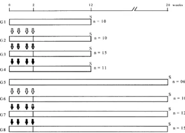
Experimental design. The experimental design is presented in Fig. 1. Male rats of similar body weights (220–230 g) at the beginning of the experiment were allocated to four experimen-tal groups, one control (untreated) and three groups which re-ceived, during the first 2 weeks of the experiment, four i.p. injections of MNU (two injections/week) at the following doses: 20 mg/kg of body weight (total of 80 mg/kg), 40 mg/kg (total of 160 mg/kg) or 60 mg/kg (total of 240 mg/kg). After
carcinogen treatment, they were kept on basal diet and water ad
libitum until sacrifice at the 12th and 20th weeks of the experi-ment, when they were killed by exsanguination under ether
an-T
Franchi et al. Cancer Sci | March 2003 | Vol. 94 | no. 3 | 241
esthesia. Complete gross examination was performed for detection of tumor masses. The thymus, spleen, bone marrow (right femur), lymph nodes (cervical and mesenteric) and liver were collected for histological analysis. The liver and spleen were weighed immediately after removed and blotted dry. The 20th week of the study was established as the latest moment for observation, taking in consideration the information that MNU at 160 mg/kg of body weight i.p. induces leukemia and malig-nant lymphoma in 10–20% of exposed male Fischer 344 rats before this time point.10, 14) Accordingly, the 12th week was
pro-posed as a convenient point for detection of possible preneo-plastic lesions.
Histological and immunohistochemical analysis. The liver, spleen, thymus, lymph nodes, the right femur and any tumor mass found were fixed in 10% buffered formalin during 48 h. After fixation, the right femur was decalcified, washed in tap water
and sectioned. Paraffin-embedded sections (5 µm thickness)
were stained with hematoxylin and eosin (H&E) for histologi-cal analysis. Sections of a lymphoma found in the mediastinum of one animal were submitted to immunohistochemical staining with anti-cytokeratin antibodies (M3515, DAKO Co., Carpinte-ria, CA) for identification of epithelial remnants within the tu-moral mass to confirm the thymic origin of the neoplasia. Sections of the liver were also submitted to immunohistochemi-cal detection of putative preneoplastic foci of hepatocytes that express the enzyme glutathione S-transferase, placental form (GST-P) with anti-GST-P antibodies (MBL, Nagoya). The im-munohistochemical studies were performed using the
avidin-bi-otin method.15) The anti-cytokeratin and the anti-GST-P
antibodies were used at the concentrations of 1:300 and 1:1000, respectively.
Hematological evaluation and bone marrow cytology. During necropsies, peripheral blood samples were collected in vacu-tainer tubes with 15% EDTA and differentially quantified through a T890 Coulter counter for the following: leukocyte, erythrocyte and platelet counts, hematocrit, hemoglobin con-centration, mean corpuscular volume, mean corpuscular hemo-globin and mean corpuscular hemohemo-globin concentration. Cytological smears of the left femur bone marrow were pre-pared immediately after necropsies and stained by Leishman’s method.16)
Statistical analysis. Analysis of variance (ANOVA) was used to evaluate the differences among the body weights and the abso-lute and relative weights of liver and spleen of the groups treated with the four different doses of MNU. The differences
of incidence of lesions among the groups were assessed by ap-plying the Goodman test.17) Differences were considered
signif-icant when P<0.05. Results
Body weights. The lower (80 mg/kg) and intermediate (160 mg/ kg) MNU-dosed groups gained body weight progressively dur-ing the experiment, and at the end of the study (20th week) they did not differ significantly from the control group. At this time point, however, the highest dose group (240 mg/kg) presented
a mean body weight significantly lower (−17%) than the
con-trol.
Lympho-hematopoietic organs. At the 20th week, the mean rela-tive spleen weight of the highest dose group was significantly
increased (+21%) when compared to the other groups.
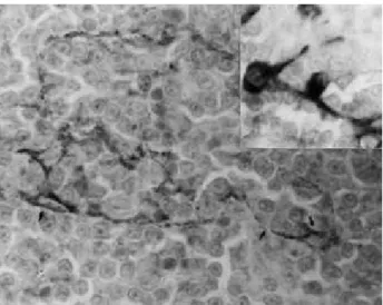
How-ever, no conspicuous spleen histological alteration was apparent in these animals. An animal treated with 240 mg/kg presented a large mediastinal lymphoma, which diffusely infiltrated the thoracic wall and the diaphragm, involving the liver and the spleen. Under H&E staining, this was a medium-sized cell type
thymic lymphoma18) composed predominantly of monotonous
sheets of lymphoblastic cells and a few macrophages which gave the appearance, at low magnification, of a “starry sky” pattern (Fig. 2). The thymic origin of this lymphoma was con-firmed by the demonstration of cytokeratin-positive probable remnants of the thymic Hassal’s bodies within the tumor mass (Fig. 3). No other gross alteration of the thymus was found in any animal in the experiment.
Other malignant and benign tumors were found histologically in the LHS in the high- and intermediate-dose MNU-treated an-imals (Table 1). Three incipient thymic lymphomas occurred in the animals treated with 240 mg/kg of MNU; they were unilat-eral intralobular neoplasias that did not enlarge the organ (Fig. 4). Two animals, treated respectively with 240 mg/kg and 160 mg/kg, developed spleen hemangiomas and one animal treated with 160 mg/kg presented spleen angioectasias.
At both 12th and 20th weeks, the hematological parameters
were within the range of reference values for control rats.19)
Also, no animal in this study presented conspicuous histologi-cal alterations in the lymph nodes or in the bone marrow of the right femur. Accordingly, no alterations in the cytological pat-tern of the left femur bone marrow smears were verified in any group at the 12th and 20th weeks. The bone marrow smears showed intact and high cellularity of the granulocytic,
erythro-Fig. 1. Experimental design. MNU=N-methyl-N-nitrosourea. MNU 80 mg/kg i.p. (4×20 mg), MNU 160 mg/kg i.p. (4×40 mg), MNU 240 mg/kg i.p. (4×60 mg). n, effective number of animals; S, sacrifice.
242 Franchi et al.
cytic and megakaryocytic cell lineages irrespective of whether the animal was treated with MNU or not.
Liver. At the 20th week, the mean absolute and relative liver weights did not differ among the groups, irrespective of MNU treatment. Although no conspicuous histological alterations were seen in the liver of any group, every animal exposed to 240 mg/kg of MNU showed a few preneoplastic GST-P-posi-tive minifoci in the liver, with 3 –4 hepatocytes each.
Discussion
Putative MNU-related effects were verified only at the 20th week of the study in the animals exposed to the highest dose (240 mg/kg), as shown by significantly diminished mean body weight and tumor development in the thymus and spleen. The 12th week seemed to be too early for detection of MNU-related LHS alterations; preneoplastic incipient lesions were registered only at the 20th week, synchronously with well-established thy-mus and spleen neoplasia. As will be discussed below, these early lesions corresponded to intra-lobular thymic lymphoma,
spleen angioectasias and GST-P+-altered foci of hepatocytes,
which should be considered as putative markers of the MNU-initiated carcinogenesis in the Wistar rat.
The thymic lymphomas found in this study at a 27% inci-dence should be assumed as dependent on MNU, because these tumors did not occur in control rats or in intermediate- and lower-dose treated animals, and the rats that developed tumors were younger than 28 weeks of age, when spontaneous tumors
are not common in this species.20) In one animal the neoplasia
was very aggressive and infiltrated thoracic and abdominal or-gans, but in three other rats, the microscopically diagnosed thy-mic lymphomas showed only intra-lobar involvement, i.e., they were in a very early stage of development. These neoplastic growths were lymphoblastic lymphomas, like the early MNU-induced thymic lymphomas described in other rat strains.18, 21)
The incidence of thymic lymphomas induced by nitrosoureas in rats has been reported as variable, depending on the strain and on the duration of the study. Fischer 344 rats treated with a total dose of 240 mg/kg of MNU — the same as the highest dose of the present study— for 6 weeks and sacrificed 18 weeks
later presented a 97% incidence of thymic lymphomas.12)
Spra-gue-Dawley animals treated intra-gastrically with 360 mg/kg of MNU showed a 100% incidence of thymic lymphoma at the
15th week.21) After treatment with 400 µg/ml of
propylni-trosourea (PNU) dissolved in drinking water for 28 weeks, the incidence of thymic lymphomas was different among Fischer 344 (incidence of 98%), Wistar/Furth (71%), Sprague-Dawley (29%), ACI/Ms (23%), Donryu (24%) and Long-Evans (10%) rats.22) Therefore, although relatively low, the 27% incidence of
thymic lymphoma observed in the highest-dose group at the 18th week after the end of the MNU-treatment points to a me-dium-term carcinogenic influence of that chemical in the Wistar strain.
MNU acts as a direct carcinogenic alkylating agent, and
per-sistent O6-methylguanine adducts have been proposed to be the
critical lesion in a variety of MNU-induced tumors. Tissues more susceptible to the carcinogenic effect of nitrosoureas have 3 to 18 times less activity of the DNA repair enzyme alkyl-transferase than tissues less susceptible to these agents.23 – 25)
When transgenic mice express the human O6
-alkylguanine-DNA alkyltransferase in the thymus they present a reduced in-cidence (from 58% to 4%) of thymic lymphomas after MNU exposure, that is, the thymus is protected from developing
neo-Fig. 3. Diffuse lymphoblastic thymic lymphoma—probable remnants of thymic Hassall’s epithelial bodies within the lymphoma: cells and re-ticular structures positive for AE-1/AE3 cytokeratins. (Immunostaining by the avidin-biotin peroxidase method, 1000×)
Table 1. Incidences of (pre)neoplasia in the thymus and spleen of male Wistar rats at the end of the experiment (20th week)
Organ/lesion Experimental groups1)
Effective number of animals
G0 G80 G160 G240
6 10 10 (%) 15 (%)
Thymus
Lymphoma 0 0 0 4 (27)
Spleen
Angiectasia 0 0 0 1 (7)
Hemangioma 0 0 1 (10) 1 (7)
Liver
GST-P+ foci2) 0 0 0 15 (100)3)
1) G0, G80, G160 and G240=control and MNU-treated animals with 80, 160 and 240 mg/kg, respectively.
2) GST-P+ foci=foci of altered hepatocytes expressing the enzyme
glu-tathione S-transferase, placental form.
3) A few foci with 3–4 positive hepatocytes each.
Franchi et al. Cancer Sci | March 2003 | Vol. 94 | no. 3 | 243
plasia.23) Therefore, the susceptibility of the rodent thymus to
develop earlier and more aggressive MNU-dependent tumors seems to depend particularly on its limited capability for DNA
repair through the enzyme O6-alkylguanine-DNA
alkyltransfer-ase.23, 26)
Since MNU has a broad spectrum of target organs, the possi-bility exists that if the experiment had been extended in time, tumors would have developed in other tissues.10, 26) The present
study, however, focused only on LHS tumors because they could be used as early markers of the carcinogenic influence of MNU on the Wistar strain. The development of putatively pre-neoplastic liver GST-P+ foci of hepatocytes was also evaluated,
because this has been an important parameter in the rat
alterna-tive medium-term bioassays for carcinogenesis.5, 27) In the
present case, GST-P+ foci were associated only with the
high-est dose of MNU, being rare and very small, in concordance with the weak MNU initiation activity in the liver.28)
The low total incidence (2/25, 8%) of spleen hemangiomas in the intermediate and high-dose treated groups makes it diffi-cult to establish a causal relationship of these benign lesions
with MNU treatment. However, since spleen hemangiomas oc-curred only in MNU-treated animals and are reported to be of very rare occurrence in young rats,29) it seems likely that they
were also dependent on MNU exposure. The finding of angio-ectasia in the spleen of another animal also exposed to the in-termediate dose of MNU supports the possibility that the spleen vessels are also a target of the carcinogen toxicity. In this con-text, it can be assumed that spleen vascular ectasia is a
preneo-plastic lesion of hemangiomas, as proposed by others.30)
The rapid development of thymus and spleen (pre)neoplasia after exposure to a relatively high dose level of MNU indicates that the outbred Wistar male rat is as susceptible as other rat strains to the carcinogenic influence of that carcinogen, devel-oping marker lesions in the lympho-hematopoietic system that can be used as end-points in alternative carcinogenesis proto-cols.
The Fundação de Amparo à Pesquisa do Estado de São Paulo (FAPESP, No. 98 /03035-8) and the Conselho Nacional de Pesquisas (CNPq, No. 300705/81-6) supported this study.
1. Bannasch P, Griesemer RA. Long-term assays for carcinogenicity in animals. In: Montesano R, Bartsch H, Vainio H, Wilbourn J, Yamasaki H, editors. Long-term and short-term assays for carcinogens: a critical appraisal. IARC Sci Publ 1986; 83: 13–83.
2. Chhabra RS, Huff JE, Schwetz BS, Selkirk J. An overview of prechronic and chronic toxicity/carcinogenicity experimental study designs and criteria used by the National Toxicology Program. Environ Health Perspect 1990; 86: 313–21.
3. Boorman GA, Maronpot RR, Eustis SL. Rodent carcinogenesis bioassay: past, present and future. Toxicol Pathol 1994; 22: 105–11.
4. Ito N, Imaida K, Tsuda H, Shibata M, Aoki T, de Camargo JLV, Fukushima S. Wide-spectrum initiation models: possible applications to medium-term multiple organ bioassays for carcinogenesis modifiers. Jpn J Cancer Res 1988; 79: 413–7.
5. Shirai T, Hirose M, Ito N. Medium-term bioassays in rats for rapid detection of the carcinogenic potential of chemicals. In: McGregor DB, Rice JM, Venitt S, editors. The use of short- and medium-term tests for carcinogens and data on genetic effects in carcinogenic hazard evaluation. IARC Sci Publ 1999; 146: 251–72.
6. Hagiwara A, Tanaka H, Imaida K, Tamano S, Fukushima S, Ito N. Correla-tion between medium-term multi-organ carcinogenesis bioassay data and long-term observation results in rats. Jpn J Cancer Res 1993; 84: 237–45. 7. Brazilian Institute of Environmental and Renewable Natural Resources
(IBAMA). Normative Act No.84, October 15 (1996) (in Portuguese). 8. Spinardi ALT, Kaneno R, Rodrigues MAM, Salvadori DMF, Rocha NS,
Barbisan LF, Ribeiro LR, de Camargo JLV. Natural killer activity in a me-dium-term multi-organ bioassay for carcinogenesis. Jpn J Cancer Res 1999;
90: 101–7.
9. Moreira ELT, de Camargo JLV, Rodrigues MAM., Barbisan LF, Salvadori DMF. Dose- and sex-related carcinogenesis by N-bis(2-hydroxypropyl)nitro-samine in Wistar rats. Jpn J Cancer Res 2000; 91: 368–74.
10. Uwagawa S, Tsuda H, Inoue T, Tagawa Y, Aoki T, Kagawa M, Ogiso T, Ito N. Enhancing potencial of 6 different carcinogens on multi-organ tumorigen-esis after initial treatment with N-methyl-N-nitrosourea. Jpn J Cancer Res 1991; 82: 1397–405.
11. Tsuda H, Fukushima S, Imaida K, Kurata Y, Ito N. Organ specific promoting effect of phenobarbital and saccharin in induction of thyroid, liver and uri-nary bladder tumors in rats after initiation with N-nitrosomethylurea. Cancer Res 1983; 43: 3292–6.
12. Mizoguchi M, Naito H, Kurata Y, Shibata M-A, Tsuda H, Wild CP, Montesano R, Fukushima S. Influence of aging on multi-organ carcinogene-sis in rats induced by N-methyl-N-nitrosourea. Jpn J Cancer Res 1993; 84: 139–46.
13. Kociba RJ, Kociba GJ. Assessment of toxicologic effects upon bone marrow and related tissues. In: Jones TC, Ward JM, Mohr U, Hunt RD, editors. Monographs on the pathology of laboratory animals (hemopoietic system). Berlin-Heidelberg: Springer-Verlag; 1990. p.79–87.
14. Ito N, Shirai T, Hasegawa R. Medium-term bioassays for carcinogens. In:
Vainio H, Magee PN, McGregor DB, McMichael AJ, editors. Mechanisms of carcinogenesis in risk identification. Lyon: International Agency for Re-search on Cancer (IARC); 1992. p. 353–88.
15. Hsu SM, Raine L, Fanger N. Use of avidin-biotin-peroxidase complex (ABC) and unlabeled antibody (PAP) procedures. J Histochem Cytochem 1981; 29: 577–80.
16. Luna LG. Histopathologic methods and color atlas of special stains and tis-sues artifacts. Washington: American Histolabs; 1992. p. 275–6.
17. Goodman LA. On simultaneous confidence intervals for multinomial propor-tions. Technometrics 1965; 7: 247–54.
18. Ogiu T. T-Cell lymphoma, thymic origin, rat. In: Jones TC, Ward JM, Mohr U, Hunt RD, editors. Monographs on pathology of laboratory animals (He-mopoietic System). Berlin-Heidelberg: Springer-Verlag; 1990. p. 286–92. 19. Harkness JE, Wagner JE. The biology and medicine of rabbits and rodents.
Media: Williams & Wilkins; 1995. p. 95.
20. Hayashi S, Nonoyama T, Miyajima H. Spontaneous non-thymic cell lympho-mas in young Wistar rats. Vet Pathol 1989; 26: 326–32.
21. Koestner AW, Ruecker FA, Koestner A. Morphology and pathogenesis of tu-mors of the thymus and stomach in Sprague-Dawley rats following intragas-tric administration of methylnitrosourea (MNU). Int J Cancer 1977; 20: 418–26.
22. Shisa H, Hiai H. Genetically determined susceptibility of Fischer 344 rats to propylnitrosourea-induced thymic lymphomas. Cancer Res 1985; 45: 1483– 7.
23. Dumenco LL, Allay E, Norton K, Gerson SL. The prevention of thymic lym-phomas in transgenic mice by human O6-alkylguanine-DNA alkyltrans-ferase. Science 1993; 59: 219–22.
24. Gerson SL, Miller K, Berger NA. O6 alkylguanine-DNA alkyltransferase ac-tivity in human myeloid cells. J Clin Invest 1985; 76: 2106–14.
25. Fong LY, Jensen DE, Magee PN. DNA methyl-adduct dosimetry and O6-alkylguanine-DNA alkyl transferase activity determinations in rat mammary carcinogenesis by procarbazine and N-methylnitrosourea. Carcinogenesis 1990; 11: 411–7.
26. Swenberg JA, Koestner A, Wechsler W, Brunden MN, Abe H. Differential oncogenic effects of methylnitrosourea. J Natl Cancer Inst 1975; 54: 86–95.
27. Moore MA, Tsuda H, Tamano S, Hagiwara A, Imaida K, Shirai T, Ito, N. Marriage of a medium-term liver model to surrogate markers— a practical approach for risk and benefit assessment. Toxicol Pathol 1999; 27: 237–42.
28. Yoshida Y, Tatematsu M, Takaba K, Iwasaki S, Ito, N. Target organ specific-ity of cell proliferation induced by various carcinogens. Toxicol Pathol 1993;
21: 436–42.
29. Losco P. Normal development, growth and aging of the spleen. In: Mohr U, Dungworth DL, Capen CC, editors. Pathobiology of the aging rat. Washing-ton: International Life Sciences Institute; 1992. p. 75–93.