In vivo
antithrombotic properties of
a heparin from the oocyte test cells
of the sea squirt
Styela plicata
(Chordata-Tunicata)
Laboratório de Tecido Conjuntivo, Hospital Universitário Clementino Fraga Filho and Programa de Glicobiologia, Instituto de Bioquímica Médica,
Universidade Federal do Rio de Janeiro, Rio de Janeiro, RJ, Brasil L. Cardilo-Reis*,
M.C.M. Cavalcante*, C.B.M. Silveira and M.S.G. Pavão
Abstract
In the ascidian Styela plicata, the oocytes are surrounded by two types of accessory cells named follicle cells and test cells. A heparin-like substance with an anticoagulant activity equivalent to 10% of mam-malian heparin and about 5% as potent as the mammam-malian counterpart for the inhibition of thrombin by antithrombin was isolated from the oocyte test cells. In the present study, we compared the antithrombotic and hemorrhagic effects of sea squirt oocyte test cell heparin with those of porcine heparin in rat models of venous thrombosis and blood loss. Intravenous administration of the oocyte test cell heparin to Wistar rats (both sexes, weighing ~300 g, N = 4 in each group) at a dose of 5.0 mg/kg body weight, which produced a 1.8-fold increase in plasma activated partial thromboplastin time, inhibited thrombosis by 45 ± 13.5% (mean ± SD) without any bleeding effect. The same dose of porcine heparin inhibited thrombosis by 100 ± 1.4%, but produced a blood loss three times greater than that of the saline-treated control. However, 10-fold reduction of the dose of porcine heparin to 0.5 mg/ kg body weight, which produced a 5-fold increase in plasma-activated partial thromboplastin time, inhibited thrombosis by 70 ± 13% with-out any bleeding effect. The antithrombotic properties of a new heparin isolated from test cells of the sea squirt S. plicata, reported here for the first time, indicate that, although sea squirt oocyte test cell heparin was a poor anticoagulant compared to porcine heparin, it had a significant antithrombotic effect without causing bleeding.
Correspondence
M.S.G. Pavão
Instituto de Bioquímica Médica CCS, UFRJ
21941-590 Rio de Janeiro, RJ Brasil
Fax: +55-21-2562-2090 E-mail: [email protected] *These authors contributed equally to this study.
Research supported by CNPq, FAPERJ and the NIH Fogarty International Center (R03 TW05775). M.S.G. Pavão is the recipient of a research fellowship from CNPq.
Received February 16, 2006 Accepted August 3, 2006
Key words
•Ascidian •Heparin
•Experimental thrombosis •Antithrombotics •Hemorrhage
Introduction
Heparin is a highly sulfated glycosami-noglycan composed of disaccharide repeats of hexuronic acid (α-L-iduronic acid or ß-D-glucuronic acid) linked 1,4 to α -D-glucosa-mine. Heparin is a heterogeneous mixture of
polymers with a similar backbone. The het-erogeneity results from variations of D-glu-cosamine sulfation (N-acetylated, N-sulfated, O-sulfated at C6 and/or C3) and of the uronic acid residue (O-sulfated or not at C2) (1).
[GlcNAc(6SO4)-GlcA-GlcNS(3SO4)-IdoA
(2SO4)-GlcNS(6SO4)] (2), heparin has a
po-tent anticoagulant activity (3). In the pres-ence of heparin, the rates of inhibition of thrombin, factor IXa, and factor Xa by anti-thrombin are increased ~1000-fold (4), so that inhibition is essentially instantaneous. Heparin has been used clinically for almost 70 years for the prevention of thromboem-bolic events frequently observed after gery, especially pelvic and orthopedic sur-gery (5,6).
In vertebrates, heparin is present in se-cretory granules of mast cells and basophils and is released only when mast cells de-granulate in response to extracellular signals (1,2,7). Among invertebrates, heparins with different structures, molecular weights and anticoagulant activity have been reported in mollusks, crustacean and annelid (8-11). More recently, a heparin with structure simi-lar to the mammalian counterpart, but with differences in the degree of sulfation has been identified in the intracellular granules of oocyte test cells of the ascidian (com-monly known as sea squirt) Styela plicata
(12). Oocyte test cells are accessory cells located in the perivitelline space of ascidian oocytes, where they remain during egg de-velopment until hatching. Sea squirt oocyte test cell heparin is composed mainly of the disaccharide [α-L-IdoA(2SO4)-1→ 4ß-D-GlcN(SO4)(6SO4)-1]n, similar to
mamma-lian heparin. About 25% of the disaccharide [α-L-IdoA-1→4ß-D-GlcN(SO4)(6SO4)-1]n is also present (12). When compared to mam-malian heparin, sea squirt oocyte test cell heparin has 10% of its anticoagulant activity measured by activated partial thromboplas-tin time (aPTT). In addition, it is about 5% as potent as the mammalian counterpart for the inhibition of thrombin by antithrombin. How-ever, it activates heparin co-factor II (HCII) to the same extent as mammalian heparin, as indicated by the IC50 for thrombin inhibition
in the presence of the inhibitor HCII (12). The prevalence of trisulfated disaccharides
containing iduronic acid and N-sulfated glu-cosamine, the lack of glucuronic acid and N-acetylgalactosamine-containing disaccha-rides, the antithrombin-mediated thrombin inhibitory activity, and the intracellular lo-calization of the ascidian glycosaminogly-can clearly demonstrate that sea squirt oo-cyte test cell heparin belongs to the heparin family.
In the present study, we compared the antithrombotic and hemorrhagic effects of sea squirt oocyte test cell and porcine hep-arins in vivo using rat models. We showed that the oocyte test cell heparin has a lower but significant antithrombotic activity and a lower bleeding effect when compared to porcine heparin.
Material and Methods
Extraction and purification of the test cell heparin
Adult specimens of S. plicata were col-lected from Guanabara Bay, Rio de Janeiro, RJ, Brazil, and maintained in an aerated aquarium. The gonads of several ascidians were carefully separated from other tissues under magnifying lenses, and the eggs iso-lated as described (12). The glycosaminogly-cans were extracted from the eggs by papain digestion and ethanol precipitation and the heparin was purified by anion-exchange chromatography on a Mono Q column (Amersham Biosciences, São Paulo, SP, Brazil) as described (12).
Inhibition of factor Xa by antithrombin in the presence of sea squirt oocyte test cell heparin
final concentrations of reactants included 50 nM human antithrombin (Haematologic Technologies Inc., Essex Junction, VT, USA), 15 nM human factor Xa (Haemato-logic Technologies) and 0-1000 µg/mL hep-arin in 100 µL 20 mM Tris/HCl, 0.15 M NaCl, and 1.0 mg/mL polyethylene glycol, pH 7.4 (Tris/PEG buffer). Factor Xa was added to initiate the reaction. After 60 s at room temperature, 500 µL 100 µM N-meth- oxycarbonyl-D-norleucyl-glycyl-L-arginine-4-nitranillide-acetate (Roche, Mannheim, Germany) in Tris/PEG buffer was added and absorbance at 405 nm was measured for 100 s. The rate of change of absorbance was proportional to the amount of factor Xa ac-tivity remaining in the incubation mixture. No inhibition occurred in control experi-ments in which factor Xa was incubated with antithrombin in the absence of heparin, nor did inhibition occur when factor Xa was incubated with heparin alone in the concen-tration range tested. Porcine intestinal mu-cosa heparin (porcine-Hep, 180 units/mg; Sigma-Aldrich, St. Louis, MO, USA) and Nadroparin (average anti-Xa activity 112 units/mg; Sanofi, Vitry-sur Seine, France) were used as standards.
Animal procedures
The animal studies were carried out in accordance with the Brazilian Animal Pro-tection Law and the Institutional Guidelines for Animal Care and Experimentation.
Ex-vivo anticoagulant action measured by activated partial thromboplastin time
The effect of sea squirt oocyte test cell heparin on coagulation was determined in Wistar rats (both sexes, ~300 g body weight) anesthetized with an intramuscular injection of 100 mg/kg ketamine (Cristália, São Paulo, SP, Brazil) and 16 mg/kg xylazine (Bayer S/ A, São Paulo, SP, Brazil), and supplemented as needed. The right carotid artery was
iso-lated and cannuiso-lated with a 22-gauge cath-eter (Jelco, Johnson & Johnson Medical Ltda., São José dos Campos, SP, Brazil) for blood collection and administration of the heparin. Blood (~500 µL) was collected into 2.8% sodium citrate (9:1, v/v) for aPTT determination before and 10, 20, 30, 40, 50, and 60 min after intravenous administration in a bolus of 0.5 mg/kg of porcine heparin or 5 mg/kg of sea squirt oocyte test cell heparin. At least 4 animals were used in each group.
In vivo antithrombotic effect
the thrombus in the absence of heparin.
Bleeding
The bleeding effect was determined in Wistar rats (both sexes, ~300 g body weight) anesthetized with a combination of xylazine and ketamine, as described above. A can-nula was inserted into the right carotid artery
for the administration of heparin (0.5 or 5.0 mg/kg). After 5 min the rat tail was cut 3 mm from the tip and carefully immersed in 40 mL distilled water at room temperature. Blood was collected for 60 min and the blood loss measured on the basis of hemo-globin content of the water solution by spec-trophotometry (14). The volume of blood was deduced from a standard curve based on blood dilution by absorbance at 540 nm. At least 4 animals were used per group.
Statistical analysis
Data are reported as means ± SD. Com-parisons between two groups were made by the t-test and the differences were consid-ered significant when P < 0.05.
Results
Anti-factor Xa activity of oocyte test cell heparin
The data in Figure 1 show that the anti-factor Xa activity of the ascidian heparin is equivalent to 1.1% of porcine heparin and 40% of Nadroparin, based on total weight. The IC50 for factor Xa inhibition by
anti-thrombin, estimated by the curves in Figure 1, was about 1.0, 2.5, and 90 µg/mL for porcine heparin, nadroparin and test cell-heparin, respectively.
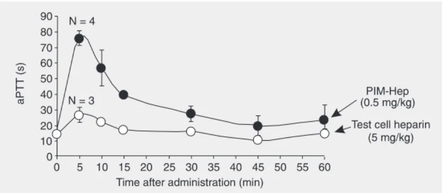
In vivo anticoagulant effect
Figure 2 shows that aPTT increased up to ~5-fold during the first 5 min after injection of porcine heparin. After 5 min, aPTT started to decrease and remained 2-fold higher than the control value for the next 60 min. Oocyte test cell heparin induced a less intense change in aPTT values, with a maximum increase of 1.8-fold at 5 min, returning to normal after 15 min. No significant change in aPTT was observed after the administration of test cell heparin doses lower than 5 mg/kg (data not shown). Figure 1. Anti-factor Xa properties of porcine and oocyte test cell heparin. Human
antithrom-bin (50 nM) was incubated with human factor Xa (15 nM) in the presence of 0 to 1000 µg/mL of the different heparins. After 60 s, the remaining factor Xa activity was determined with the chromogenic substrate N-methoxycarbonyl-D-norleucyl-glycyl-L-arginine-4-nitranillide-ac-etate (100 µM) and absorbance at 405 nm was recorded for 100 s (∆A405/min). PIM-Hep =
porcine heparin (filled circles), Nadroparin (triangles), oocyte test cell heparin (open circles). Results represent the average of two separate experiments.
Effect on thrombosis
The effect of ascidian and mammalian heparins on thrombosis was investigated us-ing an experimental venous thrombosis mo-del in rats. A single injection of 0.5 mg/kg of porcine heparin in a bolus, given 5 min before the thrombogenic stimulus with rab-bit brain thromboplastin induced about 70% inhibition of thrombosis (Figure 3). Admin-istration of the same dose of oocyte test cell heparin did not produce any inhibition of thrombosis (Figure 3). Porcine heparin ad-ministered at 5 mg/kg produced 100% inhi-bition of thrombosis, whereas the same dose of oocyte test cell heparin reduced thrombo-sis by only 45%. We also evaluated the effect of the mammalian and ascidian hep-arins on thrombosis at the dose of 2.0 mg/kg. At this dose, porcine heparin inhibited throm-bosis by 100%, whereas oocyte test cell heparin had no effect (data not shown).
Hemorrhagic effect
The hemorrhagic effect of porcine and oocyte test cell heparins was assessed after intravascular administration based on blood loss in a rat cut-tail bleeding assay. At the antithrombotic dose of 5 mg/kg body weight, oocyte test cell heparin did not modify the blood loss compared with rats receiving sa-line (Table 1). The blood loss increased ~2.6-fold in rats receiving the same dose of por-cine heparin (Table 1). The dose of porpor-cine heparin needed to achieve 70% inhibition of thrombosis (0.5 mg/kg) did not increase the blood loss.
Discussion
In a previous study, heparin with a differ-ent sulfation pattern and anticoagulant activ-ity was isolated from the oocyte test cells of the ascidian S. plicata (12). Here we as-sessed the antithrombotic and bleeding ef-fects of this heparin after intravascular
ad-ministration to rats, using experimental mod-els of venous thrombosis and bleeding.
The anti-factor Xa activity of the test cell heparin was about 40 times lower than that of a low-molecular weight heparin (Nadrop-arin), which has an average anti-factor Xa activity of 112 units/mg. Therefore, the anti-factor Xa of the oocyte test cell heparin can be estimated to be about 2.8 units/mg. The anticoagulant activity obtained by the in vi-tro experiments reported here (Figure 1) and in the previous study (12) agrees with the results obtained in in vivo experiments (Fig-ure 2), showing that the oocyte test cell heparin has a very low anticoagulant activ-ity. A dose of 5 mg/kg body weight of the oocyte test cell heparin is required to pro-duce a 1.8-fold increase in aPTT in rat plasma.
Figure 3. Antithrombotic activity of porcine heparin (PIM-Hep) or oocyte test cell heparin. Anti-thrombotic activity was deter-mined using a stasis thrombo-sis model in rats after intrave-nous administration of the dif-ferent heparins in a bolus (see Material and Methods). Mean thrombus weight was obtained for each group and then reported as percent weight in the absence of polysaccharide. Percent inhibition of thrombosis (mean ± SD, N = 4) is reported as a function of heparin concentration. *P < 0.05 for the comparisons indicated by the horizontal lines (Student t -test).
Table 1. Hemorrhagic effect of mammalian and oocyte test cell heparins.
Treatment Blood loss (µL)
Saline 32 ± 5.5 Porcine heparin (0.5 mg/kg) 21 ± 5 Porcine heparin (5.0 mg/kg) 87 ± 19 Test cell heparin (5.0 mg/kg) 36 ± 6
Porcine heparin at a dose 10 times lower produced a 5-fold increase in aPTT.
The antithrombotic activity of heparin is associated with its ability to induce inhibi-tion of thrombin and factor Xa by antithrom-bin. It has been shown more recently in animal experiments that anti-factor Xa ac-tivity is a prerequisite, although not suffi-cient by itself, for a thrombosis-preventing effect (15-17). In fact, oocyte test cell hepa-rin inhibited thrombosis to a much lesser extent than porcine heparin at the same dose. At the highest dose tested (5 mg/kg body weight), test cell heparin inhibited thrombo-sis by only 45% compared to 100% inhibi-tion obtained with the same dose of porcine heparin. This result is probably associated with the very low anti-IIa and anti-factor Xa activities of test cell heparin.
Usually, low-molecular weight heparins, that contain high anti-factor Xa and low anti-IIa activities, have a lower bleeding ef-fect than unfractionated heparin (18-21). However, the mechanism by which heparins and other sulfated glycosaminoglycans con-tribute to bleeding is unknown. In a previous study (22), we demonstrated that there is a dissociation of the anticoagulant action,
an-tithrombotic activity and bleeding effect in the case of other glycosaminoglycans. We showed that for dermatan sulfates with dif-ferent sulfation patterns an increase in HCII activity does not result in a parallel increase of the antithrombotic potency or of the bleed-ing effect (22). Similarly, the effect of an-other class of sulfated polysaccharides, namely sulfated galactans, on coagulation, bleeding and thrombosis is also not coupled (23). On the other hand, our results regard-ing the effect of heparins on bleedregard-ing indi-cated that a reduction of the anticoagulant activity of heparin abolished its bleeding effect. However, significant antithrombotic activities (70 and 45% thrombin inhibition for mammalian and oocyte test cell heparin, respectively; Table 1) with no bleeding ef-fect were observed even when oocyte test cell heparin wasadministered at high doses. We have reported here the antithrom-botic properties of a new heparin molecule from the oocyte test cells of S. plicata. Our results indicate that for this oocyte test cell heparin there is a parallelism between the anticoagulant and antithrombotic activities that are dissociated from the hemorrhagic effect.
References
1. Kjellén L, Lindahl U. Proteoglycans: structures and interactions.
Annu Rev Biochem 1991; 60: 443-475.
2. Conrad HE. Heparin binding proteins. San Diego: Academic Press; 1998.
3. McLean J. The thromboplastic action of cephalin. Am J Physiol
1916; 41: 250-257.
4. Jordan RE, Oosta GM, Gardner WT, Rosenberg RD. The kinetics of hemostatic enzyme-antithrombin interactions in the presence of low molecular weight heparin. J Biol Chem 1980; 255: 10081-10090. 5. Leyvraz PF, Richard J, Bachmann F, Van Melle G, Treyvaud JM,
Livio JJ, et al. Adjusted versus fixed-dose subcutaneous heparin in the prevention of deep-vein thrombosis after total hip replacement.
N Engl J Med 1983; 309: 954-958.
6. Poller L. Therapeutic ranges in anticoagulant administration. Br Med J 1985; 290: 1683-1686.
7. Straus AH, Nader HB, Dietrich CP. Absence of heparin or heparin-like compounds in mast-cell-free tissues and animals. Biochim Biophys Acta 1982; 717: 478-485.
8. Cassaro CM, Dietrich CP. Distribution of sulfated mucopolysaccha-rides in invertebrates. J Biol Chem 1977; 252: 2254-2261. 9. Medeiros GF, Mendes A, Castro RA, Bau EC, Nader HB, Dietrich
CP. Distribution of sulfated glycosaminoglycans in the animal king-dom: widespread occurrence of heparin-like compounds in inverte-brates. Biochim Biophys Acta 2000; 1475: 287-294.
10. Burson SL, Fahrenbach MJ, Frommhagen LH, Riccardi BA, Brown RA, Brockman JA, et al. Isolation and purification of mactins hepa-rin-like anticoagulants from mollusca. J Am Chem Soc 1956; 78: 5874-5878.
11. Rahemtulla F, Lovtrup S. The comparative biochemistry of inverte-brate mucopolysaccharides - VI. Crustacea. Comp Biochem Physiol B 1976; 53: 15-18.
13. Vogel GM, Meuleman DG, Bourgondien FG, Hobbelen PM. Com-parison of two experimental thrombosis models in rats effects of four glycosaminoglycans. Thromb Res 1989; 54: 399-410.
14. Herbert JM, Héralt JP, Bernat A, Van Amsterdan RGM, Vogel GMT, Lormeau JC, et al. Biochemical and pharmacological properties of SANORG 32701. Comparison with the “synthetic pentasaccharide (SR 90107/org 31540)” and standard heparin. Circ Res 1996; 79: 590-600.
15. Holmer E, Mattsson C, Nilsson S. Anticoagulant and antithrombotic effects of heparin and low molecular weight heparin fragments in rabbits. Thromb Res 1982; 25: 475-485.
16. Thomas DP, Merton RE, Barrowcliffe TW, Thunberg L, Lindahl U. Effects of heparin oligosaccharides with high affinity for antithrombin III in experimental venous thrombosis. Thromb Haemost 1982; 47: 244-248.
17. Thomas DP, Merton RE, Gray E, Barrowcliffe TW. The relative antithrombotic effectiveness of heparin, a low molecular weight heparin, and a pentasaccharide fragment in an animal model.
Thromb Haemost 1989; 61: 204-207.
18. Bagge L, Wahlberg T, Holmer E, Tyden H, Nystrom SO, Malm T.
Low-molecular-weight heparin (Fragmin) versus heparin for antico-agulation during cardiopulmonary bypass in open heart surgery, using a pig model. Blood Coagul Fibrinolysis 1994; 5: 265-272. 19. Colwell CW Jr. Recent advances in the use of low molecular weight
heparins as prophylaxis for deep vein thrombosis. Orthopedics 1994; 17 (Suppl): 5-7.
20. Haas S, Flosbach CW. Prevention of postoperative thromboembo-lism with Enoxaparin in general surgery: a German multicenter trial.
Semin Thromb Hemost 1993; 19 (Suppl 1): 164-173.
21. Hirsh J. Comparison of the relative efficacy and safety of low molec-ular weight heparin and unfractionated heparin for the treatment of venous thrombosis. Haemostasis 1996; 26 (Suppl 4): 189-198. 22. Vicente CP, Zancan P, Peixoto LL, ves-Sa R, Araujo FS, Mourao
PA, et al. Unbalanced effects of dermatan sulfates with different sulfation patterns on coagulation, thrombosis and bleeding. Thromb Haemost 2001; 86: 1215-1220.
23. Farias WR, Nazareth RA, Mourao PA. Dual effects of sulfated D-galactans from the red algae Botryocladia occidentalis preventing thrombosis and inducing platelet aggregation. Thromb Haemost