Roles of Klf5 Acetylation in the Self-Renewal
and the Differentiation of Mouse Embryonic
Stem Cells
Tong Zhao1☯, Chang Liu1☯, Lingyi Chen1,2*
1State Key Laboratory of Medicinal Chemical Biology, Collaborative Innovation Center for Biotherapy, 2011 Collaborative Innovation Center of Tianjin for Medical Epigenetics and College of Life Sciences, Nankai University, Tianjin, China,2State Key Laboratory of Molecular Oncology, Cancer Institute/Hospital, CAMS, Beijing, China
☯These authors contributed equally to this work. *[email protected]
Abstract
Transcription factor Krüppel-like factor 5 (Klf5) plays important roles in the formation of the inner cell mass (ICM) and the trophectoderm during embryogenesis, as well as the self-renewal and the differentiation of mouse embryonic stem cells (ESCs). Acetylation of KLF5 has been shown to reverse the transcriptional activity of KLF5 in human epidermal cells and prostate cancer cells. Whether Klf5 acetylation contributes to the lineage specification in the blastocyst and pluripotency maintenance in ESCs remains unexplored. Here, we showed the ubiquitous expression of acetylated Klf5 in the ICM and the trophectoderm, ruling out the possibility that differential acetylation status of Klf5 leads to the lineage specifi-cation in the blastocyst. We found that K358Q mutation, mimicking acetylation, enhances the transcriptional activity of Klf5 for pluripotency genes in ESCs, and that K358Q Klf5 is more potent in pluripotency maintenance and in somatic cell reprogramming, compared to K358R Klf5. In ESCs, Klf5 acetylation, stimulated by TGF-βsignaling, is involved in enhanc-ing Sox2 expression. Moreover, upon ESC differentiation, acetylation of Klf5 facilitates the suppression of many differentiation genes, except for that K358Q Klf5 activatesCdx2, pro-moting trophectodermal differentiation. In summary, our results revealed the regulatory functions of Klf5 acetylation in ESC self-renewal and differentiation.
Introduction
During mouse preimplantation development, two distinct cell lineages, the inner cell mass (ICM) and the trophectoderm (TE), are first established in the blastocyst. The pluripotent ICM cells mainly contribute to the development of the fetus, while the TE develops into the placenta [1,2]. In addition, under properin vitroculture conditions, the ICM gives rise to embryonic stem cells (ESCs). ESCs self-renew indefinitely and maintain the potential to differentiate to all cell types in the body [3,4]. Thus, ESCs have great application potential in regenerative OPEN ACCESS
Citation:Zhao T, Liu C, Chen L (2015) Roles of Klf5 Acetylation in the Self-Renewal and the
Differentiation of Mouse Embryonic Stem Cells. PLoS ONE 10(9): e0138168. doi:10.1371/journal. pone.0138168
Editor:Qiang Wu, National University of Singapore, SINGAPORE
Received:July 30, 2015
Accepted:August 27, 2015
Published:September 15, 2015
Copyright:© 2015 Zhao et al. This is an open access article distributed under the terms of the
Creative Commons Attribution License, which permits unrestricted use, distribution, and reproduction in any medium, provided the original author and source are credited.
Data Availability Statement:All relevant data are within the paper and its Supporting Information files.
medicine. Understanding the molecular mechanisms underlying ESC self-renewal and pluripo-tency is critical to the field of regenerative medicine.
It has been revealed that a unique transcriptional regulation network is essential for pluripotency maintenance of ESCs [5,6]. Oct4, Sox2 and Nanog, forming a feed-forward self-regulating circuitry, are the core components of the transcriptional regulation network for pluripotency [7–10]. Krüppel like factors, Klf2, Klf4 and Klf5, are also key players in the tran-scriptional regulation network of pluripotency [11–16]. They are highly expressed in mouse ESCs, and down-regulated upon differentiation. These three Krüppel-like factors share redun-dant functions in pluripotency maintenance and establishment. ESC colony morphology and alkaline phosphatase (AP) positivity are lost upon simultaneous knockdown of all three Klf fac-tors, while knockdown of individual Klf factor does not affect ESC colony morphology or AP positivity [11]. Both Klf2 and Klf5 can substitute for Klf4, together with Oct4, Sox2 and c-Myc, to reprogram somatic cells [17]. Moreover, the genomic binding profiles of these three Klf fac-tors are highly overlapped [11].
However, other evidences suggest that Klf5 has unique roles in pluripotency regulation. In contrast to Jiang’s work mentioned above [11], others have demonstrated that knockdown or knockout of Klf5 alone leads to down-regulation of pluripotency gene expression, as well as spontaneous ESC differentiation [13,15]. The conflicting results might be explained by the dif-ferences in knockdown efficiency and experimental settings. During ESC differentiation, Klf5 suppresses mesodermal differentiation, while Klf4 represses the expression of endodermal genes [18]. All these data suggest that Klf5 is essential for the maintenance of pluripotency in ESCs, and its function cannot be fully compensated by Klf2 and Klf4. In addition to its role in pluripotency maintenance, Klf5 is also involved in TE development. Klf5 is expressed in the ICM and the TE, as well as theirin vitrocounterparts, ESCs and trophectoderm stem cells (TSCs). In contrast, Klf2 and Klf4 are only expressed in ESCs, but not in TSCs. Consistently, Klf5knockout embryos die before implantation and have defects in both the ICM and the TE. No ESC lines can be derived fromKlf5knockout ICM cells. Knockout ofKlf5also reduces the expression ofCdx2, a key transcription factor for TE development, in TE cells, resulting in implantation failure [14,19]. Klf5 is required for the expression ofCdx2in TE cells, while it activates pluripotency gene expression in ESCs. How the transcriptional activity and down-stream targets of Klf5 is regulated in these two distinct cell types, remains elusive.
It has been reported that the transcriptional activity of Klf5 is regulated by various post-transcriptional modifications, such as phosphorylation, acetylation, ubiquitination and sumoy-lation [20–27]. The expression level of KLF5 is tightly regulated by the ubiquitin-proteasome system. And WWP1 has been identified as an E3 ubiquitin ligase for KLF5 [24]. Phosphoryla-tion of KLF5 enhances its interacPhosphoryla-tion with CREB binding protein (CBP), and hence increases its transcriptional activity [21]. Sumoylation may regulate the cytoplasmic and nuclear localiza-tion of KLF5 [26]. In addition, both sumoylation and acetylation of KLF5 affects the transcrip-tional complexes that KLF5 associates with, switching transcriptranscrip-tional activity of KLF5 between activator and repressor [20,27,28]. Most strikingly, in the HaCaT epidermal cell line, trans-forming growth factor-β(TGF-β) mediated acetylation of human KLF5 at lysine 369 reverses the transcriptional activity of KLF5, from a transcriptional repressor to a transcriptional activa-tor for thep15(CDKN2B) gene. Consequently, the pro-proliferative factor KLF5 becomes anti-proliferative upon activating TGF-βsignaling [20,28]. The opposing function of KLF5 regu-lated by K369 acetylation has also been demonstrated in prostate tumorigenesis [29].
Given the critical regulatory function of KLF5 acetylation, we investigated the role of Klf5 acetylation in the differentiation of the ICM and the TE, as well as in maintaining the pluripo-tency of mouse ESCs. We first demonstrated that Klf5, unacetylated (unAc-) and acetylated (Ac-) Klf5 are expressed in both ICM and TE cells, at similar levels. It implies that the Competing Interests:The authors have declared
segregation of the ICM and the TE is not caused by differential level of Ac-Klf5. Next, we showed that the K358Q (KQ) Klf5 mutant, mimicking Ac-Klf5, promotes ESC self-renewal more efficiently than the K358R Klf5 mutant (KR, mimicking unAc-Klf5). Acetylation of Klf5, regulated by TGF-βsignaling, increases Sox2 protein expression. In addition, during ESC dif-ferentiation, KQ Klf5, but not KR Klf5, is able to suppress the expression of differentiation genes, except forCdx2. KQ Klf5 facilitates the activation ofCdx2in embryoid bodies (EBs), implying that Klf5 acetylation might be required for the TE developmentin vivo. All these data suggest a regulatory role of Klf5 acetylation in the self-renewal and the differentiation of mouse ESCs.
Materials and Methods
Ethics statement
This study was carried out in strict accordance with the recommendations in the Guide for the Care and Use of Laboratory Animals of the National Institutes of Health. The protocols and the use of mice for this research were approved by Nankai Animal Care and Use Committee.
Cell culture
ESCs and TSCs were cultured as described previously [30]. In brief, V6.5 mouse ESCs were cul-tured in growth medium consisting of 85% DMEM (Invitrogen), 15% fetal bovine serum (Hyclone), 2 mM L-glutamine, 5000 units/ml penicillin and streptomycin, 0.1 mM non-essen-tial amino acids (Invitrogen), 0.1 mM 2-mercaptoethanol (Sigma), and 1000 units/ml LIF (Chemicon). TSCs were cultured in TSC culturing medium containing 80% RPMI 1640, 20% fetal bovine serum (Hyclone), 25 ng/ml Fgf4 (Sigma), 1 ng/ml heparin (Sigma), 0.1 mM 2-mer-captoethanol (Sigma), 1 mM sodium pyruvate (Invitrogen), 2 mM L-glutamine (Invitrogen), 5000 units/ml penicillin and streptomycin (Invitrogen).
Embryo culture
Female ICR mice (4–6 weeks) were induced to superovulate by intraperitoneal injections of 5 IU of pregnant mare serum gonadotropin (PMSG, Calbiochem) and 48 h later 5 IU human chorionic gonadotropin (hCG, Sigma). Then females were paired with ICR males overnight and checked for vaginal plugs the following morning. Two-cell embryos were flushed from ovi-ducts at 42–48 h post-hCG and cultured in groups of 20–30 in a 50μL drop of potassium
sim-plex optimization medium (KSOM) with amino acids (Millipore) covered by mineral oil (Sigma, for embryo culture) in a 37°C incubator with 6.5% CO2. All experiments were
per-formed with groups of more than 10 embryos and repeated three times.
Immunofluorescence
Generation of stable Klf5 knockdown or overexpression ESC Lines
Gene knockdown was performed with the pSuper-puro system (Oligoengine) following the manufacturer’s instruction. The 19-nt targeting sequences for Klf5 was GACCATGCGTAA CACAGAT. WT, KR and KQ Klf5 were cloned into the pCAGIPuro expression plasmid (a gift from Dr. Hitoshi Niwa). Empty pCAGIPuro, WT and mutant Klf5 overexpression, shGFP, and shKlf5 plasmids were transfected into V6.5 ESCs with Lipofectamine 3000. After 7- to 10-day puromycin or hygromycin selection, single colonies were picked, and knockdown or overexpression efficiencies were determined by quantitative RT-PCR.Colony forming assay
Mouse ESCs (500 cells per well of a 6-well plate) were cultured under differentiation condition (without feeders and LIF) for four days. Cells were washed once with PBS solution and incu-bated with alkaline phosphatase substrate kit III (Vector) for 20 min at room temperature. AP-positive colony number was counted under the microscope.
Embryoid body differentiation
To form EBs, ESCs were cultured in 30μL hanging drops (1000 cells/drop, ESC medium
with-out LIF), and EBs were collected on day 4.
Quantitative RT-PCR
Total RNA was extracted from cells using the RNeasy mini kit (Qiagen). cDNA synthesis was performed using the Tanscriptor First Strand cDNA Synthesis Kit (Roche) with random prim-ers according to the manufacturer’s instruction. PCR reactions were performed with FastStart Universal SYBR Green Master (Roche) in a Bio-Rad iQ5 system. PCR cycling conditions were 95°C for 2 min, 40 cycles of 95°C for 15 s, 58°C for 15 s, and 72°C for 30s, and then a melting curve of the amplified DNA was acquired. Quantification of target genes was normalized toβ -Actin. Primer information was listed inS1 Table.
Western blot
Cells were lysed in lysis buffer (Beyotime), and protein concentration was measured using a BCA protein assay kit (Beyotime) to ensure equal loading. The samples were resolved by SDS-PAGE, followed by transferring onto a PVDF membrane (Millipore). Membranes were probed with anti-Sox2, anti-Klf5 (GeneTex), anti-β-Tubulin (Huada), anti-Oct3/4 (Santa Cruz Biotechnology), anti-Nanog (Bethyl Laboratories), anti-Flag (sigma), and anti-Klf5 antibodies from Jin-Tang Dong’lab. Bound primary antibodies were recognized by HRP-linked second-ary antibodies (GE Healthcare). Immunoreactivity was detected by ECL Plus (Beyotime). Digi-tal images were taken with by the automatic chemiluminescence imaging analysis system (Tanon). The intensity of bands was quantified with the Quantity One analysis software (Bio-Rad).
Reprogramming assay
Statistical analysis
Data were analyzed by two-tailed Student's t test unless specified in figure legends. When reus-able two-factor analysis of variance was performed, it was indicated in figure legends. Statisti-cally significant p values were indicated in figures as follows:, p<0.01;, p<0.05. Averages
and standard deviations of at least three independent experiments were shown in figures when it is applicable.
Results
The expression levels and acetylation levels of Klf5 are similar in the
ICM and the TE
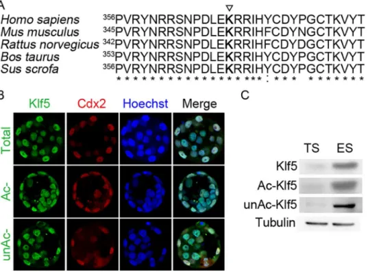
Acetylation of human KLF5 at lysine 369 switches its transcriptional activity from a repressor to an activator for thep15gene in the HaCaT epidermal epithelial cell line and prostate cancer cells [20,28,29]. Whether this acetylation site is conserved in other species, and plays critical regulatory function in other biological processes, such as the segregation of the ICM and the TE, remains to be explored. We first analyzed the homology of the Klf5 proteins across differ-ent species. Indeed, Klf5 is highly conserved among human, chimpanzee, Rhesus monkey, dog, cow, mouse, rat, chicken, zebrafish, and frog. For example, human and mouse Klf5 share 87% identity. Moreover, peptide sequences around the known acetylation site of human KLF5 (K369) have even higher homology across species, almost identical for human, mouse, rat, cow, and pig Klf5 (Fig 1A). The highly conserved sequence at the acetylation site implies that the regulatory function of Klf5 acetylation is likely conserved in these species. In addition, through the sequence alignment, the lysine residue 358 of mouse Klf5 protein is identified as a potential acetylation site.
We then tested whether Klf5 has differential acetylation status in ICM and TE cells, leading to the segregation of these two types of cells? We examined the expression of Klf5, Ac- and unAc-Klf5) in the blastocyst by immunofluorescence. Consistent with previous reports [14, 19], Klf5 is ubiquitously expressed in the blastocyst. In addition, no biased expression of Ac-Klf5 and unAc-Ac-Klf5 in ICM and TE cells was observed (Fig 1B). We further examined the expression of Klf5, Ac- and unAc-Klf5 in ESCs and TSCs, thein vitrocounterparts of the ICM and the TE. Again, all three forms are present in ESCs and TSCs. Even though ESCs express more Klf5 than TSCs, the ratios of Ac-Klf5 to total Klf5 in ESCs and in TSCs are about the same (Fig 1C). The similar acetylation levels of Klf5 in the ICM and the TE, as well as theirin vitrocounterparts, rule out the possibility that differential acetylation status of Klf5 facilitates the differentiation of the ICM and the TE.
Klf5 acetylation promotes the self-renewal of mouse ESCs
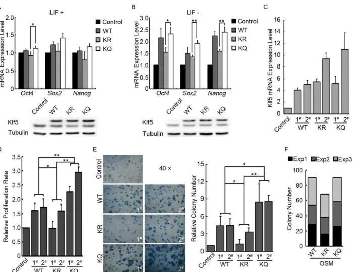
any difference among WT, KR, and KQ Klf5. To better detect the functional difference of WT and mutant Klf5, we repeated the overexpression experiment, except for that LIF was with-drawn from the ESC medium upon transfection to allow spontaneous ESC differentiation. Under this experimental condition, the activation effect of Klf5 onOct4,Sox2, andNanog became obvious. More importantly, WT and KQ Klf5 activate the key pluripotency genes more efficiently, compared to KR Klf5. And Western blot demonstrated the similar expression levels of WT, KR and KQ Klf5 (Fig 2B). These data suggested that acetylation of Klf5 enhances its transcriptional activity in ESCs.
We then asked whether acetylation of Klf5 regulates the proliferation and the self-renewal of ESCs. Stable ESC lines overexpressing WT, KQ, and KR Klf5 were first constructed, and the expression levels ofKlf5in different cell lines were quantified (Fig 2CandS1 Fig). While cul-turing these cell lines, we noticed significant variation of proliferation rates. At a similar expres-sion level ofKlf5(WT 1# and 2#, KQ 1#, and KR 1#), KQ Klf5 ESCs proliferates faster than ESCs expressing WT and KR Klf5. Even when the variation of Klf5 expression level was taken into account, reusable two-factor analysis of variance revealed that the effects of WT, KR, and KQ Klf5 on ESC proliferation are significantly different. Moreover, the proliferation rate of ESCs is correlates withKlf5expression level. The clones expressing moreKlf5(KQ 2# and KR 2#) grow faster than the clones expressing lessKlf5(KQ 1# and KR 1#), respectively (Fig 2D). A similar result was also observed in colony forming assays. KQ Klf5 ESCs form more AP Fig 1. Klf5 expression in the blastocyst, ESCs and TSCs.(A) Comparison of different species Klf5 protein sequences around the human KLF5 acetylation site. The numbers mark the coordination of the first shown residues“P”in Klf5 proteins of different species. The triangle highlights the conserved lysine residue 369 that is acetylated in human KLF5. (B) Immunofluorescence staining of total, acetylated (Ac-), and unacetylated (unAc-) Klf5 in the blastocyst. Cdx2 expression was shown to mark TE cells, and nuclei were stained by Hoechst. (C) Western blot of total, Ac-, and unAc-Klf5 in mouse ESCs and TSCs. Tubulin was used as a loading control.
positive colonies than ESCs expressing WT and KR Klf5, while KR Klf5 ESCs give rise to the least AP positive colonies (Fig 2E).
Now we have demonstrated that acetylation of Klf5 facilitates the activation of key pluripo-tency genes, and promotes ESC proliferation and self-renewal. Does Ac-Klf5 outperform unAc-Klf5 in somatic cell reprogramming? To address this question, WT, KQ, or KR Klf5, together with Oct4, Sox2, and c-Myc, were used to reprogram mouse embryonic fibroblasts (MEFs). It appears that reprogramming with WT and KQ Klf5 yields more induced pluripotent Fig 2. Functions of Ac-Klf5 in self-renewal and pluripotency regulation.(A) The effect of WT, KR, and KQ Klf5 overexpression on the expression of pluripotency genes in mouse ESCs. Empty pCAGIPuro, Flag-tagged WT, KR and KQ Klf5 overexpression plasmids were transfected into V6.5 ESCs with Lipofectamine 3000. Two days after transfection, cells were harvested and subjected to RNA purification or Western blot. The top panel shows RNA quantification results (averages and standard deviations from three independent experiments were plotted), and the bottom panel shows Western blots to demonstrate the similar expression levels of Flag-tagged WT, KR and KQ Klf5. (B) The effect of WT, KR, and KQ Klf5 overexpression on the expression of pluripotency genes during mouse ESC differentiation. Experiments were carried out as described in (A), except for that LIF was withdrawn from the medium upon transfection. Averages and standard deviations from three independent experiments were plotted. (C) Relative expression level of total Klf5 mRNA in control, WT, KR, and KQ Klf5 overexpression ESC lines. Averages and standard deviations from three independent experiments were plotted. (D)
Proliferation rates of control, WT, KQ, and KR Klf5 overexpression ESCs. 1.5×10⁵Cells were seeded in one well of a 6-well plate, and 48 hours later, total cell number was counted to measure the proliferation rate. Averages and standard deviations from three independent experiments were plotted. P values were calculated by reusable two-factor analysis of variance. (E) Colony forming assays for control, WT, KR, and KQ Klf5 overexpression ESCs growing without LIF and feeder cells. The left panel shows the image of AP stained colonies of each ESC line, and the quantification result of colony number is plotted in the right panel. Averages and standard deviations from three independent experiments were plotted. P values were calculated by reusable two-factor analysis of variance. (F) Reprogramming ability of WT, KR, and KQ Klf5 in combination with Oct4, Sox2 and c-Myc. MEFs were reprogrammed by WT, KQ, or KR Klf5, together with Oct4, Sox2, and c-Myc. Accumulated iPS colony numbers of three independent experiments (labeled with three different colors) are shown in the plot.
stem (iPS) cell colonies, compared to reprogramming with KR Klf5, even though the difference is not statistically significant (Fig 2F).
TGF-
β
signaling activates acetylation of Klf5 in mouse ESCs
We have shown the function of Klf5 acetylation in mouse ESCs, how Klf5 acetylation is regu-lated in ESCs remains to be explored. It has been demonstrated that TGF-βsignaling induces acetylation of KLF5 in cultured noncancerous epithelial cells, as well as prostate cancer cell lines [20,29]. To test whether TGF-βsignaling also regulates Klf5 acetylation in ESCs, we treated V6.5 ESCs with TGF-βor a TGF-βinhibitor SB525334. When TGF-βwas added, the acetylation level of Klf5 is markedly increased, while other pluripotency factors, including Klf5 itself, do not show any significant changes, regardless in the presence or absence of LIF. Con-versely, the treatment with SB525334 suppresses Klf5 acetylation, while total Klf5 expression level remains stable. Inhibition of TGF-βsignaling also down-regulates the expression of pluri-potency factors, Oct4, Sox2, and Nanog (Fig 3A). These data suggest that TGF-βsignaling promotes Klf5 acetylation in ESCs, and that TGF-βsignaling is involved in maintaining the expression of pluripotency genes in ESCs.
To address whether TGF-βsignaling activates the expression of Oct4, Sox2, and Nanog through acetylation of Klf5, WT and KQ Klf5 were overexpressed in V6.5 ESCs, and the cells were treated with SB525334. Overexpression of WT Klf5 does not affect the down-regulation of Oct4, Sox2, and Nanog proteins caused by inhibition of TGF-βsignaling. However, overex-pression of KQ Klf5 prevents the supoverex-pression of Sox2 protein by SB525334 treatment, while the expression of Oct4 and Nanog still decrease upon inhibition of TGF-βsignaling (Fig 3B). These data indicate that TGF-βsignaling promotes Klf5 acetylation to enhance Sox2 expres-sion. We then asked whether acetylated Klf5 regulatesSox2expression at the transcription level or at the post-transcriptional steps. The expression levels ofOct4,Sox2, andNanog mRNA were analyzed in ESCs overexpressing WT and KQ Klf5, with or without SB525334 treatment. Consistent with Western blot result, in control ESCs, inhibition of TGF-βsignaling suppresses the mRNA levels ofOct4,Sox2, andNanog. The downregulation ofNanogmRNA, but notOct4, by SB525334 treatment, can be prevented by both WT and KQ Klf5 overexpres-sion. WT Klf5 appears to be more potent in maintainingSox2mRNA expression than KQ Klf5, when TGF-βsignaling is inhibited (Fig 3C). These data suggest that acetylated Klf5 enhancesSox2expression post-transcriptionally.
Acetylated Klf5 suppresses differentiation genes, but activates
Cdx2
,
during EB differentiation
andBmp4), endodermal (Gata4andSox17) and mesodermal markers (BrachuryandHand1) in day 4 EBs, while ectodermal markers,NestinandPax6, are not significantly affected by downregulation ofKlf5(Fig 4B). When WT, KR, and KQ Klf5 were overexpressed to rescue the EB differentiation abnormality caused byKlf5knockdown, KQ Klf5 outperforms WT and KR Klf5 in maintaining the expression of pluripotency genes. However, KQ and WT Klf5 have similar functions in suppressing differentiation genes in EBs, except forCdx2andSox17, while KR Klf5 has negligible effect in differentiation gene suppression. Overexpression of WT and KR Klf5 do not affect the expression ofCdx2andSox17, while KQ Klf5 activatesCdx2and repressesSox17in EBs (Fig 4C). All these data suggest that acetylation of Klf5 is essential for its transcriptional activity in differentiated cells.
Discussion
Klf5 is not only required for the development of the ICM, but also for the formation of the functional TE [14,19]. In these two distinct types of cells, certain genes regulated by Klf5 should be expressed differentially. For example,Cdx2is activated in TE cells, and repressed in ICM cells, while the expression ofOct4andNanogare restricted to the ICM [31–34]. How does Klf5 reverse its transcriptional activity in ICM and TE cells? The opposing functions of unAc-KLF5 and Ac-KLF5 in epidermal cells and prostate cancer cells led us to hypothesize that acetylation of Klf5 may switch the transcriptional output during the segregation of the ICM and the TE. However, when we examined the expression of total, Ac-, and unAc-Klf5 in the blastocyst, no biased expression patterns of all types of Klf5 were observed. Rather, total, Ac-, and unAc-Klf5 are evenly expressed in the TE and the ICM. Therefore, the opposing tran-scriptional output is not regulated by acetylation of Klf5. Other post-trantran-scriptional modifica-tions of Klf5 might account for the reversed transcriptional activity of Klf5 in the TE and the ICM. It has been shown that in C2C12 cells, sumoylated KLF5 interacts with unliganded per-oxisome proliferator-activated receptor-δ(PPAR-δ) and co-repressors to form transcription-ally repressive regulatory complexes, whereas unsumoylated KLF5 is associated with liganded PPAR-δand CBP to activate gene expression [27]. Alternatively, the transcriptional activity of Klf5 might be controlled by different transcription factors and regulators available in TE and ICM cells.
Acetylation of Klf5 is detectable in mouse ESCs. In contrast to KLF5 acetylation in epidermal cells and prostate cancer cells, acetylation of Klf5 in ESCs only enhances its transcriptional activ-ity, rather than switching from a transcriptional repressor to an activator. KQ Klf5, mimicking the acetylated status, is more potent than the unacetylated KR Klf5 mutant, in activating pluripo-tency genes, stimulating the proliferation and self-renewal of ESCs, and reprogramming somatic cells. Despite the functional difference, acetylation of Klf5 is regulated by TGF-βsignaling in ESCs, as well as in epidermal cells and prostate cancer cells [20,29]. Interestingly, overexpression of KQ Klf5 antagonizes the downregulation of Sox2 protein by inhibition of TGF-β. It is possible that Ac-Klf5 might form a complex with Sox2 protein, and stabilize Sox2 protein. Another possi-bility is that a downstream target gene activated by Ac-Klf5 might enhance Sox2 protein stapossi-bility. How KQ Klf5 stabilizes Sox2 protein needs to be further investigated.
days, and then harvested for subsequent Western blot experiments. The nonspecific band in the anti-Klf5 blot is marked by an asterisk. (C) The effect of WT and KQ Klf5 overexpression on the repression of pluripotency gene transcription by TGF-βsignaling. Cells were treated as described in (B), and harvested for RNA purification and quantitative RT-PCR analysis. Averages and standard deviations from three independent experiments were plotted.
During ESC differentiation, Ac-Klf5 remains stable, even slightly elevated, while total Klf5 decreases, suggesting that the fraction of Klf5 being acetylated is increased. Again, acetylation of Klf5 enhances its transcriptional activity, regardless being an activator for pluripotency genes, or being a repressor for differentiation genes. The regulation ofCdx2by Klf5 is different from other differentiation genes. In contrast to other differentiation genes,Cdx2is activated by KQ Klf5, consistent with thein vivodata that Klf5 is required forCdx2expression in TE cells Fig 4. Functions of acetylated Klf5 in mouse ESC differentiation.(A) Expression of total, Ac-, and unAc-Klf5 during ESC differentiation. The nonspecific band in the anti-Klf5 blot is marked by an asterisk. V6.5 ESCs were cultured without LIF and feeder cells. Cells were harvested on day 0, 2, 4, and 6, and subjected to Western blot assay. (B) Klf5 knockdown affects the expression of pluripotency genes and differentiation genes in day 4 EBs. Day 4 EBs from shGFP control ESCs and two independent stable Klf5 knockdown ESC clones were harvested for quantitative RT-PCR analysis. Averages and standard deviations from three independent experiments were plotted. (C) Rescue effect of WT, KR, and KQ Klf5 inKlf5knockdown ESCs.shKlf5targeting sequences in WT, KR, and KQ Klf5 were mutated to render them resistant toshKlf5knockdown. Stable ESC lines overexpressing shKlf5-immune WT, KR, and KQ Klf5 were established in the Klf5 knockdown ESCs. Day 4 EBs from these ESCs were harvested for quantitative RT-PCR analysis. Averages and standard deviations from three independent experiments were plotted. P values were calculated by reusable two-factor analysis of variance.
[14]. In TE cells, Klf5, together with other transcription factors, might form a transcriptional activation complex to stimulateCdx2expression. In other lineages of differentiated cells, Klf5 might interact with a distinct set of transcription factors to repress transcription. Alternatively, it is also possible that Klf5 is a strong activator forCdx2, and that the up-regulation of other differentiation genes is due to the down-regulation of pluripotency genes upon Klf5 knock-down. WT and KR Klf5 fail to activateCdx2in EBs, implying that Klf5 acetylation might be necessary to activateCdx2in TE cells.
In summary, our study elucidates the function of Klf5 acetylation in embryo development, ESC self-renewal and differentiation. In these biological processes, acetylation of Klf5 increases its transcriptional activity, rather than switching from a repressor to an activator. TGF-β sig-naling promotes acetylation of Klf5. Detailed mechanisms for acetylation and deacetylation of Klf5 require further investigation.
Supporting Information
S1 Fig. Confirmation of the overexpression of WT, KR and KQ Klf5 in overexpression clones.Cell lysates from WT, KR and KQ Klf5 stable overexpression clones were subjected to Western blot. The clone 1# and 2# of WT, KR and KQ overexpression clones were used inFig 2C–2E.
(TIF)
S1 Table. Primer sequences used in this study. (DOC)
Acknowledgments
We thank Dr. Jin-Tang Dong for providing total Klf5, Ac-Klf5, unAc-Klf5 antibodies.
Author Contributions
Conceived and designed the experiments: LC. Performed the experiments: TZ CL. Analyzed the data: TZ CL LC. Contributed reagents/materials/analysis tools: TZ CL LC. Wrote the paper: TZ CL LC.
References
1. Cockburn K, Rossant J. Making the blastocyst: lessons from the mouse. J Clin Invest. 2010; 120 (4):995–1003. Epub 2010/04/07. 41229 [pii]doi:10.1172/JCI41229PMID:20364097; PubMed Central PMCID: PMC2846056.
2. Zernicka-Goetz M, Morris SA, Bruce AW. Making a firm decision: multifaceted regulation of cell fate in the early mouse embryo. Nat Rev Genet. 2009; 10(7):467–477. PMID:19536196. doi:10.1038/ nrg2564
3. Evans MJ, Kaufman MH. Establishment in culture of pluripotential cells from mouse embryos. Nature. 1981; 292(5819):154–156. PMID:7242681.
4. Martin GR. Isolation of a pluripotent cell line from early mouse embryos cultured in medium conditioned by teratocarcinoma stem cells. Proc Natl Acad Sci U S A. 1981; 78(12):7634–7638. PMID:6950406.
5. Jaenisch R, Young R. Stem cells, the molecular circuitry of pluripotency and nuclear reprogramming. Cell. 2008; 132(4):567–582. Epub 2008/02/26. S0092-8674(08)00115-3 [pii]doi:10.1016/j.cell.2008. 01.015PMID:18295576.
6. Young RA. Control of the embryonic stem cell state. Cell. 2011; 144(6):940–954. Epub 2011/03/19. S0092-8674(11)00071-7 [pii]doi:10.1016/j.cell.2011.01.032PMID:21414485; PubMed Central PMCID: PMC3099475.
8. Loh YH, Wu Q, Chew JL, Vega VB, Zhang W, Chen X, et al. The Oct4 and Nanog transcription network regulates pluripotency in mouse embryonic stem cells. Nat Genet. 2006; 38(4):431–440. PMID: 16518401.
9. Chen X, Xu H, Yuan P, Fang F, Huss M, Vega VB, et al. Integration of external signaling pathways with the core transcriptional network in embryonic stem cells. Cell. 2008; 133(6):1106–1117. PMID: 18555785. doi:10.1016/j.cell.2008.04.043
10. Kim J, Chu J, Shen X, Wang J, Orkin SH. An extended transcriptional network for pluripotency of embryonic stem cells. Cell. 2008; 132(6):1049–1061. PMID:18358816. doi:10.1016/j.cell.2008.02.039
11. Jiang J, Chan YS, Loh YH, Cai J, Tong GQ, Lim CA, et al. A core Klf circuitry regulates self-renewal of embryonic stem cells. Nat Cell Biol. 2008; 10(3):353–360. PMID:18264089. doi:10.1038/ncb1698
12. Zhang P, Andrianakos R, Yang Y, Liu C, Lu W. Kruppel-like factor 4 (Klf4) prevents embryonic stem (ES) cell differentiation by regulating Nanog gene expression. J Biol Chem. 2010; 285(12):9180–9189. Epub 2010/01/15. M109.077958 [pii]doi:10.1074/jbc.M109.077958PMID:20071344; PubMed Central PMCID: PMC2838337.
13. Parisi S, Passaro F, Aloia L, Manabe I, Nagai R, Pastore L, et al. Klf5 is involved in self-renewal of mouse embryonic stem cells. J Cell Sci. 2008; 121(Pt 16):2629–2634. Epub 2008/07/26. jcs.027599 [pii]doi:10.1242/jcs.027599PMID:18653541.
14. Ema M, Mori D, Niwa H, Hasegawa Y, Yamanaka Y, Hitoshi S, et al. Kruppel-like factor 5 is essential for blastocyst development and the normal self-renewal of mouse ESCs. Cell Stem Cell. 2008; 3 (5):555–567. Epub 2008/11/06. S1934-5909(08)00456-6 [pii]doi:10.1016/j.stem.2008.09.003PMID: 18983969.
15. Long X, Singla DK. Inactivation of Klf5 by zinc finger nuclease downregulates expression of pluripotent genes and attenuates colony formation in embryonic stem cells. Mol Cell Biochem. 2013; 382(1–
2):113–119. Epub 2013/06/20. doi:10.1007/s11010-013-1724-5PMID:23780512.
16. Yeo JC, Jiang J, Tan ZY, Yim GR, Ng JH, Goke J, et al. Klf2 is an essential factor that sustains ground state pluripotency. Cell Stem Cell. 2014; 14(6):864–872. Epub 2014/06/07. S1934-5909(14)00152-0 [pii]doi:10.1016/j.stem.2014.04.015PMID:24905170.
17. Nakagawa M, Koyanagi M, Tanabe K, Takahashi K, Ichisaka T, Aoi T, et al. Generation of induced plu-ripotent stem cells without Myc from mouse and human fibroblasts. Nat Biotechnol. 2008; 26(1):101–
106. PMID:18059259.
18. Aksoy I, Giudice V, Delahaye E, Wianny F, Aubry M, Mure M, et al. Klf4 and Klf5 differentially inhibit mesoderm and endoderm differentiation in embryonic stem cells. Nat Commun. 2014; 5:3719. Epub 2014/04/29. ncomms4719 [pii]doi:10.1038/ncomms4719PMID:24770696.
19. Lin SC, Wani MA, Whitsett JA, Wells JM. Klf5 regulates lineage formation in the pre-implantation mouse embryo. Development. 2010; 137(23):3953–3963. Epub 2010/10/29. dev.054775 [pii]doi:10. 1242/dev.054775PMID:20980403; PubMed Central PMCID: PMC2976279.
20. Guo P, Dong XY, Zhang X, Zhao KW, Sun X, Li Q, et al. Pro-proliferative factor KLF5 becomes anti-pro-liferative in epithelial homeostasis upon signaling-mediated modification. J Biol Chem. 2009; 284 (10):6071–6078. Epub 2008/12/06. M806270200 [pii]doi:10.1074/jbc.M806270200PMID:19056724; PubMed Central PMCID: PMC2649083.
21. Zhang Z, Teng CT. Phosphorylation of Kruppel-like factor 5 (KLF5/IKLF) at the CBP interaction region enhances its transactivation function. Nucleic Acids Res. 2003; 31(8):2196–2208. Epub 2003/04/12. PMID:12682370; PubMed Central PMCID: PMC153738.
22. Matsumura T, Suzuki T, Aizawa K, Munemasa Y, Muto S, Horikoshi M, et al. The deacetylase HDAC1 negatively regulates the cardiovascular transcription factor Kruppel-like factor 5 through direct interac-tion. J Biol Chem. 2005; 280(13):12123–12129. Epub 2005/01/26. M410578200 [pii]doi:10.1074/jbc. M410578200PMID:15668237.
23. Miyamoto S, Suzuki T, Muto S, Aizawa K, Kimura A, Mizuno Y, et al. Positive and negative regulation of the cardiovascular transcription factor KLF5 by p300 and the oncogenic regulator SET through interac-tion and acetylainterac-tion on the DNA-binding domain. Mol Cell Biol. 2003; 23(23):8528–8541. Epub 2003/ 11/13. PMID:14612398; PubMed Central PMCID: PMC262669.
24. Chen C, Sun X, Guo P, Dong XY, Sethi P, Cheng X, et al. Human Kruppel-like factor 5 is a target of the E3 ubiquitin ligase WWP1 for proteolysis in epithelial cells. J Biol Chem. 2005; 280(50):41553–41561. Epub 2005/10/15. M506183200 [pii]doi:10.1074/jbc.M506183200PMID:16223724.
26. Du JX, Bialkowska AB, McConnell BB, Yang VW. SUMOylation regulates nuclear localization of Krup-pel-like factor 5. J Biol Chem. 2008; 283(46):31991–32002. Epub 2008/09/11. M803612200 [pii]doi:10. 1074/jbc.M803612200PMID:18782761; PubMed Central PMCID: PMC2581587.
27. Oishi Y, Manabe I, Tobe K, Ohsugi M, Kubota T, Fujiu K, et al. SUMOylation of Kruppel-like transcrip-tion factor 5 acts as a molecular switch in transcriptranscrip-tional programs of lipid metabolism involving PPAR-delta. Nat Med. 2008; 14(6):656–666. Epub 2008/05/27. nm1756 [pii]doi:10.1038/nm1756PMID: 18500350.
28. Guo P, Zhao KW, Dong XY, Sun X, Dong JT. Acetylation of KLF5 alters the assembly of p15 transcrip-tion factors in transforming growth factor-beta-mediated inductranscrip-tion in epithelial cells. J Biol Chem. 2009; 284(27):18184–18193. Epub 2009/05/08. M109.007096 [pii]doi:10.1074/jbc.M109.007096PMID: 19419955; PubMed Central PMCID: PMC2709394.
29. Li X, Zhang B, Wu Q, Ci X, Zhao R, Zhang Z, et al. Interruption of KLF5 acetylation converts its function from tumor suppressor to tumor promoter in prostate cancer cells. Int J Cancer. 2015; 136(3):536–546. Epub 2014/06/17. doi:10.1002/ijc.29028PMID:24931571; PubMed Central PMCID: PMC4232457.
30. Chen L, Yabuuchi A, Eminli S, Takeuchi A, Lu CW, Hochedlinger K, et al. Cross-regulation of the Nanog and Cdx2 promoters. Cell Res. 2009; 19(9):1052–1061. Epub 2009/07/01. cr200979 [pii]doi:10. 1038/cr.2009.79PMID:19564890.
31. Strumpf D, Mao CA, Yamanaka Y, Ralston A, Chawengsaksophak K, Beck F, et al. Cdx2 is required for correct cell fate specification and differentiation of trophectoderm in the mouse blastocyst. Develop-ment. 2005; 132(9):2093–2102. PMID:15788452.
32. Nichols J, Zevnik B, Anastassiadis K, Niwa H, Klewe-Nebenius D, Chambers I, et al. Formation of plu-ripotent stem cells in the mammalian embryo depends on the POU transcription factor Oct4. Cell. 1998; 95(3):379–391. PMID:9814708.
33. Chambers I, Colby D, Robertson M, Nichols J, Lee S, Tweedie S, et al. Functional expression cloning of Nanog, a pluripotency sustaining factor in embryonic stem cells. Cell. 2003; 113(5):643–655. PMID: 12787505.