N U S A N T A R A B I O S C I E N C E ISSN: 2087-3948
Vol. 7, No. 2, pp. 73-76 E-ISSN: 2087-3956
November 2015 DOI: 10.13057/nusbiosci/n070203
Short Communication:
Isolation and biochemical characterization of
Rhizobium
strains from
nodules of lentil and pea in Tarai agro-ecosystem, Pantnagar, India
SANDEEP PRAKASH UPADHAYAY1,♥, NAVNEET PAREEK1, GAURAV MISHRA1,2,♥♥ 1Department of Soil Science, College of Agriculture , G. B. Pant University of Agriculture and Technology, Pantnagar, India-263 145.
email: [email protected]
2Scientist, Rain Forest Research Institute, Jorhat, Assam, India-785001.
email: [email protected]
Manuscript received: 18 August 2015. Revision accepted: 31 August 2015.
Abstract.Upadhayay SP, Pareek N, Mishra G. 2015. Isolation and biochemical characterization of Rhizobium strains from nodules of lentil and pea in Tarai agro-ecosystem, Pantnagar, India. Nusantara Bioscience 7: 73-76.Root nodules were collected from young and healthy seedling ofPisum sativum L andLens culinarisL from the field at different locations of Norman E. Borloug Crop Research Center, G.B.P.U.A. & T., Pantnagar, Uttarakhand state, India. FifteenRhizobium strains were isolated from the root nodule of P. sativum andL. culinarisand characterized by standard tests. All strains were gram-negative and did not absorb red color when cultured in YEMA containing congo red. In the ketolactose test yellowish zone of Cu2O not found. Also isolates showed either poor or no growth on the glucose peptone medium after one day which is indicating character of rhizobia Thirteen isolates were fast grower and only two were slow growers which is confirm by bromothymol blue test. Results confirmed that isolated strains wereRhizobium.
Keywords:Lentil, pea, root nodules,Rhizobium,YEMA
INTRODUCTION
Pulses are unique, having god gifted ability to fix nitrogen in symbiosis with rhizobial soil bacteria. Role of this symbiosis is clearly defined; in nodules bacteria reduce atmospheric nitrogen to ammonia for plant nitrogen nutrition. So, pulses are known to increase nitrogen status in soil and improving the soil fertility (Riah et al. 2014). There are about 750 genera of legumes (Young and Haukka 1996). Although most rhizobia are host specific, but it is also true that several different bacterial species are also isolated from a single legume species and it is only from limited hosts which have been examined as far as micro-symbionts are concerned (Arora et al. 2001). Lentil is known for its human nutrition ability and to maintain soil fertility (Rashid et al. 2014) while pea for its short term and highly beneficial nature (Wadhwa et al. 2011). There is huge amount of literature available, reporting rhizobia from different pulses (Riah et al. 2014; Wadhwa et al. 2011; Hou et al. 2009 and many others) but limited studies are there about the biochemical characterization of rhizobia inhabiting lentil and pea. Rhizobia are characterized on the basis of biochemical tests. So, this study was aimed to isolate and identify rhizobia on the basis of biochemical tests from root nodules of lentil and pea for better agriculture growth.
MATERIALS AND METHODS
Sample collection
Plants samples along with roots and nodules were collected at 50 days after sowing from various pea and lentil fields at Norman E. Borloug Crop Research Center, G.B.P.U.A. & T., Pantnagar, Uttarakhand, India. The roots along with mature nodules were thoroughly washed in running water until the removal of adhering soil particles. Big sized and pink colored nodule preferably on tap root were selected and transported to the laboratory for further investigation by following the method given by Vincent (1974).
Isolation ofRhizobiumfrom root nodules
Selected root nodules were dipped in 0.1% mercuric chloride (HgCl2) solution for 30 sec and later washed
successively ten times with sterilized distilled water to remove the traces of toxic HgCl2. Surface sterilized
N U S A N T A R A B I O S C I E N C E 7 (2): 73-76, November 2015
74
Biochemical tests
Biochemical tests such as Congo red test (Vincent 1974), Ketolactose test (Bernaerts and De Ley 1963), Bromothymol blue (BTB) agar test (Somasegaran and Hoben 1994) and glucose-peptone agar test (Kleczkowska et al. 1968) were done to differentiate Rhizobium and
Agrobacterium.
RESULTS AND DISCUSSIONS
A total of 15 bacterial strains were isolate from root nodules of Lentil (Lens culinaris L.) and Pea (Pisum sativum L.). Phenotypically isolated rhizobial colonies were cream colored with slime/mucoid transparent appearance on CRYEMA plates with marked distinction from red colored colonies of Agrobacterium (Figure 1).
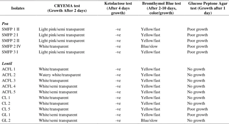
Further conformity of rhizobia was performed on ketolactose agar showed, all 15 isolates were negative for the production of 3 ketolactose from lactose no yellow zone of Cu2O around the colonies were observed (Figure 3)
which was indicative of Agrobacterium (Table 1). Production of 3-ketolactose from lactose is limited to species of Agrobacterium. Similarly, Gachande and Khansole (2011) isolated Rhizobium japonicum and Brady
Rhizobium japonicum colonies, which were circular in shape with whitish pink color on CRYEMA medium. Absence of 3 ketolactose in rhizobial colonies on CRYEMA was also in agreement with earlier work carried out by Sadowsky et al. (1983) and Sharma et al. (2010).
While further confirming these all fifteen isolates showed either poor or no growth on the glucose peptone
medium after one day indicating character of rhizobia as a conventional rule (Figure 5).Rhizobium strains growth are found poor on glucose peptone agar media while the other bacteria grow well on this media. Further purified isolates were classified as fast (turn medium yellow) and slow growing (turn medium blue) rhizobia on YEMA supplemented with BTB (Figure 4). The rhizobial isolates in the current study were further tested on YEMA plates containing BTB indicated that thirteen fast growing isolates (SMFP 1 II, SMFP 2 I, SMFP 2 II,
SMFP 3 I, ACFL 1, ACFL 2, ACFL 3, ACFL 4, ACFL 5, CL 1, CL 2, CL 5, GL 1) were found to produce yellow colonies due to acid production on the medium with high amount of mucus after 2 days of incubation (Table 1). Whereas, remaining two isolates along with reference strains SMFP 2 IV and GL 2 produced blue color colonies, which indicated the presence of alkali producers, considered as slow growing rhizobia. The use of YEMA-BTB medium for categorizing indigenous root nodulating fast and slow growing rhizobia based on acid/alkali production was also carried out by Saeki et al. (2005) in Vietnam and Sharma et al. (2010) in India. On BTB agar plates both fast and slow growing rhizobia formed circular, convex, colonies. The isolates were classified tentatively as fast (medium turn yellow) and slow growers (medium turn blue) based on their reaction on the yeast extract mannitol agar supplemented with bromothymol blue (Somasegaran and Hoben 1994). These isolates were similar in terms of reaction on the YEMA (BTB) when compared with reference strains which produced yellow and blue color in fast and slow growing strains, respectively according to Hungria et al. (2001).
Table 1.Results of different tests ofRhizobialisolates
Isolates CRYEMA test
(Growth After 2 days)
Ketolactose test (After 4 days
growth)
Bromthymol Blue test (After 2-10 days,
color/growth)
Glucose Peptone Agar test (Growth after 1
day)
Pea
SMFP 1 II Light pink/semi transparent -ve Yellow/fast Poor growth
SMFP 2 I Light pink/semi transparent -ve Yellow/fast Poor growth
SMFP 2 II Light pink/semi transparent -ve Yellow/fast Poor growth
SMFP 2 IV White/transparent -ve Blue/slow Poor growth
SMFP 3 I Light pink/semi transparent -ve Yellow/fast Poor growth
Lentil
ACFL 1 White/transparent -ve Yellow/fast No growth
ACFL 2 Watery white/transparent -ve Yellow/fast No growth
ACFL 3 White/transparent -ve Yellow/fast No growth
ACFL 4 White/semi transparent -ve Yellow/fast No growth
ACFL 5 White/semi transparent -ve Yellow/fast No growth
CL 1 White/transparent -ve Yellow/fast No growth
CL 2 White/transparent -ve Yellow/fast No growth
CL 5 White/transparent -ve Yellow/fast Poor growth
GL 1 White/semi transparent -ve Yellow/fast Poor growth
UPADHAYAY et al.–Characterization ofRhizobium strains from Pantnagar, India 75
Our findings congruence with Gachande and Khansole (2011)as reported for soybean rhizobia. Sadowsky et al. (1983) also reported that production of 3 ketolactose from lactose is limited to species ofAgrobacterium, a genus that is closely related to Rhizobium. Mahana et al. (2000) also showed negative chemical reaction for 3 ketolactose productions by isolates of Rhizobium from Vigna mung. Similarly Shetta et al. (2011) mentioned that Rhizobium
strains failed to absorb congo red stain in the CRYEMA medium. Rhizobium is symbiotic bacteria which form nodule in leguminous plant. ButAgrobacterium infects the root and forms false nodule (pseudo nodule) therefore, biochemical tests are essential to differentiate Rhizobium
and Agrobacterium. All the biochemical test and cited literature suggested that isolated strains wereRhizobium.
In conclusion, all 15Rhizobium strains did not absorb red color when cultured in YEMA containing congo red medium. Pseudo-nodule forming bacteria Agrobacterium
utilized congo red but Rhizobium strains didn’t utilize
congo red. This test is essential to differentiateRhizobium
andAgrobacterium. Other biochemical tests confirmed that isolated strains wereRhizobium.
Figure 2.Different isolates ofRhizobium strains on YEMA
Figure 1.Growth ofRhizobium strains on CRYEMA media Figure 3. Ketolactose test (No yellow ring of Cu2O around the growth ofRhizobium)
Figure 4.Bromthymol Blue test (Yellow and Blue color change of media after 2. 10 days).
N U S A N T A R A B I O S C I E N C E 7 (2): 73-76, November 2015
76
REFERENCES
Arora NK, Kang S. C, Maheshwari D. K. 2001. Isolation of siderophore producing strains ofRhizobium melilotiand their biocontrol potential against Macrophomina phaseolina that causes charcoal rot of groundnut. Curr Sci 81: 673-677.
Bernaerts MJ, De Ley J. 1963. A biochemical test for crown gall bacteria. Nature (London) 197: 406-407.
Gachande BD, Khansole GS. 2011. Morphological, cultural and biochemical characteristics of Rhizobium japonicum syn and
Bradyrhizobium japonicumof soybean. Biosci Discov 2: 1-4. Hou BC, Wang ET, Li Y, Jia RZ, Chen WF, Man CX, Sui XH, Chen WX.
2009. Rhizobial resource associated with epidemic legumes in Tibet. Microb Ecol 57: 69-81.
Hungria M, Campo RJ, Chueire LMO, Grange L, Megías M. 2001. Symbiotic effectiveness of fast-growing rhizobial strains isolated from soybean nodules in Brazil. Biol Fert Soils 33: 387-394. Kleczkowska J, Nutman PS, Skinner FA, Vincent JM. 1968. The
identification and classification of Rhizobium. In: Gibbs BM, Shapton DA (eds). Identification Methods for Microbiologists, Part B. Academic Press, London.
Mahana SK, Garg R, Parvateesam M. 2000. Cultural and biochemical characteristics of root nodule bacteria from induced mutants ofVigna mungL.Seed Pathology. Printwell Publications, Jaipur.
Rashid MH, Gonzalez J, Young JPW, Wink M. 2014. Rhizobium leguminosarum is the symbiont of lentils in the Middle East and Europe but not in Bangladesh. FEMS Microbiol Ecol 87: 64-77.
Riah N, Bena G, Heulinb KA, Lajudieb P, Laguerre G. 2014. Genotypic and symbiotic diversity of Rhizobium populations associated with cultivated lentil and pea in sub-humid and semi-arid regions of Eastern Algeria. Syst Appl Microbiol DOI: 10.1016/j.syapm.2013.12.008
Sadowsky MJ, Keyser HH, Bohlool BB. 1983. Biochemical characterization of fast-and slow-growing rhizobia that nodulate soybeans. Int J Syst Bacteriol 33: 716-722.
Saeki Y, Kaneko A, Hara T, Suzuki K, Yamakawa T, Nguyen M.T, Nagatomo Y, Akao S. 2005. Phylogenetic analysis of soybean-nodulating rhizobia isolated from alkaline soils in Vietnam. Soil Sci Plant Nutr 51: 1043-1052.
Sharma MP, Srivastava K, Sharma SK. 2010. Biochemical characterization and metabolic diversity of soybean rhizobia isolated from Malwa region of Central India. Plant Soil Environ 56: 375-383. Shetta ND, Al-Shaharani TS, Abdel-Aal M. 2011. Identification and
characterization of Rhizobiumassociated with woody legume trees grown under Saudi Arabia condition. Am Eur J Agric Environ Sci 10 (3): 410-418.
Somasegaran P, Hoben HJ. 1994. Handbook for Rhizobia: Methods in legume-RhizobiumTechnology. Springer-Verlag, New York. Vincent JM. 1974. Root-nodule symbiosis withRhizobium. In: Quispel A
(ed.). Biology of Nitrogen Fixation. North-Holland Publishing Co., Amsterdam.
Wadhwa K, Dudeja SS, Yadav RK. 2011. Molecular diversity of native rhizobia trapped by five field pea genotypes in Indian soils. J Basic Microbiol 51: 89-97.