Submitted14 March 2016 Accepted 11 May 2016 Published9 June 2016
Corresponding author Bhim P. Singh,
bhimpratap@gmail.com
Academic editor Paul Tulkens
Additional Information and Declarations can be found on page 13
DOI10.7717/peerj.2103
Copyright 2016 Zothanpuia et al.
Distributed under
Creative Commons CC-BY 4.0
OPEN ACCESS
Detection of antibiotic-resistant bacteria
endowed with antimicrobial activity from
a freshwater lake and their phylogenetic
affiliation
Zothanpuia1, Ajit K. Passari1, Vijai K. Gupta2and Bhim P. Singh1
1Department of Biotechnology, Mizoram University, Aizawl, Mizoram, India 2Molecular Glyco-biotechnology Group, University of Ireland, Galway, Ireland, UK
ABSTRACT
Antimicrobial resistance poses a serious challenge to global public health. In this study, fifty bacterial strains were isolated from the sediments of a freshwater lake and were screened for antibiotic resistance. Out of fifty isolates, thirty-three isolates showed resistance against at least two of the selected antibiotics. Analysis of 16S rDNA sequencing revealed that the isolates belonged to ten different genera, namely
Staphylococcus(n=8), Bacillus(n=7), Lysinibacillus(n=4),Achromobacter(n=3),
bacterium(n=3),Methylobacterium(n=2),Bosea(n=2), Aneurinibacillus(n=2),
Azospirillum(n=1), Novosphingobium(n=1). Enterobacterial repetitive intergenic
consensus (ERIC) and BOX-PCR markers were used to study the genetic relatedness among the antibiotic resistant isolates. Further, the isolates were screened for their antimicrobial activity against bacterial pathogens viz.,Staphylococcus aureus
(MTCC-96), Pseudomonas aeruginosa(MTCC-2453) and Escherichia coli(MTCC-739), and
pathogenic fungi viz., Fusarium proliferatum (MTCC-286), Fusarium oxysporum
(CABI-293942) andFusarium oxy. ciceri(MTCC-2791). In addition, biosynthetic genes (polyketide synthase II (PKS-II) and non-ribosomal peptide synthetase (NRPS)) were detected in six and seven isolates, respectively. This is the first report for the multifunctional analysis of the bacterial isolates from a wetland with biosynthetic potential, which could serve as potential source of useful biologically active metabolites.
SubjectsMicrobiology
Keywords BOX-PCR, Antibiotic susceptibility, PKS II, ERIC-PCR, NRPS, 16S rRNA gene
INTRODUCTION
Bacteria play a vital role in benthic food web, nutrient recycling and decomposition of various organic compounds in aquatic environments (Fischer, Wanner & Pusch,2002). Sediment is a special habitat among the aquatic ecosystem and the numbers of microbes are much higher than the corresponding water bodies (Zinger et al.,2011). The bacteria isolated from such ecosystems have an ecological significance like resistance to antibiotic which would have been adopted in the due course of selection processes (Nair, Chandramohan & Loka-Bharathi,1992;Silva & Hofer,1995).
This can be tackled by the discovery of new antibiotics having an alternate mode of action which can eliminate disease causing pathogenic microbes. Screening of microorganisms from their natural habitat is an important step for the isolation of therapeutic compounds (Newman & Cragg,2012). As a result, researchers are trying to look for new organisms which have the potential to produce novel antibiotics from unexplored habitats (Oskay, Tamer & Azeri,2004). Aquatic microorganisms are of special interest as they have not been exploited extensively compared to terrestrial microbes (Zhang et al.,2005).
Many molecular techniques have been developed in recent years for assessing genomic diversity of bacteria. Molecular identification of bacteria was performed by 16S rRNA gene sequencing as it is most conserved region and less prone to mutations (Kaushlesh et al.,
2012). PCR fingerprinting methods like enterobacterial repetitive intergenic consensus (ERIC)-PCR and BOX-PCR has been extensively used to study genetic relationship as they have discriminatory capability in differentiating different genera of bacteria (Versalovic, Koeuth & Lupski, 1991;Rademaker et al., 2000). Tamdil is a reservoir freshwater lake situated 110 kms from Aizawl, the capital of Mizoram, North East India. The lake is reconstructed as a part of building fishing reservoirs by the Fisheries Department, Government of Mizoram, and is one of the 115 wetlands in India identified under the National Wetland Conservation Programme (vide D.O.No.J/2201/01/10-CS(W)).
The aim of the present study was to assess the diversity of cultivable bacteria from the sediments of the freshwater Tamdil Lake, to study their antimicrobial activities, their resistant to frequently used antibiotics and genetic relationship among the organisms. Investigating the bacterial population in fresh water sediments is of great importance in the general understanding of the aquatic ecosystem.
MATERIALS AND METHODS
Sample collection
Water sediment samples were collected from five different locations of Tamdil Lake (23◦ 44′20.4′′N and 92◦57′10.8′E) during the month of April and May 2013 in a sterile screw capped tube and brought into Molecular Microbiology and Systematics Laboratory, Department of Biotechnology, Mizoram University (Fig. S1). The samples were stored at 4◦
C till processed.
Isolation and antibiotic susceptibility profiling
nitrofurantoin (200µg) were used. Antibiotic sensitivity was observed by measuring the diameters of the inhibition zone in mm and categorized as resistant, intermediate and sensitive to antibiotics.
Genomic DNA extraction, 16S rRNA gene amplification and phylogenetic analysis
Genomic DNA was isolated by using bacterial DNA extraction kit (Invitrogen Life technologies) as per manufacturer’s protocol. The 16S rRNA gene fragment was amplified by using universal primers—PA: 5′
-AGA GTT TGA TCC TGG CTC AG-3′
) and PH: 5′
-AAG GAG GTG ATC CAG CCG CA-3′
(Qin et al.,2009). Reaction was carried out in a total volume of 25µl consisting 1.0µl of genomic DNA (50 ng), 0.2µl of each primer (10 pmol), 2.0µl of deoxynucleotide triphosphates (2.5 mM each), 2.5µl of 1X PCR buffer, 0.2µl of Taq DNA polymerase (1 U/µl) and 15.9µl MilliQ grade water. PCR was performed on Veriti thermal cycler (Applied Biosystem, Singapore) under following conditions: initial denaturation at 95◦
C for 4 min, followed by 30 cycles of denaturation at 94◦
C for 30 s, annealing at 57.5◦C for 40 s and extension at 72◦C for 1.3 min with a final extension step at 72◦
C for 10 min. A negative control reaction mixture without DNA template of bacteria was also included with each set of PCR reactions. The amplified PCR product was run on 1.5% agarose gel and visualized under gel documentation system Bio-Rad XR+system (Hercules, CA, USA). The amplified products were purified using Purelink
PCR Purification Kit (Invitrogen Life technologies) and were sequenced commercially at Sci-Genome Labs Pvt. Ltd, India.
ERIC-PCR fingerprinting The primer sequences ERIC-1R (5′
-CACTTAGGGGTCCTCGAATGTA-3′
) and ERIC-2F (5′
-AAGTAAGTGACTGGGGTGAGCG-3′
) were used to amplify the regions of bacterial genome positioned between the ERIC sequences as described by Versalovic, Koeuth & Lupski (1991). PCR amplification was carried out on a Veriti thermal cycler (Applied Biosystem, Singapore) in a total reaction volume of 25µl. The reaction mixture consist of DNA template (50 ng)—2.5µl, 10X reaction buffer—2.5µl, dNTP mix (10 mM)—2µl, 10 pmol of each primer (ERIC 1R and ERIC 2F), 1µl of MgCl2 (25 mM), and 2 U of Taq DNA polymerase (In-vitrogen, USA). PCR was performed under following conditions: initial denaturation at 95◦
C for 7 min and then subjected to 30 cycles of denaturation at 94◦
C for 1 min, annealing at 50◦
C for 1 min and extension at 65◦
C for 8 min with a final extension step at 65◦C for 16 min. A negative control reaction mixture without DNA template of bacteria was also included with each set of PCR reactions. The amplified products were separated by electrophoresis on a 2% agarose gel using 1X TAE buffer. The PCR bands were analyzed under UV light and documented using a BioRad Gel Doc XR+
system (Hercules, CA, USA).
BOX-PCR fingerprinting
BOXA1R PCR fingerprinting was done using primer sequences BOXA1R (5′
-CTACGG CAAGGCGACGCTGACG-3′
DNA, 2.5µl of 10X Taq Buffer, 1.5µl of MgCl2, 2.0µl of 2.5 mM dNTPs (2.5 mM), 1µl of 10 pmol BOXA1R primer, 1µl of DMSO (10%), 0.5µl of BSA (10 mg/ml) and 1µl of 2 U Taq DNA polymerase. The DNA was amplified under the following conditions: initial denaturation at 95◦
C for 7 min followed by 30 cycles at 94◦
C for 1 min, at 55◦
C for 1 min, and at 65◦
C for 8 min with a final extension step at 65◦
C for 10 min. The amplified products were separated and visualized as stated above.
Screening for antibacterial activity
The selected isolates were screened for their antibacterial activity against three pathogenic bacterial strains; Staphylococcus aureus(MTCC-96),Pseudomonas aeruginosa (MTCC-2453) and Escherichia coli(MTCC-739), all were obtained from the Microbial Type Culture Collection, Institute of Microbial Technology (IMTECH), Chandigarh. Pure isolates were grown in nutrient broth for extract preparation. The grown cultures were centrifugation at 8,000 rpm for three min and the supernatant was used for screening of antimicrobial activity by using agar well diffusion method (Saadoun & Muhana,2008). The test pathogenic bacteria were spread on nutrient agar plate and wells were prepared using sterile cork borer of 6 mm diameter. A total of 50µl clear supernatant of bacterial isolates were dispensed into each wells and the plates were incubated at 37◦
C for 24 h. The antimicrobial activities of the isolates were observed by measuring the diameter of the inhibition zone around each well.
Screening for antifungal activity
All the isolates were screened for their antagonistic activity against three plant pathogenic fungi viz.Fusarium proliferatum(MTCC-286),Fusarium oxysporum(CABI-293942) and
F.usarium oxy. ciceri(MTCC-2791) by dual culturein vitroassay (Bredholdt et al.,2007). All plates were inoculated at 28◦
C for seven days and percentage of inhibition was calculated by using the formula:C−T/C×100, where,Cis the colony growth of fungal pathogen in
control, andT is the colony growth in dual culture.
Detection of biosynthetic gene sequences (PKS II and NRPS) The potential antagonistic isolates were subjected for the amplification of genes for KS domains of Polyketide synthase (PKS-II) and the adenylation domains of non-ribosomal peptide synthetase (NRPS). NRPS gene fragments were amplified using degenerate primers: A3F 5′
-GCSTACSYSATSTACACSTCSGG-3′
and A7R 5′
-SASGTCV CCSGTSGCGTAS-3′
(Ayuso-Sacido & Genilloud,2005). The degenerate primers, KS1F 5′
-TSGCSTGCTTGGAYGCSATC-3′
and KS1R 5′
-TGGAANCCGCCGAABCCTCT-3′
, were used for amplifying PKS-II (Yuan et al.,2014). The PCR products were visualized under gel documentation system as stated above.
PKS II (50µL)
For PKS II gene amplification, we used 3µL of template DNA, 5µL 10X buffer, 1µL of MgCl2 (25 mM), 1µL DMSO (10%), 5µL of dNTP (2.5 mM), 1.8µL each primer (10
mM) and Taq DNA polymerase (2U). PCR conditions as follows: 5 min at 95◦
C, followed by 35 cycles of 1 min at 95◦C, 1 min 30 s at 58◦C and 2 min at 72◦C, followed by a 10-min extension at 72◦
NRPS (50µL)
For NRPS gene amplification, we used 3µL template DNA, 5µL 10X buffer, 1µL of MgCl2
(25 mM), 1µL DMSO (10%), 5µL dNTP (2.5 mM) 2µL each primer (10 mM) and Taq DNA polymerase (2U). PCR conditions as follows: 5 min at 95◦
C, followed by 35 cycles of 1 min at 95 ◦C, 2 min at 59◦C and 4 min at 72◦C, followed by 10 min extension at 72◦C.
Statistical Analysis
DNA Sequences were compared with NCBI GenBank database using BlastN search program and sequences were aligned using the Clustal W software packaged in MEGA 5.05 (Thompson et al.,1997;Kumar et al.,2012). Suitable model was selected according to the lowest BIC (‘‘Bayesian Information Criterion’’) and the highest AIC (‘‘Akaike Information Criterion’’) scores. The phylogenetic tree was constructed by Neighbour joining method using MEGA 5.05 with Kimura 2-parameter model (R=1.53) (Kimura,1980), taking
E. coli as an out group. The robustness of the phylogenetic tree was tested by bootstrap
analysis using 1,000 replicates usingp-distance model (Felsenstein,1985).
Polymorphic DNA band were recorded in binary form i.e., 1 in case of presence of band and 0 when there is no band, to generate a binary matrix (Sneath & Sokal,
1973) for ERIC and BOX. The binary matrix was used to calculate the Simple Matching (SM) coefficient, phylogenetic tree was constructed using the Unweighted Pair Group Method with Arithmetic Mean (UPGMA) method (Lopez & Alippi,2009) using Numerical Taxonomy SYStem (NTSYS version 2.2).
RESULTS
Isolation of bacteria
A total of fifty cultivable bacteria were isolated from the sediment of Tamdil Lake, exhibiting distinct colony characteristics like size, opacity, pigmentation and texture. The colonies of bacterial strains were soft, sticky and also observed with white, red and yellow colours. The microscopic analysis indicates that 67% of the isolates were gram positive and 33% were gram negative bacteria.
Antibiotic sensitivity assay
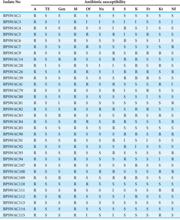
A total of 12 standard known antibiotics were used to screen bacterial strains for their antibiotic sensitivity pattern (Table 1). Out of 50 bacteria examined, 33 strains showed resistance to at least two of the antibiotics tested. All the selected isolates showed resistance against methicillin and ampicillin (100% each). Most of the isolates were susceptible to tetracycline (n=32) followed by norfloxacin (n=29), genatamycin (n=27), furazolidone
(n=24) and ketoconazole (n=23). The parentage degree of resistance to erythromycin,
Table 1 Antibiotic sensitivity profile of bacterial isolates against 12 tested standard antibiotics.
Isolate No Antibiotic susceptibility
A TE Gen M Of Nx E S K Fr Kt Nf
BPSWAC1 R S S R S S S S S S S S
BPSWAC3 R S I R I I S I I S S I
BPSWAC4 R S S R S S I R S I S S
BPSWAC5 R S S R R S R I S R S S
BPSWAC6 R S S R S S S R S S I S
BPSWAC7 R S S R R S S S S S S R
BPSWAC9 R S S R S S R S R R R S
BPSWAC14 R S R R S S R R R S S S
BPSWAC20 R I S R S I I S R S R S
BPSWAC26 R S S R R S I R R R S R
BPSWAC39 R S S R S S S R R R S S
BPSWAC41 R S S R R S R S S S R I
BPSWAC79 R S S R S S R I S R S S
BPSWAC80 R S S R S S S S S I S S
BPSWAC81 R S I R S S S S S S S R
BPSWAC82 R S S R S R R S R R S S
BPSWAC83 R S R R S S S R R S R S
BPSWAC84 R S S R R S R R S S S R
BPSWAC85 R S S R S S R S S S S S
BPSWAC90 R S S R S S S R R S R R
BPSWAC91 R S S R S S R I S I S S
BPSWAC92 R S S R S S S R I S S S
BPSWAC93 R S S R S S R S S S S R
BPSWAC94 R S S R S S S R S S I R
BPSWAC107 R S S R S S S S R S S S
BPSWAC108 R S S R S R R S S S R R
BPSWAC109 R S R R S S R R R S S S
BPSWAC110 R S S R R S S S S S S S
BPSWAC111 R S S R S S I S S S R R
BPSWAC112 R S R R S S S I R S S S
BPSWAC113 R S S R S S S S S S S S
BPSWAC114 R S S R S S S S S S S S
BPSWAC115 R S S R I S I S S S R S
Notes.
Degree of sensitivity: >10 mm, Sensitive; 5.0–9.9 mm, intermediate; 0.0–4.9 mm, resistant.
Gen, Gentamicin (10µg); Nx, Norfloxcin (30µg); T, Tetracycline (30µg); A, Ampicillin (10µg); E, Erythromycin (15µg); S, Streptomycin (30µg); M, methicillin (5µg); Of, ofloxacin (5µg); K, Kanamycin (5µ); Fr, Furazolidone (50µg); Kt, Ketoconazole (50µg); Nf, Nitrofurantoin (200µg).
ERIC-PCR fingerprinting
Figure 1 Dendrogram generated from (A) ERIC-PCR and (B) BOX-PCR genomic fingerprints of bacterial isolates using Ntsys 2.0.
by ERIC-PCR divided the isolates into two clusters (A & B). Cluster A was bigger and divided into 2 sub-clusters (A1 and A2). Cluster A consist of 25 isolates belonging to the genusStaphylococcus,Lysinibacillus, Azospirillum, Bacillus, Bacterium and Aneurinibacillus.
Cluster B consist of 8 isolates comprising different genera belonging toNovosphingobium,
Bosea, Methylobacterium and Achromobacter.Isolates BPSWAC82 and BPSWAC93 showed
100% similarity and were identified asMethylobacteriumbased on 16S rRNA gene sequence. This result agreed with the phylogenetic tree of the 16S rDNA with bootstrap supported value of 88%.
BOX-PCR fingerprinting
BOX-PCR fingerprinting of a total of 33 isolates showed specific patterns corresponding to particular genotypes and size recognizable bands were between <100 bp to 3 kb. A dendrogram generated from BOX-PCR analysis comprised of two major clusters (A & B). Cluster A was larger containing 24 isolates whereas cluster B consist of nine isolates. Cluster A is divided into 2 sub-cluster (A1 and A2).StaphylococcusandLysinibacilluswere grouped together in cluster A1 whereasAchromobacter,BacillusandAneurinibacilluswere grouped together in cluster A2. Cluster B is also divided into two sub-cluster (B1 and B2) comprising of different genera belonging toBacterium, Azospirillum, Methylobacterium, Bosea and
Novosphingobium(Fig. 1B). BOX-PCR analysis demonstrated high discriminatory ability
by constructing genus-specific clusters that were able to differentiate all the different genera reported in this study.
Evaluation of antimicrobial activity
The selected 33 isolates based on their antibiotic susceptibility profile were tested for antimicrobial activities against bacterial pathogens (Staphylococcus aureus, Pseudomonas
Table 2 In vitroantagonistic activity of selected bacterial isolates against fungal and bacterial pathogens and detection of biosynthetic genes.
Isolate no. NCBI accession no
Percentage of inhibition (PI±SD) Zone of inhibition in mm (ZI±SD) PKS II NRPS
F. oxysporum F. oxy. ciceri F. proliferatum E. coli P. aeruginosa S. aureus
BPSWAC9 KM243385 0.00a 0.00a 0.00a 9±0.1a 6±0.29a 9±0.18a − −
BPSWAC14 KM405299 60.50±0.00bc 64.38±0.17bc 0.00a 9±0.1a 12±0.2bc 10±0.05bc + +
BPSWAC20 KM405305 0.00a 58.9±0.26bde 0.00a 10±0.1bc 8±0.10bde 9±0.2a − +
BPSWAC82 KR857324 73.68±0.012bde 61.64±0.02bdfg 73.68±0.12bc 9±0.05a 12±0.15bc 9±0.05a + +
BPSWAC83 KR857325 65.78±0.28bdfg 0.00a 0.00a 7±0.25bde 8±0.05bde − − −
BPSWAC84 KR857326 0.00a 39.72±0.001bdfhi 47.36±0.02bde 9±0.0a 12±0.3bc 10±0.2bc + +
BPSWAC90 KT232317 47.36±0.03bdfhi 45.2±0.02bdfhj 47.36±0.14bde 8±0.02bdf 7±0.15bdef − + +
BPSWAC108 KT429618 60.50±0.03bc 39.72±0.09bdfhi 47.36±0.08bde 9±0.02a 6±0.15a 9±0.1a + +
BPSWAC109 KT429619 50±0.045bdfhj 0.00a 0.00a 9±0.1a 9±0.05bdeg 11±0.1bd + +
Notes.
Mean (±SD) followed by the same letter(s) in each column are not significantly different atP< 0.05 using Duncan’s new multiple range test, (+) and (−) indicates the presence
and absence of PKS II and NRPS genes.
andF. oxy. ciceri). Out of 33 isolates, nine strains exhibited antibacterial activity against two tested pathogens (Table 2). Only two isolates BPSWAC82 and BPSWAC108 showed positive activity against all the bacterial and fungal pathogens. Isolate BPSWAC82 showed highest activity against F. oxysporum(73.68%) andF. proliferatum (73.68%), whereas BPSWAC14 was found high activity against F. oxy.ciceri(64.38%). On the other hand, isolate BPSWAC20 had acute activities against bacterial pathogensE. coli(10 mm), whereas BPSWAC109 exhibited highest activity againstS. aureus(11 mm) (Table 2).
Detection of PKS and NRPS genes in selected strains
Six isolates out of 33 strains visualised band in type II polyketide synthases (PKS-II) with an amplification size of 600 bp (Fig. S2), whereas nonribosomal peptide synthetases (NRPS) genes were detected in seven isolates (21.2%) with expected size of 700 bp (Fig. S2). Isolates BPSWAC14, 82, 84, 90, 108 and 109 showed positive amplification products with both the degenerate primers for PKSII and NRPS respectively (Table 2). Isolates BPSWAC21 showing band against NRPS primers also indicated the highest antimicrobial activities against E. coli pathogens. Isolate BPSWAC14 identified as
Novosphingobium sp. and showed antimicrobial biosynthetic potential in both genes.
This isolates was found as rare genera among them and may be useful for isolation of natural products.
Sequence alignment and phylogenetic analysis
The isolated 33 strains were sequenced by amplification of 16S rRNA gene. All the partial 16S rRNA sequences were aligned using BLAST analysis and deposited in NCBI GeneBank having an accession no. The results showed that the isolates were classified into ten different genera. Majority of the isolates belongs toStaphylococcus (25%),followed byBacillus(21%),
Lysinibacillus(12%),Achromobacter (9%),Bacterium(9%)Methylobacterium(6%),Bosea
Figure 2 Pie chart showing the distribution of bacteria in water sediment of Tamdil Lake.
16S rRNA gene sequences by BlastN exhibited high level of sequence 98–100% similarity confirmed that seven isolates could be members of genusStaphylococcus. The sequences of the three isolates (BPSWAC5, BPSWAC6 and BPSWAC9) showed high identity (99%) to the genus Bacteriumwhereas isolate BPSWAC14 and BPSWAC26 exhibited high identity (88%) to the genusNovosphingobiumandAchromobacter respectively. Maximum-likelihood and neighbor-joining methods were used for the construction of phylogenetic tree. The phylogetic tree generated by both methods showed that allStaphylococcusstrains as well as other genera exceptNovosphingobiumsp. forms a major clade I along with the type strains retrieved from databases. Most of the rare genera likeNovosphingobium
sp.,Boseasp. andMethylobacteriumsp. clustering to form another clade II in Maximum
likelihood tree (Fig. 3B) under the bootstrap value of 30% respectively. However, the neighbour joining tree did not clusterBoseasp. andMethylobacteriumsp. together in clade II (Fig. 3A).
Nucleotide sequence accession numbers
Figure 3 (A) Neighbor-joining phylogenetic tree based on 16S rRNA gene of bacteria identified from Tamdil Lake. (B) Maximum likelihood phy-logenetic tree based on 16S rRNA genes. Numbers at branches indicate bootstrap values of neighbour-joining analysis (>50%) from 1,000 replicates.
DISCUSSION
There is an urgent need of new and novel antimicrobials with the development of multiple drug resistant microbes (Wise,2008). Several studies were carried out by various researchers to investigate the occurrence and distribution of antibiotics resistant bacteria in water ecosystems (Baya et al.,1986;Herwig, Gray & Weston,1997;Mudryk & Skorczewski,1998). The chances of finding new bioactive compounds is much higher from the bacteria isolated from unexplored habitats (Bredholdt et al.,2007) which have become significant for discovering novel compounds (Saadoun & Gharaibeh,2003).
the earth’s biomass and are a key ecological niche for novel microorganisms (Whitman, Coleman & Wiebe,1998). We reported 50 cultivable bacteria isolated from a wetland located in Mizoram, Northeast India. Microscopic analysis confirmed 67% as gram positive and 33% as gram negative bacteria which is in agreement with the finding ofZhuang et al.
(2003) andGontang, Fenical & Jensen(2007).
All the isolates were determined for their antibiotics susceptibility profiling using twelve standard antibiotic impregnated discs. We detected significant antibiotic resistance to most of the antibiotics under investigation. Thirty-three isolates were selected which showed resistance to methicillin and ampicillin (100% each). All the isolates were resistant to ampicillin supported by the findings of Falcao et al. (2004),Scoaris et al.
(2008) andReboucas et al.(2011) and were susceptible to tetracycline (96.6%). Among 33 isolates,Bacterium(BPSWAC9),Novosphingobium(BPSWAC14),Methylobacterium
organophilium (BPSWAC82),Lysinibacillus sp.(BPSWAC83),Bosea sp.(BPSWAC84),
Aneurinibacillus aneurinilyticus (BPSWAC108) andBacillus sonorensis (BPSWAC109)
showed resistant against 6 out of 12 antibiotics tested and might be a good candidate for further investigation. To best of our knowledge, this is the first time reported that
Novosphingobium sp. (BPSWAC14),Methylobacterium organophilium(BPSWAC82), and
Bosea sp.(BPSWAC84) showed multi antibiotics resistance. Multiple drug resistance of
these isolates could be due to some pollutants in the lakes. Multiple bacterial resistances to antibiotics had earlier been reported in aquaculture environments (Hatha et al.,2005).
The genomic relatedness of the selected isolates was studied using ERIC and BOX-PCR fingerprinting. The dendrogram generated by ERIC-BOX-PCR divided the isolates into two groups (A and B), Staphylococcus, Lysinibacillus, Azospirillum, Bacillus, Bacterium
and Aneurinibacillusfalls under cluster A. Cluster B consist ofNovosphingobium, Bosea,
Methylobacterium and Achromobacter, which was in agreement with the findings of
De-Bruijn (1992). ERIC-PCR has previously demonstrated useful for genotyping Vibrio
parahaemolyticusas well, isolated from aquatic system in North China (Xu et al.,2016).
The dendrogram generated by BOX-PCR also consist of cluster A and B.Staphylococcus, Lysinibacillus, Achromobacter,BacillusandAneurinibacilluswere grouped together in cluster A. Cluster B of different genera belonging toBacterium, Azospirillum, Methylobacterium,
Bosea and Novosphingobiumwhich was in agreement with the findings ofLee et al.(2012).
BOX-PCR fingerprinting shows that it is very useful technique to differentiate between very closely related bacterial strains identification and has been applied to study the great genetic diversity at species level (Versalovic, Koeuth & Lupski,1991). In this study, we observed that genetic variation was very high among the 33 isolates, when analyzed by BOX-PCR fingerprinting and among themStaphylococcussp. was the dominant species (25%). The finding from our study was in agreement with previously reports ofAli(2014).
A significant antimicrobial activity of bacteria was detected in this study against both Gram positive and Gram negative bacteria. Interestingly, those seven isolates that showed most resistance against antibiotics also showed antagonistic activity also. Two isolateMethylobacterium organophilum(BPSWAC82) andAneurinibacillus aneurinilyticus
(73.68%). In addition, the anti fungal activity ofMethylobacteriumspp. againstFusarium
udum, F. oxysporum, Pythium aphanidermatum, andSclerotium rolfsiiwas also reported
(Poorniammal, Sundaram & Kumutha,2009).Bacillus sonorensis(BPSWAC109) exhibited highest activity against S. aureuswhich was in accordance with (Rakesh et al.,2011). A significant antibacterial activity of the genusBacilluswas also reported recently (Etyemez & Balcazar,2016) and several species ofBacillusproduce antimicrobial peptides which are commercially available (Leaes et al.,2016).
Nonribosomal peptides and polyketides are two different families of natural products which are the major source of pharmaceutical products (Walsh,2004), synthesised by nonribosomal peptide synthetases (NRPS) and polyketide synthases (PKS) respectively (Khater, Anand & Mohanty,2016). Detection of these genes has been generally used for assessing biosynthetic potential of culturable and non-culturable microorganisms (Minowa, Araki & Kanehisa,2007). In this study, PKS type II and NRPS genes were detected in six isolates and seven isolates respectively. Interestingly, biosynthetic genes were detected in those isolates showing antimicrobial activities which further proved the existence of biosynthetic gene clusters and may be responsible for the production of antimicrobial secondary metabolites. Both PKS II and NRPS genes were detected in the isolated strains likeBacillus sonorensis(BPSWAC 109),Aneurinibacillus aneurinilyticus(BPSWAC 108),lysinibacillus fusiformis(BPSWAC 90),Bosea sp.(BPSWAC 84),Methylobacterium
organophilium (BPSWAC 82) andNovosphingobium sp(BPSWAC 14). Earlier studies
reported the presence of biosynthetic genes like PKS and NRPS inBacillusspp. and some other firmicutes (Straight et al.,2007;Aleti, Sessitsch & Brader,2015) but, to our knowledge this is the first time for the report of PKS II and NRPS gene inNovosphingobiumandBoseasp.
All the bacterial isolates were characterized by sequencing the 16S rRNA gene; most isolates showed 98–100% identity with NCBI BlastN sequences. Bacteria (n=33) isolated
from the sediment samples were diverse and represented two bacterial phyla (Proteobacteria and Firmicutes). Microorganisms belonging to Firimicutes (gram positive) were the leading group in the samples which was in accordance with the studies ofZhuang et al.(2003) and
Gontang, Fenical & Jensen(2007) of marine sediments but in contrast with the findings of gram negative bacteria as a dominant bacteria isolated from lake water (Panneerselvam & Arumugam,2012). Maximum-likelihood and neighbor-joining methods showed that all
Staphylococcusstrains as well as other genera exceptNovosphingobiumsp. forms a major
clade I with an exception ofBacteriummorphologically similar withStaphylococcussp. and hence clustered together along with the type strains. Most of the rare genera like
Novosphingobiumsp.,Boseasp.and Methylobacteriumsp. clustering to form another clade
II, consistent with the findings of previous studies (Shukla et al.,2011;Ganesan,2013).
CONCLUSION
further proved the potential for the production of these compounds was further verified by the detection of PKS and NRPS biosynthetic genes. This is the first reported occurrence of two rare isolates (NovosphingobiumandBoseasp.) from a wetland with biosynthetic potential, which can be exploited for the search of biologically active metabolites
ACKNOWLEDGEMENTS
The authors are thankful to Dr. Anthonia O’Donovan, School of Natural Sciences, National University of Ireland, Galway for the critical reading and the language editing of the manuscript. Authors are thankful to North Eastern Hill University, Shillong, for SEM.
ADDITIONAL INFORMATION AND DECLARATIONS
Funding
Funding was provided by the Department of Science and Technology (DST), Government of India, New Delhi (for funding as young scientist project to BPS (No: SERB/F/2501/2013-14)) and the Department of Biotechnology, Ministry of Science & Technology, India, for establishment of the DBT-BIF Centre and the DBT-State Biotech Hub in the Department, which has been used for the present study. The funders had no role in study design, data collection and analysis, decision to publish, or preparation of the manuscript.
Grant Disclosures
The following grant information was disclosed by the authors:
Department of Science and Technology, Government of India: SERB/F/2501/2013-14. Department of Biotechnology, Ministry of Science & Technology, India.
Competing Interests
Vijai K. Gupta is an Academic Editor for PeerJ.
Author Contributions
• Zothanpuia and Ajit K. Passari performed the experiments, contributed
reagents/mate-rials/analysis tools, wrote the paper, prepared figures and/or tables.
• Vijai K. Gupta analyzed the data, reviewed drafts of the paper.
• Bhim P. Singh conceived and designed the experiments, analyzed the data, contributed
reagents/materials/analysis tools, wrote the paper, reviewed drafts of the paper, editing of the manuscript.
DNA Deposition
Supplemental Information
Supplemental information for this article can be found online athttp://dx.doi.org/10.7717/ peerj.2103#supplemental-information.
REFERENCES
Aleti G, Sessitsch A, Brader G. 2015.Genome mining: prediction of lipopeptides and polyketides fromBacillusand related Firmicutes.Computational and Structural
Biotechnology Journal13:192–203DOI 10.1016/j.csbj.2015.03.003.
Ali HH. 2014.Isolation and identification ofStaphylococcusbacteria from fish of fresh water and its antibiotics sensitivity in mosul city.Basrah Journal of Veterinary Research1:33–42.
Ayuso-Sacido A, Genilloud O. 2005.New PCR primers for the screening of NRPS and PKS-I systems in actinomycetes: detection and distribution of these biosyn-thetic gene sequences in major taxonomic groups.Microbial Ecology49:10–24 DOI 10.1007/s00248-004-0249-6.
Baya AM, Barayton PR, Brown VL, Grimes DJ, Russek-Cohen E, Colwell RR. 1986. Coincident plasmids and antimicrobial resistance in marine bacteria isolated from polluted and unpolluted Atlantic Ocean samples.Applied and Environmental
Microbiology51:1285–1292.
Bredholdt H, Galatenko OA, Engelhardt K, Fjaervik E, Terekhova LP, Zotchev SB. 2007.Rare actinomycete bacteria from the shallow water sediments of the Trondheim fjord, Norway: isolation, diversity and biological activity.Environmental
Microbiology9:2756–2764DOI 10.1111/j.1462-2920.2007.01387.x.
Brown AE. 2005.Reidy PE, Fornango JS, eds.Benson’s microbiological applications:
laboratory manual in general microbiology. New York: McGraw-Hill Inc.
De-Bruijn FJ. 1992.Use of repetitive (repetitive extragenic palindromic and enterobac-terial repetitive intergenic consensus) sequences and the polymerase chain reaction to fingerprint the genomes of Rhizobium meliloti isolates and other soil bacteria.
Journal of Applied Microbiology58:2180–2187.
Etyemez M, Balcazar JL. 2016.Isolation and characterization of bacteria with antibac-terial properties from Nile tilapia (Oreochromis niloticus).Research in Veterinary 105:62–64DOI 10.1016/j.rvsc.2016.01.019.
Falcao JP, Brocchi M, Proenca-Modena JL, Acrani GO, Correa EF, Falco DP. 2004. Virulence characteristics and epidemiology ofYersinia enterocoliticaand Yersiniae other thanY. pseudotuberculosisandY. pestisisolated from water and sewage.Journal
of Applied Microbiology96:1230–1236DOI 10.1111/j.1365-2672.2004.02268.x.
Felsenstein J. 1985.Confidence limits of phylogenies: an approach using the bootstrap.
Evolution39:783–791DOI 10.2307/2408678.
Ganesan B. 2013.Molecular Characterization of camphor utilizing bacterial isolates from refinery sludge and detection of target loci-Cytochrome P-450 cam mono oxygenase (cam C gene) by PCR and gene probe.SpringerPlus2:Article 170 DOI 10.1186/2193-1801-2-170.
Gontang EA, Fenical W, Jensen PR. 2007.Phylogenetic diversity of Gram-positive bacteria cultured from marine sediments.Applied and Environmental Microbiology 73:3272–3282DOI 10.1128/AEM.02811-06.
Hatha M, Viverkanandam AA, Joice GJ, Chistol GJ. 2005.Antibiotic resistance patterns of mobile aeromonds from farm raised fresh fish.International Journal of Food
Microbiology98:131–134DOI 10.1016/j.ijfoodmicro.2004.05.017.
Herwig RP, Gray JP, Weston DP. 1997.Antibacterial resistant bacteria in surficial sediments near salmon net-cage farms in Puget Sound, Washington.Aquacult 149:263–283DOI 10.1016/S0044-8486(96)01455-X.
Huang T, Zheng Y, Yan Y, Yang L, Yao Y, Zheng J, Wu L, Wang X, Chen Y, Xing J, Yan X. 2016.Probing minority population of antibiotic-resistant bacteria.Biosensors and
Bioelectronics80:323–330DOI 10.1016/j.bios.2016.01.054.
Kaushlesh KY, Shariqa M, Sarad KM, Narsuddin AM, Khurshid A. 2012.Molecular characterization of cellulose degrading bacteria on the basis of 16S rRNA. Interna-tional Journal of Advances in Applied Sciences27:80–92.
Khater S, Anand S, Mohanty D. 2016.In silicomethods for linking genes and secondary metabolites: the way forward.Synthetic and Systems Biotechnology1(2):80–88 DOI 10.1016/j.synbio.2016.03.001.
Kimura M. 1980.A simple method for estimating evolutionary rates of base substitutions through comparative studies of nucleotide sequences.Journal of Molecular Evolution 16:111–120DOI 10.1007/BF01731581.
Kumar S, Stecher G, Peterson D, Tamura K. 2012.MEGA-CC: computing core of molecular evolutionary genetics analysis program for automated and iterative data analysis.Bioinformatics28:2685–2686DOI 10.1093/bioinformatics/bts507.
Leaes FL, Velho RV, Caldas DGG, Ritter AC, Tsai SM, Brandelli A. 2016.Expression of essential genes for biosynthesis of antimicrobial peptides of Bacillus is modulated by inactivated cells of target microorganisms.Research in Microbiology167:83–89 DOI 10.1016/j.resmic.2015.10.005.
Lee LH, Cheah YK, Syakima AMN, Shiran MS, Tang TL, Lin HP, Hong K. 2012. Analysis of Antarctic proteobacteria by PCR fingerprinting and screening for antimicrobial secondary metabolites.Genetics and Molecular Research11:1627–1641 DOI 10.4238/2012.June.15.12.
Lopez AC, Alippi AM. 2009.Diversity ofBacillus megateriumisolates cultured from
hon-eys.LWT-Food Science and Technology42:212–219DOI 10.1016/j.lwt.2008.05.001.
Martinez JL, Baquero F. 2014.Emergence and spread of antibiotic resistance: setting a parameter space.Upsala Journal of Medical Science119:68–77
Minowa Y, Araki M, Kanehisa M. 2007.Comprehensive analysis of distinctive polyketide and nonribosomal peptide structural motifs encoded in microbial genomes.Journal
of Molecular Biology368:1500–1617DOI 10.1016/j.jmb.2007.02.099.
Mudryk Z, Skorczewski P. 1998.Antibiotic resistance in marine neustonic and plank-tonic bacteria isolated from theGdansk Deep.Oceanologia40:Article 125.
Nair S, Chandramohan D, Loka-Bharathi PA. 1992.Differential sensitivity of pigmented and non-pigmented marine bacteria to metals and antibiotics.Water Research 26:431–434DOI 10.1016/0043-1354(92)90042-3.
Newman DJ, Cragg GM. 2012.Natural products as sources of new drugs over the 30 years from 1981 to 2010.Journal of Natural Products75:311–335 DOI 10.1021/np200906s.
Oskay AM, Tamer U, Azeri C. 2004.Antibacterial activity of some actinomycetes isolated from farming soil of Turkey.African Journal of Biotechnology4:441–446.
Panneerselvam A, Arumugam G. 2012.Isolation and identification of bacteria from lake water in and around ranipet area, vellore district.International Journal of
Pharmaceutical and Biological Archives3:1008–1011.
Poorniammal R, Sundaram SP, Kumutha K. 2009.In vitrobiocontrol activity of Methy-lobacterium extorquens against fungal pathogens.International Journal of Plant Protection2:59–62.
Qin S, Li J, Chen HH, Zhaoand GZ, Zhu WY. 2009.Isolation, Diversity and antimicro-bial Activity of Rare Actinobacteria from medicinal plants of tropical rain forests in Xishuangbanna, China.Applied and Environmental Microbiology75:6176–6186 DOI 10.1128/AEM.01034-09.
Rademaker JL, Hoste B, Louws FJ, Kersters K, Swings J, Vauterin L, Vauterin P, De-Bruijn FJ. 2000.Comparison of AFLP and rep-PCR genomic fingerprint-ing with DNA-DNA homology studies: Xanthomonas as a model system.
In-ternational Journal of Systematic and Evolutionary Microbiology50:665–677
DOI 10.1099/00207713-50-2-665.
Rakesh OD, Pathak R, Dhaker AS, Arora R, Kumar R, Rajaram R, Gautam HK. 2011.Isolation, characterization, and bioactivity of deep sea bacteria with special reference to induction of antibacterial and antioxidant metabolites following gamma irradiation.Canadian Journal of Pure and Applied Sciences5:1363–1370.
Reboucas RH, De-Sousa OV, Lima AS, Vasconcelos FR, De-Carvalho PB, Vieira RH. 2011.Antimicrobial resistance profile ofVibriospecies isolated from marine shrimp farming environments (Litopenaeusvannamei) at Ceara, Brazil.Environmental
Research111:21–24DOI 10.1016/j.envres.2010.09.012.
Robert AP, Lorraine F, Walter M, Ronald MR. 2009.Laboratory exercises in Microbiology. 3rd edition. Hoboken: John Wiley and Sons, Inc.
Saadoun I, Muhana A. 2008.Optimal production conditions, extraction, partial purifica-tion and characterizapurifica-tion of inhibitory compounds produced byStreptomyces Ds104 isolate against multi-drug resistantCandida albicans.Current Trends in Biotechnology
and Pharmacy 2:402–432.
Scoaris DO, Colacite J, Nakamura CV, Nakamura TU, Filho BAA, Filho BPD. 2008. Virulence and antibiotic susceptibility ofAeromonasspp. isolated from drinking water.Antonie van Leeuwenhoek93:111–122DOI 10.1007/s10482-007-9185-z. Shukla P, Shipra S, Rajeev S, Rwat AK. 2011.Sequencing of 16S rRNA gene for
iden-tification ofStaphylococcusspecies in water sample.African Journal of Microbiology
Research5:5142–5146DOI 10.5897/AJMR11.313.
Silva AL, Hofer E. 1995.Escherichia coliisolated from salt-water fish: resistance to drug and colicinogeny.Biomedical Letters51:175–181.
Sneath PHA, Sokal RR. 1973.Numerical taxonomy: the principles and practice of
numeri-cal classification. San Francisco: W. H. Freeman and Company.
Straight PD, Fischbach MA, Walsh CT, Rudner DZ, Kolter R. 2007.A singular en-zymatic megacomplex fromBacillus subtilis.Proceedings of National Academy of
Sciences of the United States of America104:305–310DOI 10.1073/pnas.0609073103.
Thompson JD, Gibson TJ, Plewniak F, Jeanmougin F, Higgins DG. 1997.The Clustal X windows interface: flexible strategies for multiple sequence alignment aided by quality analysis tools.Nucleic Acids Research24:4876–4882.
Versalovic J, Koeuth T, Lupski JR. 1991.Distribution of repetitive DNA sequences in eubacteria and application to fingerprinting of bacterial genomes.Nucleic Acids
Research19:6823–6831DOI 10.1093/nar/19.24.6823.
Walsh CT. 2004.Polyketide and nonribosomal peptide antibiotics: modularity and versatility.Science303:1805–1810DOI 10.1126/science.1094318.
Whitman WB, Coleman DC, Wiebe WJ. 1998.Prokaryotes: the unseen majority.
Proceedings of National Academy of Sciences of the United States of America 95:6578–6583DOI 10.1073/pnas.95.12.6578.
Wise R. 2008.The world wide threat of antimicrobial resistance.Current Science 95:181–187.
Xu X, Cheng J, Wu Q, Zhang J, Xie T. 2016.Prevalence, characterization, and antibiotic susceptibility ofVibrio parahaemolyticusisolated from retail aquatic products in North China.BMC Microbiology16:32DOI 10.1186/s12866-016-0650-6.
Yuan M, Yu Y, Li HR, Dong N, Zhang XH. 2014.Phylogenetic diversity and biological activity of actinobacteria isolated from the chukchi self marine sediments in the Arctic Ocean.Marine Drugs12:1281–1297DOI 10.3390/md12031281.
Zhang L, An R, Wang J, Sun N, Zhung S, Kuai J. 2005.Exploring novel bioactive compounds from marine microbes.Current Opinion in Microbioogy8:276–281 DOI 10.1016/j.mib.2005.04.008.