REVIEW
Regulation of Spatiotemporal Patterns by
Biological Variability: General Principles and
Applications to
Dictyostelium discoideum
Miriam Grace, Marc-Thorsten Hütt*
School of Engineering and Science, Jacobs University Bremen, Bremen, Germany
*m.huett@jacobs-university.de
Abstract
Spatiotemporal patterns often emerge from local interactions in a self-organizing fashion. In biology, the resulting patterns are also subject to the influence of the systematic differences between the system’s constituents (biological variability). This regulation of spatiotemporal
patterns by biological variability is the topic of our review. We discuss several examples of correlations between cell properties and the self-organized spatiotemporal patterns, together with their relevance for biology. Our guiding, illustrative example will be spiral waves of cAMP in a colony ofDictyostelium discoideumcells. Analogous processes take place in diverse situations (such as cardiac tissue, where spiral waves occur in potentially fatal ventricular fibrillation) so a deeper understanding of this additional layer of self-orga-nized pattern formation would be beneficial to a wide range of applications. One of the most striking differences between pattern-forming systems in physics or chemistry and those in biology is the potential importance of variability. In the former, system components are essentially identical with random fluctuations determining the details of the self-organization process and the resulting patterns. In biology, due to variability, the properties of potentially very few cells can have a driving influence on the resulting asymptotic collective state of the colony. Variability is one means of implementing a few-element control on the collective mode. Regulatory architectures, parameters of signaling cascades, and properties of struc-ture formation processes can be "reverse-engineered" from observed spatiotemporal pat-terns, as different types of regulation and forms of interactions between the constituents can lead to markedly different correlations. The power of this biology-inspired view of pattern for-mation lies in building a bridge between two scales: the patterns as a collective state of a very large number of cells on the one hand, and the internal parameters of the single cells on the other.
Author Summary
Pattern formation is abundant in nature—from the rich ornaments of sea shells and the diversity of animal coat patterns to the myriad of fractal structures in biology and
pattern-OPEN ACCESS
Citation:Grace M, Hütt M-T (2015) Regulation of Spatiotemporal Patterns by Biological Variability: General Principles and Applications toDictyostelium discoideum. PLoS Comput Biol 11(11): e1004367. doi:10.1371/journal.pcbi.1004367
Editor:Herbert Levine, Rice University, UNITED STATES
Published:November 12, 2015
Copyright:© 2015 Grace, Hütt. This is an open access article distributed under the terms of the
Creative Commons Attribution License, which permits unrestricted use, distribution, and reproduction in any medium, provided the original author and source are credited.
Funding:Our work on variability was funded by the Deutsche Forschungsgemeinschaft (DFG; grant HU 937/4) and the center for Mathematics, Modeling, and Computing (MAMOC) at Jacobs University Bremen. The funders had no role in study design, data collection and analysis, decision to publish, or preparation of the manuscript.
Competing Interests:The authors have declared
forming colonies of bacteria. Particularly fascinating are patterns changing with time, spa-tiotemporalpatterns, like propagating waves and aggregation streams. Bacteria form large branched and nested aggregation-like patterns to immobilize themselves against water flow. The individual amoeba inDictyostelium discoideumcolonies initiates a transition to a collective multicellular state via a quorum-sensing form of communication—a cAMP signal propagating through the community in the form of spiral waves—and the subse-quent chemotactic response of the cells leads to branch-like aggregation streams. The the-oretical principle underlying most of these spatial and spatiotemporal patterns is self-organization, in which local interactions lead to patterns as large-scale collective”modes”
of the system. Over more than half a century, these patterns have been classified and ana-lyzed according to a”physics paradigm,”investigating such questions as how parameters regulate the transitions among patterns, which (types of) interactions lead to such large-scale patterns, and whether there are "critical parameter values" marking the sharp, spon-taneous onset of patterns. A fundamental discovery has been that simple local interaction rules can lead to complex large-scale patterns. The specific pattern "layouts" (i.e., their spa-tial arrangement and their geometric constraints) have received less attention. However, there is a major difference between patterns in physics and chemistry on the one hand and patterns in biology on the other: in biology, patterns often have an important functional role for the biological system and can be considered to be under evolutionary selection. From this perspective, we can expect that individual biological elements exert some con-trol on the emerging patterns. Here we explore spiral wave patterns as a prominent exam-ple to illustrate the regulation of spatiotemporal patterns by biological variability. We propose a new approach to studying spatiotemporal data in biology: analyzing the correla-tion between the spatial distribucorrela-tion of the constituents’properties and the features of the spatiotemporal pattern. This general concept is illustrated by simulated patterns and experimental data of a model organism of biological pattern formation, the slime mold Dictyostelium discoideum. We introduce patterns starting from Turing (stripe and spot) patterns, together with target waves and spiral waves. The biological relevance of these pat-terns is illustrated by snapshots from real and theoretical biological systems. The principles of spiral wave formation are first explored in a stylized cellular automaton model and then reproduced in a model ofDictyosteliumsignaling. The shaping of spatiotemporal patterns by biological variability (i.e., by a spatial distribution of cell-to-cell differences) is demon-strated, focusing on twoDictyosteliummodels. Building up on this foundation, we then discuss in more detail how the nonlinearities in biological models translate the distribution of cell properties into pattern events, leaving characteristic geometric signatures.
Introduction
Purpose of this review
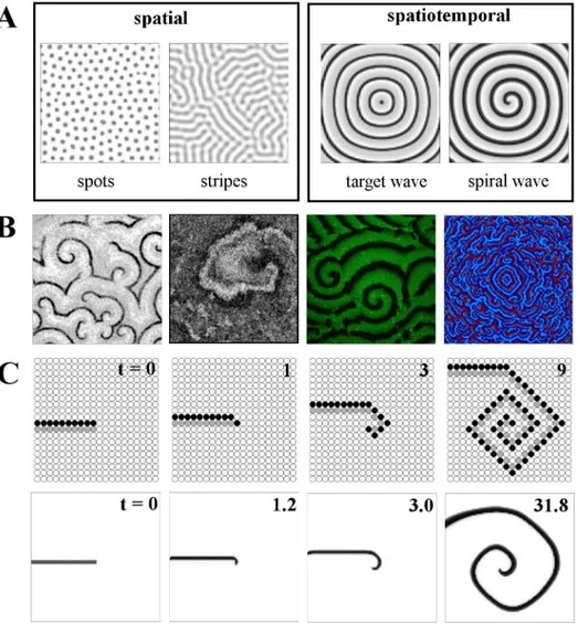
Turing patterns represent one of the major categories of patterns that may be observed. Another important and widespread type is the travelling wave; such patterns are periodic and vary dynamically with time. These wave patterns manifest in biology in situations such as the circular "target" wavefronts produced by the sinoatrial node in the heart, or the open-ended, spiral waves of signaling found in the development of the frogXenopus laevis[8]. Examples of both these wave types are shown inFig 1A. While target waves require an oscillating "pace-maker" cell in their center, spiral waves have a self-sustaining spiral core and typically originate from the breaking of the wavefront of target waves. The deep link between excitability and wave propagation allows us to look at the underlying mechanisms in an algorithmic fashion: A highly stylized, minimal model of excitable dynamics can reproduce both the schematics of
Fig 1. Introduction to pattern types and spiral wave formation.A: (Left to right) spatial and
spatiotemporal pattern examples. Spot and stripe Turing patterns both in coupled Schnakenberg elements; target wave formation from a central pacemaker and established spiral wave, both in coupled FitzHugh-Nagumo oscillators. B: Snapshots of spiral wave patterns from diverse biological systems: (left to right) cAMP signaling in aDictyostelium discoideumcolony, local contraction in neonatal rat cardiac monolayer cultures,
MinD protein density in a lipid bilayer and simulated cytokine levels in a two-dimensional grid of cells. See Acknowledgments for image sources. C (upper row): The update rules of the minimal three-state cellular automaton model lead to spiral wave formation, when applied to an open wave front (consisting of a layer of excited cells, depicted in black, and an adjacent layer of refractory cells, depicted in gray). Lower row: a similar numerical experiment for the model from [6,7].
wave propagation and the basic principle of the transition from an open wave front to a self-sustained spiral (seeFig 1C). In this review, we are particularly concerned with understanding aspects of such transitions: in particular, how they are regulated by features of the nonlinear relationships governing the system at hand.
Spatiotemporal patterns have been a highly successful perspective in the study of complex systems for two main reasons: (1) Patterns often fall into a small number of generic categories (e.g., aggregation patterns or propagating waves [9]). They provide fundamental links between local interaction rules and large-scale collective behaviors [10]. The types of interactions and the nonlinearities of the reactions establishing such large-scale patterns are ubiquitous, with reaction-diffusion systems providing the most prominent example; see, e.g., [11]. The predic-tion derived from the observapredic-tion of patterns of biochemical agents and corresponding bio-chemical mechanisms implementing a reaction-diffusion system (e.g., in hydra development and head-region regeneration [12]) constitutes another example of the enormous power that lies in the observation of patterns in real systems and the subsequent dissection (or deconstruc-tion) of these patterns towards an identification of local interaction rules guided by our under-standing of self-organization. (2) Patterns are abstract, generic objects that can establish (or make visible) unexpected parallels between diverse biological, technological, and social sys-tems. The ubiquity of spiral wave patterns across many systems and many scales, from molecu-lar-level interactions to ecosystem-wide responses [13], is a good example of this unifying capacity of a pattern perspective.Fig 1Bcontains some examples of spiral waves in real biologi-cal systems.
As high-throughput analysis techniques provide ever more quantitative and often spatially resolved biological data (see, e.g., [14–16] as examples of such technological advances), we now have the means to explore the merits of a biology-inspired view, in which the (statistical) details of the patterns matter. The essential features of a biologically oriented perspective on pattern formation are (firstly) that in the majority of pattern-forming biological systems, the patterns often, though not exclusively (see, e.g., [17] for a critique of an indiscriminately adaptionist perspective on biology), serve a purpose and therefore can be considered to be evolutionarily optimized. A variety of examples exist: the size of spiral waves regulates the size of multicellular aggregates in subsequent stages of aggregation in the slime moldDictyostelium discoideum. Bacteria stabilize themselves via aggregation against water flow. Another slime mold,Physarum plasmodium, employs pattern formation to sample space for food sources. Even circadian rhythms in plants can have a complex underlying spatiotemporal organization, leading to propagating waves along leaves, coordinating photosynthetic activity [18]. The stomatal cells of plants respond with complex patterns (termed "stomatal patchiness") to changes in the envi-ronment, which effectively implement a density classification algorithm [19].
Noise has always been at the forefront of interest in the analysis of nonlinear systems. For biological systems, over the last few years, intrinsic and extrinsic noise in gene expression [20–
Secondly, it is plausible, and supported by diverse evidence [25–29], that in biology, the exact spatial layout of the patterns (among the diverse pattern arrangements possible in this systemic configuration) is selected by the distribution of cell properties (i.e., by biological vari-ability), rather than solely—like in physics or chemistry—by spontaneous fluctuations [30]. In this way, spatiotemporal patterns are becoming ever more relevant for computational biology and systems biology because the intracellular "implementation" of such optimized collective states can be investigated. The patterns can serve as a "microscope" for the underlying single-cell mechanisms of regulation.
Understanding how variability (i.e., the magnitude of cell-to-cell differences) shapes collec-tive states (i.e., the emerging patterns) is instrumental to a biology-oriented view of self-organi-zation. We here review the role of variability in the self-organization of biological systems. Our key example is the establishment of spiral wave patterns in excitable media, which is used to model such phenomena as, e.g., the self-organization ofDictyostelium discoideum[15,31–33], cardiac arrhythmia [34] and even the epidemic spreading of infectious diseases (see, e.g., [35,36]). We thus explore the relationship between variability in cell properties and features of spiral wave patterns for a variety of mathematical models.
Basic principles of spiral wave formation and the influence of biological
variability
In biology, variability in the form of cell-to-cell differences (in some biologically relevant parameter) can be expected to contribute more significantly than stochastic noise to the hetero-geneity in a biological system (see, e.g., [29] for experimental evidence and [37–39] for theoreti-cal arguments). The potential importance of variability in biologitheoreti-cal pattern formation is one of the most striking differences between biological systems and those in physics or chemistry, where system components are essentially identical and random fluctuations are the only factor determining the details of the self-organization process and the resulting patterns. This has far-reaching implications for our understanding of biological systems, in which patterns are linked to function. The patterns frequently constitute a (precursor of a) collective systemic mode and are thus subject to evolutionary selection. Variability may be seen as a mechanism by which individual elements shape these collective modes.
We start out with an example to illustrate how the distribution of cell properties can influ-ence self-organized spatiotemporal patterns. Spiral waves can be generated in a variety of dif-ferent ways: one of the most accessible basic mechanisms can be shown with an extremely simple model of excitable dynamics.Fig 1introduces the main mechanistic idea of spiral wave pattern formation, illustrates their ubiquity in biological systems and provides a first example of how they are influenced by biological variability. The model used is a three-state cellular automaton on a two-dimensional lattice. Each lattice site is either susceptibleS, excitedE, or refractoryR. An excitable (susceptible) elementSacquires the stateE, if there is an excitationE in its four-element neighborhood; a refractory elementRreturns to the susceptible stateSafter one time step (see "Models" for details). InFig 1C, upper row, an open-ended wave front (first snapshot) curls up to form a spiral wave. In spite of the geometrical artifacts due to the simple four-element neighborhood and the simplistic state space, the general principle is clearly visi-ble: the initial conditions of a wavefront segment (black elements) directly followed by a layer of refractory lattice sites (gray elements) trigger two forms of patterns, a propagating wave front (oriented upwards in the figure) and the open end of the wave front giving rise to first an additional excited element and then to a curved wave segment, which subsequently propagates outwards in a spiral shape.
A more sophisticated model (by [7]), which we will refer to as the "Levine model," brings the whole scenario much closer to biological reality. It was originally formulated to account for the basic mechanisms behind spiral wave formation inDictyostelium discoideum. Technically, it is an ordinary differential equation (ODE)—cellular automaton hybrid. It uses effective degrees of freedom (excitability, refractory phase), along with biologically plausible variables (the local concentration of the intercellular signaling substance, cAMP) and parameters (e.g., the feedback strength, describing how an external cAMP signal affects cAMP production) to account for the most important properties of the actual biological system. This model has also been used in [37] and [33]. The lower row inFig 1Cshows a similar open-wavefront simulation using the Levine hybrid model, indicating that even with this more complex model, the behav-ior of spiral wave formation is qualitatively the same.
InDictyostelium, such spiral waves represent a chemotactic signal guiding each cell towards an aggregation site, at which the formation of a multicellular structure is initiated. The distribu-tion of spiral waves thus directly affects the fate of each cell. The spiral wave density (qualita-tively speaking) translates into the cell counts of the multicellular mounds. With spiral waves, a pathological dynamical state of cardiac tissue, identifying the spatial distributions of cell prop-erties that lead to reduced spiral wave probability would be advantageous.
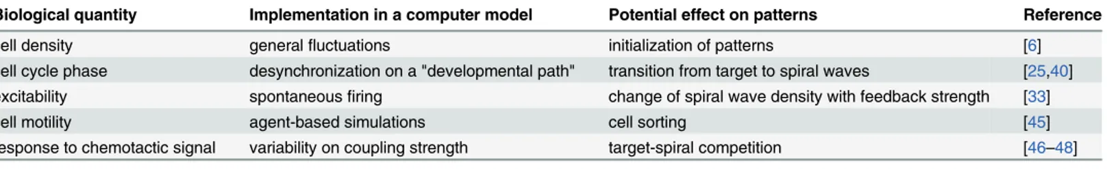
The growing accessibility ofDictyosteliummutant data and their resulting pattern-level phe-notypes, through studies such as [44], also enhance the prospects of gaining insight into pattern formation in this model organism via mathematical models that incorporate detailed and real-istic biological knowledge.Table 1contains some examples of biological properties in Dictyos-telium, with their corresponding methods of implementation in a computer model and the potential effects on patterns that result.
These quantities are, of course, interdependent. Cell density [6] and excitability [33], for example, vary with the current stage in the cell cycle [25,40]. That responses ofDictyostelium to chemotactic stimuli are cell-specific has been shown in [29]. One method of incorporating variability intoDictyosteliummodels is the use of agent-based models, with applications to pro-cesses such as cell sorting [45]. The role of coupling strength was investigated in [46] and [47]; effects such as target-spiral competition were shown in [48].
Different cell properties will influence the exact layout of the emerging patterns in different ways. Similarly, different forms of cellular interactions and signal processing will modulate the effects of this biological variability in different ways. When such interdependences are well understood, the statistical properties of the patterns can thus serve as a "microscope" for the underlying principles of regulation.
Effect of Variability: A Model Study
The impact of variability on spiral wave patterns is best visualized by an event perspective, in which target wave centers and spiral wave tips are considered "spatiotemporal pattern events." The temporal sequences and spatial distributions of these pattern events can then be compared with the spatial distribution of cell properties. A cluster of oscillatory elements is a likely candi-date for emission of target waves. The additional pattern events emerging from this region are then a consequence of the proximity to other such pacemaker regions and the "roughness" of the propagating wave front, which is the result of the sequences of excitability encountered by the propagating wave front—up to the point at which target waves break up into spirals, in one possible mechanism.
As an illustration of the wide range of geometrical influences biological variability has on the emerging layout of spiral wave patterns, we use different models of excitable media (with varying levels of biological detail), simulate spiral wave patterns, reconstruct events (using the
Table 1. Sources of variability affecting details of macroscale patterns inDictyosteliumand their representations in computer-based mathematical
models.
Biological quantity Implementation in a computer model Potential effect on patterns Reference
cell density generalfluctuations initialization of patterns [6]
cell cycle phase desynchronization on a "developmental path" transition from target to spiral waves [25,40] excitability spontaneousfiring change of spiral wave density with feedback strength [33]
cell motility agent-based simulations cell sorting [45]
response to chemotactic signal variability on coupling strength target-spiral competition [46–48]
methods from [49]), and thus translate the spatiotemporal patterns into a "pattern event plot," in which such underlying geometries are clearly discernible.
How do these "event plots" work? Using algorithms acting upon the space-time cube of the (simulated or measured) data, the centers of target waves, as well as the centers and orienta-tions of spiral waves are identified (details about the algorithm are provided in [49]). Such "event plot" representations of spatiotemporal patterns as sequences of events are not new and can be employed even in the case of homogeneous systems. In [50], they have been employed to study spiral breakup in calcium dynamics. In [33], the identification of spiral wave tips via phase singularities was a prerequisite for studying changes of pattern statistics with the strength of the cAMP feedback loop. However, here, in the presence of variability, the geometric con-straints on pattern events arising from the specific nonlinearities become visible. In [25], this has been worked out in detail for the receptor desensitization-based ODE model of Dictyoste-liumsignaling, referred to here as the "Goldbeter model" [40].
We performed such pattern-based investigations for the "hodgepodge machine" [51,52], the FitzHugh-Nagumo oscillator as a generic model of excitable dynamics (S2 Fig) [39,53,54] and theDictyosteliummodels from [7] and [40]. We discuss the results from the latter two models in detail here. The underlying cell property is, on a general level, the level of excitability of the individual elements forming the coupled system. The distribution of this property and its dynamical features, possibly modulated by other properties, determine the development of spi-ral waves.
The hodgepodge machine from [51] is a more gradual version of the three-state "forest fire" cellular automaton. Itsnstates (a typical value ofnis 100) are traversed in a monotonously increasing fashion. Two parameters, the "excitability" and the "sensitivity," dictate the impact of neighboring cells (sensitivity) and the steepness of the excitatory response (excitability), respec-tively. This minimalistic cellular automaton model of excitable dynamics displays high positive correlations between target waves and the distribution of excitability, as well as between spiral waves and the distribution of sensitivity [52].
landscapes (see [48]). These contrasting pattern event sequences illustrate the great versatility of this simple model.
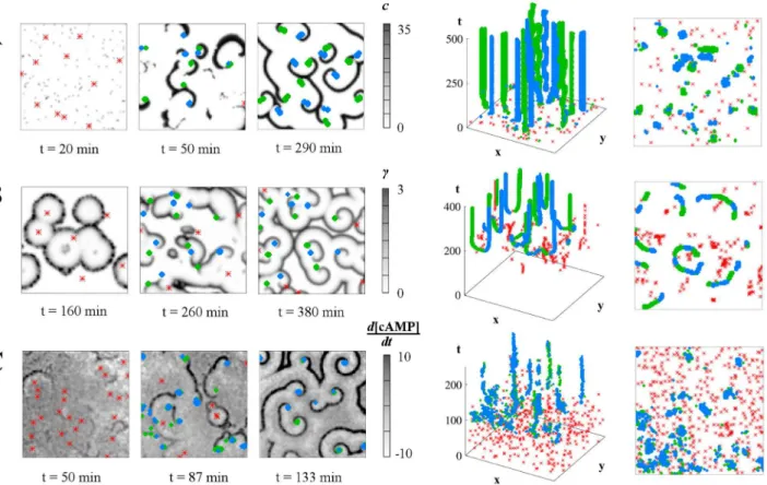
Figs2and3illustrate the main features of these results. InFig 2(see alsoS2 Fig) the general method of pattern event reconstruction and pattern event plots is demonstrated, both for simu-lated (A and B) and real (C)Dictyosteliumdata. The underlying geometry of the pattern events, as well as similarities and differences between the spatiotemporal arrangements of events, become clearly discernible in these event plots. The effects of the two different models for Dic-tyosteliumpattern formation are visible here: the striking geometrical shaping of the arrange-ment of spiral wave in the Goldbeter model from [40] (2B) contrasts with the conversion of a target wave pattern into a dense spiral wave pattern for the Levine model from [7] (2A). In the latter, the cell property we vary is the capacity to spontaneously enter the excited state and the mechanism of spiral development appears to be based on the breaking of a target wavefront by an unexcitable region. The geometric details of the possible mechanism are explored in [37]. In the experimentalDictyosteliumdata inFig 2C, spirals form at a later stage, likely due to the interacting target wavefronts that precede their appearance. This event sequence closely resem-bles that inFig 2B.
The twoDictyosteliummodels display very different geometric relationships between pat-tern events. The pairwise appearance of (left- and right-handed) spirals is more pronounced in
Fig 2Athan inFig 2B. Both models, as well as the experimental data (Fig 2C), show a clear
Fig 2. Event identification in simulated and real spiral wave patterns.Left column: snapshots of the lattices; middle column: corresponding space-time
event plots; right column: top-down views of the event plots. Target wave origins are red asterisks, left- and right-handed spiral waves are blue and green diamonds, respectively. A: In the "Levine" model, spiral waves evolve early on due to colliding wavefronts, without a sustained target-wave phase. B: Development of spiral waves from the interaction of target waves in the "Goldbeter" model with a developmental path. C: Development of spiral waves from target waves in experimentalDictyostelium discoideumdata. All simulated lattices are 100x100 and experimental data is rescaled to the same size.
target wave phase preceding the spiral formation, although this phase is most pronounced in the Goldbeter model in 2B. Also, there are strong differences in spiral wave density (the num-ber of spiral waves per area). However, even at first glance, the biologically more detailed model (Fig 2B) shows a much stronger geometric arrangement of spiral waves than the more schematic model (Fig 2A).
In our previous work on the "Levine model" from [7], we observed a pronounced anti-corre-lation between spiral waves and a key cell property, the firing rate, and particularly a clustering of spiral wave tips in regions devoid of spontaneously firing "pacemaker" cells. Similar results are shown here inFig 3A: the spatial locations of the pacemakers remain fixed (grey crosses) and we study the distribution of spiral wave tips across a large number of simulation runs (red-scale signal plotted in the lower row). Although it is hard to discern any clear relationship between pacemaker locations and spiral tip occupancy for single runs (upper row), averaging occupancy over many runs shows the greater density in the central "avenue" free of pacemak-ers: a clearly visible anticorrelation between the spatial distribution of pacemaker cells and the spatial distribution of spiral wave probabilities. Some features of this distribution can also be understood from a simple geometrical model based on triplets of pacemaker cells [37].
The clearest geometrical shaping of the spiral waves is observed in the Goldbeter model from [40], where variability is coupled to the dynamics via a developmental path. The develop-mental path moves each element through different dynamical regimes: (non-excitable)steady state!excitable!oscillatory!excitable. There, spiral wave tips cluster at characteristic dis-tances from their parent target wave centers: the tips map out the Voronoi diagram around a specific fraction of cells functioning as effective pacemaker cells. These effective pacemakers
Fig 3. Effect of "pacemaker" location in the "Levine" model from [6,7].Pacemaker locations are shown as grey or black crosses and spiral tip
occupancy in the red scale. A: A conserved pacemaker pattern leads to different spiral wave patterns of excitability (upper row). Spiral tip occupancy over 1,000 runs favors locations free of pacemakers. The probability of observing a spiral wave at a particular spatial site is determined by the spatial distribution of cell properties. A simple pacemaker grouping gives rise to a less coherent, more variable tip occupancy in the Levine model compared to the clear geometric shaping in the "Goldbeter" model (C), based on 250 runs. D: Quantification of the relationships between pattern types in the Goldbeter model: over 100 runs, the correlation coefficient of (left to right) target wave origins to time offsets on the developmental path, spiral tips to target origins and spiral tips to time offsets; against the radius of the Gaussian filter.
are those cells that are in the oscillatory regime when the majority of other cells are already in the excitable regime, thus ensuring wave propagation [25]. The pattern events accompanying the evolution of a sample Goldbeter lattice are highlighted in the accompanying animation (S1 Video).
In the top-down (i.e., time-collapsed) views of the event plots, also shown inFig 2, these geometrical constraints among the pattern elements become apparent. In particular, in the Goldbeter model, the Voronoi diagram-like relationship between the spiral tips and their par-ent target wave origins are suggested even on this single-run level. Comparing simulation results for the three models shown in Figs2,3andS2 Fig, it is striking how different these geo-metrical constraints in the emerging spatiotemporal patterns are, both in the characteristic sequence of events and in the spatial density and distribution of asymptotic patterns. This sim-ple examsim-ple already shows the potential capacity of spatiotemporal patterns to discriminate between possible underlying mechanisms of regulation. However, it should be noted that these examples are based on specific parameter constellations, and a complete evaluation of the model-specific differences requires a comprehensive exploration of the parameter spaces. InFig 3B and 3C, we continue our comparison of the Levine and Goldbeter models by exploring their behavior in response to the same initial pacemaker pattern, our cell property here. With the same fixed distribution of five pacemakers, we allow a certain amount of ran-domness in both systems over many runs. In the case of the Levine model, there is a certain probability of a pacemaker firing per timestep. In the Goldbeter model, the time offsets on the developmental path are randomly allocated. In both cases, pacemakers may randomly emerge in addition to the fixed ones. In the Goldbeter system, the fixed pacemakers are given a time advantage over the random pacemakers, while the small additional number in the Levine sys-tem makes the setup more comparable in this respect to the Goldbeter syssys-tem. The resulting spiral occupancy images further highlight the dramatic differences between these models, with the clear Voronoi diagram-type tracing of the spiral tips in the Goldbeter model in contrast to the diffuse anticorrelation in the Levine model, which becomes clearer with clustered pace-maker elements, as inFig 3A.
The different mathematical descriptions of excitable systems shape the spatial distribution of pattern elements in systematically different ways. More quantitative methods for exploring these constraints (via point process statistics; cf. [55]) have been discussed in [25]. Even the computation of correlation coefficients between pattern events can provide a first quantitative indication of their spatial relationships: inFig 3Dwe compute such coefficients for the Goldb-eter model with varying values of the Gaussian filter applied to the datasets. Using a fixed value of the quantity of the time offset on the developmental path,Δ, over 100 runs, we plot the mean values of the Spearman correlation coefficients against the value of the Gaussian filter (error bars are the standard error of the mean). Although the resulting quantities are small, they reflect the qualitative relationships observed in the event plots. We note a positive correla-tion between target wave origin occupancy and time offset, a negative relacorrela-tionship between spi-ral tip occupancy and target origin occupancy, and an apparent resulting anticorrelation between spiral tips and time offsets that is a maximum at our intermediate spatial scale and becomes less pronounced at larger scales.
the interface of computational systems biology and pattern formation is what we were referring to earlier as patterns serving as "microscopes" for the underlying regulatory principles.
We have seen that the distribution of cell properties affects the distribution of pattern ele-ments and thus the details of the asymptotic pattern. How can variability, as a parameter mea-suring the amount of cell-to-cell differences, affect the patterns themselves? In [48] it was shown that the density of spiral waves changes with variability in a resonance-like fashion, resembling the classical phenomenon of diversity-induced stochastic resonance [57,58]. The underlying mechanisms here are a strong dependence of the competition between target waves and spiral waves on variability.
Additionally, the core idea of patterns as functionally important and evolutionarily advanta-geous collective states leads us to expect deep relationships between regulatory components and properties of spatiotemporal patterns. For the case ofDictyostelium, such relations should also reveal themselves in dedicated experiments, e.g., by varying the amount of cell-to-cell vari-ability and studying the effect on pattern formation or by altering levels of specific regulatory components by mutagenesis and observing differences in pattern formation to the wild-type.
Furthermore, the findings summarized here can serve as a theoretical framework for reverse-engineering the fundamental regulatory mechanisms underlying the observed spatio-temporal patterns.
Outlook
We have here introduced the principles of pattern predictability in biological systems, illus-trated by a range of mathematical models and real experimental data. Taken together, the results described and reviewed here provide strong evidence supporting the general hypothesis that single-element properties are systematically mapped onto patterns and thus conserved across processes of self-organization (as opposed to being enslaved and "deleted" by the collec-tive). The results reviewed here show that the initial properties of potentially very few cells have a driving influence on the resulting asymptotic collective state of the colony.
Although most of our examples so far have discussed pattern formation on regular lattices, the concept of pattern predictability can, in principle, be extended to other topological architec-tures, such as networks. The study by Marr and Hutt [59] is an example of how variability can reorganize patterns on networks: shortcuts inserted into a regular (ring-like) network architec-ture induce a transition from Wolfram class IV dynamics to Wolfram class III dynamics in binary cellular automata on graphs. The“variability”in these“small-world graphs”[60] deviat-ing from regular networks lies in the degree (number of neighbors) of each element. With the work on Turing patterns on graphs [4] and related work [61,62], a new paradigm for the inter-pretation of dynamics on graphs is currently emerging: topology-compatible“collective modes”that establish themselves in a graph due to the interplay of topological and dynamical parameters. Waves organizing around hubs (highly connected nodes) are a striking example of such collective modes [63]. Networks are increasingly recognized as a powerful and intuitive way to represent real biological processes (see, e.g. [64,65]), with a vast range of applications extending from gene metabolism [66] to ecological processes [67].
In the specific examples of spiral wave patterns discussed above, we want to highlight the importance of biological variability and the regulatory information contained in spatiotempo-ral patterns. However, we do not want to suggest that the complex relationship between the nonlinearities of the model, the nature and spatial distribution of cell properties, and the result-ing spatiotemporal patterns is well understood and can lead to unique conclusions (e.g., about the relevant nonlinearities, given the distribution of cell properties). The details of this relation-ship remain far from clear.
Understanding the role of variability in pattern formation relies on disentangling the role of discrete element properties from that which may be an innate property of the system even without discretization, as is the case for homogeneous systems, such as the example explored in [50]. However, exploring the role of variability is appropriate in biological systems, as discrete-ness is an inevitable constituent of such systems: they are all composed of individual, interact-ing parts, whether at the molecular or the organismal scale.
Incorporating the spatiotemporal organization of biological systems is a major challenge for systems biology. The field is now making the transition from a purely temporal understanding of biological processes (the "well-stirred test-tube" perspective) to full spatiotemporal descrip-tions (see e.g., [69–71]). This, together with recent findings on the theoretical side, has reinvig-orated the interest in classical models of spatiotemporal pattern formation.
One of the aims of systems biology is to establish the architectures and kinetics of signaling pathways and intracellular regulations in an iterative process between modelling and experi-ment. However, systems biology currently fails to exploit the very large pool of macroscopic observations represented by spatiotemporal patterns, which potentially provide important insight into intracellular regulation processes. Spatiotemporal patterns form within single cells or in a population of cells according to the intrinsic laws of protein–protein interactions, intra-cellular feedback loops and (on the multiintra-cellular level) cell–cell communication. The patterns change systematically with the parameters of regulation. When guided by a suitable mathemat-ical model, the detailed layout of spatial and spatiotemporal patterns can reveal the properties of the underlying regulatory system.
The very recent publication by [72] is an ideal case study of this capacity of self-organized spatiotemporal patterns. By representing the intricate regulatory network responsible for the chemotactic response of individualDictyosteliumcells as a stylized excitable system (mathe-matically formulated as interconnected FitzHugh-Nagumo oscillators), a wealth of experimen-tal information can be qualitatively explained, including the dynamics of adenylyl cyclase A in response to steps of external cAMP and the reason for the production of extracellular phosphodiesterases.
We are convinced that the general framework of pattern predictability has a vast range of additional applications, beyond the systems shown inFig 1A. Analyzing pattern predictability can be envisioned, e.g., for cell sorting in inDictyostelium[73], wound healing [74,75], predic-tion of Turing patterns [3], design of patterns in a broad range of self-organized processes and prediction of patterns in social and socioeconomical systems [76].
Background
Theoretical investigations of biological variability
influence of variability and stochastic contributions has been described, e.g., from the point of view of synchronization [79] and from the network point of view [80].
The effect of variability on spatiotemporal chaos has been studied in [81–83]. Additionally, various numerical investigations have explored variability-induced pattern formation, com-pared to and in cooperation with noise using arrays of coupled oscillators [28,58,84]. The fact that, similarly to noise, variability can induce patterns or trigger a transition from one pattern to another has been a focus of research in the 1990s and early 2000s. In the more theoretically oriented studies, our biologically motivated term "variability" often appears as“diversity,” “ dis-order,”or“quenched noise.”For coupled FHN oscillators, it has been shown that variability can induce both a phase transition from oscillatory to excitable behavior [28] and, in a subexci-table system, pattern formation in the form of spiral waves of excitation [58]. Pattern complex-ity has been shown to be highest at intermediate variabilcomplex-ity [84]. Such studies culminated in the phenomenon of diversity-induced resonance [57,58], where the response of an excitable or bistable system to a subthreshold stimulus is optimal at intermediate levels of diversity. In [85] such a diversity-induced resonance was found in simulations of calcium dynamics, showing that calcium waves propagate optimally at intermediate cell-to-cell variability.
In recent work on excitable dynamics and spiral wave patterns, there is a strong trend to take into account realistic structural geometries in order to better understand the observed spa-tiotemporal patterns (see, e.g., [86]). On the methodological side, target and spiral wave identi-fication techniques have been developed and implemented [49,87,88] and simulation methods have been advanced [89]. The authors of [90] also discuss how spiral tip identification in the FHN model influences the reconstructed patterns of meandering spiral waves. The use of gen-eralized recurrence plots for reconstructing the phase diagram of nonlinear spatiotemporal sys-tems from a limited set of observations has been described by [91]. In spite of its historical roots in the 1960s and 1970s, the explanation of spiral wave patterns still receives an enormous amount of scientific attention. The reason is 2-fold: (1) Our understanding of pattern forma-tion processes still has severe gaps on the theoretical side; in particular, the routes from homo-geneous patterns to fully established spiral waves are surprisingly dependent on the details of the system at hand. (2) It is currently being noticed that (precisely due to the dependence of patterns on the regulatory details within the system) a deep and detailed analysis of spiral wave patterns can help access the underlying principles of regulation. Detailed theoretical studies have provided evidence that the correlations between cell property distributions and patterns strongly depend on the regulatory mechanisms at the level of individual cells (e.g., comparing the results from [37] and [25]).
Returning to the purely theoretical description of properties of spatiotemporal patterns, it would of course be a major step forward if some aspects of the predictability of patterns from cell properties could also be understood analytically. Two approaches are particularly promis-ing for tacklpromis-ing this question. Firstly, in [92] a mathematical framework has been developed for describing diffusion and annihilation of spiral wave tips within a simple kinematical model. Secondly, in [93] a response theory of (spiral wave) patterns under small perturbations has been formulated, in particular, the sensitivity of the spiral’s drift velocity.
Dictyostelium
pattern formation
A model system for which regulation by variability could be a principal mechanism (see, e.g., [7,40]) is the slime moldDictyostelium discoideum. In this paradigmatic example of biological pattern formation, individual amoeba cells aggregate under the influence of the chemotactic signal cAMP and form a multicellular organism [99]. This process is initiated by nutrient dep-rivation; this causes single cells to emit cAMP into their environment. These molecules are detected by neighboring cells via highly specific surface receptors [100], initiating the intracel-lular autocatalytic synthesis of additional cAMP by the enzyme adenylyl cyclase (ACA) and its subsequent secretion into the environment. Time-delayed receptor desensitization and halting of ACA activity are involved in the following refractory period. Extracellular cAMP is degraded by membrane-bound and secreted phosphodiesterase, which is, on the other hand, regulated by its inhibitor. The coupling of the underlying reaction kinetics with diffusion results in wave propagation. As long as the local cAMP concentration increases with time, the cells react with positive chemotaxis, resulting in periodic movement perpendicular to the wave front, i.e., towards the origin of the chemical signal.
As more details become available concerning the molecular network responsible for cAMP oscillations inDictyostelium(e.g., [101,102]), modelling efforts increase in their corresponding complexity (see, e.g., [103,104], which are typical of this transition towards a more "systems biology" approach). In terms of the localDictyosteliumresponse to the cAMP gradient, prog-ress is still being made in the understanding of the small-scale decisions underlying pattern formation [105]. Another evolving avenue of interest isDictyostelium’s contribution to under-standing the origins of multicellularity [106,107]; such efforts are spurred on by the sequencing ofDictyostelium discoideum’s genome [108]. These recent examples illustrate the continually increasing, vast amount of research that has been done over more than four decades into many aspects of theDictyosteliumlife cycle. In particular, several findings of the last few years have added new ideas to the view that in the case ofDictyostelium, biological variability is responsi-ble for certain stages of symmetry breaking in the usual course of the developmental cycle (local pattern initiation, spatial distribution of cell streams, and distribution and proportions of differentiated cell types).
are self-sustaining continuous structures, which preserve their stability, to a certain extent out-side the excitable regime. This allows for maintenance of the aggregation process under devel-opmentally and environmentally conditioned changes. In contrast, target waves require the periodic activity of oscillatory regions (i.e. pacemakers).
Strong support for the hypothesis that cell properties can indeed affect the collective pat-terns inDictyosteliumhas come from the recent observation that the direction and magnitude of a cell’s response to a signal pulse is an individual cell property, which remains constant in time [29]. In that work the behavior of single cells under periodic cAMP signals was analyzed and it is observed that the characteristics of the gradient sensing response of an individual cell at a certain time point strongly correlate with those of the same cell at a later time point. A recent paper [15] provides further support for our hypotheses, demonstrating that Dictyoste-liumcells’internal cAMP concentrations oscillate at a frequency determined by intracellular machinery. The authors found that experimentally observed rhythmic cAMP synthesis could only be replicated successfully in their mathematical model when the stochastic pulsing of indi-vidual cells in response to subthreshold cAMP levels was included in the model. The implica-tion is that organized group dynamics in aDictyosteliumpopulation depend on the random behavior of individual cells (see also [109]). Another study finds that isogenicDictyostelium cells have diverse sensitivities to cAMP, and that this may facilitate collective behavior [110]. Similarly, in [72] the roles of noise and variability inDictyosteliumcAMP signaling were explored in a phenomenological FitzHugh-Nagumo model. Here, variability was incorporated in the form of individual cells’thresholds of response to extracellular cAMP. The results sug-gested that stochastic noise can account for population-level cAMP oscillations, while variabil-ity in this cell property alone cannot. These recent works, in addition to greatly enhancing the experimental accessibility of the system (via the direct measurement of cAMP concentrations), emphasize the role of individual cells and the debate whether it is the activity of dedicated pace-maker cells or the spontaneous firing of random cells which constitutes the main driving mech-anisms behind the initiation of patterns during this early phase (see also [111]). While the work from [15] emphasizes the latter aspect, the results of [29] suggest an important role for the former, and it is likely that both noise and variability interact in the shaping of spatiotem-poral patterns.
These diverse sets of questions, all of which are related to biological variability and their influence on pattern formation, illustrate the importance and timeliness of establishing a biol-ogy-oriented perspective on spatiotemporal patterns.
In the following, we will focus on two additional mathematical models. The first is a highly detailed model of pattern formation inDictyostelium, which was initially formulated in [112]; we also refer to this model as the "Goldbeter" model. This model incorporates relevant biologi-cal details, such as the relay of suprathreshold cAMP pulses and autonomous cAMP oscilla-tions, and the phosphorylation-dependent modification of the cAMP receptor, in the same way that current modeling attempts in systems biology would (see, e.g., [104,113]). Standard model reduction techniques (such as time scale separation) yielded a three-dimensional model that has also been elegantly used to analyze spatiotemporal patterns [40]. The dynamical vari-ables are the fraction of active cAMP receptors and the concentrations of external and intracel-lular cAMP, respectively (see "Models" for details).
A novel ingredient organizing the interplay between biological variability and the resulting patterns in a system is the concept of a "developmental path." This is a specific parameter drift with time which couples small cell-to-cell property differences to the cell’s dynamical behavior, thus turning them into "organizers" of the spatiotemporal patterns. The "developmental path" concept was proposed by Lauzeral and coworkers [40], in the context of the model of Dictyoste-lium discoideumpattern formation developed by Martiel and Goldbeter [112] (see also [114]). It was created as a hypothetical mechanism of the development of heterogeneity from homoge-neous conditions for the synthesis of spiral waves of cAMP inDictyostelium. According to this concept, the cells in a population ofDictyosteliumundergo time-dependent changes in cell properties, and intercellular variation in these properties places the cells at differing points along this developmental path. The path in [40] results from sigmoidal variations in the maxi-mum activity of adenylate cyclase and the rate of extracellular cAMP degradation. The varia-tion from cell to cell in the cell cycle when starvavaria-tion begins is a possible origin of the different time offsets of the cells on the path. Such a path would be expected to occur in a higher-dimen-sional space in reality, but even in this simple form can provide an adequate representation of the actual biological dynamics. We consider the developmental path form of the Goldbeter model as an example of predicting patterns arising from a biologically plausible source of vari-ability. Furthermore, we discuss the extension of the concept to the FHN model, showing that the principle is versatile and can be applied to diverse biological models as a method of generat-ing heterogeneity.
Models
Here we describe the models used throughout this review. The symbolr2represents the dis-cretized Laplacian for 2-D diffusion, and we use the five-point Laplacian except in the case of the Levine model, where the eight-point Laplacian is used.Dis the value of the respective diffu-sion coefficient.
Schnakenberg model. This simple model [2] appears inFig 1A, as an example of a driver of Turing-type patterns. Here,c1= 0.05,c-1= 1.0,c3= 1.0,Du= 1.0,Dv= 20.0, andγ= 2.0. The value of the parameterc2determines whether the system gives rise to spots (c2= 1.00) or stripes (c2= 1.57). The initial values ofuandvare randomly varied between -0.01 and 0.01.
@u
@t ¼gðc1 c 1uþc3u 2
vÞ þDur
2
u
@v
@t ¼gðc2 c3u 2
vÞ þDvr
2
v
ð1Þ
FitzHugh-Nagumo model. The FitzHugh-Nagumo (FHN) model [53,54] is widely used in studies of excitable media, as it is a low-dimensional model that can display a wide range of dynamics, including excitable, oscillatory, quiescent, and bistable behavior. Here it provides examples of target and spiral wave types (Fig 1A), spiral formation from a target wave due to interruption of the wavefront by less excitable elements (S1 Fig), and as an example of model-specific pattern events (S2 Fig). The FHN equations describing a 2-D lattice with diffusive cou-pling of the elements are:
@ui;j
@t ¼
1
εðða ui;jÞðui;j 1Þui;j vi;jÞ þDr
2
ui;j
dvi;j
dt ¼bui;j gvi;jþci;jðtÞ
whereui,jrepresents the voltage-like variable at each lattice site andvi,jis the recovery variable. ci,j(t) is the parameter that may be subject to variability, either through a Gaussian distribution (“static”variant) or via a developmental path. The static variant appears inFig 1A,S1andS2A
Figs, while the developmental-path variant appears inS2B Fig.
Each FHN element may be excitable, oscillatory, or in a non-excitable steady state, depend-ing on the model parameters. Here, the parameters have the valuesa= -1,g=b= 0.12,ε= 1 andD= 0.1. A value ofΔx= 1.0 is used for the discretization.
For the static variant, there is no developmental path. ForFig 1A,c= 0.025 for the target wave andc= 0.021 for the spiral wave. ForS1 Fig,c= 0.024 and an additional fraction equal to 0.400 of all elements are assignedc= 0.030, which is of lower excitability. InFig 1AandS1 Fig, the central pacemaker element is assignedc= 0.000. ForS2A Fig, values ofcare randomly drawn from a Gaussian distribution, with mean value 0.024 and standard deviation 0.006. Varying the value of c changes the dynamical properties of the element; inS2A Figthe ele-ments are mainly oscillatory, but some are also excitable and steady-state.
The developmental-path variant is analogous to the corresponding Goldbeter model, but incorporates variation in only one parameter,c:
ci;jðtÞ ¼ c0tanh½
t tcþDti;j
Tc
; ð3Þ
where the time offsetsΔti,jdiffer between elements (seeEq 4) andtis the“global time.”The value ofc0determines the dynamical regimes traversed in the temporal evolution ofci,j(t). Here it has the value 0.02, and the lattice values ofci,j(t) vary between approximately -0.02 at the start of the developmental paths and 0.02 at the end, with a value ofci,j(t)0 resulting in the maximal oscillatory frequency. The parameterstcandTcdescribe the shape of the path inc, heretc= 1000 andTc= 50.
Each FHN element is assigned a time offsetΔti,jon this path, which distributes the values of cover the lattice. The time offsets are created as above:
PðDti;jÞ ¼ 1
Dexp Dti;j
D
; ð4Þ
whereΔis a desynchronization parameter which controls the spread of time offsetsΔti,j; here Δ= 500.
Three-state cellular automaton encoding excitable dynamics. This model appears inFig 1Cas an example of the development of a spiral wave from an open-ended planar wavefront. It consists of three discrete states for each node (susceptibleS, excitedE, refractoryR), which are updated synchronously in discrete time steps according to a set of update rules allowing for sig-nal propagation: (1) a susceptible nodeSbecomes an excited nodeE, when a direct neighbor is in the excited state; (2) an excited nodeEenters the refractory stateR; (3) a node regenerates (R!S) afterrtime steps. The parameterris the deterministic refractory period of the system; here,r =1.
architectures in studies of self-organized criticality. Other variants of three-state excitable dynamics have been used to describe epidemic spreading (see, e.g., [118,119]).
Levine model from [7] and [33]. Here we discuss the "Levine" model ofDictyostelium pat-tern formation introduced in [7] (see also [33,37]). We will use the form from [33]. This model appears in Figs1C,2B,3A and 3B. The model is a hybrid cellular automaton-ODE model for Dictyostelium, in which the cells have discrete internal states coupled to two continuous exter-nal variables: the local values of extracellular cAMP concentration and excitability. The rates of change of these variables are
@ci;j
@t ¼ Gci;jþrFsi;jðtÞ þDr
2
ci;j
dEi;j
dt ¼Zþbci;j
ð5Þ
wherecis the extracellular cAMP concentration,Γis the constant of extracellular degradation of cAMP mediated by phosphodiesterase andrFis the rate of cAMP production. Additionally, si,jis the internal cell state which controls cAMP production, taking the value of 1 for afiring state and 0 for a quiescent state,ηis the intrinsic excitability increase andβis the genetic feed-back factor which affects the sensitivity to signals from other cells.
The excitability increases monotonically and has an upper limitemax. In the ready and rela-tive refractory states, activation can take place if the cAMP concentration exceeds the threshold ti,j:
ti;jðtÞ ¼ cmax A
t tþTARP
ð1 E
i;jÞ ð6Þ
whereτis the time elapsed since the cell entered the relative refractory phase and has a
maxi-mum value ofTRRPfor the ready state, andA= (TRRP+TARP)(cmax-cmin)/TRRP). Here,TRRPand TARPare the relative and absolute refractory periods, respectively. A pacemaker has a certain probabilitypFoffiring per time step. The resulting randomfiring events lead to amplification of the heterogeneity of excitability in the system through feedback. ForFig 1C, pacemakers emerge randomly, as in [33] and the fraction of pacemaking elementsε= 0.50,β= 0.20, the starting excitability =emax/2 andpF= 0.002. For Figs2A,3A and 3B, static distributions of pacemakers are used and the starting excitability =η= 0.0. For Figs2Aand3A,β= 0.20,pF= 0.002 andε= 0.195 and 0.190 respectively. ForFig 3B,β= 0.30,ε= 0.001 (in addition to the
five assigned pacemakers) andpF= 0.05. Other parameter settings are as in [33].
Goldbeter model from [40,112]. This model ofDictyosteliumpattern formation appears in Figs2Band3C, and in the accompanying event plot animation (S1 Video). It was developed by Martiel and Goldbeter [112] (see also [114]). We here discuss the reduced, three-dimen-sional version of the original model with nine dynamic variables, both from [112]. Each lattice point represents a group of ten cells whose properties are synchronized.
The three dynamic variables are the total fraction of active cAMP receptor (ρT) and the nor-malized concentrations of intracellular (β) and extracellular (γ) cAMP:
drT
dt ¼ f1ðgÞrTþf2ðgÞð1 rTÞ;
db
dt ¼ qsFðrT;g;aÞ ðkiþktÞb;
@g
@t ¼ ðktb=hÞ kegþDgr
2
g;
with
f1ðgÞ ¼
k1þk2g
1þg ;f2ðgÞ ¼
k1L1þk2L2cg
1þcg
FðrT;g;aÞ ¼ aðlyþεY
2
Þ
1þaþεY2ð1þ
aÞ;Y ¼
rTg
1þg:
ð8Þ
This model is based on reversible desensitization of the cAMP receptors on the surface of cells. Extracellular cAMP may bind to the active receptor; this activates adenylate cyclaseσ,
converting intracellular ATP to cAMP. The activity of the receptors depends upon the extracel-lular cAMP concentration and the fraction of active receptors. Intracelextracel-lular cAMP diffuses out of the cell and can be hydrolyzed by intra- or extracellular phosphodiesteraseke.
The "developmental path" concept was proposed by Lauzeral and coworkers [40], in the context of the three-dimensional model. According to this mechanism, specified cell properties follow a defined trajectory over time, with this variation leading cells successively through qui-escent, excitable, oscillatory and excitable regimes of dynamical behavior. Desynchronization of the cells’properties on this path then provides the necessary cell-to-cell differences for spiral waves to form. This path incorporates variation in adenylate cyclase and phosphodiesterase, in correspondence to experimental observations. The developmental path concept thus provides a mechanism for the generation of variability from homogeneous initial conditions. This model was shown to successfully reproduce the sequence of macroscale patterning observed in experimentalDictyosteliumcolonies.
Here we discuss developmental path 3 of [40], in whichσandkeare varied sigmoidally:
sðtÞ ¼savþsamptanh t tsþDti;j ts
keðtÞ ¼kavþkamptanh
t tkþDti;j
Tk
:
ð9Þ
wheretis the "global time" and the parameterstσandTσ; andtkandTkdescribe the shape of the variations inσandkrespectively. The lattice elements (representing synchronized groups
of cells) are assigned time offsetsΔti,jon this path, which distributes the values ofσ(t) andke(t)
over the lattice. As in [40], each time offset is randomly drawn from an exponential distribu-tion:
Pðti;jÞ ¼ 1
Dexp
ti;j
D
: ð10Þ
In Figs2B,3D, and the accompanying animation (S1 Video),Δ= 25; in 3C,Δ= 15. Other parameter settings are as described in [40].
Supporting Information
S1 Fig. Development of spiral waves from a central pacemaker in the FitzHugh-Nagumo (FHN) model due to the presence of elements of lower excitability, leading to curvature of the planar wavefront.The entirety of the lattice is excitable, but a fraction of 0.4 of the ele-ments have lower excitability.
(EPS)
of spiral waves from the interaction of target waves with areas of lower excitability in the "static" FHN model. B: Development of spiral waves from target waves in the FHN model with a developmental path. Spirals originate within the zones of target wave entrainment, apparently through the wave-in-wave mechanism.
(EPS)
S1 Video. Animation of the spatiotemporal evolution of a lattice of "Goldbeter" elements, as their time offsets move along a developmental path in the parametersσandke.Pattern events are represented by red asterisks (target wave origins) and left- and right-handed spiral tips (blue and green diamonds, respectively). Left side: development of theu-field with overlaid detected events; right side: space-time plot of the pattern events.
(AVI)
Acknowledgments
We are grateful to our experimental and theoretical collaborators for kindly providing snap-shots of spiral wave patterns: Satoshi Sawai (University of Tokyo, Japan) for theDictyostelium discoideumdata used in Figs1Band2C; Gil Bub (University of Oxford, England) for the rat cardiac cell culture data; Simon Kretschmer and Petra Schwille (Max Planck Institute of Bio-chemistry, Germany) for the Min data; Cilie W. Feldager, Sirin Gangstad, and Ala Trusina (Center for Models of Life, Niels Bohr Institute, Denmark) for the cytokine data, all forFig 1B.
References
1. Turing AM. The chemical basis of morphogenesis. Phil Trans R Soc B. 1952; 237(641):37–72.
2. Schnakenberg J. Simple chemical reaction systems with limit cycle behaviour. J Theor Biol. 1979; 81 (3):389–400. PMID:537379
3. Maini P, Painter K, Chau HP. Spatial pattern formation in chemical and biological systems. J Chem Soc Faraday Trans. 1997; 93(20):3601–3610.
4. Nakao H, Mikhailov AS. Turing patterns in network-organized activator-inhibitor systems. Nat Phys.
2010; 6(7):544–550.
5. Economou AD, Ohazama A, Porntaveetus T, Sharpe PT, Kondo S, Basson MA, et al. Periodic stripe formation by a Turing mechanism operating at growth zones in the mammalian palate. Nat Genetics. 2012; 44(3):348–351. doi:10.1038/ng.1090PMID:22344222
6. Kessler D, Levine H. Pattern formation inDictyosteliumvia the dynamics of cooperative biological entities. Phys Rev E Stat Nonlin Soft Matter Phys. 1993; 48(6):4801–4804.
7. Levine H, Aranson I, Tsimring L, Truong TV. Positive genetic feedback governs cAMP spiral wave for-mation inDictyostelium. Proc Natl Acad Sci USA. 1996; 93(13):6382–6386. PMID:8692824
8. Lechleiter J, Girard S, Peralta E, Clapham D. Spiral calcium wave propagation and annihilation in
Xenopus laevisoocytes. Science. 1991; 252(5002):123–126. PMID:2011747
9. Levine H, Ben-Jacob E. Physical schemata underlying biological pattern formation—examples, issues and strategies. Phys Biol. 2004; 1(2):P14–P22.
10. Mikhailov AS, Calenbuhr V. From cells to societies: models of complex coherent action. Springer; 2002.
11. Kondo S, Miura T. Reaction-diffusion model as a framework for understanding biological pattern for-mation. Science. 2010; 329(5999):1616. doi:10.1126/science.1179047PMID:20929839
12. Nakamura Y, Tsiairis CD, Ozbek S, Holstein TW. Autoregulatory and repressive inputs localizeHydra Wnt3to the head organizer. Proc Natl Acad Sci USA. 2011; 108(22):9137–9142. doi:10.1073/pnas. 1018109108PMID:21576458
13. Rietkerk M, Van de Koppel J. Regular pattern formation in real ecosystems. Trends Ecol Evol. 2008; 23(3):169–175. doi:10.1016/j.tree.2007.10.013PMID:18255188
14. Darzacq X, Yao J, Larson DR, Causse SZ, Bosanac L, de Turris V, et al. Imaging transcription in living
cells. Ann Rev Biophys. 2009; 38(1):173–196.
16. Taniguchi D, Ishihara S, Oonuki T, Honda-Kitahara M, Kaneko K, Sawai S. Phase geometries of two-dimensional excitable waves govern self-organized morphodynamics of amoeboid cells. Proc Natl Acad Sci USA. 2013; 110(13):5016–5021. doi:10.1073/pnas.1218025110PMID:23479620
17. Gould SJ, Lewontin RC. The spandrels of San Marco and the Panglossian paradigm: a critique of the adaptationist programme. Proc R Soc B: Biological Sciences. 1979; 205(1161):581–598.
18. Rascher U, Hütt M, Siebke K, Osmond B, Beck F, Lüttge U. Spatiotemporal variation of metabolism in
a plant circadian rhythm: the biological clock as an assembly of coupled individual oscillators. Proc Natl Acad Sci USA. 2001; 98(20):11801–11805. PMID:11573013
19. Peak D, West JD, Messinger SM, Mott KA. Evidence for complex, collective dynamics and emergent, distributed computation in plants. Proc Natl Acad Sci USA. 2004; 101(4):918–922. PMID:14732685
20. Raser JM, O’Shea EK. Noise in gene expression: origins, consequences, and control. Science. 2005; 309(5743):2010–2013. PMID:16179466
21. Elowitz MB, Levine AJ, Siggia ED, Swain PS. Stochastic Gene Expression in a Single Cell. Science. 2002; 297(5584):1183–1186. PMID:12183631
22. Swain PS, Elowitz MB, Siggia ED. Intrinsic and extrinsic contributions to stochasticity in gene expres-sion. Proc Natl Acad Sci USA. 2002; 99(20):12795–12800. PMID:12237400
23. Maamar H, Raj A, Dubnau D. Noise in Gene Expression Determines Cell Fate inBacillus subtilis. Sci-ence. 2007; 317(5837):526–529. PMID:17569828
24. MacArthur BD, Ma’ayan A, Lemischka IR. Systems biology of stem cell fate and cellular reprogram-ming. Nat Rev Mol Cell Biol. 2009; 10(10):672–681. doi:10.1038/nrm2766PMID:19738627
25. Geberth D, Hütt M. Predicting the distribution of spiral waves from cell properties in a developmental-path model ofDictyosteliumpattern formation. PLoS Comput Biol. 2009; 5(7):e1000422. doi:10.
1371/journal.pcbi.1000422PMID:19593362
26. Hilgardt C, Müller SC, Hütt M. Reconstruction of cellular variability from spatiotemporal patterns of
Dictyostelium discoideum. Nonlinear Biomed Phys. 2007; 1(1):10. PMID:17908287
27. Snijder B, Pelkmans L. Origins of regulated cell-to-cell variability. Nature. 2011; 12(2):119–125.
28. Glatt E, Gassel M, Kaiser F. Variability-induced transition in a net of neural elements: From oscillatory
to excitable behavior. Phys Rev E Stat Nonlin Soft Matter Phys. 2006; 73(6):066230. PMID:
16906969
29. Samadani A, Mettetal J, Van Oudenaarden A. Cellular asymmetry and individuality in directional
sens-ing. Proc Natl Acad Sci USA. 2006; 103(31):11549–11554. PMID:16864788
30. Sagués F, Garca-Ojalvo J. Spatiotemporal order out of noise. Rev Mod Phys. 2007; 79(3):829–882.
31. Durston A.Dictyostelium discoideumaggregation fields as excitable media. J Theor Biol. 1973; 42 (3):483–504. PMID:4358315
32. Gross J, Peacey M, Trevan D. Signal emission and signal propagation during early aggregation in Dictyosteliumdiscoideum. J Cell Science. 1976; 22(3):645–656. PMID:1035221
33. Sawai S, Thomason PA, Cox EC. An autoregulatory circuit for long-range self-organization in Dictyos-teliumcell populations. Nature. 2005; 433(7023):323–326. PMID:15662425
34. Pertsov AM, Davidenko JM, Salomonsz R, Baxter WT, Jalife J. Spiral waves of excitation underlie reentrant activity in isolated cardiac muscle. Circ Res. 1993; 72(3):631–650. PMID:8431989
35. Johansen A. Spatio-temporal self-organization in a model of disease spreading. Phys D. 1994; 78 (3):186–193.
36. van Ballegooijen WM, Boerlijst MC. Emergent trade-offs and selection for outbreak frequency in
spa-tial epidemics. Proc Natl Acad Sci USA. 2004; 101(52):18246–18250. PMID:15604150
37. Geberth D, Hütt M. Predicting spiral wave patterns from cell properties in a model of biological self-organization. Phys Rev E Stat Nonlin Soft Matter Phys. 2008; 78(3):1–9.
38. Tang J, Yi M, Chen P, Luo J, Ma J, Xia H. The influence of diversity on spiral wave in the cardiac tis-sue. Europhys Lett. 2012; 97(2):28003.
39. Grace M, Hütt M. Predictability of spatio-temporal patterns in a lattice of coupled FitzHugh-Nagumo
oscillators. J R Soc Interface. 2013; 10(81):20121016–20121016. doi:10.1098/rsif.2012.1016PMID:
23349439
40. Lauzeral J, Halloy J, Goldbeter A. Desynchronization of cells on the developmental path triggers the formation of spiral waves of cAMP duringDictyosteliumaggregation. Proc Natl Acad Sci USA. 1997; 94(17):9153–9158. PMID:9256451
42. Loose M, Fischer-Friedrich E, Ries J, Kruse K, Schwille P. Spatial regulators for bacterial cell division self-organize into surface waves in vitro. Science. 2008; 320(5877):789–792. doi:10.1126/science.
1154413PMID:18467587
43. Gangstad SW, Feldager CW, Juul J, Trusina A. Noisy transcription factor NF-κB oscillations stabilize
and sensitize cytokine signaling in space. Phys Rev E Stat Nonlin Soft Matter Phys. 2013; 87 (2):022702. PMID:23496543
44. Sawai S, Guan XJ, Kuspa A, Cox EC. High-throughput analysis of spatio-temporal dynamics in Dic-tyostelium. Genome Biol. 2007; 8(7):R144. PMID:17659086
45. Palsson E, Othmer HG. A model for individual and collective cell movement inDictyostelium discoi-deum. Proc Natl Acad Sci USA. 2000; 97(19):10448–10453. PMID:10984537
46. Glatt E, Gassel M, Friedemann K. Variability-sustained pattern formation in subexcitable media. Phys.
Rev. E 75.2 (2007): 026206.
47. Lee KJ, Cox EC, Goldstein RE. Competing patterns of signaling activity inDictyostelium discoideum. Phys. Rev. Lett. 1996; 76(7):1174–1177. PMID:10061652
48. Grace M, Hütt MT. Pattern competition as a driver of diversity-induced resonance. Eur Phys J B. 2014; 87(2):1–9.
49. Geberth D, Hütt M. Combining spiral and target wave detection to analyze excitable media dynamics. Phys A. 2010; 389(2):249–258.
50. Falcke M, Bär M, Lechleiter J, Hudson J. Spiral breakup and defect dynamics in a model for
intracellu-lar Ca2+dynamics. Physica D: Nonlinear Phenomena. 1999; 129(3):236 –252.
51. Dewdney AK. Computer Recreations: The hodge-podge machine makes waves. Sci Am. 1988; 225:104–107.
52. Geberth D, Hilgardt C, Hütt MT. Systematics of spatiotemporal heterogeneity: Regulation of large-scale patterns by biological variability. Nova Acta Leopold. 2009; 357:145–159.
53. FitzHugh R. Impulses and physiological states in theoretical models of nerve membrane. Biophys J. 1961; 1(6):445–466. PMID:19431309
54. Nagumo J, Arimoto S, Yoshizawa S. An active pulse transmission line simulating nerve axon. Proc
IRE. 1962; 50(10):2061–2070.
55. Illian J, Penttinen A, Stoyan H. Statistical Analysis and Modelling of Spatial Point Patterns. Chiches-ter: Wiley-Interscience; 2008.
56. Dormann D, Vasiev B, Weijer CJ. Propagating waves controlDictyostelium discoideum morphogene-sis. Biophys Chem. 1998; 72(1–2):21–35. PMID:9652084
57. Tessone C, Mirasso C, Toral R, Gunton J. Diversity-induced resonance. Phys Rev Lett. 2006; 97 (19):194101. PMID:17155633
58. Glatt E, Gassel M, Kaiser F. Variability-sustained pattern formation in subexcitable media. Phys Rev
E Stat Nonlin Soft Matter Phys. 2007; 75(2 Pt 2):026206. PMID:17358404
59. Marr C, Hütt MT. Similar impact of topological and dynamic noise on complex patterns. Phys Lett A. 2006; 349(5):302–305.
60. Watts DJ, Strogatz SH. Collective dynamics of "small-world" networks. Nature. 1998; 393(6684):440– 2. PMID:9623998
61. Moretti P, Muñoz MA. Griffiths phases and the stretching of criticality in brain networks. Nat Commun.
2013;4.
62. Ódor G. Rare regions of the susceptible-infected-susceptible model on Barabási-Albert networks.
Phys Rev E Stat Nonlin Soft Matter Phys. 2013; 87(4):042132. PMID:23679396
63. Müller-Linow M, Hilgetag CC, Hütt MT. Organization of excitable dynamics in hierarchical biological networks. PLoS Comput Biol. 2008; 4(9):e1000190. doi:10.1371/journal.pcbi.1000190PMID:
18818769
64. Alm E, Arkin AP. Biological networks. Curr Opin Struct Biol. 2003; 13(2):193–202. PMID:12727512
65. Mason O, Verwoerd M. Graph theory and networks in biology. IET Syst Biol. 2007; 1(2):89–119. PMID:17441552
66. Beber ME, Hütt MT. How Do Production Systems in Biological Cells Maintain Their Function in
Changing Environments? In: Robust Manuf Control. Springer; 2013. p. 3–16.
67. Bascompte J. Networks in ecology. Basic Appl Ecol. 2007; 8(6):485–490.
68. Falcke M, Levine H. Pattern selection by gene expression inDictyostelium discoideum. Phys Rev
![Fig 3. Effect of "pacemaker" location in the "Levine" model from [6,7]](https://thumb-eu.123doks.com/thumbv2/123dok_br/16335133.188481/10.918.62.779.119.490/fig-effect-pacemaker-location-levine-model.webp)
