Submitted 28 February 2015 Accepted 15 June 2015 Published14 July 2015
Corresponding author
Youhe Gao, gaoyouhe@bnu.edu.cn
Academic editor Paula Soares
Additional Information and Declarations can be found on page 8
DOI10.7717/peerj.1082
Copyright 2015 Zhang et al.
Distributed under
Creative Commons CC-BY 4.0
OPEN ACCESS
Urinary microRNA can be concentrated,
dried on membranes and stored at room
temperature in vacuum bags
Fanshuang Zhang1, Xiaoyu Cheng1, Yuan Yuan1, Jianqiang Wu1and Youhe Gao1,2
1Department of Pathophysiology, Institute of Basic Medical Sciences Chinese Academy of Medical
Sciences, School of Basic Medicine Peking Union Medical College, Beijing, China
2Department of Biochemistry and Molecular Biology, Gene Engineering and Biotechnology
Beijing Key Laboratory, Beijing Normal University, Beijing, China
ABSTRACT
Urine accumulates traces of changes that occur in the body and can potentially serve as a better biomarker source. Urinary microRNA is a promising class of non-invasive disease biomarkers. However, long-term frozen human urine samples are not a good source for the extraction of urinary microRNA. In this paper, we demonstrate that urinary microRNA can be concentrated, dried on membranes and stored in vacuum bags at room temperature for several months. The amount of total RNA on the membranes after storage at room temperature for three months was unchanged. The levels of miR-16 and miR-21 exhibited no significant differences (P=0.564, 0.386). This simple and economical method makes the large-scale storage of clinical samples of urinary microRNA or other nucleic acids possible.
Subjects Biochemistry, Molecular Biology, Urology
Keywords Urinary microRNA, Concentration, Preservation, Nylon membrane, Room temperature
INTRODUCTION
The fundamental property of a biomarker is change. Whereas plasma typically maintains a homeostatic internal environment, urine tends to reflect changes occurring inside the body. This property potentially makes urine a better biomarker source than plasma (Gao, 2013;Li, Zhao & Gao, 2014). In addition, urine can be obtained in large quantities using non-invasive procedures, and various components of urine are relatively stable given that they have been incubated in the bladder at 37 ◦
C for several hours.
ailments such as malignancies of the prostate, bladder and kidney as well as other urologic diseases which have already been extensively studied and reviewed (Catto et al., 2011;Mlcochova et al., 2014;Ralla et al., 2014;Yang et al., 2013;Zhang et al., 2014). The preservation of urinary microRNA is the foundation of the application of urinary microRNA as the biomarker source for diagnoses. In 2013, the stability of microRNAs in urine and urine storage conditions were evaluated byMall et al. (2013)and this study demonstrated that microRNA was relatively stable in the harsh urinary environment, even at varied temperatures and after ten freeze–thaw cycles. However, the median total RNA concentration of urine is 94µg/L (with 129 interquartile range) in contrast to
concentrations of 308µg/L (with 104 interquartile range) in plasma and 47,240µg/L
(with 73,180 interquartile range) in breast milk (Weber et al., 2010).
Recently, a new type of commercial urine preservation tube was invented for the preservation of urine samples. However, the tubes are quite expensive, and this simple preservation method without concentration requires a significant amount of space.Jia et al. (2014)introduced a method for directly adsorbing urinary proteins onto polyvinylidene difluoride (PVDF) membranes that can be subsequently dried and stored; however, other urinary components were not considered. Based on this method, an alternative nylon membrane, which exhibits stronger binding affinities for nucleic acids including DNA and RNA via electrostatic interaction, was assessed for its ability to concentrate and preserve urinary microRNA.
MATERIALS AND METHODS
Ethics statement
The purpose of this paper is to examine the use of nylon membranes to preserve and concentrate urinary nucleic acids. Collecting urine does not harm the donor at all. Instead of using individual urine samples, mixed urine samples were used in this study, so personal information did not documented. The specimens were collected after obtaining verbal informed consent from the participants or written consent from the participants who were willing to sign. The consent procedure including verbal or written consent and research protocol were approved by the Institute of Basic Medical Sciences Medical Ethics Committee of Peking Union Medical College (Project No. 040-2014). All of the records of consent by the participants were documented by the authors.
Urine collection
Urine samples from 10 healthy participants were collected and stored at 4◦C and subsequently combined. All of the urine samples were centrifuged at 5,000×g for 30 min
at 4◦
C. The supernatant was aliquoted into 50 mL sample tubes and stored at−80◦C
until use. The participants included four males and six females ranging from 24 to 30 years in age.
Urinary nucleic acids preservation on the membranes
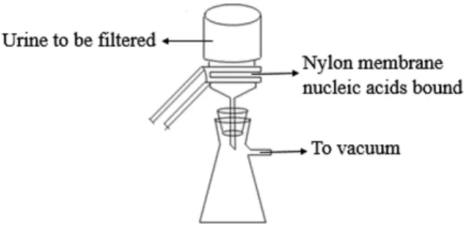
Figure 1 The scheme of microRNA concentration and preservation.
BNBZF810S, Pall, NY) and the medium-speed qualitative filter papers were cut according to the diameter of the vacuum suction filter bottle (10 cm2filter area). In Step 2, three to four sheets of wet filter paper were placed onto the vacuum suction filter bottle, and one wet nylon membrane was placed immediately onto the filter papers to avoid the generation of bubbles. In Step 3, the vacuum suction filter bottle was installed and connected to the vacuum pump. In Step 4, the collecting bottle was then loaded with urine supernatant. In Step 5, the vacuum pump was opened to allow the urine to pass through the nylon membrane in a drop-wise fashion. In Step 6, the intensity of the vacuum pressure was set to approximately 5 kPa with an initial velocity of approximately 1 to 2 drops per second. As time passed, the speed of the drops was reduced, so a higher intensity vacuum pressure was required. The total filtration process time was 5 to 10 min, and the intensity of the vacuum pressure was 5 to 50 kPa, depending on the volume of the urine. In Step 7, after the urinary nucleic acids were adsorbed onto the nylon membranes, the membranes were placed into the 56◦
C drying oven for 3 to 5 min to complete the drying process or left to dry at room temperature. In Step 8, the dry membranes were placed between aseptic sealing bags and then sealed using a vacuum sealer. Tags were added to the dry membranes that contained the basic information on the participants and the unique number of the membranes, which could be cross-referenced with additional information relevant to the urine sample. Finally, the membranes were stored at−80◦C or room temperature. A flow
chart explaining the process is presented inFig. 1.
Urinary RNA extraction
RNA is more labile than DNA and ribonuclease (RNase) is known to be present in the body fluids (Reddi & Holland, 1976;Zhao et al., 2015). Consequently, RNA molecules on the urinary nucleic acid-bound dry membranes were examined in this paper.
The urinary nucleic acid-bound dry membranes were cut into small pieces and placed in clean tubes. Total RNA, including microRNAs, was extracted by using TRIzol reagent (Invitrogen, Carlsbad, CA) according to the manufacturer’s instructions. Briefly, 250µL
of chloroform was added, and the samples were shaken vigorously for 15 s. Then the samples were centrifuged at 12,000 rpm for 15 min at 4◦
C. The fraction from the top aqueous phase was obtained and transferred into new 1.5-mL tubes. Next, 600µL of
was centrifuged for 10 min at 12,000 rpm at 4◦C. After removing the ethanol, the pellet was dissolved in RNase-free water and quantified with a NanoDrop spectrophotometer (Thermo Fisher Scientific). Finally, the sample was stored at−80◦C for further analysis
by RT-qPCR.
MicroRNA quality control
The quality of the microRNA was evaluated on a Bioanalyzer 2100 instrument (Agilent, Santa Clara, CA, USA) using small RNA kit.
Reverse transcription of urinary microRNA
U6, miR-16 and miR-21 were reverse transcribed using synthetic primers and the Promega GoScriptTMReverse Transcription kit (Promega Corporation, Madison, USA) according to the manufacturer’s protocol. After combining the experimental primers with an equal amount of template RNA, the primers and template mix were thermally denatured at 70◦C for 5 min and chilled on ice for 5 min. The primers and template mix were added to the reaction mix on ice. Following an initial annealing at 25◦
C for 5 min, the reaction was incubated at 42◦C for up to one hour. Then, the reaction was incubated in a controlled-temperature heat block at 70◦
C for 15 min. The reaction product was placed on ice or stored at−20◦C until use.
Real-time PCR of urinary microRNA
To quantify urinary microRNA, real-time PCR amplification was performed in a 20-µl
re-action volume on a Bio-Rad real-time PCR system according to the manufacturer’s proto-cols. The real-time PCR reactions were set up in triplicate in 8-tube PCR strips (AXYGEN, Union City, CA 94587, USA) and briefly centrifuged. Real-time PCR conditions included one cycle of initial activation for 10 min at 95◦C followed by 40 cycles of denaturation for 15 s at 95◦
C, annealing for 10 s at 95◦
C and extension for 30 s at 55◦
C. The melting program was performed at 55◦
C to 94.5◦
C at a heating rate of 1◦
C per 5 s. The levels of the target microRNAs were normalized using U6, and the relative level of microRNA was determined using theΔΔCt method. All of the samples were tested in triplicate.
Statistical analysis
All statistical analysis was performed with the SPSS software (version 16, IBM), and all tests were two-sided with a 0.05 significance level. The total RNA amounts on the membranes before and after three months of storage at room temperature were compared using
t-tests. TheCTvalues of microRNA before and after three months of storage at room temperature were compared using Mann–Whitney U tests to account for differences in variance between groups.
RESULTS
Loading capacity of the nylon membranes
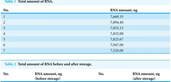
Table 1 Total amount of RNA.
No. RNA amount, ng
1 7,669.35
2 7,894.40
3 7,832.15
4 7,832.00
5 7,825.67
6 7,947.00
7 7,520.00
Table 2 Total amount of RNA before and after storage.
No. RNA amount, ng
(before storage)
No. RNA amount, ng
(after storage)
1 10,853.10 5 11,370.33
2 11,867.70 6 11,173.53
3 11,793.60 7 12,396.78
4 10,544.40 8 12,742.29
through a new nylon membrane to detect urinary nucleic acids not adsorbed by the first membrane. The urinary nucleic acid-bound membranes were dried completely in a 56◦C drying oven, and RNA was extracted and quantified. Experiments repeated in triplicate indicated that the average amount of RNA extracted from the first nylon membrane was 4,442.1667±333 ng, whereas 759.1±21.83 ng of RNA was extracted from the second
nylon membrane. A comparison of the nucleic acid adsorption amounts from the first and second membranes revealed that most of the RNA was adsorbed on the first membrane. The binding rate of urinary RNA was 85.41% [4,442/(4,442+759)].
Reproducibility of urinary RNA preservation
The adsorption of 50 mL of mixed centrifuged urine on 10 cm2nylon membrane was repeated 7 times. Then, the membranes were placed into the 56◦
C drying oven for 5 min to complete the drying process. The total RNA that was adsorbed onto each nylon membrane was extracted and quantified with a NanoDrop spectrophotometer. As shown inTable 1, the average amount of total RNA from 7 repeats was 7,788.65±145.9 ng, and
the coefficient of variation was 0.0187.
The amount of urinary microRNA stored at room temperature for three months was unchanged
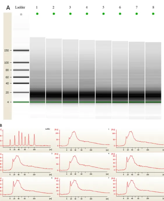
Figure 2 (A) Gel electrophoresis analysis of small RNA fractions on an Agilent small RNA chip. (B) Fluorescence intensity of small RNA fractions at various sizes (nt).Lane 1–4 represented the membranes before 3 month storage. Lane 5–8 represented the membranes after 3 month storage.
extracted and quantified with a NanoDrop spectrophotometer (Table 2). The average amount of the total RNA from 4 independent experiments from membranes before room temperature storage was 11,264.70±666.23 ng, and the coefficient of variation
was 0.0232. The average amount of total RNA after 3 months of room temperature storage was 11,920.73±766.56 ng, and the coefficient of variation was 0.0219. No significant
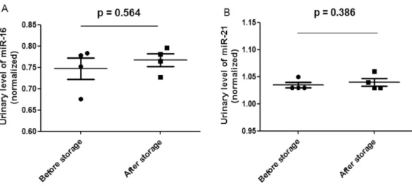
Figure 3 Scatter plots of urinary levels of miR-16 (A) and miR-21 (B) in the before storage group (n=4) and the after storage group (n=4).MicroRNA levels (Log10 scale atY-axis) were normalized
to U6. The line represents the median value.
values were proportional to urinary microRNA quantity (Mall et al., 2013), and U6 was reported as an internal normalization control (Mlcochova et al., 2014). Our data indicate that U6 was detectable in the urine of all of the 8 samples, and no significant difference in
CTvalues (P=1.000) was observed before and after storage. Additionally, the miR-16 and miR-21CTvalues did not significantly differ before and after storage (Fig. 3).
DISCUSSION
MicroRNA has been proposed as a promising class of biomarkers (Kroh et al., 2010;Mall et al., 2013;Mitchell et al., 2008). This study exclusively examined total RNA and microRNA levels on the urinary nucleic acid-bound membranes.
Although it has not been widely used until recently, urinary DNA is a potential source of material for molecular diagnosis (Chan et al., 2003;Goessl et al., 2000;Zhang et al., 1999). However, the storage of fresh urine at temperatures of 4◦
Generally, 200µL or 400µL fresh urine will be used for each RNA extraction. Because
of the low concentration of urinary RNA, it often costs several extractions to have enough RNA for reverse transcription. The method described here provides an economical way to concentrate and preserve urinary RNA. Different urine samples may have different amount of RNA on the membrane. However, no matter how much RNA could be recovered, as long as the amount of templates used for PCR reaction remained the same, the target microRNAs can still be quantified and compared.
Urine is a better source for biomarker research (Gao, 2013). The current study was the first to report the use of nylon membranes to concentrate and preserve urinary nucleic acids. The nucleic acids preserved on nylon membranes could be extracted and subse-quently analyzed using traditional downstream analytical applications, such as PCR, real time PCR, nucleic acid microarray or other nucleic acid test methods. This method allows for the preservation of urine samples from all consenting patients during every stage of dis-ease development or even for those consenting healthy individuals for prospective studies.
ADDITIONAL INFORMATION AND DECLARATIONS
Funding
This work was supported by the National Basic Research Program of China
(2012CB517606, 2013CB530805), Key Basic Research Program of the Ministry of Science and Technology of China (No. 2013FY114100), 111 Project (B08007), PUMC Youth Fund and the Fundamental Research Funds for the Central Universities (33320140133). The funders had no role in study design, data collection and analysis, decision to publish, or preparation of the manuscript.
Grant Disclosures
The following grant information was disclosed by the authors:
National Basic Research Program of China: 2012CB517606, 2013CB530805.
Key Basic Research Program of the Ministry of Science and Technology of China: 2013FY114100.
111 Project: B08007. PUMC Youth Fund.
Fundamental Research Funds for Central Universities: 33320140133.
Competing Interests
The authors declare there are no competing interests.
Author Contributions
• Fanshuang Zhang conceived and designed the experiments, performed the experiments,
analyzed the data, contributed reagents/materials/analysis tools, wrote the paper, prepared figures and/or tables.
• Xiaoyu Cheng researched the overestimation of the concentration of urinary RNA
• Yuan Yuan contributed reagents/materials/analysis tools. • Jianqiang Wu analyzed the data.
• Youhe Gao conceived and designed the experiments, reviewed drafts of the paper.
Human Ethics
The following information was supplied relating to ethical approvals (i.e., approving body and any reference numbers):
Institute of Basic Medical Sciences Medical Ethics Committee of Peking Union Medical College—Project No. 040-2014.
Supplemental Information
Supplemental information for this article can be found online athttp://dx.doi.org/ 10.7717/peerj.1082#supplemental-information.
REFERENCES
Bartel DP. 2004.MicroRNAs: genomics, biogenesis, mechanism, and function.Cell116:281–297
DOI 10.1016/S0092-8674(04)00045-5.
Bordelon H, Russ PK, Wright DW, Haselton FR. 2013.A magnetic bead-based method for concentrating DNA from human urine for downstream detection.PLoS ONE8:e68369
DOI 10.1371/journal.pone.0068369.
Catto JW, Alcaraz A, Bjartell AS, De Vere White R, Evans CP, Fussel S, Hamdy FC,
Kallioniemi O, Mengual L, Schlomm T, Visakorpi T. 2011.MicroRNA in prostate, bladder, and kidney cancer: a systematic review.European Urology59:671–681
DOI 10.1016/j.eururo.2011.01.044.
Chan AK, Chiu RW, Lo YM, Clinical Sciences Reviews Committee of the Association of Clinical B. 2003.Cell-free nucleic acids in plasma, serum and urine: a new tool in molecular diagnosis.
Annals of Clinical Biochemistry40:122–130DOI 10.1258/000456303763046030.
Gao Y. 2013.Urine—an untapped goldmine for biomarker discovery?Science China Life Sciences
56:1145–1146DOI 10.1007/s11427-013-4574-1.
Goessl C, Krause H, Muller M, Heicappell R, Schrader M, Sachsinger J, Miller K. 2000.
Fluorescent methylation-specific polymerase chain reaction for DNA-based detection of prostate cancer in bodily fluids.Cancer Research60:5941–5945.
Hilhorst M, Theunissen R, Van Rie H, Van Paassen P, Tervaert JW. 2013.DNA extraction from long-term stored urine.BMC Nephrology14:238DOI 10.1186/1471-2369-14-238.
Jia L, Liu X, Liu L, Li M, Gao Y. 2014.Urimem, a membrane that can store urinary proteins simply and economically, makes the large-scale storage of clinical samples possible.Science China Life Sciences57:336–339DOI 10.1007/s11427-013-4582-1.
Kainz P, Seifriedsberger M, Strack HB. 1989.A modified primer extension procedure for specific detection of DNA-RNA hybrids on nylon membranes.Analytical Biochemistry179:366–370
DOI 10.1016/0003-2697(89)90146-2.
Khandjian EW. 1986.UV crosslinking of RNA to nylon membrane enhances hybridization signals.
Molecular Biology Reports11:107–115DOI 10.1007/BF00364822.
Kroh EM, Parkin RK, Mitchell PS, Tewari M. 2010.Analysis of circulating microRNA biomarkers in plasma and serum using quantitative reverse transcription-PCR (qRT-PCR).Methods
Li M, Zhao M, Gao Y. 2014.Changes of proteins induced by anticoagulants can be more sensitively detected in urine than in plasma. Science China Life Sciences57:649–656
DOI 10.1007/s11427-014-4661-y.
Mall C, Rocke DM, Durbin-Johnson B, Weiss RH. 2013.Stability of miRNA in human urine supports its biomarker potential.Biomarkers in Medicine7:623–631DOI 10.2217/bmm.13.44.
Mitchell PS, Parkin RK, Kroh EM, Fritz BR, Wyman SK, Pogosova-Agadjanyan EL, Peterson A, Noteboom J, O’Briant KC, Allen A, Lin DW, Urban N, Drescher CW, Knudsen BS,
Stirewalt DL, Gentleman R, Vessella RL, Nelson PS, Martin DB, Tewari M. 2008.
Circulating microRNAs as stable blood-based markers for cancer detection.Proceedings of the National Academy of Sciences of the United States of America 105:10513–10518
DOI 10.1073/pnas.0804549105.
Mlcochova H, Hezova R, Stanik M, Slaby O. 2014.Urine microRNAs as potential noninvasive biomarkers in urologic cancers.Urologic Oncology32:41.e1–41.e9
DOI 10.1016/j.urolonc.2013.04.011.
Pall Corporation. 2015. Nylon membrane.Available athttp://www.pall.com/main/ oem-materials-and-devices/product.page?lid=gri78lty.
Ralla B, Stephan C, Meller S, Dietrich D, Kristiansen G, Jung K. 2014.Nucleic acid–based biomarkers in body fluids of patients with urologic malignancies.Critical Reviews in Clinical Laboratory Sciences51:200–231DOI 10.3109/10408363.2014.914888.
Reddi KK, Holland JF. 1976.Elevated serum ribonuclease in patients with pancreatic cancer.
Proceedings of the National Academy of Sciences of the United States of America73:2308–2310
DOI 10.1073/pnas.73.7.2308.
Taylor GR. 1985.Southern transfer of native DNA using nylon membranes.Analytical Biochemistry148:524–526DOI 10.1016/0003-2697(85)90262-3.
Thurston SJ, Saffer JD. 1989.Ultraviolet shadowing nucleic acids on nylon membranes.Analytical Biochemistry178:41–42DOI 10.1016/0003-2697(89)90353-9.
Veltri RW, Makarov DV. 2006.Nucleic acid–based marker approaches to urologic cancers.
Urologic Oncology24:510–527DOI 10.1016/j.urolonc.2006.07.002.
Weber JA, Baxter DH, Zhang S, Huang DY, Huang KH, Lee MJ, Galas DJ, Wang K. 2010.The microRNA spectrum in 12 body fluids.Clinical Chemistry 56:1733–1741
DOI 10.1373/clinchem.2010.147405.
Yang Y, Xiao L, Li J, Kanwar YS, Liu F, Sun L. 2013.Urine miRNAs: potential biomarkers for monitoring progression of early stages of diabetic nephropathy.Medical Hypotheses81:274–278
DOI 10.1016/j.mehy.2013.04.031.
Yokota M, Tatsumi N, Tsuda I, Takubo T, Hiyoshi M. 1998.DNA extraction from human urinary sediment.Journal of Clinical Laboratory Analysis12:88–91
DOI 10.1002/(SICI)1098-2825(1998)12:2<88::AID-JCLA3>3.0.CO;2-F.
Zhang DZ, Lau KM, Chan ES, Wang G, Szeto CC, Wong K, Choy RK, Ng CF. 2014.Cell-free urinary microRNA-99a and microRNA-125b are diagnostic markers for the non-invasive screening of bladder cancer.PLoS ONE9:e100793DOI 10.1371/journal.pone.0100793.
Zhang J, Tong KL, Li PK, Chan AY, Yeung CK, Pang CC, Wong TY, Lee KC, Lo YM. 1999.
Presence of donor- and recipient-derived DNA in cell-free urine samples of renal transplantation recipients: urinary DNA chimerism.Clinical Chemistry45:1741–1746.