in
Saccharomyces cerevisiae
Ewan P. Plant1¤a, Phuc Nguyen1¤b, Jonathan R. Russ1, Yvette R. Pittman2, Thai Nguyen3¤c, Jack T. Quesinberry4, Terri Goss Kinzy2, Jonathan D. Dinman1
*
1Department of Cell Biology and Molecular Genetics, University of Maryland, College Park, Maryland, United States of America,2Department of Molecular Genetics, Microbiology and Immunology, University of Medicine and Dentistry of New Jersey (UMDNJ) Robert Wood Johnson Medical School, Piscataway, New Jersey, United States of America,3The Science and Technology Center at Eleanor Roosevelt High School, Greenbelt, Maryland, United States of America,4Huntington High School, Huntington, Maryland, United States of America
Background.Decoding of mRNAs is performed by aminoacyl tRNAs (aa-tRNAs). This process is highly accurate, however, at
low frequencies (1023– 1024) the wrong aa-tRNA can be selected, leading to incorporation of aberrant amino acids. Although
our understanding of what constitutes the correct or cognate aa-tRNA:mRNA interaction is well defined, a functional
distinction between near-cognate or single mismatched, and unpaired or non-cognate interactions is lacking.Methodology/
Principal Findings. Misreading of several synonymous codon substitutions at the catalytic site of firefly luciferase was
assayed inSaccharomyces cerevisiae. Analysis of the results in the context of current kinetic and biophysical models of
aa-tRNA selection suggests that the defining feature of near-cognate aa-aa-tRNAs is their potential to form mini-helical structures with A-site codons, enabling stimulation of GTPase activity of eukaryotic Elongation Factor 1A (eEF1A). Paromomycin specifically stimulated misreading of near-cognate but not of non-cognate aa-tRNAs, providing a functional probe to
distinguish between these two classes. Deletion of the accessory elongation factor eEF1Bcpromoted increased misreading of
near-cognate, but hyperaccurate reading of non-cognate codons, suggesting that this factor also has a role in tRNA
discrimination. A mutant of eEF1Ba, the nucleotide exchange factor for eEF1A, promoted a general increase in fidelity,
suggesting that the decreased rates of elongation may provide more time for discrimination between aa-tRNAs. A mutant form of ribosomal protein L5 promoted hyperaccurate decoding of both types of codons, even though it is topologically distant
from the decoding center.Conclusions/Signficance.It is important to distinguish between near-cognate and non-cognate
mRNA:tRNA interactions, because such a definition may be important for informing therapeutic strategies for suppressing these two different categories of mutations underlying many human diseases. This study suggests that the defining feature of near-cognate aa-tRNAs is their potential to form mini-helical structures with A-site codons in the ribosomal decoding center. An aminoglycoside and a ribosomal factor can be used to distinguish between near-cognate and non-cognate interactions.
Citation: Plant EP, Nguyen P, Russ JR, Pittman YR, Nguyen T, et al (2007) Differentiating between Near- and Non-Cognate Codons inSaccharomyces cerevisiae. PLoS ONE 2(6): e517. doi:10.1371/journal.pone.0000517
INTRODUCTION
Accurate transmission of biological information is a central requirement at all levels of life. In cells, one aspect of this process is the faithful translation of the genetic code from DNA into protein. The intermediaries in the last stage of this process include
mRNA, tRNAs, ribosomes and many trans-acting factors. The
protein coding information of an mRNA is formatted as codons. The anticodon loops of aminoacyl tRNAs (aa-tRNAs) form base-pairing interactions with the codons. This enables ribosomes to add amino acids sequentially to the nascent protein. aa-tRNAs that can participate in standard Watson-Crick interactions with the first two bases in a codon and can form either canonical or non-Watson-Crick pairs at the third or ‘‘wobble’’ position are designated cognate-tRNAs [1,2]. In contrast, tRNAs that do not meet these requirements are commonly referred to as near- and non-cognate tRNAs. Utilization of near- and non-cognate tRNAs is called misreading or a missense error. Misreading occurs with low frequencies of 1023and 1024per codon ([3] and references within).
The 64 codons encode 20 different amino acids and three termination signals. In cases where one amino acid is represented by multiple codons, some tRNAs can decode more than one codon. This redundancy is facilitated by tRNA modifications and by wobble base-pairing between the anticodon and the codon (reviewed in [4]). Our understanding of how the ribosome achieves such a high degree of specificity has been facilitated by both kinetic and structural analyses in bacteria (reviewed in [5–7]). In vitro
kinetic analyses using ribosomes, tRNAs, and the bacterial tRNA binding factor EF-Tu have broken down the process of aa-tRNA selection into a series of discrete steps (reviewed in [6] ). These studies have identified two stages (k22and k7) in this process
that favor rejection of aa-tRNAs whose anticodon loops cannot base-pair with codons. A mutational analysis demonstrated that
Academic Editor:Alfred Lewin, University of Florida, United States of America ReceivedApril 10, 2007;AcceptedMay 18, 2007;PublishedJune 13, 2007 This is an open-access article distributed under the terms of the Creative Commons Public Domain declaration which stipulates that, once placed in the public domain, this work may be freely reproduced, distributed, transmitted, modified, built upon, or otherwise used by anyone for any lawful purpose. Funding:This work was supported by grants from the National Institutes of Health to JDD (GM058859 and AI064307), and to TGK (GM057483).
Competing Interests:The authors have declared that no competing interests exist.
* To whom correspondence should be addressed.E-mail: [email protected] ¤aCurrent address: Laboratory of Hepatitis and Related Emerging Agents, Division of Emerging and Transfusion-Transmitted Diseases, Office of Blood Research and Review, Center for Biologics Evaluation and Research (CBER), FDA, Bethesda, Maryland, United States of America
¤bCurrent address:National Institute of Allergy and Infectious Diseases, National Institutes of Health, Bethesda, Maryland, United States of America
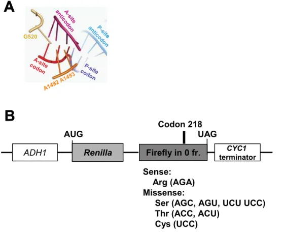
two independent mechanisms corresponding to these two steps are required for utilization of cognate aa-tRNAs [8]. Structural, biophysical, and computational analyses also show a mechanism for positive selection of cognate aa-tRNAs[7–10]} that emphasizes the geometry of base pairing at the ribosomal decoding center [11]. Formation of an appropriately configured mini-helix in the decoding center generates an RNA minor-groove, enabling interaction with three critical bases of the small subunit rRNA. Formation of this mini-helix stimulates A1492 and A1493 of the small subunit rRNA to flip out into the minor groove forming a complex arrangement of hydrogen bonds with the tRNA/ mRNA backbones in concert with G530 (Figure 1A). This in turn stimulates a conformational change in the aa-tRNA that transduces the information from the decoding center to activate the GTPase activity of EF-Tu (reviewed in [12]). The energy barrier for flipping out of A1492 and A1493 is sufficiently small for correct binding of aa-tRNA to shift the equilibrium in favor of the subsequent steps [7]. Aminoglycoside antibiotics such as paromo-mycin stimulate misreading by binding to the decoding center, displacing A1492 and A1493. This forces these bases to mimic the ‘‘flipped out’’ conformations that they normally assume in response to the mini-helix formation by a cognate codon:antico-don pair (reviewed in [5]). Together, these kinetic and biophysical mechanisms ensure the accurate utilization of cognate aa-tRNAs.
An unsettled issue remains the precise distinction between ‘‘near-cognate’’ and ‘‘non-cognate’’ tRNAs, especially in eukar-yotes. This is important since the rational design or utilization of therapeutics may exploit the functional differences that exist between these two classes of tRNAs. One recent study has suggested that the relative abundances of bacterial aa-tRNAs plays a significant role in translational accuracy [3]. By this model, highly abundant aa-tRNAs are more likely than low abundance aa-tRNAs to misread codons that are decoded by other low abundance aa-tRNAs. The current study using the yeast
Saccharomyces cerevisiaesupports this competition model. However it also suggests that this is not sufficient to explain the functional differences between near- and non-cognate aa-tRNAs. Using a series of seven substitutions of a codon in the catalytic site of firefly luciferase, we show that a second critical distinction lies in the ability to form hydrogen bonding interactions at all three positions between the aa-tRNA anticodon loop and the codon in the decoding center. This is likely the result of changes in formation of the codon:anticodon mini-helix. Thus, transient formation of the mini-helix allows the rRNA and tRNA conformational changes required for activation of the GTPase activity of eEF1A, the eukaryotic homolog of EF-Tu. This is supported by the demonstration of paromomycin stimulated misreading by aa-tRNAs that are capable of forming a transient
Renilla
Firefly in 0 fr.
ADH1
terminatorCYC1AUG
UAG
Sense:
Arg (AGA)
Missense:
Ser (AGC, AGU, UCU UCC)
Thr (ACC, ACU)
Cys (UCC)
Codon 218
A -site anticod
on P
-site anticod
on
A-site
codon P-site
codon
G520
A149 2 A1493
A
B
Renilla
Firefly in 0 fr.
ADH1
terminatorCYC1A -site anticod
on P
-site anticod
on
A-site
codon P-site
codon
G520
A
A-site anticod
on P
-site anticod
on
A-site
codon P-site
codon
G520
A
B
A149 2 A1493
Figure 1. The decoding center and dual-luciferase reporters for determining rates of translational misreading in yeast. Panel A. The codon:anticodon mini-helix in the decoding center is stabilized by base-pairing at all three positions of the mini-helix favoring A-minor interactions with flipped out bases G520, A1492 and A1493. PyMol (Delano Scientific, LLC) was used to generate this figure based on the coordinates 1IBM in the RSCN Protein Data Bank [40].Panel B.In all missense reporters, transcription is initiated from the yeastADH1promoter, and terminated at a sequence from theCYC139UTR. The luciferase genes fromRenillaand firefly are cloned in frame to produce a fusion of the two proteins. The sense reporter has the AGA codon encoding arginine at amino acid residue 218 in the catalytic site of firefly luciferase. Missense reporters contain the indicated mutations at this position, which encode the indicated amino acids. Efficiencies of missense suppression were calculated by dividing the ratio of firefly/Renilla luciferase generated from cells harboring the missense test vectors by the ratio of firefly to Renilla luciferase generated from cells harboring the sense control plasmid.
interaction. This identifies paromomycin as a functional probe to distinguish between near- and non-cognate aa-tRNAs. The hypothetical roles in this process played by eEF1A and its
associated factors, eEF1Ba and eEF1Bc, were also investigated.
The results suggest that the GTPase activity of eEF1A is preferentially stimulated by near-cognate codon:anticodon inter-actions, and point to discrete functional regions of the protein. Studies of eEF1Ba, the catalytic subunit of the guanine nucleotide exchange factor (GEF) required for recycling of GTP-bound eEF1A, are consistent with a kinetic model in which limiting concentrations of eEF1A:aa-tRNA:GTP ternary complex should decrease rates of protein synthesis, resulting in increased selection against both near- and non-cognate aa-tRNAs. Interestingly, deletion of the proposed regulatory subunit of the GEF complex, eEF1Bc, tended to promote increased misreading of near-cognate codons and decreased misreading of non-cognate codons. This suggests eEF1Bcmay have a regulatory function. In a final series of experiments, the potential role of the fungus-specific elongation factor eEF3 in translational fidelity was indirectly assayed through analysis of a series of mutants in the ribosomal protein L5 (rpL5). rpL5 forms part of the ribosome binding site for this factor [13]. Like the eEF1Bamutant, the ability of an rpL5 mutant to promote enhanced fidelity at both non- and near-cognate codons suggests that slowing rates of elongation by disrupting the synergy between eEF1A and eEF3 results in increased selection against both near-and non-cognate aa-tRNAs.
RESULTS
Baseline and paromomycin-stimulated rates of
missense suppression suggests a functional
difference between near- and non-cognate tRNAs
Introduction of a missense mutation into the active site of an enzyme followed by quantitative measurement of the restoration of enzymatic activity can provide a basis to monitor translational error rates. The arginine at position 218 of firefly luciferase is located in the active site and is required for enzymatic activity. Mutation of the corresponding AGA arginine codon to either the AGC or UCU serine codon was previously used to monitormissense error rates of ribosome bound chaperone mutants inS.
cerevisiae[14]. The current study employed a bicistronic luciferase reporter system to monitor suppression of a series of missense mutations. This could functionally distinguish translational fidelity effects due to the inherent translatability of different codons and subsequently correlate these with trans-acting influences. In this assay, the gene encoding firefly luciferase is fused in frame with
a downstream Renilla luciferase gene. Test plasmids harbored
missense codons at position 218 of the firefly luciferase gene (Figure 1B, ‘‘Missense’’). The control was identical except that it contained the wild-type AGA codon at this position (Figure 1B, ‘‘Sense’’). Even though the two luciferase proteins are fused, the activity from each can be measured independently as they utilize different substrates. Rates of misreading were calculated by dividing the ratio of firefly luciferase activity to Renilla luciferase activity generated from the missense vector in strains harboring the indicated mutant allele by the ratio generated with the sense plasmid. The results were statistically tested as previously described [15].
Previous studies have shown that missense errors occur with frequencies on the order of 1024in bothE. coliand inS. cerevisiae
[3,16–18]. Consistent with the literature, the seven missense mutations assayed at position 218 demonstrated ratios of firefly to
Renilla luciferase activities that were reduced by approximately 4 orders of magnitude in all cases (Table 1). Although rates of misreading are low and the differences between test and control
samples are small, the sensitivity of the assay and rigor of the statistical methods enable meaningful analysis of the data. Inspection of these data revealed that rates of misreading varied over an approximately 4.5-fold range. The inability of any one amino acid (i.e. Ser, Cys, or Thr) to disproportionately influence apparent missense incorporation suggests that the assay monitored incorporation errors rather than altered luciferase activity arising from incorporation of any specific amino acid. The data reveal that the common element among the three most stimulatory codons is the potential to form a stable GNC base pair at the second position. The observation of different error rates among the four serine codons (which are decoded by different tRNA families) suggests that relative tRNA abundances are also important.
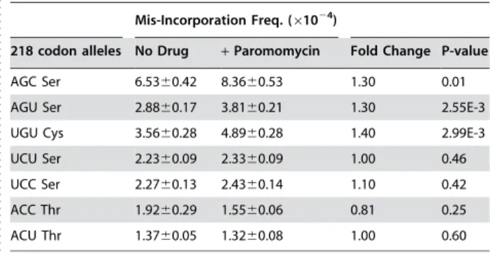
Formation of the mini-helix in the decoding center stimulates flipping out of the small subunit bases A1492 and A1493 that in combination with G530 stabilizes this structure (Figure 1A). Paromomycin binding to the decoding center of the small ribosomal subunit displaces A1492 and A1493, thus enhancing the frequency of missense errors (reviewed in [6]). It has been hypothesized that not only does the formation of the mini-helix displace A1492 and A1493, but once displaced they sterically position the interribose bonds to maintain A-form helices [19]. In the current study, translational misreading errors were significantly stimulated by paromomycin at codons that are capable of forming complete mini-helices with arginyl-tRNAs: UGU, AGU and AGC (Figure 2). In contrast, paromomycin did not stimulate misreading with anticodons unable to form the mini-helix, e.g. UCU, UCC, ACU and ACC. These findings suggest a functional definition for near- versus non-cognate codon:anticodon interactions. This potential to form base pairing interactions at all three positions, which is possibly nucleated by a strong Watson-Crick base pair at the second position would allow transient formation of the mRNA:tRNA mini-helix. We propose this as the defining feature of a near-cognate interaction. In contrast, non-cognate interac-tions are defined by their lack of potential to form the mini-helix.
eEF1A mutants generally affect utilization of
near-cognate aa-tRNAs
In eukaryotes, a ternary complex composed of eEF1A, aa-tRNA, and GTP delivers the aa-tRNA to the A-site of the ribosome. When an aa-tRNA containing the correct anticodon is sampled by the ribosome, a signal is transmitted through the body of the tRNA. This stimulates GTP hydrolysis by eEF1A and subsequent accommodation of the tRNA into the ribosomal A-site (reviewed
Table 1.Baseline levels and effects of paromomycin on suppression of missense mutations at codon 218 of firefly luciferase.
. . . .
Mis-Incorporation Freq. (61024)
218 codon alleles No Drug +Paromomycin Fold Change P-value
AGC Ser 6.5360.42 8.3660.53 1.30 0.01 AGU Ser 2.8860.17 3.8160.21 1.30 2.55E-3 UGU Cys 3.5660.28 4.8960.28 1.40 2.99E-3 UCU Ser 2.2360.09 2.3360.09 1.00 0.46 UCC Ser 2.2760.13 2.4360.14 1.10 0.42 ACC Thr 1.9260.29 1.5560.06 0.81 0.25 ACU Thr 1.3760.05 1.3260.08 1.00 0.60 Wild-type codon is AGA Arginine.
doi:10.1371/journal.pone.0000517.t001
...
....
...
...
....
...
...
....
...
...
....
...
...
....
...
...
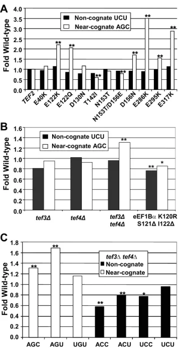
in [5,6]). eEF1A is encoded by the essentialTEF1andTEF2genes inS. cerevisiae. A set of TEF2mutants expressed in a tef1Dtef2D genetic background [20] were assayed with respect to their effects on misreading using the AGC and UGU serine codons to monitor misreading of near- and non-cognate codons, respectively. The results show allele-specific responses specifically to near-cognate codons. Strains bearing one of six alleles (E122K, E122Q, D156N, E286K, E295K, and E317K) promoted enhanced misreading of the near-cognate AGC Ser codon, but not of non-cognate UCU Ser (Figure 3A, Table 2, p,0.01). In contrast, a strain expressing the T142I and to the lesser extent N153T/D156E mutant were better able to distinguish between the cognate AGA and near-cognate AGC codons. This enhanced fidelity did not extend to the
non-cognate UCU codon (Figure 3A, Table 2, p,0.01). In
summary, it appears that near-cognate codons and not non-cognate are able to influence eEF1A activity in an allele-specific manner.
A mutation in eEF1B
a
promotes general
hyperfidelity
After hydrolysis of GTP, the eEF1A:GDP complex is released
from the ribosome. The eEF1Basubunit (encoded byTEF5) is the
essential nucleotide exchange factor responsible for catalytic activity in eEF1A recycling [21]. Analysis of the role of the
eEF1Ba protein capitalized on the availability of the K120R
S121DI122Dmutant form of the protein that altered or deleted
residues involved in critical interactions with the nucleotide binding pocket of eEF1A [22]. This mutant strain was previously shown to enhance translational fidelity by promoting lower levels of readthrough at all three stop codons [23]. In the current study,
the K120R S121D I122D eEF1Ba mutant showed modest but
consistent enhanced fidelity at both the UCU and AGC non- and near-cognate codons respectively (Figure 3B, Table 2, p,0.01). As discussed below, we hypothesize that this may be due to limiting concentrations of eEF1A:GTP.
Codon-specific misreading in the absence of eEF1B
c
In yeast, the eEF1 complex contains a third non-essential subunit, eEF1Bc. Yeast cells express two isoforms of eEF1Bc, encoded bythe TEF3 and TEF4 genes. The N-terminus of Tef3p exhibits
structural similarity to gluthathione-S-transferases and is thought to participate in the regulation of elongation during stress [24,25]. Deletion of either eEF1Bcisoform alone had no significant effects on misreading at either the AGC near-cognate or UGU non-cognate codons (Figure 3B, Table 2). However deletion of both
eEF1Bc-encoding genes caused a statistically significant increase in mis-incorporation at near- but not at non-cognate codons (Figure 3B, Table 3). These results suggest that these two proteins have redundant activities and that this factor functions in ensuring translational fidelity. To investigate this phenomenon further, misreading in the absence of eEF1Bc(tef3Dtef4D) was assayed at the remaining 5 missense codons. Although misreading of the near-cognate AGU Ser codon was also enhanced, there was no effect on recognition of the ‘near-cognate’ UGU Cys codon (Table 3, Figure 3C). In contrast, although recognition of the UCU ‘non-cognate’ codon was not affected, recognition of the other non-cognate codons (ACC, ACU, and UCC) was signifi-cantly enhanced by the absence of eEF1Bc.
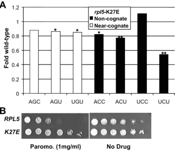
The K27E mutant of ribosomal protein L5 promotes
a general enhancement of fidelity
The three-site model of the ribosome posits that preventing simultaneous occupancy of the ribosomal A- and E-sites by aa-tRNA and deacylated-aa-tRNAs respectively helps to coordinate the elongation cycle (reviewed in [26]). In fungi, the unique essential elongation factor 3 (eEF3) facilitates eEF1A-dependent A-site binding of aa-tRNA and has ATP-dependent activity required for the release of deacylated tRNA from the E site [27]. Although ribosomal protein L5 (rpL5) is far from the decoding center, it has been shown to interact with eEF3 [13]. Previous studies have characterized five temperature-sensitive alleles of the yeastRPL5
gene encoding rpL5 [28,29]. A preliminary assay of five rpl5
mutant strains using the near- (AGC) and non- (UCU) cognate reporters indicated that the K27E mutant of L5 tended to be generally hyperaccurate (data not shown). Analysis of all 7 missense codons revealed that the K27E mutant generally promoted greater levels of translational accuracy (Figure 4A, Table 3). Dilution spot assays revealed that rpL5-K27E is resistant to paromomycin (Figure 4B), consistent with the notion that this mutant is antagonistic to the action of the drug. As discussed below, these findings suggest an indirect role for rpL5 in translational decoding. This may be through its association with eEF3 and account for some differences in A-site fidelity between bacteria and fungi
DISCUSSION
A significant number of human diseases are caused by missense mutations. Understanding the functional differences between mutations and drugs that result in near- versus non-cognate
I-U Base pair N H H H N O O R O H H H N N N N R I U N N N N N H CH3 H H H H R
R O O
O
mcm5U-C N3-N3,
4-carbonyl-amino C U CH2 C O O
H2C N
N N N O H H H H H H R R O O O U U U-U 2-carbonyl-N3, 4-carbonyl-N3
G C I
U G U
• •• • • •
U C U
mcm5A G U
• • •
• •
U C U
mcm5A G C
• • •
•
• •• ••
tRNA
Arg2tRNA
Arg3tRNA
Arg3N H I -C N N H H H H H R R O O O - - -• •• • • • • • • • • • • • • • •• •• N I -C - -• • • • • •• • •• • • • • • • • • •• • • • • • •• ••
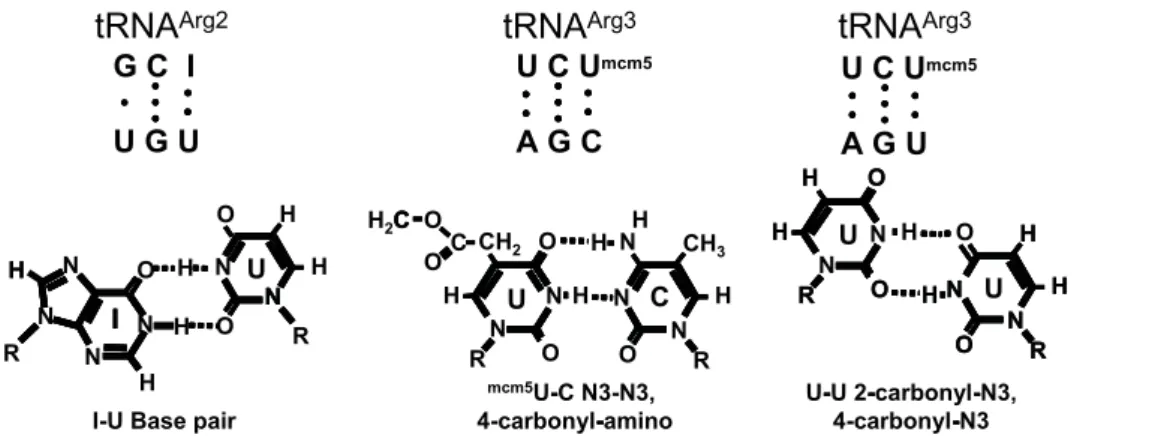
Figure 2. Proposed basis for near-cognate codon-anticodon interactions. Top.Base pairing between tRNAArg2anticodon and UGU codon (left), and between tRNAArg3anticodon and the AGC and AGU codons.Bottom.INU, UNC, and UNU base pairing.
codon usage may inform strategies for therapeutic interventions. In the current study, codon misreading was examined in the yeast eukaryotic model system using a dual luciferase reporter in which the AGA Arg codon in the firefly luciferase catalytic site was changed to AGC, AGU, UCU, UCC (serine); UGU (cysteine); and ACC or ACU (threonine). Each of these substitutions has a polar sidechain like arginine, but lacks the positive charge. The consistent 4-order of magnitude decrease in firefly luciferase activity with these substitutions agrees with previously
character-ized missense error rates. The finding no one amino acid alters firefly luciferase activity outside a range supports that the assay monitors misincorporation of arginine. The observation that the frequency of Arg misincorporation varied over an approximately 4.5-fold range independent of the identity of the encoded amino acid suggests that misincorporation frequency is determined by other factors.
In each species, some codons are used more frequently than others. The codon bias is especially clear in highly expressed genes. Studies inE. colihave led to the suggestion that codon bias minimizes the deleterious effects of aberrant decoding [30,31]. Examination of codon bias in highly expressed yeast genes (see [32]) reveals that both the AGU and AGC Ser codons occur at a low frequency of approximately 5% of serine in the yeast genome. All of the other codons employed in this study occur with significantly higher frequencies. UGU is the most frequently used Cys codon at 84% in highly transcribed genes. Its substitution promoted even higher rates of miscoding than AGU Ser at 5% codon frequency. Thus, it is clear that codon frequency is not the sole determinant of translational fidelity. A recent study inE. coli
suggested that competition between different tRNAs is the underlying factor influencing misreading error rates [3]. In yeast, gene copy number for individual tRNA species correlates with initial estimates of relative tRNA content in normally growing cells, allowing the number of functional genes encoding cognate tRNAs for each codon to be used as a proxy measure of tRNA abundance [33,34]. Examination of yeast tRNA gene copy numbers [35] reveals that AGA Arg is decoded by the highly
abundant tRNAArg3 (11 copies). In support of the tRNA
competition model, the codons that intrinsically promoted higher levels of misreading were decoded by lower abundance tRNAs, while those at the lower end of the range are decoded by more abundant tRNAs. For example, the AGU and AGC codons are decoded by the relatively low abundance tRNASer3(4 copies), and all Cys codons are decoded by the 4 copy tRNACys. In contrast,
the UCU and UCC codons are decoded by tRNASer2(11 copies),
and the ACC and ACU codons are decoded by the high copy tRNAThr1a(11 copies). However, tRNA competition alone cannot fully explain the observed differences. The pattern becomes more apparent when the ability of near-cognate codons to base pair with anticodons of different arginyl-tRNAs is considered (Figure 2). The first two bases of the AGC and AGU Ser codons could be
recognized by themcm5UCU anticodon of the highly abundant
tRNAArg3. Base pairing at the wobble positions of these two
codons andmcm5U is also theoretically possible through N3-N3, 4-carbonyl-amino, and 2-carbonyl-N3, 4-carbonyl-N3 hydrogen bonding respectively (see Figure 2). Note that although there are three possible UNU base pairs and two possible CNU couples, the geometries of the N3-N3, 4-carbonyl-amino, and of the 2-carbonyl-N3, 4-carbonyl-N3 hydrogen pairing schemes provide the most energetically favorable topologies within the constraints of the tRNA:mRNA mini-helix. Further, it has been suggested that cmo5 modification of U34 stabilizes the shape of the anticodon
loop (reviewed in [4]). Although the C19-C19 distance between pyrimidine-pyrimidine nucleotide pairs is short, and is thus destabilizing relative to that for pyrimidine-purine base pairs, biophysical analyses suggest that bridging water molecules could
produce stable and planar U34NU3 and U34NC3 base pairs
(reviewed in [4]). Of note, although biophysical analyses of RNA duplexes indicate that UNU base-pairing is more stable than UNC pairs at pH 7.0, the observation that the AGC codon
pro-moted.2-fold more misreading than the AGU codon suggests
that this particular UNC base pair is more energetically permissible within the topological constraints imposed by the codon:anticodon
**
**
**
**
Fold Wild-ty
p
e
0.0 0.5 1.0 1.5 2.0 2.5 3.0 3.5 4.0
TEF2E40K
E122 K
E122QD130NT142IN153 T
N153 T/D1
56E
D156NE286KE295 K
E317K Non-cognate UCU
Near-cognate AGC
**
**
**
**
**
**
**
**
Non-cognate UCU Near-cognate AGC Non-cognate UCU Near-cognate AGC
**
**
**
**
A
B
Fold Wild-ty
p
e
Non-Cognate Near-Cognate
tef3Δ tef3Δ
tef4Δ
eEF1BαK120R
S121Δ I122Δ
tef4Δ
0.0 0.2 0.4 0.6 0.8 1.0 1.2 1.4 1.6
**
Non-cognate UCU Near-cognate AGC
*
**
Non-Cognate Near-Cognate Non-Cognate Near-Cognate
**
Non-cognate UCU Near-cognate AGC
*
**
C
0.0 0.2 0.4 0.6 0.8 1.0 1.8
AGC AGU UGU ACC ACU UCC
Fold W
ild-ty
p
e
**
UCU 1.2
1.4
1.6
**
**
**
*
Non-cognate Near-cognate
tef3∆tef4∆
**
**
**
**
*
Non-cognate Near-cognate
tef3∆tef4∆
Non-cognate Near-cognate
tef3∆tef4∆
Table 2.Effects of selected alleles of genes on mis-reading of non-cognate and near-cognate mutations at codon 218 of firefly luciferase.
. . . .
Non-cognate (UCU) Near-cognate (AGC)
Strain mis-incorp (61024) Fold WT P-value mis-incorp.(
61024) Fold-WT P-Value
eEF1A
WT (TEF2) 2.0460.13 1.00 1.00 7.0160.49 1.00 1.00
E40K (TEF2-3) 1.9160.18 0.94 0.56 8.1160.72 1.16 0.22
E122K (TEF2-4) 2.3360.17 1.14 0.17 15.460.55 2.20 9.76E-10
E122Q (TEF2-10) 1.7360.16 0.85 0.14 14.460.79 2.05 2.43E-6
D130N (TEF2-13) 1.6160.12 0.79 0.02 8.1660.21 1.16 0.05
T142I (TEF2-7) 1.6260.14 0.79 0.03 4.2060.48 0.60 6.64E-4
N153T (tef2-19) 2.0560.04 1.00 0.95 6.3960.14 0.91 0.06
N153T/D156E (tef2-18) 1.8760.05 0.91 0.03 5.5360.20 0.79 4.92E-3
D156N (tef2-17) 1.8560.04 0.91 0.02 11.9460.39 1.69 3.10E-7
E286K (TEF2-1) 1.8160.08 0.89 0.04 24.6460.11 3.50 3.58E-8
E295K (TEF2-9) 2.0060.20 0.98 0.87 10.960.95 1.55 3.09E-3
E317K (TEF2-2) 2.3160.04 1.13 0.06 20.160.91 2.87 1.70E-7
eEF1Ba
WT 3.1560.12 1.00 1.00 3.5060.14 1 1.00
K120R S121DI22D 2.3860.22 0.76 8.89E-3 2.9860.10 0.85 0.01
eEF1Bc
WT 1.2260.14 1.00 1.00 1.2460.09 1 1.00
tef3D 1.0060.04 0.81 0.15 1.1860.05 0.95 0.49
tef4D 1.2560.14 1.02 0.41 1.1460.06 0.92 0.41
tef3Dtef4D 1.1760.05 0.96 0.62 1.6260.03 1.31 1.09E-5
doi:10.1371/journal.pone.0000517.t002
..
...
....
...
...
....
...
...
....
...
...
....
...
...
....
...
...
....
...
...
....
...
...
....
...
...
....
...
...
....
...
Table 3.Survey of isogenic wild-type and mutant pairs of strains with seven different codons at codon 218 of firefly luciferase.
. . . .
codon mis-incorp (61024) Fold WT P-value
eEF1Bc
WT tef3Dtef4D
AGC 9.7860.31 12.860.23 1.31 1.1E-05 AGU 2.8560.21 4.8160.11 1.69 4.1E-06 UGU 2.8160.16 3.2760.18 1.16 0.08 ACC 1.3960.06 0.8060.06 0.58 2.2E-05 ACU 1.1360.05 0.8960.05 0.79 3.0E-03 UCC 2.0760.08 1.6060.14 0.78 0.02 UCU 3.0360.25 2.9060.13 0.96 0.62 RPL5
WT K27E
AGC 10.660.51 9.3960.80 0.88 0.21 AGU 3.2660.08 2.8160.05 0.86 0.05 UGU 2.9560.14 2.5060.18 0.85 0.02 ACC 1.1660.07 0.9560.04 0.82 0.02 ACU 0.9960.05 0.7660.02 0.77 1.0E-03 UCC 1.5960.11 1.7560.06 1.11 0.22 UCU 2.4760.15 1.3360.09 0.54 1.5E-07 doi:10.1371/journal.pone.0000517.t003
....
...
....
...
...
....
...
...
....
...
...
....
...
...
....
...
...
....
...
...
....
...
...
....
...
...
...
A
0 0.2 0.4 0.6 0.8 1 1.2
AGC AGU UGU ACC ACU UCC UCU
Fold w
ild-t
y
p
e
Non-cognate Near-cognate
rpl5-K27E
**
* * *
** Non-cognate Near-cognate
rpl5-K27E Non-cognate Near-cognate
rpl5-K27E
**
* * *
**
B
RPL5K27E
Paromo. (1mg/ml) No Drug
Figure 4. Characterization of alleles ofRPL5.Panel A.Effects of the K27E rpl5 mutanton misreading of seven missense codons. Effects on the indicated missense reporters are depicted as fold of isogenic wild-type cells. ** indicates p values of ,0.01. Panel B. Paromomycin dilution spot assays. Ten-fold dilutions (106R101CFU) of logarithmically
growing cells were arrayed onto H-leu medium containing paromo-mycin (1 mg/ml) or no drug control plates. Cells were grown at 25uC for 3 days.
mini-helix structure. Examination of the UGU Cys codon also reveals that it can potentially base pair at all three positions with
the ICG anticodon of tRNAArg2, which is encoded by 6 genes
(Figure 2). Although G36NU1 pairing does not normally occur in cognate codon:anticodon interactions, this has been posited to
occur at the P-site in Ty1promoted programmed+1 ribosomal
frameshifting [36]. The lower abundance of this tRNA in combination with the less stable G36NU1 base pair at the first position of this pair may account for the lower frequency of misreading of this codon as compared to AGC Ser. tRNA modifications that facilitate wobble position interactions are present in both bacterial and eukaryotic systems, supporting the hypothesis that the formation of a mini-helix may be central for the ribosome to distinguish between near- and non-cognate interactions across the kingdoms. Kramer and Farabaugh also noted that misreading was enhanced in the presence of
paromomycin for codons where there was potential for UNU
base-pairing [3]. However, the frequency of misreading varies between eukaryotes and bacteria indicating that other trans-acting factors or ribosomal components are involved. We extended our analysis to examine the effects of some of these factors.
Influence of eEF1A and associated elongation
factors in translational fidelity
Because eEF1A delivers aa-tRNA to the ribosome and cognate codon:anticodon interactions stimulate its GTPase activity, mutants of this factor could alter translational fidelity in two ways. First, altered affinity for the ribosome could affect the initial binding step. Second, changes in intrinsic GTPase activity could affect aa-tRNA stimulation threshold. Because initial binding is independent of codon:anticodon interactions, mutants affecting the first step would be expected to alter fidelity in response to both near- and non-cognate codons. Since such an outcome was not observed for any of the eEF1A mutants, it is unlikely that any of the mutants affected initial binding rates. In contrast, since codon:anticodon interactions between near-cognate aa-tRNAs might be more likely to induce tRNA structural changes than those between non-cogante aa-tRNAs, eEF1A mutants with increased intrinsic GTPase activity or with decreased activation thresholds would more likely be stimulated by near-cognate as opposed to non-cognate aa-tRNAs. Similarly, those having decreased intrinsic GTPase activity or increased activation thresholds would be more discriminatory when presented with near-cognate aa-tRNAs.
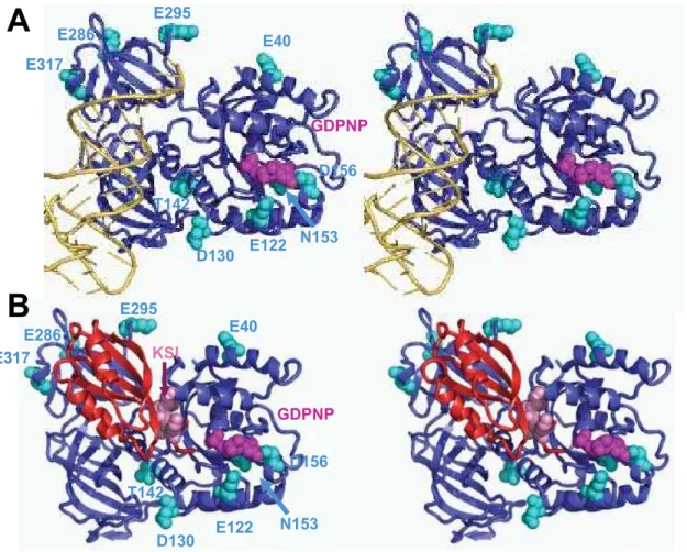
Examination of the mutants within the context of the structure
of the eEF1ANaa-tRNANGTP modeled as the ternary complex
provides some clues with regard to which mechanism may be altered in these mutants (Figure 5A). The charge reversal mutants in domain 2 proposed to be involved in binding the tRNA acceptor stem (E286K, E295K and E317K) all promoted enhanced misreading of the near-cognate AGC codon. It is tempting to speculate that these mutants may promote increased aa-tRNA dissociation rates by mimicking the structural change induced by correct tRNA:mRNA interactions and subsequently stimulating the GTPase activity of eEF1A. In contrast, the T142I mutant in domain 1 that interacts with the phosphate backbone of residue 61 at the base of the D-loop was more discriminatory, suggesting that this interaction is important for stimulation of GTP hydrolysis. Mutants in the vicinity of the GTP binding pocket had allele-specific effects on incorporation of the near-cognate tRNA. Charge reversal or neutralization of E122K or D156N strongly stimulated misreading. Perhaps the presence of additional positive charge in this region enhances GTP binding and/or GTPase
activity. In contrast, loss of a positive charge in the N153T mutant slightly inhibited misreading. Curiously, this effect was enhanced in the N153T/D156N double mutation. Last, mutations of E40 and D130 which are not closely linked to either tRNA or GTP binding did not affect missense suppression even though they were initially isolated as +1 insertion suppressor mutants. It is also striking that genetic screens have never identified fidelity mutations in domain 3, which is also proposed to interact with aa-tRNA. This suggests that these interactions are either non-essential or irrelevant to presentation at the A-site.
The K120R S121D I122D (KSI) mutant of eEF1Ba (tef5–7)
promoted increased accuracy in decoding both the ACG near-and UCU non-cognate codons. As noted above, mutants affecting fidelity in response to both near- and non-cognate codons are likely to be due to altered affinity at the initial binding step. Previous genetic analyses showed that this mutant also promoted increased fidelity at nonsense codons, slower rates of growth and protein synthesis, and hypersensitivity to translational inhibitors [23]. Figure 5B shows the X-ray crystal structure of eEF1A in
complex with the catalytic terminus of eEF1Ba. eEF1Babinds to
domain I and II of eEF1A, where KSI amino acids are located close to the nucleotide-binding pocket in domain 1 of eEF1A. The
binding site of eEF1Bato domain II overlaps the proposed
aa-tRNA binding site. This is significantly different from the bacterial homolog, and may be another way in which eukaryotes and bacteria differ in maintaining fidelity and promoting aa-tRNA delivery. We propose that these residues may facilitate exchange of GDP for GTP by eEF1A. Slowing this process would serve to limit the availability of functional ternary complexes, thus affecting binding of both near- and non-cognate codons. The effects of loss
of eEF1Bcshows a general trend of enhanced accuracy of
non-cognate codons like an eEF1Bamutant. However, the increased
misreading of a near-cognate codon is more like an eEF1A mutant. Thus, although the precise function of eEF1Bchas yet to be determined, it is reasonable to hypothesize that it may modulate eEF1Bato affect fidelity.
Ribosomal protein L5: Coordination of tRNA exit
from the E-site with aa-tRNA entrance at the A-site
The observation that the rpL5-K27E mutant generally promoted increased accuracy was initially surprising since this protein is located on the back of the central protuberance of the large subunit, far away from the ternary complex binding site, the ribosomal A-site, and the decoding center. In addition, a previous study implicated rpL5 in binding of peptidyl-tRNA, but not of aa-tRNAs [29]. Nonetheless, the rpL5-K27E mutant generally promoted increased fidelity in response to almost all of the near-and non-cognate codons tested. An intriguing explanation for the allele-specific effects observed here may come from the observa-tion that rpL5-L5 interacts with the fungal-specific elongaobserva-tion factor eEF3 [13]. eEF3 is an ATPase that interacts with eEF1A and catalyzes release of deacylated-tRNA from the ribosomal E-site [27,37]. It has been proposed that eEF3 and eEF1A work synergistically to remove deacylated tRNAs from the E-site and promote delivery of cognate aa-tRNA to the A-site [27]. It is tempting to speculate that altered eEF3 binding to the K27E form of rpL5 upsets this synergy. Similar to the model proposed for theeEF1Ba KSI mutant, the rpL5-K27E mutant might promote
enhanced accuracy at the codon recognition step.
since that work employed eight different codons at two positions of firefly luciferase. Since most were non- synonymous, the effects of different amino acid substitutions and their specific locations on the activity of the enzyme cannot be controlled for. Since different strain backgrounds are used in the studies and even in different sets of mutants analyzed in this work, variations in wild-type values observed requires the use of specific statistical methodology for accurate comparisons between datasets [15]. Further, relative tRNA abundance issues complicated the two instances where synonymous codons were used. Despite these differences, in-dependent analyses, reporter constructs and strain backgrounds showed similar levels of misreading. Furthermore, specific effects on misreading were observed for mutants of ribosomal compo-nents and key factors involved in elongation and termination. These data support the universal application of this approach to studies of translational fidelity.
Kinetics: the difference between near- and
non-cognate interactions may occur at the GTPase
activation step
As described above, studies in the bacterial system shows accuracy during translation elongation is likely a two step process involving two distinct biophysical mechanisms: initial selection and
proof-reading (reviewed in [5,38]). The first step, initial binding, is mostly determined by the interaction between the ribosome and EF-Tu, and forward and reverse rates (k1and k21) are not affected
by aa-tRNA identity [6]. However, during the next step of codon recognition, the stabilizing effects due to interactions of G530, A1492 and A1493 with the mini-helix results in dissociation rates
(k22) of near-cognate aa-tRNAs being approximately 400-fold
than those of cognate aa-tRNAs [39]. Computational modeling suggests the existence of two major energy minima at the decoding center corresponding to the flipped-in and flipped-out conforma-tions of A1492, and A1493, and that fast flipping between the two states provides a kinetic means to discriminate at the level of codon:anticodon interactions [10]. Formation of the stable mini-helix results in the physical transduction of information to the ternary complex, thus activating the endogenous GTPase of EF-Tu (k3). This step acts as a kinetic trap to select for aa-tRNAs
capable of forming the mini-helix. We propose that it is here that the difference between near- and non-cognate tRNA:mRNA interactions also occurs in eukaryotes. The potential of near-cognate interactions to form mini-helices, albeit at lower frequencies, provides the opportunity for stimulation of GTPase activation. The data also suggest that the presence of a canonical Watson-Crick base pair between N35NN92 may aid in nucleating mini-helix formation, consistent with molecular dynamics
model-E40
E122 D130 T142 E295
N153 D156 E286
GDPNP
E317
E40
E122 D130 T142 E295
N153 D156 E286
GDPNP
E317
E40
E122 D130
T142 E295
N153 D156 E286
KSI
GDPNP
E317
E40
E122 D130
T142 E295
N153 D156 E286
KSI
GDPNP
E317
A
B
Figure 5. Modeling of mutations in eEF1A and eEF1Bathat influence influence misincorporation of missense aa-tRNAs.The nucleotide exchange
ing showing that stability testing by the kink in the mRNA between the P- and A-site codons destabilizes position 2 mismatches more severely than mismatches at the first position [9]. In contrast, non-cognate interactions cannot possibly form mini-helices, and thus are incapable of forming the kinetic trap and activating GTP hydrolysis. In a kinetic analysis comparing different codon:anticodon mis-matches, one tRNA capable of participating in a non-cognate interaction was employed and stimulated GTPase activation approximately 6.7 fold less than the near-cognate codons [39]. This
is consistent with the ,4.5 fold increased rates of misreading
promoted by near-cognate codons in the current study.
Using paromomycin to functionally distinguish
between ‘near-’ and ‘non-cognate’ codon:anticodon
interactions
Binding of cognate aa-tRNA stimulates rearrangement of G530, A1492, and A1493 to establish A-minor interactions between themselves and the minor groove of the codon-anticodon helix [11,40]. Binding of paromomycin to the decoding center stimulates similar displacement of A1492 and A1493, positioning them to stabilize codon:anticodon interactions in a promiscuous manner [41], perhaps trapping them in the flipped-out state [10]. In light of the data presented here, we suggest that mini-helix formation is a precondition for paromomycin-stimulated mis-reading. Furthermore, paromomycin-enhanced and the potential for mini-helix formation are coordinately maximized in the decoding center. Thus, paromomycin has the potential to be used
as a tool to functionally distinguish between ‘near-’ and ‘non-cognate’ codons. However, this requires expansion of the system to utilize other codons for which the encoded amino acids do not result in a partially active luciferase protein. Kramer and Farabaugh observed changes in misreading with two different aminoglycosides, paromomycin and streptomycin [3]. Although more information about aminoglycoside-ribosome interactions has recently become available (reviewed in [42]) more experimental work needs to be performed to determine if any aminoglycosides other than paromomycin may be better sensors of cognate status.
MATERIALS AND METHODS
E. coli
and yeast strains and genetic methods.
E. colistrain DH5awas used to amplify plasmids. High efficiency transformations were performed as previously described [43]. TheSaccharomyces cerevisiaestrains used in this study are listed in Table 4.
The RPL5 strains were generously provided by Dr. John
Woolford. Isogenic TEF2, TEF3, and TEF4 yeast strains were
previously described [25,44,45]. Strains were cultured on YPAD or synthetic complete medium (H-) [46] and were freshly plated and incubated for two to five days at 30uC prior to transformation. Yeast were transformed with the alkali cation method [47], plated on appropriate selective media, and incubated at 30uC for four days. To assay for paromomycin sensitivity, 10-fold dilutions of logarithmically growing cells were spotted onto H-leu containing drug at a concentration of 1 mg/ml, or onto no-drug control plates, and grown at 25uC for 3 days.
Table 4.Yeast Strains used in this study
. . . .
Strain Genotype Source
JD932D MATa ade 2-1 trp1-1 ura3-1 leu2-3,112 his3-11,15 can1-100[L-AHN M1] [52]
M213 MATaleu2-3,112 his4-713 ura3-52 trp1Dlys2-20 met2-1 tef2Dtef1::LEU2+pTEF2 [20] TKY111 MATaleu2-3,112 his4-713 ura3-52 trp1Dlys2-20 met2-1 tef2Dtef1::LEU2+pTEF2-2E317K [53] TKY112 MATaleu2-3,112 his4-713 ura3-52 trp1Dlys2-20 met2-1 tef2Dtef1::LEU2+pTEF2-3E40K [53] TKY113 MATaleu2-3,112 his4-713 ura3-52 trp1Dlys2-20 met2-1 tef2Dtef1::LEU2+pTEF2-4E122K [53] TKY114 MATaleu2-3,112 his4-713 ura3-52 trp1Dlys2-20 met2-1 tef2Dtef1::LEU2+pTEF2-7T142I [53] TKY115 MATaleu2-3,112 his4-713 ura3-52 trp1Dlys2-20 met2-1 tef2Dtef1::LEU2+pTEF2-9E296K [53] TKY116 MATaleu2-3,112 his4-713 ura3-52 trp1Dlys2-20 met2-1 tef2Dtef1::LEU2+pTEF2-10E122Q [53] TKY117 MATaleu2-3,112 his4-713 ura3-52 trp1Dlys2-20 met2-1 tef2Dtef1::LEU2+pTEF2-13D130N [53] TKY278 MATaleu2-3,112 his4-713 ura3-52 trp1Dlys2-20 met2-1 tef2Dtef1::LEU2+pTEF2-17D156N [23] TKY280 MATaleu2-3,112 his4-713 ura3-52 trp1Dlys2-20 met2-1 tef2Dtef1::LEU2+pTEF2-19N153T [23] TKY282 MATaleu2-3,112 his4-713 ura3-52 trp1Dlys2-20 met2-1 tef2Dtef1::LEU2+pTEF2-18N153T D156E [23] TKY539 MATaleu2-3,112 his4-713 ura3-52 trp1Dlys2-20 met2-1 tef2Dtef1::LEU2+pTEF2-1E286K This work
TKY677 MATa ura3-52 trp1D101 lys2-801 his3D200 leu2D1 [25]
TKY678 MATa ura3-52 trp1D101 lys2-801 his3D200 leu2D1 tef3::LEU2 [25] TKY679 MATa ura3-52 trp1D101 lys2-80 his3D200 leu2D1 tef4::TRP1 [25] TKY680 MATa ura3-52 trp1D101 lys2-801 his3D200 leu2D1 tef3::LEU2 tef4::TRP1 [25] TKY235 MATaura3-52 trp1D101 lys2-801 leu2D1 met2-1 his4-713 tef5::TRP1 pTEF5 URA3 [23] TKY243 MATaura3-52 trp1D101 lys2-801 leu2D1 met2-1 his4-713 tef5::TRP1 pTEF5-7 K120R S121DI22D [23] JWY3742 MATaura3-52 trp1-D101 leu2 his3-D200 ade1 rpl5-D1::TRP1+pRS315-RPL1-HA(RPL5) [28] JWY3750 MATaura3-52 trp1-D101 leu2 his3-D200 ade1 rpl5-D1::TRP1+pRS315-RPL1-HA-1(rpl5- K27E) [28] JWY3751 MATaura3-52 trp1-D101 leu2 his3-D200 ade1 rpl5-D1::TRP1+pRS315-RPL1-HA-2(rpl5-T28A) [28] JWY3749 MATaura3-52 trp1-D101 leu2 his3-D200 ade1 rpl5-D1::TRP1+pRS315-RPL1-HA-3(rpl5-V53G) [28] JWY3752 MATaura3-52 trp1-D101 leu2 his3-D200 ade1 rpl5-D1::TRP1+pRS315-RPL1-HA-4(rpl5-G91R) [28] JWY3761 MATaura3-52 trp1-D101 leu2 his3-D200 ade1 rpl5-D1::TRP1+pRS315-RPL1-HA-5(rpl5-K289E) [28] doi:10.1371/journal.pone.0000517.t004
....
...
....
...
...
....
...
...
....
...
...
....
...
...
....
...
...
....
...
...
....
...
...
....
...
...
....
...
...
....
...
...
Plasmid Constructs
Plasmids used in this study contained a dual luciferase cassette on
a yeast vector backbone with the URA3 selectable marker. The
parental pYDL-Control plasmid containing the wild-typeRenilla
and firefly luciferase genes has been described previously [48]. Missense mutations were introduced into the arginine codon (AGA) at position 218 in the catalytic site of firefly luciferase [14] using variations on the following primers:
59-ATGCGAGAANNNGACGCAGGCAGTTCTATG-39 and
59-GCCTGCGTCN9N9N9TTCTCGCATGCCAGAGATC-39
(Integrated DNA Technology, Coralville, IA) where N denotes bases at codon 218 that were changed on the sense strand, and
N9 are the corresponding bases mutagenized on the antisense
strand. Oligonucleotide site directed mutagenesis reactions were performed using the StrataGene Quikchange II kit (La Jolla, CA) according to the manufacturer’s instructions. The seven mutants thus created are listed in Table 1. A second set of
plasmids containing the yeast TRP1 reporter were constructed
by transferring the dual luciferase cassettes from the resulting
plasmids as SpeI–XhoI fragments into p414 ADH [49].
Dual Luceriferase Assays
Transformed yeast cells were grown overnight in selective medium at 30uC to OD595of 0.8 to 1.0. Cells were pelleted by
centrifu-gation, washed twice with 0.5 ml of cold lysis buffer (phosphate
buffered saline containing 1 mM phenylmethylsulfonylfluoride), resuspended in cold lysis buffer and broken by agitation with glass beads (0.5 mm BioSpec). Lysates were clarified by centrifugation, and supernatants transferred to pre-chilled tubes. Luminescence
reactions were initiated by addition of 50ml of Promega DLR
system to 5ml of clarified cell lysates and measured using a Turner Design TD20/20 luminometer. At least three readings were taken for each assay and all assays were repeated (n = 3 – 12) until the data were normally distributed to enable statistical analyses both within and between experiments [15]. An unpaired two-sample
t-test was used to test the hypothesis that two datasets came from the same population, a rejected hypothesis indicating that the datasets were significantly different. TheP-values from this test is the estimation of the probability of an incorrect conclusion [15].
ACKNOWLEDGMENTS
We thank Ashton Trey Belew for assistance with data analysis, and Jennifer Baxter-Roshek and Rasa Rakauskaite for their technical and supervisory assistance.
Author Contributions
Conceived and designed the experiments: JD EP. Performed the experiments: EP PN JR YP TN JQ. Analyzed the data: TK JD EP JR. Contributed reagents/materials/analysis tools: TK JD PN. Wrote the paper: TK JD EP.
REFERENCES
1. Crick FHC (1966) Codon-Anticodon Pairing - Wobble Hypothesis. Journal of Molecular Biology 19: 548–555.
2. Agris PF (1991) Wobble position modified nucleosides evolved to select transfer RNA codon recognition: a modified-wobble hypothesis. Biochimie 73: 1345–1349.
3. Kramer EB, Farabaugh PJ (2007) The frequency of translational misreading errors in E. coli is largely determined by tRNA competition. RNA 13: 87–96. 4. Agris PF, Vendeix FA, Graham WD (2007) tRNA’s wobble decoding of the
genome: 40 years of modification. J Mol Biol 366: 1–13.
5. Ogle JM, Ramakrishnan V (2005) Structural Insights into Translational Fidelity. Annu Rev Biochem 74: 129–177.
6. Daviter T, Gromadski KB, Rodnina MV (2006) The ribosome’s response to codon-anticodon mismatches. Biochimie 88: 1001–1011.
7. Sanbonmatsu KY (2006) Alignment/misalignment hypothesis for tRNA selection by the ribosome. Biochimie 88: 1075–1089.
8. Cochella L, Brunelle JL, Green R (2007) Mutational analysis reveals two independent molecular requirements during transfer RNA selection on the ribosome. Nat Struct Mol Biol 14: 30–36.
9. Sanbonmatsu KY, Joseph S (2003) Understanding discrimination by the ribosome: stability testing and groove measurement of codon-anticodon pairs. J Mol Biol 328: 33–47.
10. Sanbonmatsu KY (2006) Energy landscape of the ribosomal decoding center. Biochimie 88: 1053–1059.
11. Ogle JM, Murphy FV, Tarry MJ, Ramakrishnan V (2002) Selection of tRNA by the Ribosome Requires a Transition from an Open to a Closed Form. Cell 111: 721–732.
12. Cochella L, Green R (2005) Fidelity in protein synthesis. Curr Biol 15: R536–R540.
13. Andersen CB, Becker T, Blau M, Anand M, Halic M, et al. (2006) Structure of eEF3 and the mechanism of transfer RNA release from the E-site. Nature 443: 663–668.
14. Rakwalska M, Rospert S (2004) The Ribosome-Bound Chaperones RAC and Ssb1/2p Are Required for Accurate Translation in Saccharomyces cerevisiae. Mol Cell Biol 24: 9186–9197.
15. Jacobs JL, Dinman JD (2004) Systematic analysis of bicistronic reporter assay data. Nucleic Acids Res 32: e160–e170.
16. Bouadloun F, Donner D, Kurland CG (1983) Codon-specific missense errors in vivo. EMBO J 2: 1351–1356.
17. Salas-Marco J, Bedwell DM (2005) Discrimination between defects in elongation fidelity and termination efficiency provides mechanistic insights into translational readthrough. J Mol Biol 348: 801–815.
18. Stansfield I, Jones KM, Herbert P, Lewendon A, Shaw WV, et al. (1998) Missense translation errors in Saccharomyces cerevisiae. Journal of Molecular Biology 282: 13–24.
19. Lim VI, Curran JF, Garber MB (2005) Ribosomal elongation cycle: energetic, kinetic and stereochemical aspects. J Mol Biol 351: 470–480.
20. Sandbaken MG, Culbertson MR (1988) Mutations in elongation factor EF-1a affect the frequency of frameshifting and amino acid misincorporation in
Saccharomyces cerevisiae. Genetics 120: 923–934.
21. Hiraga K, Suzuki K, Tsuchiya E, Miyakawa T (1993) Cloning and characterization of the elongation factor EF-1 beta homologue of Saccharomy-ces cerevisiae. EF-1 beta is essential for growth. FEBS Lett 316: 165–169. 22. Andersen GR, Pedersen L, Valente L, Chatterjee I, Kinzy TG, et al. (2000)
Structural basis for nucleotide exchange and competition with tRNA in the yeast elongation factor complex eEF1A:eEF1Balpha. Mol Cell 6: 1261–1266. 23. Carr-Schmid A, Valente L, Loik VI, Williams T, Starita LM, et al. (1999)
Mutations in elongation factor 1beta, a guanine nucleotide exchange factor, enhance translational fidelity. Mol Cell Biol 19: 5257–5266.
24. Jeppesen MG, Ortiz P, Shepard W, Kinzy TG, Nyborg J, et al. (2003) The crystal structure of the glutathione S-transferase-like domain of elongation factor 1Bgamma fromSaccharomyces cerevisiae. J Biol Chem 278: 47190–47198. 25. Olarewaju O, Ortiz PA, Chowdhury W, Chatterjee I, Kinzy TG (2004) The
translation elongation factor, eEF1B,plays a role in the oxidative stress response pathway. RNA Biology 1: 12–17.
26. Nierhaus KH (1990) The allosteric three-site model for the ribosomal elongation cycle: features and future. Biochemistry 29: 4997–5008.
27. Triana-Alonso FJ, Chakraburtty K, Nierhaus KH (1995) The elongation factor 3 unique in higher fungi and essential for protein biosynthesis is an E site factor. J Biol Chem 270: 20473–20478.
28. Deshmukh M, Stark J, Yeh LC, Lee JC, Woolford JL Jr (1995) Multiple regions of yeast ribosomal protein L1 are important for its interaction with 5 S rRNA and assembly into ribosomes. J Biol Chem 270: 30148–30156.
29. Meskauskas A, Dinman JD (2001) Ribosomal protein L5 helps anchor peptidyl-tRNA to the P-site inSaccharomyces cerevisiae. RNA 7: 1084–1096.
30. Stoletzki N, Eyre-Walker A (2007) Synonymous codon usage inEscherichia coli: selection for translational accuracy. Mol Biol Evol 24: 374–381.
31. Najafabadi HS, Lehmann J, Omidi M (2007) Error minimization explains the codon usage of highly expressed genes inEscherichia coli. Gene 387: 150–155. 32. Plewniak F (2007) GCG Documentation. http://www.vet.gla.ac.uk/GCGdoc/
Data_Files/codon_freq_tables.html.
33. Ikemura T (1982) Correlation between the abundance of yeast transfer RNAs and the occurrence of the respective codons in protein genes. Differences in synonymous codon choice patterns of yeast andEscherichia coliwith reference to the abundance of isoaccepting transfer RNAs. J Mol Biol 158: 573–597. 34. Percudani R, Pavesi A, Ottonello S (1997) Transfer RNA gene redundancy and
translational selection inSaccharomyces cerevisiae. J Mol Biol 268: 322–330. 35. Hani J, Feldmann H (1998) tRNA genes and retroelements in the yeast genome.
Nucleic Acids Res 26: 689–696.
37. Anand M, Chakraburtty K, Marton MJ, Hinnebusch AG, Kinzy TG (2002) Functional interactions between yeast translation elongation factors eEF1A and eEF3. J Biol Chem 278: 6985–6991.
38. Rodnina MV, Gromadski KB, Kothe U, Wieden HJ (2005) Recognition and selection of tRNA in translation. FEBS Lett 579: 938–942.
39. Gromadski KB, Daviter T, Rodnina MV (2006) A uniform response to mismatches in codon-anticodon complexes ensures ribosomal fidelity. Mol Cell 21: 369–377.
40. Ogle JM, Brodersen DE, Clemons WM, Jr, Tarry MJ, Carter AP, et al. (2001) Recognition of cognate transfer RNA by the 30S ribosomal subunit. Science 292: 897–902.
41. Carter AP, Clemons WM, Brodersen DE, Morgan-Warren RJ, Wimberly BT, Ramakrishnan V (2000) Functional insights from the structure of the 30S ribosomal subunit and its interactions with antibiotics. Nature 407: 340–348. 42. Hobbie SN, Bruell C, Kalapala S, Akshay S, Schmidt S, et al. (2006) A genetic
model to investigate drug-target interactions at the ribosomal decoding site. Biochimie 88: 1033–1043.
43. Inoue H, Nojima H, Okayama H (1990) High efficiency transformation of
Escherichia coliwith plasmids. Gene 96: 23–28.
44. Anand M, Valente L, Carr-Schmid A, Munshi R, Olarewaju O, et al. (2001) Translation elongation factor 1 functions in the yeast Saccharomyces cerevisiae. Cold Spring Harb Symp Quant Biol 66: 439–48.
45. Carr-Schmid A, Durko N, Cavallius J, Merrick WC, Kinzy TG (1999) Mutations in a GTP-binding motif of eukaryotic elongation factor 1A reduce
both translational fidelity and the requirement for nucleotide exchange. J Biol Chem 274: 30297–30302.
46. Dinman JD, Wickner RB (1994) Translational maintenance of frame: mutants of
Saccharomyces cerevisiae with altered -1 ribosomal frameshifting efficiencies. Genetics 136: 75–86.
47. Ito H, Fukuda Y, Murata K, Kimura A (1983) Transformation of intact yeast cells treated with alkali cations. J Bacteriol 153: 163–168.
48. Harger JW, Dinman JD (2003) An in vivo dual-luciferase assay system for studying translational recoding in the yeast Saccharomyces cerevisiae. RNA 9: 1019–1024.
49. Mumberg D, Muller R, Funk M (1995) Yeast vectors for the controlled expression of heterologous proteins in different genetic backgrounds. Gene 156: 119–122.
50. Andersen GR, Valente L, Pedersen L, Kinzy TG, Nyborg J (2001) Crystal structures of nucleotide exchange intermediates in the eEF1A- eEF1Balpha complex. Nat Struct Biol 8: 531–534.
51. Nissen P, Kjeldgaard M, Thirup S, Polekhina G, Reshetnikova L, et al. (1995) Crystal structure of the ternary complex of Phe-tRNAPhe, EF-Tu, and a GTP analog. Science 270: 1464–1472.
52. Tumer NE, Parikh B, Li P, Dinman JD (1998) Pokeweed antiviral protein specifically inhibits Ty1directed+1 ribosomal frameshifting and Ty1 retro-transposition inSaccharomyces cerevisiae. J Virol 72: 1036–1042.