MICOBIOTA FOLÍCOLA DE Dimorphandra wilsonii, ESPÉCIE ARBÓREA
BRASILEIRA AMEAÇADA DE EXTINÇÃO
Dissertação apresentada à Universidade Federal de Viçosa, como parte das exigências do Programa de Pós-Graduação em Fitopatologia, para obtenção do título de Magister Scientiae.
VIÇOSA
Ficha catalográfica preparada pela Seção de Catalogação e Classificação da Biblioteca Central da UFV
T
Silva, Meiriele da, 1981-
S586m Micobiota folícola de Dimorphandra wilsonii, espécie 2012 arbórea brasileira ameaçada de extinção / Meiriele da Silva
– Viçosa, MG, 2012.
x, 87f. : il. (algumas col.) ; 29cm.
Orientador: Robert Weingart Barreto.
Dissertação (mestrado) - Universidade Federal de Viçosa. Inclui bibliografia.
1. Dimorphandra wilsonii - Doenças e pragas.
2. Dimorphandra mollis - Doenças e pragas. 3. Micologia. 4. Espécies em extinção. 5. Biodiversidade. 6. Plantas em extinção. 7. Fungos - Classificação. 8. Cerrados - Brasil. I. Universidade Federal de Viçosa. II. Título.
MEIRIELE DA SILVA
MICOBIOTA FOLÍCOLA DE Dimorphandra wilsonii, ESPÉCIE ARBÓREA
BRASILEIRA AMEAÇADA DE EXTINÇÃO
APROVADA: 10 de fevereiro de 2012.
Dissertação apresentada à Universidade Federal de Viçosa, como parte das exigências do Programa de Pós-Graduação em Fitopatologia, para obtenção do título de Magister Scientiae.
____________________________ ______________________________ Prof. Maurício Dutra Costa Prof. Harold Charles Evans
____________________________ _____________________________ Prof. José Luís Bezerra Prof. Gleiber Quintão Furtado
____________________________ Prof. Robert Weingart Barreto
A minha mãe,
Por serem o meu apoio.
Dedico!
iii A Deus, por ser o meu refúgio e a minha fortaleza.
A minha mãe Maria Raimunda, pelo apoio, por acreditar em mim e me amar incondicionalmente.
A todos meus irmãos, Eduardo, Leila, Laura, Lúcia, Jéssica, Joyce, Luiza e São, pela amizade e apoio.
A minha cunhada, Flávia, e aos sobrinhos, Franciellen e Eduardo Henrique, pelo carinho e pela amizade.
A família Liparini e Pereira, pela amizade e pelo apoio sempre.
Ao Departamento de Fitopatologia da Universidade Federal de Viçosa, pela oportunidade de realização do curso de Mestrado.
Ao Conselho Nacional de Desenvolvimento Científico e Tecnológico – CNPq, pela concessão da bolsa de estudo de Mestrado.
Ao Professor Robert Weingart Barreto, pela orientação, pelos ensinamentos, pelo apoio, pelo incentivo e pela amizade.
Ao Professor Olinto Liparini Pereira, pela orientação, pelo apoio, pelos ensinamentos, pela amizade e pelo incentivo.
Aos Professores do Departamento de Fitopatologia, por terem contribuido na construção de mais um grau no meu conhecimento.
Aos colegas da Clínica de Doenças de Plantas, pelo convívio e pela amizade.
Aos amigos do Laboratório Patologia de Sementes e de Pós-Colheita, Deiziane, Camila, Danilo, André e Alexandre, pela ajuda e pela amizade.
Aos funcionários do Departamento de Fitopatologia, Délio, Braz, Rita e, Sara, pela gentileza e educação com que sempre me trataram.
Ao Fernando Fernandes, Juliana e Pedro do Jardim Botânico da Fundação Zoo-Botânica de Belo Horizonte, pela gentileza e pela ajuda nas coletas.
A administração da Floresta Nacional de Paraopeba (Flona-Paraopeba) pelo apoio na condução dos trabalhos de campo e pela gentileza.
A todos aqueles que, direta e indiretamente, contribuíram para a realização deste trabalho.
v MEIRIELE DA SILVA, filha de Maria Raimunda, nasceu na cidade de Corinto, Minas Gerais, no dia 15 de setembro de 1981.
Realizou os estudos básicos na cidade de Paraopeba, no mesmo estado.
Em 2005 iniciou o curso de graduação em Agronomia na Universidade Federal de Viçosa, UFV, graduando-se em janeiro de 2010.
RESUMO ... vii
ABSTRACT... ix
INTRODUÇÃO GERAL... 1
REFERÊNCIAS ... 5
ARTIGO 1 ... 8
According to the guidelines of Mycologia... 8
Foliicolous mycobiota of Dimorphandra wilsonii, an endangered Brazilian tree species ... 9
Abstract: ... 10
INTRODUCTION... 10
MATERIALS AND METHODS ... 12
RESULTS AND DISCUSSION ... 16
ACKNOWLEDGEMENTS ... 34
LITERATURE CITED ... 35
TABLES AND FIGURES ... 43
ARTIGO 2 ... 79
Mycotaxon (In Press)... 79
Alveariospora, a new anamorphic genus from trichomes of Dimorphandra mollis in Brazil ...80
Introduction...80
Material & methods ...80
Taxonomy ...80
Acknowledgments ...82
Literature cited ...82
vii SILVA, Meiriele da, M.Sc., Universidade Federal de Viçosa, fevereiro de 2012.
Micobiota folícola de Dimorphandra wilsonii, espécie arbórea brasileira ameaçada de extinção. Orientador: Robert Weingart Barreto. Coorientador: Olinto Liparini Pereira.
Dimorphandra wilsonii Rizzini, Fabaceae, popularmente conhecida como “faveiro de Wilson”, é espécie endêmica de Minas Gerais, que ocorre na transição do Cerrado para a Mata-Atlântica, ao norte de Belo Horizonte. Consta do anexo I da lista da Flora Brasileira Ameaçada de Extinção, nas listas da flora ameaçada de Minas Gerais e da International Union for Conservation of Nature (IUCN), onde figura como criticamente ameaçada, o nível que antecede o de extinção. Em contrapartida, Dimorphandramollis
Benth., planta taxonomicamente muito próxima do faveiro de Wilson, popularmente conhecida como “faveira” e “falso barbatimão”, é comum e amplamente distribuída pelo Cerrado brasileiro principalmente nos estados de Goiás, Mato Grosso, Minas Gerais e São Paulo. Com o objetivo de contribuir para o conhecimento sobre a micobiota associada a plantas ameaçadas de extinção no Brasil e, por conseguinte, também possivelmente ameaçadas, efetuou-se levantamento da micobiota folícola associada a D. wilsonii, e, paralelamente, de fungos folícolas que colonizam D. mollis, encontrados nas proximidades de indivíduos remanescentes de D. wilsonii. Nesse tipo de estudo micológico visa-se à geração de informação tanto na área micológica quanto na contribuição na geração de uma lista preliminar de espécies fúngicas em risco de extinção. Pretendeu-se excluir da lista de espécies fúngicas em risco de extinção aquelas que, além de ocorrerem sobre D. wilsonii, tambémocorrem sobre D. mollis. As coletas foram concentradas nos municípios de Caetanópolis, Paraopeba, Juatuba, Fortuna de Minas, Sete lagoas e Pequi. Quinze espécies fúngicas foram encontradas, descritas e ilustradas, dentre as quais oito tiveram a região ITS (Internal Transcribed Spacer) do rDNA e a região LSU (Large Subunit 28S) parcialmente sequenciadas e comparadas filogeneticamente a outras sequências disponíveis no GenBank a fim de se obter uma compressão mais completa das relações destes gêneros. Treze espécies fúngicas foram encontradas associadas a D. wilsonii: Vesiculohyphomyces cerradensis, Johansonia chapadiensis, Trichomatomyces byrsonimae, Piricauda paraguayensis, Geastrumia polystigmatis, Phillipsiella atra, Stomiopeltis suttoniae, Microcalliopsis dipterygis,
espécie nova), cf. Radulidium sp. (provável gênero novo). Seis espécies foram encontradas associadas a D. mollis: Trichomatomyces byrsonimae, Piricauda paraguayensis, Pseudocercospora sp. (provável espécie nova), Pseudocercosporella sp. (provável espécie nova), Janetia sp. 2 (provável espécie nova) e Alveariospora distoseptata (proposto como novo gênero e espécie). As espécies Vesiculohyphomyces cerradensis, Trichomatomyces byrsonimae, Piricauda paraguayensis, Geastrumia polystigmatis, Phillipsiella atra, Microcalliopsis dipterygis e Stomiopeltis suttoniae já foram relatadas ocorrendo em outras espécies de plantas tendo ampla distribuição, portanto, não tem interesse conservacionista. Quatro fungos ocorreram em ambas as espécies de Dimorphandra: Pseudocercospora sp. (provável espécie nova),
Pseudocercosporella sp. (provável espécie nova), Piricauda paraguayensis e
Trichomatomyces byrsonimae. Conclui-se que há possibilidade de que os três novos taxa associados unicamente a D. wilsonii: Janetia sp., Byssogene sp. e cf. Radulidium
ix SILVA, Meiriele da, M.Sc., Universidade Federal de Viçosa, February, 2012.
Foliicolous mycobiota of Dimorphandra wilsonii, an endangered Brazilian tree species. Adviser: Robert Weingart Barreto. Co-Adviser: Olinto Liparini Pereira.
Dimorphandra wilsonii (Fabaceae), commonly known as “faveiro de Wilson”, is an
Pseudocercospora sp. (probably new species), Pseudocercosporella sp. (probably new species), cf. Radulidium sp. (probably new genus). Six species were found associated with D. mollis: Trichomatomyces byrsonimae, Piricauda paraguayensis,
Pseudocercospora sp. (probably new species), Pseudocercosporella sp. (probably new species), Janetia sp. 2 (probably new species) and Alveariosporadistoseptata (proposed as a new genus and species). The species Vesiculohyphomyces cerradensis,
Trichomatomyces byrsonimae, Piricauda paraguayensis, Geastrumia polystigmatis,
Phillipsiella atra,Microcalliopsis dipterygis and Stomiopeltis suttoniae are notoriously polyphagous and have been reported on a variety of substrates having a wide distribution and, therefore, have no interest for conservation. Four fungi occurred on both species of Dimorphandra: Pseudocercospora sp., Pseudocercosporella sp.,
1
INTRODUÇÃO GERAL
O Cerrado é o segundo maior bioma do Brasil, depois da Amazônia, cobrindo 21% do território nacional em grande parte coincidente com o Planalto Central. O domínio original do Cerrado tem aproximadamente 2,0 milhões de km², dos quais apenas 400 mil km2 não foram degradados (Coutinho, 2002; Klink & Machado, 2005). O Cerrado apresenta distribuição contínua na área nuclear, com cerca de 1,5 milhões de km², que se estende por toda a região Centro-Oeste e o oeste da Bahia e Minas Gerais, Tocantins, ocorrendo ainda no sul do Maranhão, norte do Piauí e sul de Rondônia (Eiten, 1972). No estado de Minas Gerais, o Cerrado ocupa toda a porção centro-ocidental, cobrindo mais de 300.000 Km2 (Laca-Buendia & Brandão, 1995; Martins, 2000). Existem ainda áreas disjuntas de Cerrado encravadas entre domínios vizinhos e entre faixas de transição, como nos estados do Amapá, Amazonas, Pará, Roraima, São Paulo e norte do Paraná (Eiten, 1972; Ribeiro & Walter, 1998).
A flora do Cerrado é característica e diferenciada dos biomas adjacentes, embora muitas fisionomias compartilhem espécies com outros biomas. Além do clima, da química e física do solo, da disponibilidade de água e nutrientes, e da geomorfologia e topografia, a distribuição da flora é condicionada pela latitude, freqüência das queimadas, profundidade do lençol freático, além de inúmeros fatores antrópicos (Ribeiro & Walter, 1998). A riqueza florística do domínio Cerrado compreende segundo Mendonça et al. (1998) um total de 6671 espécies nativas, distribuídas em 170 famílias e 1144 gêneros. As famílias mais representativas no bioma Cerrado são: Fabaceae (859 espécies), seguida por Compositae (559 espécies), Orchidaceae (493 espécies), Poaceae (373 espécies), Rubiaceae (257 espécies) Melastomataceae (238 espécies), Myrtaceae (212 espécies), Euphorbiaceae (195 espécies), Malpighiaceae (128 espécies) e Lytraceae (120 espécies).
ao Cerrado biodiversidade estimada em 160.000 espécies de plantas, animais e fungos (Pennington et al., 2000).
O bioma Cerrado é uma das 25 formações vegetacionais consideradas como prioritárias para a conservação da biodiversidade mundial, denominadas de hot spots
(Myers et al., 2000; Queiroz, 2009). Os remanescentes de vegetação primária do Cerrado correspondem a somente 20% da área original ocupada pelo bioma e somente 6,2% estão localizados em áreas de preservação. A diversidade florística do Cerrado é estimada em 10.000 espécies, sendo que 4.400 são endêmicas e respondem por 1,5% da biodiversidade vegetal mundial (Myers etal., 2000).
No Brasil, a vegetação natural de Cerrado vem sendo substituída por culturas agrícolas, pastagens e espécies florestais de rápido crescimento (Sano et al. 2010; Lenza et al., 2011). Se a exploração dessas áreas nativas continuar em forte ritmo de ampliação, a vegetação do Cerrado acabará restrita às áreas de Unidades de Conservação, terras indígenas e regiões impróprias à agropecuária, fazendo com que muitas espécies de plantas, assim como animais possam ser levados à extinção (Queiroz, 2009). Um exemplo de espécie endêmica do bioma Cerrado, criticamente ameaçada devido à degradação do seu habitat é a leguminosa arbórea Dimorphandra
wilsonii Rizzini, conhecida popularmente como “faveiro de Wilson”. Um estudo
publicado recentemente relata que apenas pouco mais de dez indivíduos desta espécie arbórea são conhecidos (Fernandes et al., 2007). Todos esses exemplares ocorrendo em áreas de Cerrado já transformadas em pastagens nos municípios de Paraopeba e Caetanópolis, MG. A espécie foi incluída na lista da IUCN como estando criticamente ameaçada, o nível mais elevado de risco para a sobrevivência de uma espécie e que antecede o de extinta na natureza (Fernandes et al., 2007; Fonseca et al., 2010).
3 apenas três espécies fúngicas como ameaçadas de extinção, os liquens Cladonia
perforata A. Evans e Erioderma pedicellatum (Hue) P.M. Jorg. e o cogumelo
comestível Pleurotusnebrodensis (Inzenga) Quél. (IUCN, 2012).
A negligência com relação aos microrganismos deve-se principalmente ao fato dos mesmos não serem vistos a olho nu e pela falsa ideia de que microrganismos de modo geral possuem dispersão facilitada e, consequentemente, ampla distribuição geográfica (Griffith, 2011). Entretanto, especialmente após o uso de ferramentas moleculares no estudo da ecologia microbiana, sabe-se atualmente que populações microbianas podem ter estrutura populacional definida e endêmica (Rodriguez et al., 2004). As dificuldades práticas de se reunir evidências que justifiquem o reconhecimento de espécies fúngicas como estando ameaçadas são em parte responsáveis pela virtual ausência de fungos nas listas de espécies ameaçadas e nas medidas de conservação. Minter (2010) se referiu aos fungos, neste contexto, como “the orphans of Rio”, por terem sido negligenciados na “Conferência das Nações Unidas sobre Ambiente e Desenvolvimento Sustentável”, também conhecida como ECO 92 ou Rio 92. Um grupo de fungos para os quais seria, em princípio, possível reunir evidências científicas sobre a condição de ameaça iminente de extinção é o dos fungos fitopatogênicos altamente específicos em suas relações com espécies de plantas hospedeiras ameaçadas de extinção (Rocha et al., 2010).
A perda por extinção de uma espécie vegetal pode levar, num evento de coextinção, à extinção de um leque de organismos especializados que dependem desta planta. Eventos de coextinção são bem documentados para outros casos de interação parasitas-hospedeiro tais como: piolhos parasitas de pombos, diferentes parasitas em primatas (fungos do gênero Pneumocystis, nematóides e piolhos), vespas polinizadoras e insetos herbívoros (Koh et al., 2004; Thacker et al., 2006; Dunn et al., 2009).
A emergência do reconhecimento da necessidade de conservação dos fungos é ilustrada pela realização, em Whitby - Reino Unido (26-30 de outubro de 2009) do evento “FUNGAL CONSERVATION science, infrastructure and politics” e, posteriormente em 6 de agosto de 2010 no Royal Botanic Garden Edinburgh, no âmbito do 9th International Mycological Congress, de onde foi fundada a International Society for Fungal Conservation (http://www.fungal-conservation.org/).
da Floresta Atlântica, Coussapoa flocosa Akkermans & C.C. e listada como vulnerável (IUCN, 2012). Nesta publicação foram descritas seis novas espécies fúngicas, incluindo um gênero novo para a ciência.
5
REFERÊNCIAS
Berlund H, Jonsson BG. 2005. Verifying an extinction debt among Lichens and Fungi in northern Swedish boreal forests. Conservation Biology 19: 338–348.
Coutinho LM. 2002. O bioma do Cerrado. In: Klein, A. L. (ed.). Eugen Warming e o Cerrado brasileiro: um século depois. São Paulo: UNESP. P. 77–92.
Dunn RR, Harris C, Colewll RK, Koh LP, Sodhi NS. 2009. The sixth mass coextinction: are most endangered species parasites and mutualists? Proceedings the Royal Society 276: 3037–3045. doi: 10.1098/rspb.2009.0413
Eiten G. 1972. The Cerrado Vegetation of Brazil. Botanical Review 38: 201–341. Fernandes FM, Fonseca AG, Kaechele K, Goulart MF, Marinho W, Souza HAV,
Queiroz AR, Giorni V, Oliveira G, Rodrigues MJ, Bacelar M, Lovato MB. 2007. Tentando evitar maisuma extinção: o caso do Faveiro de Wilson (Dimorphandra wilsonii Rizz.). In: T.S. Pereira, M.L.M.N. Costa & P.W. (orgs.). Recuperandoo verde para as cidades - A Experiência dos Jardins Botânicos Brasileiros. Rio de Janeiro, RBJB. Pp. 87-98.
Fonseca MB, Franca MGC, Zonta E, Giorni V. 2010. Crescimento inicial de
Dimorphandra wilsonii (Fabaceae - Caesalpinioideae) em diferentes condições de fertilidade em solo de Cerrado. Acta Botanica Brasilica 24:322–327.
Griffith GW. 2011. Do we need a global strategy for microbial conservation? Trends in Ecology & Evolution. doi: 10.1016/j.tree.2011.10.002
IUCN. 2012. Red List of Threatened Species. Disponivel em: < http://www.iucnredlist.org >.
Klink CA, Machado RB. 2005. A conservação do Cerrado brasileiro. Megadiversidade 1:147–155.
Koh LP, Dunn RR, Sodhi NS, Colwell RK, Proctor HC, Smith VS. 2004. Species coextinctions and the biodiversity crisis. Science 305: 1632–1634.
Laca-Buendia, J.P. & Brandão, M. 1995. Composição florística e análise fitossociológica do Cerrado em Minas Gerais-I: Alto Paranaíba, Mata da corda e parte do Planalto de Araxá. Daphne 5: 7–18.
Veadeiros, Goiás, e áreas de cerrado sentido restrito do Bioma Cerrado. Revista Brasileira de Botânica 3: 247–259.
Martins, C.S. 2000. Caracterização física e fitogeográfica de Minas Gerais. In: Mendonça, M.P. & Lins, L.V. (org.) Lista vermelha das espécies ameaçadas de extinção da flora de Minas Gerais. Belo Horizonte: Fundação Biodiversistas e Fundação Zoo-Botânica 35–43.
Mendonça MP, Lins LV. 2000. Lista vermelha das espécies ameaçadas de extinção da flora de Minas Gerais. Belo Horizonte, Fundação Biodiversitas, Fundação Zôo-Botânica de Belo Horizonte. 160p.
Mendonça RC, Felfili JM, Walter BMT, Silva Junior MC, Rezende AV, Filgueiras TS, Nogueira PE. 1998. Flora vascular do Cerrado. In: Sano M & Almeida SP (eds). Cerrado: ambiente e flora planaltina: Embrapa-CPAC, p. 287-556.
Mendonça RC, Felfili JM, Walter BMT, Silva Junior MC, Rezende AV, Filgueiras TS, Nogueira PE, Fagg CW. 2008. Flora vascular do Cerrado: Checklist com 12.356 espécies. In Sano SM, Almeida SP, Ribeiro JF (eds). Cerrado: ecologia e flora: Embrapa-CPAC, Planaltina, p. 417–1279.
Minter D. 2010. Safeguarding the Future. In: Boddy L, Coleman M, eds. From Another Kingdom. Edinburgh, UK: Royal Botanic Garden Edinburgh: p. 144-153. Minttermeier AA, Robles P, Hoffmann M, Pilgrim J, Brooks T, Minttermeier CG,
Lamoreux J, Fonseca GB. 2005. Hotspots revisited: earth’s biologically richest and most endangered ecoregions p. 392.
Moore D, Nauta MM, Evans SE, Rotheroe M (eds). 2001. Fungal conservation. Cambridge. Cambridge University Press p. 272.
Myers N, Mittermeier RA, Mittermeier CG, Fonseca GAB, Kent J. 2000. Biodiversity hotspots for conservation priorities. Nature 403: 853–858.
Pennington RT, Prado DE, Pendry CA. 2000. Neotropical seasonally dry forests and Quaternary vegetation changes. Journal of Biogeography 27: 261–273.
Queiroz FA. 2009. Impactos da sojicultura da exportação sobre a biodiversidade do Cerrado. Sociedade & Natureza 21: 193–209.
7 Ribeiro JF, Walter BMT. 1998. Fitofisionomias do bioma do Cerrado. In: Sano, M. & Almeida, S.P. (eds.) Cerrado: ambiente e flora. Planaltina: Embrapa-CPAC. Pp. 89–166.
Rocha FB, Barreto RW, Bezerra JL, Neto JAAM. 2010. Foliar micobiota of
Coussapoa floccosa a highly threatened tree of the Brazilian Atlantic Forest. Mycologia 102:1241–1252.
Rodriguez RJ, Cullen D, Kurtzman CP, Khachatourians GG, Hegedus DD. 2004. Molecular methods for discriminating taxa, monitoring species, and assessing fungal diversity. In Mueller GM, Bills GF, Foster MS (eds) Biodiversity of fungi inventory and monitoring methods 1st edn. Elsevier, California, p 77–102.
Sano EE, Rosa R, Brito JLS, Ferreira LG. 2010. Land cover mapping of the tropical savanna region in Brazil. Environmental Monitoring Assessment 166:113–124. Thacker JL, Hopkins GW, Dixon AFG. 2006. Aphids and scale insects on threatened
trees: co-extinction in a minor threat. Oryx 40: 233–236.
ARTIGO 1
According to the guidelines of Mycologia
Foliicolous mycobiota of Dimorphandra wilsonii, an endangered Brazilian tree
9
Foliicolous mycobiota of Dimorphandra wilsonii, an endangered Brazilian tree
species
Meiriele da Silva1 Danilo B. Pinho1 Olinto L. Pereira1 Fernando F. Moreira2 Robert W. Barreto1
1
Departamento de Fitopatologia, Universidade Federal de Viçosa, Minas Gerais 36570-000, Brazil
2
Abstract: A survey of foliicolous fungi associated with Dimorphandra wilsonii and
Dimorphandra mollis (Fabaceae) was conducted in the state of Minas Gerais, Brazil.
Dimorphandra wilsonii is a tree species native from the Brazilian Cerrado and listed
as highly endangered of becoming extinct. Fungi strictly depending on this plant
species as a substrate are probably also on the verge of co-extinction. The study of this
mycobiota aimed at contributing towards the field of fungal conservation and
generating preliminary lists of potentially endangered fungal species is a necessary
step in such a study. Surveying the mycobiota of D. mollis, which is a common species
with a broad geographical distribution, was also considered necessary as these two
species are closely related and coexist. Therefore, fungi occurring on D. wilsonii might
also occur on the non-endangered D. mollis and hence not be in danger of
co-extinction. Fourteen fungal species were collected, identified, described and
illustrated, including: five ascomycetes [Phillipsiella atra, Johansonia chapadiensis,
Stomiopeltis suttoniae, Microcalliopsis dipterygis, Byssogene sp. nov.], eight
hyphomycetes anamorphs [Vesiculohyphomyces cerradensis, Trichomatomyces
byrsonimae, Piricauda paraguayensis, two Janetia sp. nov., Pseudocercospora sp.
nov., Pseudocercosporella sp. nov., cf. Radulidium gen. nov.] and one coelomycete
(Geastrumiapolystigmatis). Three fungi were exclusive to D. wilsonii and possibly in
endangered of extinction: Byssogene sp. nov., Janetia sp. nov. 1, and cf. Radulidium
gen. nov.
Keywords: biodiversity, Cerrado, co-extinction, fungal conservation, phylogeny, taxonomic novelties.
INTRODUCTION
The Cerrado is a Brazilian biome that is second in surface area only to the Amazon. It covers 21% of the country (2 million km2) and is largely coincident with the central plateau (Klink and Machado 2005). In Brazil, the Cerrado natural vegetation is progressively being replaced by agricultural crops, pasture and cultivated exotic forest species (Sano et al. 2010, Lenza et al. 2011). One among the many plant species occurring in this biome that are now endangered is Dimorphandra wilsonii
11 in nature (Fernandes et al. 2007). All these individuals occur in areas of Cerrado that have been turned into pastures and all occur in a small area in the neighboring municipalities of Paraopeba and Caetanópolis in the state of Minas Gerais. The species is listed in (http://www.iucnredlist.org/) as being critically endangered, the highest level of risk to the survival of a species and prior to extinction in nature.
The lack of knowledge and awareness about the fungi and the fact that most fungi are either invisible to the naked eye or produce ephemorals macroscopic fruit bodies has probably led to the mistaken impression that species belonging to the Fungi are capable of escaping environmental changes, are easily dispersed, ubiquous and are broadly spread (Griffith, 2011). Nevertheless, investigations involving surveys of environmental DNA have been indicating that microbial communities may have a well defined structure, with the populations with a high level of endemism (Rodriguez etal. 2004). Nevertheless, the practical difficulties for gathering evidence that individual fungal species are actually threatened are in part responsible for their virtual absence from lists of endangered species and from policies aimed at preventing global loss of biodiversity. Minter (2010) has referred to fungi, considering that context as “the orphans of Rio”, for this large group of organisms having been left out of the agenda in the Earth Summit (United Nations Conference on Environment and Development - UNCED) that occurred in Rio de Janeiro in June 1992. Some years ago it was conjectured by Rocha et al. (2010) that it might be possible to gather convincing scientific evidence of threat of extinction by investigating highly host-specific plant pathogenic fungi associated with endangered plant species. The loss of one plant species may lead to events of coextinction threatening a range of specialized organisms depending strictly on that species for its survival. Such events are well documented for parasite-host interactions such as: pigeon lice, primate parasites, pollinizer wasps and herbivorous insects (Koh et al. 2004, Thacker et al. 2006, Dunn etal. 2009).
In Brazil, the first work to be published addressing the issue of fungal conservation was published by Rocha et al. (2010) and involved the study of the foliage mycobiota of Coussapoa flocosa Akkermans & C.C. (Cecropiaceae) which may be in danger of coextinction because of its dependence on this rare endemic tree of the Brazilian Atlantic Forest. This study led to the discovery of six new fungal species, including a new fungal genus.
The present work aims at expanding the study started by Rocha et al. (2010) in order to encompass an additional endangered Brazilian plant species (Dimorphandra
wilsonii) and its mycobiota - which may also be potentially endangered of
coextinction. Additionally, Dimorphandra mollis Benth. which is a common species with a broad geographical distribution, closely related to D. wilsonii and which coexists with that plant in its remaining area of occurrence in nature also had its mycobiota studied. This was done in order to determine if fungi occurring on D. wilsonii also occur on the non-endangered D. mollis and hence should not be regarded as in potential danger of co-extinction.
Hence, the objectives of this study were: I) to survey and describe the foliicolous mycobiota associated with Dimorphandra wilsonii; II) to survey and describe the mycobiota of D. mollis; III) Check the possible co-occurrence of fungi found in D. wilsonii and on D. mollis and produce preliminary evidence about the possible threat to the survival of fungal species unique to D. wilsonii. The fungi that were collected are described and discussed below.
MATERIALS AND METHODS
Survey trips were conducted between 2009 and 2011, locally in the municipality of Paraopeba, Caetanópolis, Juatuba, Fortuna de Minas, Sete Lagoas and Pequi (TABLE 1). Existing information on localities of occurrence of D. wilsonii individuals in nature were provided by F. Fernandes (Fundação Zoo-Botânica de Belo Horizonte) who has
been conducting regular surveys for the remaining individuals of that species since
2003. Whenever individuals belonging to the closely related species, D. mollis, were
found growing in the vicinities of an individual of D. wilsonii,branches and foliage of
individuals belonging to that species were also collected. Dimorphandra wilsonii is
13
gray bark that is not easily detached as in D. mollis. Its leaflets are also larger than
those in D. mollis (3-5 cm long) (FIG.1). Pictures were taken in the field with a SONY DSC-H9 digital camera, samples of branches bearing foliage were collected with a long-poled prunner and dried in a plant press.
After screening in the lab, dried relevant specimens were deposited at the herbarium of Universidade Federal de Viçosa (VIC). Samples were carefully examined under a stereomicroscope (OLYMPUS SZX7), while still fresh and fungal structures were either scraped with a scalpel from tissue surfaces (when externally produced) or free-hand sections or sections prepared with the help of a freezing microtome (Microm HM 520) whenever necessary were prepared and mounted on in lactophenol or other mounting media. Observations and measurements were carried out with an OLYMPUS BX 51 light microscope fitted with a digital camera (OLYMPUS E330) and a drawing tube.
Isolations of fungi in pure culture were attempted by direct transfer of spores or other fungal structures onto plates containing VBA - vegetable broth-agar, as described by Pereira etal. (2003), with the help of a sterile fine pointed needle. Pure cultures were preserved in PCA slants or in silica-gel, as described by Dhingra and Sinclair (1996).
For scanning electron microscopy, the samples were prepared by the following protocol: samples were fixed in 2.5% glutaraldehyde + sodium cacodylate buffer (0.1mol L-1, pH 7.2) (1:1) for 1h a room temperature; washed six times in 0.1mol L-1 sodium cacodylate buffer (10 m each washing period); samples were postfixed by immersion in 1% OsO4 prepared in cacodylate buffer 0.1 mol L-1 (1:1), and kept for 4
h at 4-8 ºC then washed again six times in buffer; dehydrated by successive transfer in a graded alcohol solutions series (30, 50, 60, 70, 80, 95, 100%) and left 10min in each solution at room temperature; samples were placed in a critical point dryer (Baltec model 030) with CO2 as transition fluid; after drying the samples were coated with
gold (20 nm thick) with a sputter coater (Balzers® model FDU 010) and examined with a Carl-Zeiss Model LEO VP 1430 electron microscope.
lamps placed 35 cm above the plates) after 23 days. Color terminology followed Rayner (1970).
For the molecular phylogeny studies of five of the fungi (Janetia sp.,
Radulidium-like sp., Piricauda sp., Pseudocercospora sp. and Pseudocercosporella
sp., pure cultures were grown on PDA at 25 ºC for up to four weeks depending on their growth rate. Genomic DNA was extracted from the mycelium using Wizard® Genomic DNA Purification Kit (Promega corporation, WI, U.S.A.). For the sequencing of three of the fungal species (Mycosphaerellaceae, Phillipsiellaatra and
Trichomatomyces byrsonimae), DNA was extracted by removing fungal structures
from the plant tissue with a fine glass needle and placing them in a microtube (1.5 ml) of the extraction kit. In this case, each fungal structure was carefully examined under the highest power of a stereomicroscope in order to check for possible contamination with other fungi or mycoparasites and to exclude any plant material from the sample. PCR reactions were set-up using the following ingredients for each 25 µl reaction: 12,5 µl of DreamTaqTM PCR Master Mix 2X (MBI Fermentas, Vilnius, Lithuania), 1 µl of 10 µM of each forward and reverse primer synthesised by Invitrogen (Carlsbad, USA), a maximum of 25 ng/µl of genomic DNA, and nuclease-free water to complete the total volume.
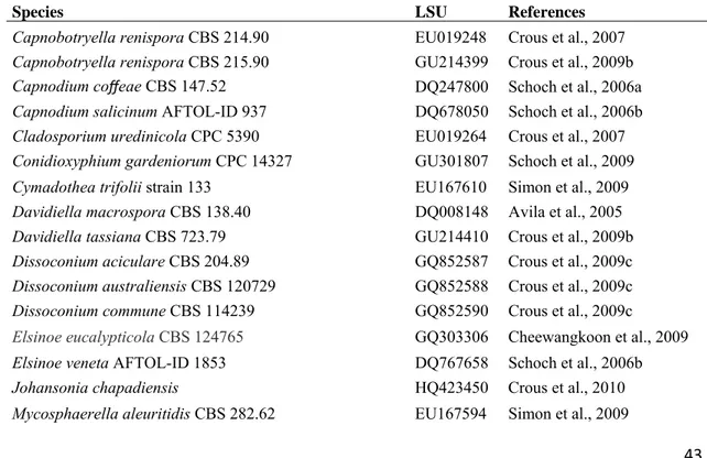
15 MegaBACE 500TM. The nucleotide sequences were read with Chromas lite 2.01 (http://www.technelysium.com.au/chromas_lite.html) and edited with the DNA Dragon software (http://www.dna-dragon.com/index.php). All sequences were checked manually and nucleotide arrangements at ambiguous positions were clarified using both primer direction sequences. New sequences will be deposited in GenBank (http://www.ncbi.nlm.nih.gov). Sequences of large subunit ribosomal of additional species were retrieved from GenBank (TABLE 2).
Consensus regions were compared against GenBank’s database using their Mega BLAST program. In addition, sequences were selected from Arzanlou et al. (2007), Crous et al. (2009b, 2009c), Frank et al. (2010), Schoch et al. (2009) and Yang et al. (2010) to reveal the phylogenetic position of the strains within Capnodiales. The closest hit sequences were then downloaded in FASTA format and aligned using the multiple sequence alignment program MUSCLE® (Edgar 2004), with default parameters in operation. MUSCLE® was implemented using the program MEGA v.5 software (Tamura et al. 2011). Alignments were checked and manual adjustments were made where necessary. Gaps (insertions/deletions) were treated as missing data.
Trees were visualized in FigTree (http://tree.bio.ed.ac.uk/software/figtree/) and exported to graphics programs. The species Elsinoë eucalypticola Cheewangkoon & Crous, E. veneta (Burkh.) Jenkins, Myriangium duriaei Mont. & Berk. and M.
hispanicum J.B. Martínezwere used as outgroup in these analyses.
RESULTS AND DISCUSSION Phylogeny
The LSU region of the sequences was used to obtain additional sequences from GenBank, which were added to the aligment. Due to the inclusion of the shorter LSU sequences of Stomiopeltis sp. (AY598919) and Stomiopeltis versicolor (Desm.) Arx (FJ147163) in the alignment, it was not possible to subject the full length of the determined LSU sequences (TABLE 2) to the analysis. The manually adjusted LSU alignment contained 84 sequences (including the outgroup sequences) and, of all 461 characters used in the phylogenetic analysis, 172 were parsimony-informative, 193 were variable and 268 were conserved. Although the ITS sequences were not used in phylogenetic analyses, they were lodged in GenBank for future studies and DNA barcode purposes.
Taxonomy
Thirteen fungal species were found associated with D. wilsonii: Byssogene sp.,
Geastrumia polystigmatis, Janetia sp. 1, Johansonia chapadiensis, Phillipsiella atra,
Piricauda paraguayensis, Microcalliopsis dipterygis, Pseudocercospora sp.,
Pseudocercosporella sp., cf. Radulidium sp., Stomiopeltissuttoniae, Trichomatomyces byrsonimae, Vesiculohyphomyces cerradensis. Five fungal species were collected in association with D. mollis: Piricauda paraguayensis, Pseudocercospora sp.,
Pseudocercosporella sp., Trichomatomyces byrsonimae and Janetia sp. 2. These fungi are described below.
17 anastomosing, net-forming, slightly undulate, composed of pale brown, flattened, thin walled, septate hyphae, 2.5–5.0 μm, smooth. Ascomata hypophyllous, superficial, solitary, discoid, no ostiolate, black, margin raised, 175–225 μm diam., 100–117.5 μm high, outer gray wall of texture globulosa becoming hyaline, pseudoparenchymatose more internally, 6-cell thick, 12.5–20 μm, dark brown. Asci bitunicate, ovate, oblong to obclavate, 37.5–65 × 25–35 μm, 8–spored. Ascospores cylindrical to ellipsoid, 20– 27.5×5–10 μm, dictyoseptate, subhyaline, guttulate, smooth.
Specimens examined: On living leaves of Dimorphandra wilsonii. BRAZIL: Minas Gerais:
Paraopeba, Fazenda Tabuleiro Grande, 27 Jul 2011, M. Silva & O.L. Pereira (VIC 31808).
Notes: The fungus described above belongs to the family Saccardiaceae by having ascomata superficially, discoid, asci parallel in a single layer, bitunicate, ascospores with two or many celled, hyaline or brown, and clearly belongs to the genus Byssogene Syd. in having dictyoseptate ascospores almost hyaline, ascomata discoid and superficial brown mycelium (von Arx and Müller 1975). Byssogene amboinensis Syd. is the single species in this genus and was previously known to occur on Eugenia sp. (Myrtaceae) in Amboina (Sydow 1922). Byssogeneamboinensis
Geastrumiapolystigmatis Bat. & M.L.Farr, Saccardoa 1: 71 (1960) FIG. 3 Colonies on living leaves, adaxial, circular to irregular, sparse. Internal mycelium indistinct. External mycelium loose, branched, 2.5–3.0 µm diam, discrete and irregularly scattered over areas of the host, septate, medium brown to sub-hyaline, smooth. Conidiomata hypophyllous, superficial, scutelate, opening by an irregular rupture, hemispherical or subglobose, black, 125–140 μm diam., 52.5–85.0 μm in height, wall composed of one-celled layer, angular, 10–12.5 μm, dark brown, smooth. Conidiophores micronematous, restricted to conidiogenous cells, developing from the hyphae at the point of their transformation into the covering wall, terminal, holoblastic, narrowly elongate ampulliform, clavate to fusiform, 8–17 μm long, 2 μm wide at the base, cells swelling at the apex, hyaline and by more or less dichotomous forking produces a cluster of clavate to fusiform outgrowths which proceed to differentiate into conidial arms after the initial cluster has been cut off from the conidiogenous cell by a septum. Conidia dry, holoblastic, solitary, formed singly at the apex of each conidiogenous cell, cheiroid, fasciculate groups of 4-10 straight to slightly curved filiform arms closely united at long pedicellate basal cell, each arm, 15–50 × 2–4 μm, apex 2.5–4 μm, base 1.0–2.5 μm, 2–7 septate, hyaline when immature becoming light brown at maturity, smooth.
Specimens examined: On living leaves of Dimorphandra wilsonii. BRAZIL: Minas Gerais:
Paraopeba, Fazenda Tabuleiro Grande, 13 Jul 2009, M. Silva, O.L. Pereira & R.W. Barreto (VIC
31771).
Notes: Geastrumia polyastigmatis Bat. & M.L. Farr is the only species in this genus and was previously known to occur on a member of the Fabaceae - Andira
jamaicensis (W. Wright) Urb. in Brazil and Republica Dominicana, as well as on
19
Janetia sp. 1 on D. wilsonii (to be proposed as a new species) FIGS. 4–5 Colonies hypophyllous, dense, brown, forming dark heads on trichomes. Internal mycelium indistinct. External mycelium superficial, up to 4 µm diam, branched, septate, brown, smooth. Conidiophores forming sporodochial clusters on the apex of trichomes, micronematous, mononematous. Conidiogenous cells holoblastic, monoblastic, denticulate, cylindrical to narrowly ampulliform, straight to slightly curved, 7.5–37.5 × 2.5–5.0 µm, reddish brown to pale brown. Conidiogenous loci apical on conidiogenous cells, flat, truncate, not darkened, unthickened, conidial secession schizolytic. Conidia dry, solitary, obclavate to cylindrical, straight to slightly curved, occasionally slightly constricted at some of the septa, 12.5–75 × 5.0–7.5 µm, rounded at the apex up to 2.5 µm wide, rounded at the base 5.0 µm, 1–9 euseptate, reddish brown to pale brown, smooth, guttulate.
In culture: On PCA, slow-growing (3.7-4.5 cm diam, after 27 days), chrysanthemoid to subcircular, flat, to convex, cottonose aerial myceliun accompanied by immersed growth within medium, dense, or without aerial mycelium and internal mycelium irradiating and sinuose and raising from the medium on radial strands. On PDA greenish centrally glaucous, followed by a periphery of pale olivaceous buff thick ring of mycelium, or olivaceous grey, pronunced diurnal zonation, under alternating light but less pronounced in the dark no sporulation. Mycelium sparser and with less pronounced diurnal zonation, in the dark reverse blueish.
Specimens examined: On living leaves of Dimorphandra wilsonii. BRAZIL: Minas Gerais:
Paraopeba, Fazenda Tabuleiro Grande, 13 Jul 2009, M. Silva, O.L. Pereira & R.W. Barreto (VIC
31772− HOLOTYPE); 19 Jul 2010, M. Silva & O.L. Pereira (VIC 31780); 19 Jul 2010, M. Silva & O.L.
Pereira (VIC 31781).
Janetia sp. 2 on D. mollis (to be proposed as a new species – distinct from above) FIGS. 6–8
Colonies hypophyllous, on trichomes, effuse, brown or dark brown, giving
humped hyphal cells, 5–6 × 3–4 µm, brown, smooth. Conidiogenous cells holoblastic, monoblastic, denticulate. Conidiogenous loci a flat-topped hump on conidiogenous
cells, 1.5–3.0 µm, unthickened, dark brown. Conidia dry, holoblastic, solitary, cylindrical, straight to slightly curved, often slightly constricted at septae, 16–35 ×
6.5–9.5 µm, apex rounded 4–5 µm , base 4.5–6.0 µm, 2–7 transversally distoseptate, eguttulate, pale brown to brown, smooth walled but faintly spirally sulcate along the
conidial length.
Specimens examined: On living leaves of Dimorphandra mollis. BRAZIL: Minas Gerais:
Paraopeba, Flona, 21 Jul 2010, M. Silva & O.L. Pereira (VIC 31812− HOLOTYPE).
Notes: The genus Janetia M. B. Ellis has 20 species characterized by producing euseptate or distoseptate, phragmosporous conidia with schizolytic secession from dematiaceous, denticulate, monoblastic or polyblastic, integrated conidiogenous cells (Calduch et al. 2002, Dornelo-Silva and Dianese 2003, Ellis 1976, Goh and Hyde 1996, Hughes 1983, Reddy et al. 2004) (TABLE 3). Janetia sp. 1 is similar to Janetia salvertiae Dornelo-Silva and Dianese, which occurs on Salvertia convallariodora St. Hil. (Vochysiaceae) and Vochysia sp. (Vochysiaceae), Janetia
euphorbiae M. B. Ellis which colonizes stems of Euphorbia tirulicallis L.
(Euphorbiaceae), and J. cubensis Matsush., described from leaves of Roystoneae regiae (Kunth) O.F. Cook (Arecaceae). Janetiasalvertiae differs from Janetia sp. 1 by having clavate, shorter and narrower conidia (15–30 × 3–5 µm) (Dornelo-Silva and Dianese 2003), while Janetiaeuphorbiae is distinguished from Janetia sp. 1 by having shorter and wider conidia (18-36 x 6-8 µm), not forming sporodochial groups on host trichomes (Ellis 1976). Although Janetia cubensis has conidia of similar morphology to those of Janetia sp. 1, Goh & Hyde (1996) suggested that it is unlikely that J.
cubensis belongs to Janetia, since it has rhexolytic conidial secession and its conidiogenous “denticles” do not appear to be bulbous (Goh and Hyde 1996; Reddy et al. 2004). Janetia sp. 1, therefore cannot be adequately placed in any known species of
Janetia and will, be described as new.
Janetia sp. 2 is somewhat similar to J. canensis B. Sutton & Pascoe, described from stems of Acacia fimbriata A. Cunn. ex Don and A. linifolia (Vent.) Willd (Fabaceae), Janetia bacilliformis Gamundí, Arambi & Giaiotti found on leaves of
21 Hughes described from leaves of Garrya fremontii Torr. (Garryaceae). Janetia canensis can be distinguished from Janetia sp 2 by having polyblastic conidiogenous cells, conidial walls which are deeply invaginated at the distosepta, smooth conidial walls and longer conidia 16–57 µm (Sutton and Pascoe 1988). Janetia bacilliformis
differs from Janetia sp. 2 by having, longer bacilliform conidia (60–156 µm long) as well as monoblastic or polyblastic, longer and wider conidiogenous cells (10–22 × 3–5 µm) and smooth conidial walls. Janetia garryae differs from Janetia sp. 2 by having euseptate and longer conidia (25–70 µm long) (Hughes 1983). Hence, the introduction of a new species is also justified for Janetia sp 2. The new species Janetia sp. 1 differs from Janetia sp. 2 by having longer conidiogenous cells (7.5–37.5 µm). It also has euseptate, smooth, longer and narrower conidia (12.5–75 × 3–5 µm).
Only Janetia sp. 1 was successfully isolated in pure culture. The investigation of sequences obtained from that taxon has indicated that it groups closely to the genus
Zasmidium Fr. in a clade that is highly supported in the family Mycosphaerellaceae. The lack of any other sequences of a member of Janetia in sequence databases and the lack of knowledge of the teleomorph connection for this genus limits a better understanding of the phylogenetic relationships for this taxon (FIG. 33).
Johansonia chapadiensis Crous, R.W. Barreto, Alfenas & R.F. Alfenas, IMA Fungus
1: 117-122 (2010) FIGS. 9–10 Colonies on living leaves, hypophyllous, sooty dots, sparsely distributed. Internal mycelium indistinct. External mycelium hypogenous, 3–5 μm diam., branched, net forming, composed of septate, pale brown, warty hyphae. Ascomata hypophyllous, superficial, solitary, discoid, sessile, non–ostiolate, 240–350 × 85–115
mostly straight to occasionally slightly curved, 110–385 × 4–5 μm, 5–14 septate, smooth, unbranched, brown, thick-walled dark brown, with rounded tips.
Specimens examined: On living leaves of Dimorphandra wilsonii. BRAZIL: Minas Gerais:
Paraopeba, Fazenda Tabuleiro Grande, 19 Jul 2010, M. Silva & O.L. Pereira (VIC 31779); 25 Jul 2011,
M. Silva & O.L. Pereira (VIC 31795).
Notes: This genus was first described by Saccardo in 1889 with Johansonia setosa as the type species. The genus is characterized by having discoid ascoma surrounded by setose hyphae and containing numerous asci which contain eight hyaline, two-celled ascospores. The genus was placed in the family Saccardiaceae (von Arx and Müller 1975) There are about 13 species accepted within this genus but only four are reported in association with members of the Fabaceae. These are:
Johansonia amadelpha (Syd.) Arx (on Hymenaea sp.), Johansonia brasiliensis Arx (on Inga sp.), Johansonia setosa (G. Winter) Sacc. (on Andira sp. and Dipteryxalata
Vog.) and Johansoniachapadiensis Crous, R.W. Barreto, Alfenas & R.F. Alfenas (on
D. mollis). The fungus found during the survey on D. wilsonii has a morphology which is equivalent to that described for J. chapadiensis (Crous et al. 2010), hence we decided to place it within this taxon. Therefore, this is the first record of J.
chapadiensis on D. wilsonii.
23 narrow column towards the conspicuously domed ascal apex. Ascospores biseriate to inordinate, fusiform to ellipsoid, 7.5–10 × 2.5–4 μm, 1–septate, upper cell slightly broader than lower cell, eguttulate, hyaline, smooth.
Specimens examined: On living leaves of Dimorphandra wilsonii. BRAZIL: Minas Gerais:
Paraopeba, Fazenda Tabuleiro Grande, 14 Jul 2011, M. Silva, O.L. Pereira & R.W. Barreto (VIC
31773); 27 Jul 2011, M. Silva & O.L. Pereira (VIC 31805); on living leaves of Dimorphandrawilsonii.
BRAZIL: Minas Gerais: Caetanópolis, Fazenda São Bento, 27 Jul 2011, M. Silva & O.L. Pereira (VIC
31807).
Notes: The genus Phillipsiella was proposed by Cooke (1878) with
Phillipsiella atra as the type species, reported on Quercusvirginiana Mill (Fagaceae) in Georgia (Sacardo 1882), based on the following features: ascoma discoid, asci bitunicate, containing numerous asci, ascospores hyaline with two cells. There are twelve species accepted within this genus but none is reported in association with members of the Fabaceae (Farr et al. 2011). Our specimens collected on D. wilsonii
are the first to be reported growing on a member of the Fabaceae and they fit well within the description of P. atra. Müller and von Arx (1962) placed the genus
Phillipsiella within Schizothyriaceae. Later, von Arx & Müller (1975) transferred
Phillipsiella to the Saccardiaceae. However, Barr (1979) revived the family
Phillipsielaceae of von Höhnel (1909) and Eriksson (1981) also discussed and accepted the Phillipsiellaceae (Katumoto 1986). Based on the analysis of large subunit ribosomal DNA gene sequences and resulting phylogeny generated in the present study (FIG. 33), it is finally clarified that Phillipsiella belongs to the Dothideomycetes (Capnodiales) and is a member of Schizothyriaceae in a clade highly supported in connection with Schizothyrium pomi (Mont. ex Fr.) v. Arx, the type species of the family Schizothyriaceae. In the same way, based on our DNA phylogeny, the genus
Johansonia also belongs to Schizothyriaceae, since Phillipsiella and J. chapadiensis
grouped in the same highly supported clade (FIG. 33).
macronematous terminally at the apex of trichomes, mononematous, cylindrical, 5.0– 62.5 × 5.0–7.5 µm, 0–5 septate, occasionally branched, brown, smooth. Conidiogenous cells terminal or intercalary, integrated, monotretic, cylindrical, 10– 25.0 × 5.0–7.5 µm, light brown. Conidiogenous loci often with conspicuous dark scars with a well-defined pore in the middle, 1-6 per cell, up to 2.5 µm diam. Conidia dry, solitary, ovoid becoming pyriform, beaked at maturity, 15–20 × 10.0–12.5 when immature becoming 32.5–60.0 × 17.5–30 µm (body) when mature, dictyoseptate, 5–6
transversally and 4–7 longitudinal septa, pale brown to dark brown, rugose, beak, 40.0–125 × 2.5–4.5 µm, brown to pale brown to subhyaline terminally, tapering to 1.5–2.5 diam at apex, 3–5 septate.
In culture: On PCA, slow-growing (0.6-1.6 cm diam, after 27 days), either of scanty floccose aerial mycelium and growing very poorly or of mostly immersed lobate, flat, dentritic mycelium, colony composed of monilioid and filamentous hyphae, striate and granulose, dark mouse gray, to iron gray, reverse dark olivaceous with slight yellow pigmentation of medium, no sporulation.
Specimens examined: On living leaves of Dimorphandra mollis. BRAZIL: Minas Gerais:
Paraopeba, Flona, 20 Jul 2010, M. Silva & O.L. Pereira (VIC 31782); on living leaves of
Dimorphandrawilsonii. Brazil, Minas Gerais: Paraopeba, Fazenda Tabuleiro Grande, 21 Jul 2010, M.
Silva & O.L. Pereira (VIC 31785).
Notes: The morphology typical of fungi in the genus Piricauda Bubák following the generic concept of Hughes (1960) and Ellis (1971) is: micronematous conidiophores developing on superficial hyphae, conidiogenous cells monotretic and tretic conidia arising singly from a pore on the conidiogenous cell. There are eight species in the genus Piricauda: P. cochinensis (Subram.) M. B. Ellis, P. cubensis
Hol.Jech. & Mercado, P. longispora Mercado, Guiné & Guarro, P. mexicana
Mercado, Heredia & Mena, P. paraguayensis (Speg) R.T. Moore, P. pseudarthriae
(Hansf.) M.B. Ellis, P. taiwanensis Matsush. and P. vulcanensis in several hosts (Ellis 1971, 1976, Sierra et al. 2005). Our species fits well with the description of P.
paraguayensis characterized by having spherical to ovoid, pale brown to dark brown conidia, with hyaline to subhyaline apical appendage 20–40 × 18–30 µm (Ellis 1971). This species was previously reported on Bignonia sp., Citharexylum sp. and Duranta
25 grouped in a clade in Dothideomycetes, Capnodiales, Mycosphaerellaceae. This is the first information to become available on the phylogenetic position of this obscure genus (FIG. 33).
Pseudocercospora sp. (to be proposed as a new species) FIGS. 17–18 Lesions on living leaves amphigenous, starting as chlorosis that later develop into necrosis in the oldest parts of leaves, irregular, brown, 1.5–8.0 mm diam, coalescing to cover the whole surface of the leaflets and leading to leaflet blight. Internal mycelium indistinct. External mycelium absent. Stromata well developed, substomatal, irregular to convex, 25–40 x 30–62.5 µm. Conidiophores hypophyllous arising from stromata, in sporodochium, cylindrical–obclavate, straight to curved or sinuous, 10.0–27.5 × 2.5–5.0 µm, 0–3 septate, unbranched, light brown, smooth, mostly restricted to the conidiogenous cells. Conidiogenouscells terminal, holoblastic, integrated, mostly cylindrical, light brown. Conidiogenous loci terminal, inconspicuous, truncate, 1–2.5 µm diam, neither thickened nor darkened. Conidia dry, solitary, cylindrical, mostly curved, 17.5–87.5 × 3.0–4.0 µm, truncate at the base, tapering at the end to a subacute apex, 1–12 septate, hilum unthickened and not darkened, subhyaline to olivaceous, guttulate, smooth.
In culture: slow-growing (2.1-2.8 cm diam, after 27 days), slightly to pronouncedly lobate at edges, flat to low convex and pale olivaceous grey cerebriform centrally, surrounded with a ring of honey colored immersed mycelium followed by a narrow ring of olivaceous mycelium, followed by a periphery of pale olivaceous grey mycelium, diurnal zonation either pronounced or subtle, reverse followed by a centre of mycelium leaden black, followed by a periphery of leaden gray mycelium, no sporulation.
Specimens examined: On living leaves of Dimorphandra wilsonii. BRAZIL: Minas Gerais:
Paraopeba, Fazenda Tabuleiro Grande, 14 Jul 2009, M. Silva, O.L. Pereira & R.W. Barreto (VIC
31774−HOLOTYPE); 25 Jul 2011, M. Silva & O.L. Pereira (VIC 31797); on living leaves of
Dimorphandramollis. BRAZIL: Minas Gerais: Paraopeba, Flona, 15 Jul 2009, M. Silva & O.L. Pereira
Notes: Pseudocercospora is one of the largest genera of anamorphic fungi including more than 1.200 species (Kirk et al. 2008). Approximately 200 are parasitic on members of the Fabaceae. Besides host-association, conidiogenous loci and hila, presence or absence of pigmentation in conidiophores and conidia are the main morphological characters used in the taxonomy of genus Pseudocercospora (Crous and Braun 2003). The fungus on Dimorphandra was compared with species of
Pseudocercospora reported on hosts phylogenetically close to Dimorphandra - the
Dimorphandra-group (Banks and Lewis 2009) including Burkea Benth.,
Erythrophleum Afzel. Ex R. Br., Mora Schomb. ex Benth., Pachyelasma Harms,
Stachyothyrsus Harms and Sympetalandra Stapf. Only one species of
Pseudocercospora is know to occur on a member of this group, Pseudocercospora
erythrophlei Z.Q. Yuan reported on leaves of Erythrophleum chlorostachys Baill (Yuan 1996). This is the first report of Pseudocercospora on a member of
Dimorphandra and the fungus found in this study clearly differs from P. erythrophlei
in having shorter conidiophores and conidia (10–27 µm, 17.5–87.5 µm respectively), and because of the absence of geniculate conidiogenous cells with short “denticles”, thus justifying the proposition of the new species. Morphology of Pseudocercopora
on D. wilsonni and on D. mollis are identical and sequence analysis has confirmed that isolates obtained from the two hosts belong to the same species which will be proposed as new (FIG. 33). The Pseudocercospora on Dimorphandra spp. grouped very closedly to Pseudocercospora bixae (Allesch. & F. Noack) Crous, Alfenas & R.W. Barreto in the phylogenetic analysis, nevertheless, P. bixae is a parasite of a distantly related host (Bixa orellana L.) belonging to a different host-family (Bixaceae) and has a clearly distinct morphology - longer and narrower conidiophores (15–60 × 2.5–3.5 µm), longer conidia (25–60 µm long) (Chupp 1954) and is clearly not conspecific with the fungus on Dimorphandra.
27 Internal mycelium intercellular, 1.5–2.5 µm, branched, septate, pale brown. External mycelium absent. Stromata superficial, 17.5–30 x 37.5–55 µm., composed of subhyaline textura angularis cells. Conidiophores mostly restricted to conidiogenous cells, hypophyllous arising from stromata, in dense fascicles, cylindrical, straight to slightly sinuous, 10–20 × 2.5–5.0 µm, 0–1 septate, unbranched, hyaline, smooth,. Conidiogenuous cells integrated, terminal, holoblastic, cylindrical, hyaline. Conidiogenous loci, truncate, up to 2.5 µm diam, neither thickened nor darkened. Conidia dry, solitary, cylindrical, slightly curved to curved, 22.5–57.5 × 4–6 µm, truncate at the base, apex rounded, 2–7 septate, hilum unthickened and not darkened, hyaline to olivaceous, guttulate, smooth.
In culture: slow-growing (2.0-3.0 cm diam, after 27 days), slightly lobate edges, flat, immersed on media at periphery, aerial mycelium buff cottonous either followed with an outer ring with irregular portions of buff or honey cottonose mycelium (on PCA) or followed with a narrow ring of white cottonous mycelium over a layer of olivaceous gray colony, followed by a periphery of pale olivaceous gray mycelium (on PDA), reverse either rosy buff (on PCA) on prinrose alternating with with leaden black (on PDA), no sporulation.
Specimens examined: On living leaves of Dimorphandra wilsonii. BRAZIL: Minas Gerais:
Paraopeba, Fazenda Tabuleiro Grande, 26 Jul 2011, M. Silva & O.L. Pereira (VIC 31788−HOLOTYPE);
26 Jul 2011, M. Silva & O.L. Pereira (VIC 31798, VIC 31804); on living leaves of Dimorphandra
mollis. BRAZIL: Minas Gerais: Paraopeba, Flona, 15 Jul 2009, M. Silva & O.L. Pereira (VIC 31776);
20 Jul 2010, M. Silva & O.L. Pereira (VIC 31783); 08 Feb 2011, M. Silva (VIC 31789); on living
leaves of Dimorphandra wilsonii. BRAZIL: Minas Gerais: Juatuba, 07 Feb 2011, M. Silva (VIC
31786); on living leaves of Dimorphandrawilsonii. BRAZIL: Minas Gerais: Fortuna de Minas, 07 Feb
2011, M. Silva (VIC 31787); 08 Feb 2011, M. Silva (VIC 31793); on living leaves of Dimorphandra
wilsonii. BRAZIL: Minas Gerais: Sete Lagoas, 09 Feb 2011, M. Silva (VIC 31790); 09 Feb 2011, M.
Silva (VIC 31791); 09 Feb 2011, M. Silva (VIC 31792).
Braun. Pseudocercosporella astragali and P. cystisi are easily distinguished from
Pseudocercosporella on Dimorphandra by having longer and narrower conidia (15–70 × 2.5–4 µm, 50–125 × 1–4 µm respectively) while P. ougeniae has longer conidia (35)60–90(115) µm and narrower conidiophore 3-4 µm. Pseudocercosporatephrosiae
has longer conidiophores 50–100(125) µm and smaller conidia 15–45 µm than the fungus on Dimorphandra (Braun 1995, Chupp 1954) (TABLE 4). Additionally, considering the usual significant levels of host-specificity found for this group of fungi, it is hence considered adequate to place the fungus on Dimorphandra in a species to be proposed as new. The overlapping in DNA sequence analysis (FIG. 33) together with the common morphology found for Pseudocercosporella specimens on
D. wilsonni and on D. mollis clearly shows that they belong to the same specimens.
cf. Radulidium (to be proposed as a new genus) FIGS. 21–23 Colonies hypophyllous, effuse, brown, on trichomes. Internal mycelium absent. External mycelium superficial, 3–4 µm, branched, septate, pale brown, smooth. Conidiophores formed apically on trichomes, in loose groups, semi-macronematous, cylindrical, 12.5–37.5 × 4.0–5.0 µm, 0–2 septate, occasionally branched, light brown, smooth. Conidiogenous cells holoblastic, terminal or intercalary, integrated, cylindrical, 10.0–15.0 × 4.5–5.0 µm, light brown. Conidiogenous loci with protuberant dark scars 3–8 per cell, up to 1 µm diam, darkened 1 µm. Conidia dry, solitary, fusiform to cylindrical, 15–35 × 5.0–7.5 µm, 0–3 septate, hilum unthickened, not darkened, eguttulate, light brown, verruculose.
In culture: slow-growing (3.7-4.0 cm diam, after 27 days), circular, low convex, floccose aerial mycelium centrally with faint diurnal zonation, followed by narrow periphery of dense rosy buff immersed myceliun , ochraceous reverse, no sporulation.
Specimen examined: On living leaves of Dimorphandra wilsonii. BRAZIL: Minas Gerais:
Paraopeba, Fazenda Tabuleiro Grande, 26 Jul 2011, M. Silva & O.L. Pereira (VIC 31803− HOLOTYPE).
Notes: According Arzanlou et al. (2007) the anamorphic genus
Ramichloridium Stahel ex de Hoog accommodates a wide range of species which
29 species belonging to this genus and also to segregate some new genera from
Ramichloridium. The fungus on D. wilsonii is morphologically and phylogenetically closer to species belonging to the Ramichloridium segregates Radulidium Arzanlou,
W. Gams & Crous, Veronaea Cif. & Montemart. and Veronaeopsis Arzanlou & Crous
(Arzanlou et al. 2007). Radulidium differs from the fungus on D. wilsonii by having conidiophores with an apical part forming a pale brown, generally straight axis,
crowded, prominent, blunt denticles, unthickened scars and hila on conidia
predominantly aseptate. In Veronaea, differently from the fungus on D. wilsonii
conidiophores are long, conidiogenous cells are faintly pigmented, scars are unthickened and conidia are smooth. Veronaeopsis differs from the fungus on D. wilsonii by having conidiogenous cells with densely crowded, prominent denticles and smooth conidia (Arzanlou et al. 2007). Hence, the introduction of a new genus seems to be justified.
Stomiopeltis suttoniae (J. M. Mend.) Luttrell, Mycologia 38:572 (1946) FIG. 24 Colonies on living leaves, hypophyllous, sparse, fuliginous, covering part of the leaf. Internal mycelium not observed. External mycelium hypogenous, anastomosing, net-forming, slightly undulate, composed of pale brown, flattened, thin walled, septate hyphae, 2.5–5.0 μm, smooth. Ascomata hypophyllous, superficial, solitary, scutelate, ostiolate, 100–350 μm diam., composed of meandrically interwoven hyphae (textura epidermoidea), black. Pseudoparaphyses filiform, 2–2.5 μm, septate, unbranched, hyaline. Asci bitunicate, radially arranged, clavate to ovate–oblong, 35– 55 × 10–15 μm, 8–spored. Ascospores obovoid to ellipsoid, 10–17.5 × 4–5.0 μm, 1– septate, with the upper cell slightly broader, guttulate, hyaline, smooth.
Specimens examined: On living leaves of Dimorphandra wilsonii. BRAZIL Minas Gerais:
Paraopeba, Fazenda Tabuleiro Grande, 14 Jul 2009, M. Silva, O.L. Pereira, R.W. Barreto (VIC 31809,
VIC 31810); on living leaves of Dimorphandra wilsonii. BRAZIL: Minas Gerais: Caetanópolis,
Fazenda São Bento, 15 Jul 2009, M. Silva, O.L. Pereira, R.W. Barreto (VIC 31811).
celled, hyaline ascospores (von Arx and Müller 1975). The fungus on D. wilsonii fits well with the description of S. suttoniae (Mendonza) Luttrell (Batista 1959, Luttrell 1946). This species was previously reported on Suttonia lessertiana (A. DC.) Mez in Hawaii and on Erythroxylum sp. in association with Micropeltis gravataensis Bat. & Vital and Hymenopeltis erythroxylii Bat. & Vital in Brazil (Batista 1959). This is the first report of this fungus on D. wilsonii (Fabaceae). Although recorded sporadically, the few exisiting records of this easily overlooked species suggest that this is a broadly spread species having a wide host-range.
Trichomatomyces byrsonimae Dornelo-Silva & Dianese, Mycologia 96: 879–884 (2004)FIGS. 25–27
Colonies minute, hypophyllous, dense, fuligenous, dark brown to black, forming spirally arranged heads on the apex of trichomes. Internal mycelium absent. External mycelium superficial, 2.5–5.0 µm, branched, septate, light brown, smooth. Conidiophores crowded on apex of trichomes, single, micronematous, straight to curved, 11.0–21.5 × 4.0–5.0 µm, 0–2 septate, unbranched, dark brown, smooth. Condiogenous cells integrated, polyblastic, sympodial, 7.5–15.0 × 3.5–5.0 µm, 0–2 septate, brown. Conidiogenous loci indistinct. Conidia dry, solitary, elliptic-fusiform, slightly unilaterally curved, 12.5–25 × 7.5–12.5 µm, walls thinner and smooth on one side and thicker and roughened on the other, apex acute and base truncate, 1–4 septate, constricted at the septa on one (thinner-walled) side but not on the other (thicker-walled), brown, eguttulate, striated.
Specimens examined: On living leaves of Dimorphandra wilsonii. BRAZIL: Minas Gerais:
Paraopeba, Fazenda Tabuleiro Grande, 13 Jul 2009, M. Silva, O.L. Pereira & R.W. Barreto (VIC
31769); on living leaves of Dimorphandra mollis. BRAZIL: Minas Gerais: Paraopeba, Flona, 15 Jul
2009, M. Silva & O.L. Pereira (VIC 31777); on living leaves of Dimorphandra wilsonii. BRAZIL:
Minas Gerais: Caetanópolis, Fazenda São Bento, 19 Jul 2010, M. Silva & O.L. Pereira (VIC 31778); on
living leaves of Dimorphandra wilsonii. BRAZIL: Minas Gerais: Paraopeba, Fazenda Tabuleiro
Grande, 26 Jul 2011, M. Silva & O.L. Pereira (VIC 31799); 26 Jul 2011, M. Silva & O.L. Pereira (VIC
31 Notes: Trichomatomyces byrsonimae (Bat. & Peres) Dornelo-Silva & Dianese is recorded here for the first time on D. wilsonii and D. mollis. This species was originally described as Piricaudabyrsonimae Bat. & Peres, on Byrsonimabasiloba A. Juss (Malpighiaceae), and later transferred to a new genus - Trichomatomyces - by Dornelo-silva and Dianese (Batista et al. 1962, Dornelo-Silva and Dianese 2004). Our fungus fitted well within the description of T. byrsonimae given by Dornelo-Silva and Dianese (2004). The material examined by latter authors was growing on foliar trichomes of Qualea grandiflora Mart. (Vochysiaceae). Although poorly known and only recorded three times, it is already clear that this fungus is not host-specialized since for each record the host-genus belonged to a distinct family. All records are from the Cerrado and it is possible that this fungus is restricted to this biome. The analysis of DNA sequences placed Trichomatomyces byrsonimae in the proximity of
Stomiopeltis in Micropeltidaceae (FIG. 33), nevertheless, as there is little information available in DNA sequence banks for this and related fungi, it is early for conjecturing on the phylogenetic affinities of Trichomatomyces.
Vesiculohyphomyces cerradensis Armando, Pereira-Carvalho & Dianese, Mycol. Res. 113: 261-274 (2009) FIGS. 28–29
Colonies hypophyllous, brown, sooty. Internal mycelium absent. External mycelium superficial, 4–5 µm, branched, septate, brown, smooth. Conidiophores hypophyllous, solitary, macronematous, cylindrical, 255–362.5 × 5.0–7.5 µm, 9–16 septate, unbranched, dark brown, smooth. Conidiogenous cells in terminal clusters of verticillate rings on conidiophores, discrete, polyblastic, an inflated subsphaerical vesicle-like head on short and somewhat crooked stalk, 5.0–12.5 × 4.25–7.5 µm, light brown. Conidiogenous loci 2–5 per cell, 1.5–2.0 µm diam, black. Conidia dry, grouped, fusiform to cymbiform, 12.5–39.5 × 5–6.5 µm, conidial apex acute, 2–2.5 µm, base rounded, 4–5.0 µm, 1–4 euseptate, hilum slightly protuberant, dark brown, eguttulate, striate when mature.
Specimens examined: On living leaves of Dimorphandra wilsonii. BRAZIL: Minas Gerais: