FACULDADE DE MEDICINA
Programa de Pós-Graduação em Saúde da Criança e do Adolescente
CRESCIMENTO FACIAL VERTICAL
APÓS A ADENOTONSILECTOMIA EM
RESPIRADORES ORAIS: O QUE
ESPERAMOS É O QUE
ENCONTRAMOS?
BERNARDO QUIROGA SOUKI
BELO HORIZONTE - MG
BERNARDO QUIROGA SOUKI
CRESCIMENTO FACIAL VERTICAL
APÓS A ADENOTONSILECTOMIA EM
RESPIRADORES ORAIS: O QUE
ESPERAMOS É O QUE
ENCONTRAMOS?
Tese apresentada ao Programa de Pós-Graduação em
Saúde da Criança e do Adolescente, da Faculdade de
Medicina da UFMG, como requisito parcial à obtenção do
grau de Doutor em Ciências da Saúde.
Orientador: Prof. Dr. Jorge Andrade Pinto
Co-orientadora: Profa. Dra. Helena Maria Gonçalves Becker
BELO HORIZONTE - MG
+) (
, ( - . /
% ( & 0 1 2
3 24 ( 5 2 .
6 7 8( 9 : 0 1 :
0
; 24 . < + 6 = 0>
, 4 $ ? @ 2A . B C /
-. CC & 0 1 2 CCC 9 :
0 1 : 0 CD 6<
Programa de Pós-Graduação em Ciências da Saúde Área de Concentração em Saúde da Criança e do Adolescente
Reitor: Prof. Ronaldo Tadêu Pena
Vice-Reitora: Profª. Heloisa Maria Murgel Starling
Pró-Reitora de Pós-Graduação: Profª. Elizabeth Ribeiro da Silva
Pró-Reitor de Pesquisa: Prof. Carlos Alberto Pereira Tavares
Diretor da Faculdade de Medicina: Prof. Francisco José Penna
Vice-Diretor da Faculdade de Medicina: Prof. Tarcizo Afonso Nunes
Coordenador do Centro de Pós-Graduação: Prof. Carlos Faria Santos Amaral
Subcoordenador do Centro de Pós-Graduação: João Lúcio dos Santos Jr.
Chefe do Departamento de Pediatria: Profª. Maria Aparecida Martins
Coordenador do Programa de Pós-Graduação em Ciências da Saúde – Área de Concentração em Saúde da Criança e do Adolescente: Prof. Joel Alves Lamounier
Subcoordenadora do Programa de Pós-Graduação em Medicina - Área de Concentração em Pediatria: Profª. Ana Cristina Simões e Silva
Colegiado do Programa de Pós-Graduação em Ciências da Saúde – Área de Concentração em Saúde da Criança e do Adolescente:
Profª. Ivani Novato Silva
Prof. Jorge Andrade Pinto
Profª. Lúcia Maria Horta Figueiredo Goulart
Profª. Maria Cândida Ferrarez Bouzada Viana
Prof. Marco Antônio Duarte
Profª. Regina Lunardi Rocha
À minha amada esposa Barbra, grande incentivadora deste ideal acadêmico. Sem o seu apoio este projeto não chegaria ao final.
Às minhas queridas filhinhas Ana Clara e Nina, de quem tanto tempo roubei para dedicar a este trabalho.
AGRADECIMENTOS
Ao Dr. Jorge Andrade Pinto pela oportunidade a mim confiada ao assumir a responsabilidade desta orientação, abrindo portas para este meu projeto pessoal. A liberdade e confiança por ele me oferecidas, bem como a objetividade na sua competente orientação me fizeram crescer academicamente. Reconhecerei eternamente esta oportunidade.
À minha co-orientadora Helena Maria Gonçalves Becker pela carinhosa acolhida no Ambulatório do Respirador Oral do HC-UFMG, lidando sempre de forma empolgada e amiga nas questões relativas aos nossos projetos
acadêmicos, incluindo esta tese. Muitíssimo obrigado por tudo!!!
À Letícia Paiva Franco, médica otorrinolaringologista que participou ativamente de toda a coleta de dados para esta tese, operando de forma competente as nossas crianças. O seu interesse pelo bom andamento do meu trabalho, como se fosse a sua própria tese, nunca será esquecido. Você foi incrível!!!
À ortodontista Giovana Batista Pimenta, companheira de projetos acadêmicos à tanto tempo, muito obrigado pela ajuda na coleta dos dados
e elaboração dos artigos. Este trabalho também é seu!!!
À Gleicilene Silva Chaves pela competente ajuda na coleta de dados, controlando os retornos dos pacientes pós-cirúrgicos, assim como monitorando a adesão dos núcleos familiares daquelas crianças que estavam na fila de espera do SUS. A maneira alegre e disponível que você sempre nos ajudou ficará aqui registrada. Boa sorte nos seus projetos futuros.
A todos os residentes de Otorrinolaringologia do HC-UFMG, graduandos bolsistas do AROUFMG e funcionárias do Hospital São Geraldo do HC-UFMG, que contribuíram com a coleta de dados, o meu muito obrigado.
Ás alergologistas Juliana e Marisa, colegas de AROHC-UFMG agradeço a ajuda na coleta de dados, bem como a cordialidade na relação profissional semanal.
Ao cirurgião-dentista Sidney M. Williams agradeço a disponibilidade de revisar a redação dos artigos em língua inglesa.
Aos Drs. Paulo Camargos, Dauro Oliveira, Celso Becker e Paulo Fernando que participaram da banca de qualificação, trazendo importantes sugestões
para esta tese.
APRESENTAÇÃO
Este trabalho se refere à tese apresentada ao Programa de Pós-Graduação em Saúde da Criança e do Adolescente da Faculdade de Medicina da Universidade Federal de Minas Gerais (UFMG) e representa requisito parcial para a obtenção do título de doutor.
Os questionamentos que motivaram as investigações apresentadas nesta tese, bem como os dados para a sua elaboração, surgiram no Ambulatório do Respirador Oral do Hospital das Clínicas da UFMG (AROHC-UFMG). Tal projeto teve as suas atividades iniciadas em novembro de 2002, sendo aprovado pelo Comitê de Ética e Pesquisa da UFMG (COEP-UFMG) com o parecer ETIC 291/03 sob o título “Estudo das alterações otorrinolaringológicas, fonoaudiológicas, alergológicas, ortodônticas e posturais do respirador oral”.
A proposta primária do AROHC-UFMG é a avaliação interdisciplinar de crianças respiradoras orais. Após a anamnese completa, conduzida por otorrinolaringologistas, as crianças são submetidas a exames clínico e complementar por profissionais das áreas de Otorrinolaringologia, Alergologia, Ortodontia e Fonoaudiologia, visando diagnosticar os fatores etiológicos da disfunção respiratória e dar o encaminhamento e/ou orientações terapêuticas.
Até o dia 20 de agosto de 2009, após quase 7 anos de atividades, o AROHC-UFMG atendeu 639 crianças com idade variando entre 2 anos e 8 meses a 12 anos e 9 meses. A média de idade é de 6 anos e 6 meses. Deste total, 364 (56,96%) eram do sexo masculino e 275 (43,04%) do sexo feminino. A indicação de cirurgia para a desobstrução das vias aéreas superiores foi dada para 286 crianças (44,75%).
1) “Prevalence of malocclusion among mouth breathing children: do expectations meet reality?”
2) “Changes in vertical dentofacial morphology after adeno-/tonsillectomy during deciduous and mixed dentitions mouth breathing children - one year follow up study”
3) “Vertical facial growth following adeno-/tonsillectomy: changing concepts?”
O primeiro artigo (Capítulo 1.1) foi elaborado a partir dos dados coletados durante os primeiros cinco anos de funcionamento do AROHC-UFMG. Ele traz um levantamento epidemiológico sobre a prevalência de más oclusões em um centro de referência para respiradores orais. A reconhecida associação entre a respiração oral e algumas alterações dentofaciais (má oclusão de classe II, mordida aberta anterior e mordida cruzada posterior), faz com que os clínicos tenham a expectativa de encontrar más oclusões na maioria das crianças respiradoras orais. Da mesma forma, é fácil imaginar que o grau de obstrução das vias aéreas superiores tenha associação com a prevalência das referidas más oclusões. Nos primeiros anos de funcionamento do AROHC-UFMG, os profissionais envolvidos com o atendimento perceberam que a expectativa de encontrar más oclusões nas crianças examinadas não era plenamente contemplada. Surgiu, assim, a necessidade de estudar de maneira academicamente formal este assunto, especialmente em uma grande amostra de respiradores orais. Este primeiro artigo foi publicado na revista International
Journal of Pediatric Otorrinolaryngology, no volume 73, disponível online em 12
de março de 2009. Os seus dados principais foram apresentados, na forma de pôster, no XIX ENT World Congress, recebendo o prêmio de Melhor Trabalho na categoria Otorrinopediatria.
Journal of Pediatric Otorrinolaryngology, recebendo o número
IJPORL-D-09-00411.
No terceiro artigo (Capítulo 3.3) é feita uma reflexão sobre o conceito consensual de que as crianças submetidas à desobstrução cirúrgica das vias aéreas superiores adquirem um crescimento facial vertical mais próximo da normalidade. Este artigo será enviado para a publicação na revista Angle
Orthodontist, após a publicação do Artigo 2, em função deste último servir de
referencial metodológico.
Além dos capítulos referentes aos artigos, esta tese traz um capítulo de Considerações Iniciais onde são introduzidos os temas a serem estudados, além da descrição do Objetivo da tese. No capítulo de Considerações Finais é feita uma breve síntese dos achados e são apresentadas as conclusões. Nos Anexos são trazidas 1) a aprovação desta pesquisa pelo Comitê de Ética em Pesquisa da Universidade Federal de Minas Gerais, 2) a versão em PDF da publicação do Artigo 1 e 3) o comprovante de aceitação do Artigo 2 pela revista
International Journal of Pediatric Otorhinolaryngology.
As citações apresentadas em cada um dos três artigos encontram-se com numeração “entre colchetes” [ ], na seqüência que aparecem nos texto, conforme normas das revistas para qual eles foram encaminhados. A lista de referências bibliográficas encontra-se ao final de cada artigo.
RESUMO
Introdução: A associação entre a respiração oral e o crescimento dentofacial
tem sido descrita na literatura há pelo menos 150 anos. Apesar de uma série de conceitos a respeito deste tema estar consolidado na mente dos clínicos, é lícito questionar se a expectativa criada pelos dados apresentados previamente corresponde à realidade. O que esperamos é o que encontramos? Assim, esta tese teve como objetivo 1) levantar a prevalência de más oclusões associadas com a respiração oral e estudar a sua associação com os fatores obstrutivos nasais, 2) estudar o impacto da adenotonsilectomia (A+A), realizada em dois estágios do desenvolvimento oclusal, no crescimento facial vertical e 3) avaliar se a A+A realmente favorece a melhora do padrão de crescimento facial vertical, utilizando um desenho metodológico diferente, com outro tipo de grupo controle.
Métodos: Tese apresentada no formato de três artigos, com cada um deles
respondendo a cada objetivo, respectivamente. O primeiro deles apresenta um levantamento epidemiológico sobre a prevalência de más oclusões (classe II, mordida aberta anterior e mordida cruzada posterior) em uma amostra de 401 crianças respiradoras orais. Por meio de análise univariada foi estudada a associação entre a obstrução das vias aéreas superiores e essas más oclusões. O segundo artigo traz um estudo sobre o crescimento facial vertical, após 1 ano da A+A, em dois estágios do desenvolvimento da oclusão (dentaduras decídua e mista). No terceiro artigo é feita uma avaliação do crescimento facial vertical após a A+A em 39 crianças respiradoras orais (TG). O grupo controle (CG), composto por crianças respiradoras orais com indicação de A+A, foi pareado com o TG em relação à faixa etária, estágio de desenvolvimento da oclusão, gênero e padrão facial vertical.
Resultados: Artigo 1 - A idade média da amostra era de 6 anos e 6 meses
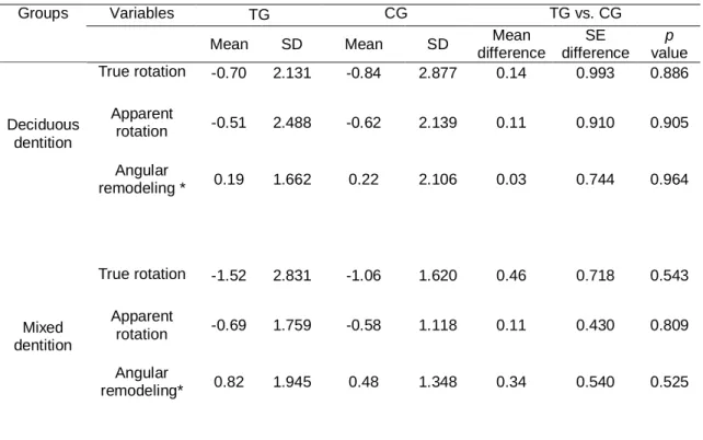
dentaduras decídua e mista e 48% dos indivíduos durante a dentadura permanente. Nas dentaduras mista e permanente a mordida aberta anterior e a má oclusão de classe II foram muito prevalentes. Mais do que 50% das crianças respiradoras orais apresentavam uma relação inter-arcos dentários normal nos três planos do espaço. A análise univariada não mostrou associação estatisticamente significativa entre o tipo de obstrução (hiperplasia por adenóide/amígdala ou presença de rinite) e más oclusões (classe II, mordida aberta anterior e mordida cruzada posterior). Artigo 2 - Após 1 ano de acompanhamento, nenhuma diferença estatisticamente significativa no crescimento facial vertical foi observada nos grupos submetidos a A+A na dentaduras decídua ou mista, comparativamente aos seus grupos controle obstruídos. Exceção feita à divergência maxilo-mandibular durante a fase de dentadura decídua. Artigo 3 - Crescimento facial significativo (p<0,000) foi encontrado para todas as medidas lineares em TG e CG. Uma redução da proporção do terço inferior da face em relação à altura facial total, da inclinação do plano mandibular em relação à base craniana e da divergência maxilo-mandibular, bem como um aumento da proporção da altura facial posterior em relação à altura facial anterior total, aconteceu em TG e CG. Não houve diferença estatisticamente significativa entre a rotação mandibular do TG e CG.
Conclusões:
. A prevalência de mordida cruzada posterior foi maior na população de respiradores orais do que na população geral, independentemente dos estágios de desenvolvimento da oclusão.
. A prevalência de mordida aberta anterior e de má oclusão de classe II foi maior nas crianças mais velhas (dentaduras mista e permanente) do que nas mais novas (dentadura decídua).
. Não houve associação entre a causa da respiração oral (hiperplasia de adenóide, hiperplasia de amígdala, rinite, funcional) e a presença de má oclusão de classe II, mordida aberta anterior e mordida cruzada posterior. . A maioria das crianças respiradoras orais apresentou uma relação oclusal inter-arcos normal.
. As crianças submetidas a A+A tiveram um crescimento facial predominantemente horizontal, similar à normalidade descrita na literatura. . As crianças que permaneceram obstruídas por 1 ano também tiveram um crescimento facial predominantemente horizontal.
SUMMARY
Introduction: The association between nasal impairment and dentofacial
morphology has been studied for more than a century. Controversies still exist about this subject, despite a lot of information is available on the literature. Therefore, the purpose of this PhD thesis was to evaluate if expectations meet reality regarding some assumptions previously established on clinicians’ minds. Three points were investigated: 1) epidemiological report on the prevalence of malocclusion among a group of children consecutively admitted at a referral mouth breathing (ENT) center, studying the association of such malocclusions and upper airway obstructive factors, 2) the impact of respiration normalization on vertical dentofacial growth during two stages of dental development after adeno-/tonsillectomy (T&A) and 3) the impact of respiration normalization on vertical dentofacial growth after adeno-/tonsillectomy (T&A), controlling the results with a matched group of untreated mouth breathing children.
Methods: The work described in this thesis consists of three papers. Each one
answering each objective listed above. The first paper reports a cross-sectional, descriptive study, carried out at an Outpatient Clinic for Mouth-Breathers. Dental inter-arch relationships and nasal obstructive variables of 401 children were diagnosed and the appropriate cross tabulations were done. In the second paper, linear and angular cephalometric measurements, as well as superimposing tracings of serial lateral cephalograms of 39 patients in the treatment group were compared with those of 31 untreated mouth breathing controls. Cephalometric records in the treatment group comprised registrations made at baseline before surgery (T0), and then at approximately 1 year post-operatively (T1). Corresponding registrations were available for the control group, with baseline cephalometric radiographs taken approximately 1 year before the second one (T0 and T1, respectively). Treated and untreated individuals were divided into deciduous and mixed dentition groups to aid identification of an optimum timing for normalizing the respiration after T&A, under a vertical dentofacial perspective. In the third paper the impact of T&A on the vertical dentofacial growth is revisited after an untreated group of mouth breathing children served as controls.
from 2 to 12 years. All subjects were evaluated by otorhinolaryngologists to confirm mouth breathing habit. Adenoid/tonsil obstruction was detected in 71.8% of this sample, regardless of the presence of rhinitis. Allergic rhinitis alone was found in 18.7% of the children. Non obstructive mouth breathing was diagnosed in 9.5% of this sample. Posterior crossbite was detected in almost 30% of the children during primary and mixed dentitions and 48% in permanent dentition. During mixed and permanent dentitions, anterior open bite and class II malocclusion were highly prevalent. More than 50% of the mouth breathing children carried a normal inter-arch relationship in the sagital, transversal and vertical planes. Univariate analysis showed no significant association between the type of the obstruction (adenoids/tonsils obstructive hyperplasia or the presence of allergic rhinitis) and malocclusions (class II, anterior open bite and posterior crossbite).
Paper #2 - After one year of follow up, no statistically significant difference on
vertical dentofacial growth was observed in deciduous or mixed dentitions treatment groups compared to same stage untreated control groups. The reduction of the divergence (NL-MP) between maxilla and mandible was statistically significant greater for adeno-/tonsillectomy group during primary dentition.
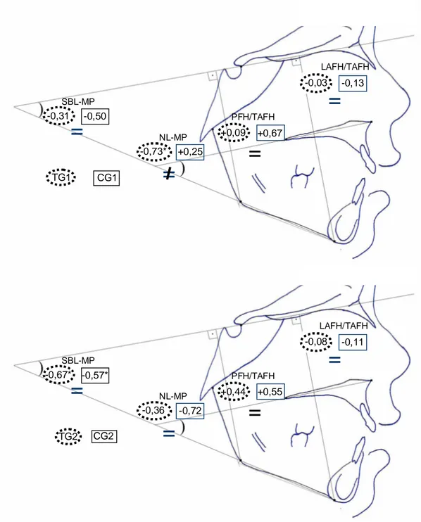
Paper #3 - Statistically significant growth (p<0.000) was found for all linear
measurements (SBL-Go, SBL-Me, NL-Me) in both groups (TG and CG). A reduction in LAFH/TAFH, SBL-MP and NL-MP, as well as an increase in PFH/TAFH, were the growth mean behavior both in TG and CG. There was no statistically significant difference between TG and CG regarding the mandibular rotation.
Conclusions:
. The prevalence of posterior crossbite is higher in mouth-breathing children than in the general population.
. During mixed and permanent dentitions, anterior open bite and class II malocclusion were more likely to be present in mouth breathers.
. Although more children showed these malocclusions, most mouth breathing children evaluated in this study did not match the expected “mouth breathing dental stereotype.
or tonsils and the presence of rhinitis were not risk factors to the development of class II malocclusion, anterior open bite or posterior crossbite.
. Regarding the vertical dentofacial growth pattern, normalization of the mode of respiration after T&A in young children (deciduous dentition) is not more effective than in older children (mixed dentition).
. The normalization of the mode of respiration, after T&A, did not change the pattern of mandibular vertical growth, after one year, when compared to a matched untreated group of mouth breathers.
. Apparently, there is a greater clockwise rotation of the anterior portion of maxilla in adeno-/tonsillectomized children than in obstructed controls during primary dentition.
LISTA DE FIGURAS
Artigo 2
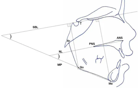
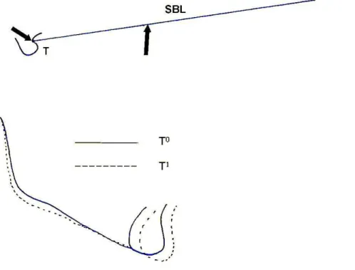
Figure 1- Cephalogram illustrating the skeletal landmarks, the
angular and linear measurements……….. 64
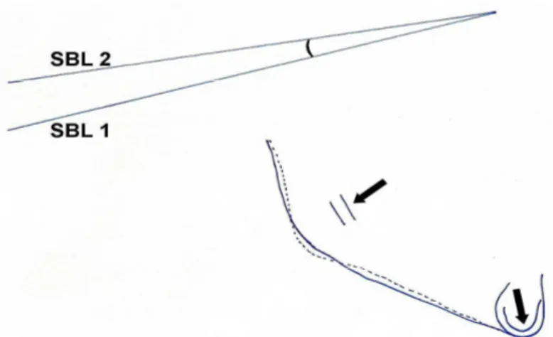
Figure 2- Mandibular true rotation evaluated by angular changes between T0 (SBL 1) and T1 (SBL 2) after the superimposition on the fiducial skeletal landmarks
indicated by arrows……… 65
Figure 3- Mandibular apparent rotation between T0 and T1.
Superimposition on the SBL at “point
T”………..…. 66
Figure 4- Net growth measured in the four groups (TG1, CG1, TG2, CG2). Negative values mean measurement reduction between T0 and T1 while positive values indicate increase………... 72
Artigo 3
Figure 1- Net growth measured in the treatment group (TG) and control group (CG). Negative values mean measurement reduction between T0 and T1 while positive values indicate
LISTA DE TABELAS
Artigo 1
Table 1- Prevalence of dental and ENT findings according to gender distribution. Number of children (n) and prevalence given in
percentage (n/N x 100%)……… 41
Table 2- Prevalence of dental and ENT findings in the deciduous. mixed and permanent dentitions. Number of children (n) and prevalence given
in percentage (n/N x 100%)……….. 43
Table 3- Univariate analysis between grouped malocclusion (dependent variable) and the obstructive causes for mouth breathing
(independent variables)………. 45
Artigo 2
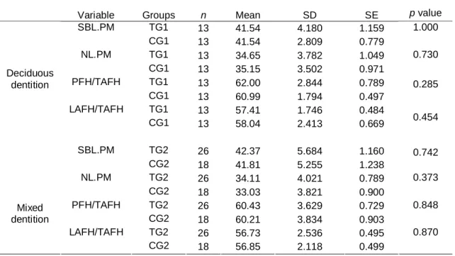
Table 1- Table 1- Independent samples t-test comparison of the baseline (T0) cephalometric angular and ratio measurements between the treatment (TG) and control (CG) groups during the two stages of dental development (deciduous and mixed dentitions)……….. 68
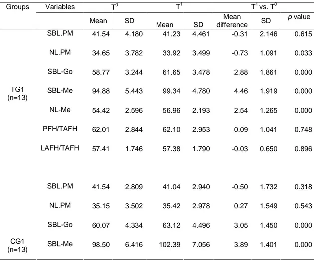
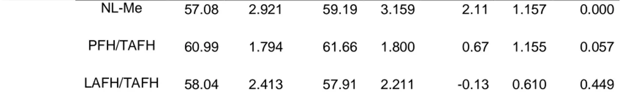
Table 2- Table 2- Paired-sample t-test comparison between changes of cephalometric measurements in T0 and T1 for the group of children submitted to T&A during deciduous dentition (TG1) and its untreated matched control group (CG1)………..………... 69
Table 3- Table 3- Paired-sample t-test comparison between changes of cephalometric measurements in T0 and T1 for the group of children submitted to T&A during mixed dentition (TG2) and its untreated
matched control group (CG2)...………... 70
Table 4- Table 4– Independent samples t-test comparison of mandibular rotation (true rotation. apparent rotation and angular remodeling) between treatment (TG) and control (CG) groups during deciduous
and mixed dentitions………... 71
Artigo 3
Table 1- Comparison of the baseline (T0) cephalometric angular, linear and ratio measurements between the TG (n=39) and CG (n=31)…….. 89
Table 2- Comparison between the treatment group (TG) and control group (CG) for changes within each pair of variable using a paired
Student’s t-test….……….. 90
Table 3- Comparison between the treatment group (TG) and control group (CG) for nominal changes in vertical facial proportions (LAFH/TAFH and PFH/TAFH) and skeletal rotation (SBL-PM and NL-PM) using χ2
test……….. 92
ÍNDICE
Capítulo 1: Considerações iniciais ... 20
1.1 A associação entre a respiração oral e o crescimento dentofacial.
1.2 Normalização da respiração, após a desobstrução cirúrgica das vias aéreas
superiores, e o crescimento facial vertical.
1.3 Adeno-/tonsilectomia na desobstrução das vias aéreas superiores: existe
uma época ideal?
1.4 Objetivo da tese ………... 29
1.5 Referências bibliográficas ... 30
Capítulo 2: Artigos ……… 33
2.1 - Artigo 1: “Prevalence of malocclusion among mouth breathing children: do
expectations meet reality?” ……….. 34
2.2 - Artigo 2: “Changes in vertical dentofacial morphology after
adeno-/tonsillectomy during deciduous and mixed dentitions mouth breathing children -
one year follow up study” ……… … 57
2.3 - Artigo 3: “Vertical facial growth following adeno-/tonsillectomy: changing
concepts?” ……….. .... 82
Capítulo 3: Considerações finais ………. .... 100
Apêndices e Anexos
Apêndice 1 – Termo de Consentimento Livre e Esclarecido ... 108 Apêndice 2 – Dados brutos do Artigo 1 ... 109 Apêndice 3 – Dados brutos dos Artigos 2 e 3 ... 118
Anexo 2: Artigo 1 – versão impressa da Revista International Journal of Pediatric Otorhinolryngology... 122 Anexo 3: Aceitação do Artigo 2 pela Revista International Journal of Pediatric
CAPÍTULO 1
1.1 A associação entre a respiração oral e o crescimento dentofacial.
O equilíbrio das funções vitais exercidas pelo sistema estomatognático, dentre elas a respiração nasal, é essencial para que haja o desenvolvimento dentofacial normal, dentro dos padrões morfológico e genético de cada indivíduo 7, 23.
Assim, a função naso-respiratória tem sido de grande interesse nas últimas décadas, devido à sua relação biológica com a forma e a função, e também por causa de sua enorme implicação clínica, para pediatras, otorrinolaringologistas, alergologistas, ortodontistas, fonoaudiólogos, fisioterapeutas e outros profissionais da área de saúde que lidam com pacientes em fase de crescimento 22.
Investigações sobre o impacto de fatores ambientais sobre o crescimento e o desenvolvimento facial têm demonstrado uma associação entre a obstrução das vias aéreas e variadas formas de más oclusões e displasias ósseas 3, 5, 9, 17,
20, 22
.
Em humanos, os estudos têm concentrado suas atenções no papel das formas etiológicas mais incidentes de obstrução respiratória causadoras da respiração oral: hiperplasia adenoideana, rinites alérgicas, hiperplasia amigdaliana, hipertrofias de conchas nasais 3, 12, 18.
Por outro lado, trabalhos clássicos com primatas não humanos confirmaram que a obstrução nasal severa à passagem de ar, artificialmente criada, pode causar uma série de más oclusões. Apesar da resposta não ser uniforme entre os animais, a abertura da boca para a realização da respiração oral gradualmente resultou em um plano mandibular mais inclinado e um ângulo goníaco mais aberto 8, 9, 10.
inferior é evertido, hiperfuncionante. A musculatura jugal é relaxada, o nariz é pequeno e pouco desenvolvido. A língua se posiciona inferior e anteriormente, entre os incisivos superiores e inferiores. Os incisivos superiores são projetados para vestibular. O olhar demonstra cansaço e a face apresenta uma expressão atoleimada 26.
Espera-se, ainda, que os respiradores orais crônicos tenham uma atresia maxilar, com tendência a um cruzamento no segmento posterior 3,20, um padrão de crescimento facial vertical excessivo17, muitas vezes com uma mordida aberta anterior e uma relação oclusal de classe II 22.
Apesar das características dentofaciais descritas acima serem aquelas que vêm à mente da maioria dos profissionais da área de saúde, quando diante de um paciente respirador oral, a literatura mostra que, do ponto de vista epidemiológico, a “fácies adenoideana” típica não é o achado mais comum nos pacientes respiradores orais. Alguns autores, inclusive, questionam a associação entre o padrão respiratório e a morfologia facial 15.
Shapiro29 concluiu que, apesar do crescente volume de artigos científicos demonstrando as relações entre a obstrução das vias aéreas superiores e o crescimento facial, os clínicos deveriam ter cuidado na indicação de terapias radicais ou na promessa de resultados ousados.
Alterações morfológicas isoladas (como o aumento da altura facial anterior inferior e a atresia dos arcos) são bastante prevalentes em respiradores orais 3,
20
, enquanto que a relação sagital inter-arcos mais encontrada é a de classe I e não a de classe II 11, 16.
Ricketts26 afirmou que a face dos respiradores orais cresce com excesso vertical devido à rotação mandibular posterior favorecida pela manutenção da boca aberta.
Apesar da controvérsia se a respiração oral é que causaria o excesso de crescimento facial vertical ou se indivíduos com morfologia facial alongada estariam mais susceptíveis à obstrução das vias aéreas superiores30, 35, é fato que existe uma forte associação entre os respiradores orais e uma face longa 5,
17
.
1.2 Normalização da respiração, após a desobstrução cirúrgica das vias
aéreas superiores, e o crescimento facial vertical.
Acreditando-se que a respiração oral favorece um crescimento facial excessivo é possível teorizar que a normalização da função respiratória, após a desobstrução cirúrgica das vias aéreas superiores, é capaz de promover uma reversão, pelo menos parcial, deste padrão perverso de crescimento facial.
Diversas publicações descreveram o impacto positivo da adenoidectomia e do aumento do fluxo de ar pelo nariz no crescimento facial vertical. Entretanto, a maioria delas14,18,19,21,36 foi produto de um mesmo estudo longitudinal, conduzido na Suécia na década de 1960, onde 38 crianças foram acompanhadas por cinco anos e o crescimento comparado com o de indivíduos sem obstrução respiratória.
Kerr, McWilliam e Linder-Aronson14 estudaram a mudança de forma e posicionamento espacial da mandíbula após a adenoidectomia, concluindo que decorridos 5 anos da normalização da respiração oral o padrão esquelético das crianças se tornou menos dolicocefálico. Eles concluíram que a mudança do padrão respiratório influenciou a rotação mandibular, bem como a sua morfologia.
Behlfelt2 estudou o efeito do aumento das amígdalas e da sua remoção cirúrgica no crescimento facial. A amostra era composta por 73 crianças com idade média de 10,1 anos. O pesquisador encontrou que crianças com hiperplasia amigdaliana têm maior prevalência de retro-inclinação de incisivos inferiores, protrusão de incisivos superiores, redução do comprimento da arcada inferior, tendência à mordida aberta anterior, aumento da sobressaliência e tendência ao cruzamento na região posterior. Na análise esquelética, estas crianças mostraram ter maior prevalência de retrognatismo mandibular e de rotação horária da mandíbula, aumento na altura facial anterior inferior e mordida aberta. Após a remoção cirúrgica das amígdalas, houve um reposicionamento dorsal da base da língua, favorecendo uma redução da atresia mandibular e da prevalência de mordida cruzada posterior. Identificou-se, também, um aumento da altura facial posterior inferior.
Woodside, Linder-Aronson e Lundstrom36 verificaram não haver diferenças na direção do crescimento maxilar no grupo de crianças adenoidectomizadas, em relação às crianças sem problemas respiratórios. O crescimento da sínfise mandibular, expresso no queixo, foi maior no grupo de crianças operadas do que no grupo controle normal.
monitoramento longitudinal de crianças que forem precocemente submetidas à adenoidectomia.
Mahoni, Karsten e Linder-Aronson21 tiveram como objetivo determinar se as alturas dentoalveolar e facial, inicialmente aumentadas nas crianças respiradoras orais, são mantidas após a adenoidectomia. As comparações feitas com um grupo de crianças respiradoras nasais, cinco anos após a cirurgia, mostraram que a redução da altura dentoalveolar dos molares superiores e da altura facial anterior inferior estão associadas à mudança do padrão respiratório de oral para nasal.
Recentemente, Zettergren-Wijk, Forsberg e Linder Aronson37 publicaram os seus achados em relação ao crescimento facial de 17 crianças submetidas à adenoidectomia para o tratamento de Síndrome da Apnéia Obstrutiva do Sono (SAOS). O padrão morfológico facial vertical das crianças portadoras de SAOS, que antes da adenoidectomia era diferente daquele encontrado nas 17 crianças-controle, sem problemas respiratórios, adquiriu características de semelhança 5 anos após sanado o problema obstrutivo.
Chama a atenção, consideração feita por Linder-Aronson, Woodside e Lündstrom19 que, sob o ponto de vista puramente científico, seria preferível ter uma amostra controle obstruída, ao invés de composta por crianças sem obstrução naso-respiratória. Entretanto tal desenho metodológico, segundo estes autores, teria limitações éticas.
1.3 Adeno-/tonsilectomia na desobstrução das vias aéreas superiores:
existe uma época ideal?
A tonsilectomia tem sido utilizada como procedimento cirúrgico há muito tempo. Em 50 a.c., Celsus já havia descrito uma técnica para tal operação. Já a adenoidectomia, por outro lado, provavelmente não havia sido executada até o final do século XIX, quando Wilhelm Meyer sugeriu que as vegetações adenoideanas eram responsáveis não somente pelos sintomas nasais, mas também pela perda de audição 6.
As duas cirurgias conjuntamente começaram a ser empregadas de maneira cada vez maior no início do século XX, quando a então popular teoria da infecção focal indicava que vários distúrbios sistêmicos, com destaque para o “reumatismo” eram causados pela doença das amígdalas e adenóide 24.
De forma exagerada, entusiastas inclusive indicavam A+A como tratamento para condições diversas como anorexia, retardo mental, enurese ou simplesmente como medida de promoção de saúde 24.
Talvez o apogeu do entusiasmo com a A+A tenha acontecido, em algumas comunidades, onde cirurgias por atacado nas populações infantis aconteciam nas próprias escolas públicas 6.
considerável de estudos eram publicados confirmando que a A+A era ineficaz24.
O preconceito em relação a A+A, particularmente no meio pediátrico, surgiu e até mesmo ficou exagerado pelas freqüentes indicações inadequadas24.
Durante os anos de 1950, um importante programa de saúde norte americano (United Mine Workers of America Health and Retirement Funds), na esperança de melhorar a qualidade de atenção e também reduzir custos, instituiu a norma de exigir uma avaliação e consentimento prévio de peritos credenciados, em relação a A+A 24.
Ao final dos anos 1960, uma considerável parcela dos livros-texto de pediatria questionava as indicações de amigdalectomia, enquanto uma revisão cética de um conceituado periódico denominou esta intervenção de “ritual cirúrgico” 24.
Em 1976, uma sugestão foi feita propondo que A+A fosse completamente suspenso, até que sua eficácia pudesse ser estabelecida em ensaios clínicos controlados 24.
Não bastasse este ambiente, que variava entre o ceticismo e condenação, o apoio a A+A continuou a existir em vários segmentos da área médica. Estudos que indicaram a associação entre a obstrução das vias aéreas superiores e as alterações no crescimento dentofacial contribuíram com o incentivo à continuidade desta técnica cirúrgica18.
Atualmente, vivemos a fase de análise de resultados e indicações mais criteriosas, baseadas em estudos científicos, porém o estigma da cirurgia ainda permanece entre alguns profissionais, especialmente da área de Pediatria.
fisiopatológica tende a agravar as seqüelas dentofaciais 26,27, 2) a maior parte do crescimento facial acontece nos primeiros anos de vida4.
Entretanto quem geralmente define a época de uma intervenção cirúrgica para a normalização da respiração oral é o médico pediatra. Como, por questões históricas, alguns destes profissionais tendem a recomendar o adiamento da A+A, tal situação é preocupante, uma vez que, até a presente data, não há um relato de estudo clínico controlado para definir a idade limítrofe para a normalização do padrão respiratório, nos casos de obstrução das vias aéreas superiores, sob uma perspectiva ortodôntica, especialmente em relação ao padrão de crescimento facial vertical.
1.4 Objetivo da tese
Diante dos fatos expostos anteriormente, o objetivo desta tese foi avaliar se as expectativas apresentadas a seguir, relacionadas à associação entre a respiração oral e o complexo dentofacial, correspondem à realidade. Ou seja, o que esperamos é o que encontramos?
Expectativa 1: A maioria das crianças respiradoras orais é portadora de má oclusão de classe II, mordida aberta anterior e mordida cruzada posterior, sendo que a gravidade da obstrução das vias aéreas superiores tem associação com estas más oclusões.
Expectativa 2: A desobstrução cirúrgica das vias aéreas superiores de respiradores orais, durante a fase de dentadura decídua, propicia um crescimento facial vertical mais favorável do que quando realizada durante a fase de dentadura mista.
1.5 Referências Bibliográficas
1 Arun T, Isik F, Sayinsu K. Vertical growth changes after adenoidectomy.
Angle Orthod. 2003; 73:146-150.
2 Behlfelt K. Enlarged tonsils and the effect of tonsillectomy: characteristics of the dentition and facial skeleton posture of the head, hyoid hone and tongue; mode of breathing. Swed Dent J 1990; suppl 72:5-35.
3 Bresolin D, Shapiro PA, Shapiro GG, Chapko MK, Dassel S. Mouth breathing in allergic children: Its relationship to dentofacial development.
Am J Orthod. 1983; 83:334-40.
4 Casselbrant ML. What is wrong in chronic adenoiditis/tonsillitis anatomical considerations. Int J Ped Otorhinol. 1999;49:S133-S135.
5 Cheng MC, Enlow DH, Papsidero M, Broadbent Jr BH, Oyen O, Sabat M. Developmental effects of impaired breathing in the face of the growing child. Angle Orthod. 1988; 58:309-320.
6 Deutsch ES. Tonsillectomy and adenoidectomy. Changing indications.
Pediatr Clin North Am. 1996; 43:1319-38.
7 Enlow DH. Crescimento facial. 3 ed. São Paulo: Artes Médicas. 553p. 1993.
8 Harvold EP, Chierici G, Vargervik K. Experiments on the development of dental malocclusions. Am J Orthod. 1972; 61:38-44.
9 Harvold EP, Tomer BS, Vargervik K, Chierici G. Primate experiments on oral respiration. Am J Orthod. 1981; 79:359-372.
10 Harvold EP, Vargervik K, Chierici G. Primate experiments on oral sensation and dental malocclusions. Am J Orthod. 1973; 63:494-508.
11 Howard CC. Inherent growth and its influence on malocclusion. J Am Dent
Assoc. 1932; 19:642-648.
12 Hulcrantz E, Larson M, Hellquist T, Ahqvist-rastad J, Jakobsson OP. Int J
Ped Otorhinolaryngol. 1991; 22:125-34.
13 Karlsen AT. Craniofacial growth differences between low and hogh MP-SN angle males: a longitudinal study. Angle Orthod. 1995; 65:341-350.
14 Kerr JS, McWilliam JS, Linder Aronson S. Mandibular form and position related to changed mode of breathing – a five-year longitudinal study.
15 Kluemper GT, Vig PS, Vig KW. Nasorespiratory characteristics and craniofacial morphology. Eur J Orthod. 1995; 17:491-495.
16 Leech HL. A clinical analysis of orofacial morphology and behavior of 500 patients attending an upper respiratory research clinic. Dent Pract. 1958; 9: 57-68.
17 Lessa FCR, Enoki C, Feres MFN, Valera FCP, Lima WTA, Matsumoto MAN. Breathing mode influence in craniofacial development. Braz J
Otorhinol. 2005; 71:156-60.
18 Linder-Aronson S. Effects of adenoidectomy on dentition and facial skeleton over a period of five years. In: Cook JT (ed) Transactions of the
Third International Orthodontic Congress. St Louis: The CV Mosby
Company, 1975; 85-100.
19 Linder-Aronson S, Woodside DG, Lundström A. Mandibular growth direction following adenoidectomy. Am J Orthod Dentof Orthop. 1986; 89:73-284.
20 Lofstrand-Tideström B, Thilander B, Ahlqvist-Rastad J, Jakobsson O, Hultcrantz E. Breathing obstruction in relation to craniofacial and dental arch morphology in 4-year-old children. Eur J Orthod. 1999; 21:323-332. 21 Mahony D, Karsten A, Linder Aronson S. Effects of adenoidectomy and
changed mode of breathing on incisor and molar dentoalveolar heights and anterior face heights. Aust Orthod J. 2004; 20:93-98.
22 McNamara JA. Influence of respiratory pattern on craniofacial growth.
Angle Orthod. 1981; 81:269-300.
23 Moss-Salentijn L. Melvin L. Moss and the functional matrix. J Dent Res. 1997; 76:1814-1817.
24 Paradise JL. Tonsillectomy and adenoidectomy. In: Bluestone CD, Alper CM, Stool SE, Arjmand EM. Pediatric otolaryngology. Chapter 61. Vol 2. 4th ed. Philadelphia: Saunders; 2003:1210-1222.
25 Pirara S, Bento RF, Camas J. Consensos e controvérsias nas indicações de adenoamigdalectomia entre pediatras e otorrinolaringologistas. Braz J
Otorhinol. 1999; 65:308-315
26 Ricketts RM. Respiratory obstruction syndrome. Am J Orthod. 1968; 54:495-514.
Cleft Palate Bulletin. 1958;8:4-5 (abstract) cited by Linder Aronson S.
Effects of adenoidectomy on dentition and facial skeleton over a period of five years. In: Cook JT (ed) Transactions of the Third International
Orthodontic Congress. St Louis: The CV Mosby Company, 1975; 85-100.
28 Schudy FF. The rotation of the mandible resulting from growth: its implications in orthodontic treatment. Angle Orthod. 1965; 35:36-50.
29 Shapiro PA. Effects of nasal obstruction on facial development. J Allergy
Clin Immunol. 1988; 81: 967-71.
30 Smith RM, Gonzales C. The relationship between nasal obstruction and craniofacial growth. Ped Clin of North America. 1989; 36:1423-34.
31 Soares JF, Siqueira AL. Introdução à estatística médica. Belo Horizonte: Coopmed Editora Médica, 2 ed., 2002. 300p.
32 Sparks CS, Jantz RL. A reassessment of human cranial plasticity: Boas revisited. Proc Nat Acad Sciences. 2002; 99:14636-14639.
33 Tollaro I, Baccetti T, Franchi L. Mandibular skeletal changes induced by early functional treatment of class III malocclusion: a superimposition study.
Am J Orthod Dentof Orthop.1995; 108:525-532.
34 Wang MK, Buschang PH, Behrents R. Mandibular rotation and remodeling changes during early childhood. Angle Orthod. 2009; 79:271-275.
35 Warren DW. Effect of airway obstruction upon facial growth. Otolaryngol
Clin North America. 1990; 23:699-712.
36 Woodside DG, Linder Aronson S, Lundström, A. Mandibular and maxillary growth after changed mode of breathing. Am J Orthod Dentof Orthop. 1991; 100:1-18.
CAPÍTULO 2
Artigos
Artigo 1
Título: Prevalence of malocclusion among mouth breathing children: do
expectations meet reality?
Autores: Bernardo Q. Souki, Giovana B. Pimenta, Marcelo Q. Souki, Leticia P.
Franco, Helena M. G. Becker and Jorge A. Pinto.
Revista: International Journal Pediatric Otorhinolaryngology
Artigo 1
Prevalence of malocclusion among mouth breathing children: do
expectations meet reality?
Bernardo Q. Soukia,b, Giovana B. Pimentaa, Marcelo Q. Soukia, Leticia P. Francoa, Helena M. G. Beckera and Jorge A. Pintoa
a
Federal University of Minas Gerais, Outpatient Clinic for Mouth-Breathers, Belo Horizonte, Brazil
b
Catholic University of Minas Gerais, School of Dentistry, Orthodontics, Belo Horizonte, Brazil
Keywords: Mouth breathing, malocclusion, adenoids, tonsils, rhinitis
Abstract
Objective: The aim of this study was to report epidemiological data on the
prevalence of malocclusion among a group of children, consecutively admitted at a referral mouth breathing otorhinolaryngological (ENT) center. We assessed the association between the severity of the obstruction by adenoids/tonsils hyperplasia or the presence of allergic rhinitis and the prevalence of class II malocclusion, anterior open bite and posterior crossbite.
Methods: Cross-sectional, descriptive study, carried out at an Outpatient Clinic
for Mouth-Breathers. Dental inter-arch relationship and nasal obstructive variables were diagnosed and the appropriate cross tabulations were done.
Results: Four hundred and one patients were included. Mean age was 6 years
(adenoids/tonsils obstructive hyperplasia or the presence of allergic rhinitis) and malocclusions (class II, anterior open bite and posterior crossbite).
Conclusions: The prevalence of posterior crossbite is higher in mouth-breathing
children than in the general population. During mixed and permanent dentitions, anterior open bite and class II malocclusion were more likely to be present in mouth breathers. Although more children showed these malocclusions, most mouth breathing children evaluated in this study did not match the expected “mouth breathing dental stereotype”. In this population of mouth breathing children, the obstructive size of adenoids or tonsils and the presence of rhinitis were not risk factors to the development of class II malocclusion, anterior open bite or posterior crossbite.
1 Introduction
The association between nasal respiratory impairment and dento-facial morphology has been studied for more than a century [1-3] and for decades it has been strongly accepted that inter-arch growth pattern can be influenced by an unbalanced muscular function on mouth breathers [4].
The knowledge that obstruction of nasal breathing most likely will perversely impact the facial growth even led some authors to propose classic terms to describe such patients as “adenoid faces” [5] , “long face syndrome” [6] and “respiratory obstruction syndrome” [7].
A stereotype of these patients, therefore, can be drawn, where an anterior open bite [8], a reduced transversal dimension [9,10], associated or not with posterior crossbite [11], and a class II malocclusion [12, 13,14] are expected.
However, as individual facial genotypes have different sensitivity on developing malocclusion, following the exposure to mouth breathing, a wide variety of inter-arch relationships can be found.
breathers and that nasal impaired respiration will ultimately result in this malocclusion. Besides that, one question arises: can we predict the outcome of these malocclusions based on the presence and on the type of airway obstructive cause which led to this deleterious habit?
Routinely, Ear, Nose and throat (ENT) specialists and general clinicians use the diagnosis of the airflow blockage by adenoids and tonsils hyperplasia as a parameter to the establishment of the treatment planning [15]. Although this axiom has been used routinely by clinicians, it has not been sufficiently tested regarding the development of malocclusion.
The aim of this study was to report epidemiological data on the prevalence of malocclusion among a group of children, consecutively admitted at a referral mouth breathing ENT center. We assessed the association between severity of the obstruction by adenoids/tonsillar hyperplasia or the presence of allergic rhinitis and the prevalence of class II malocclusion, anterior open bite and posterior crossbite.
2 Patients and methods
2.1 Population
Four hundred and forty four children consecutively referred by pediatricians and primary care physicians to the Outpatient Clinic for Mouth-Breathers, at the Hospital das Clínicas at Federal University of Minas Gerais (UFMG), Brazil, between November of 2002 and November of 2007, with the chief complaint of mouth breathing were systematically evaluated by a multidisciplinary team comprised by ENT doctors, allergologists and orthodontists, in a single day visit.
Children whose mouth breathing could not be confirmed, those who have had previous orthodontic treatment or were younger than 2 years of age were excluded from the analysis. Therefore, the sample of this cross-sectional study totaled 401 patients.
All subjects were evaluated by otorhinolaryngologists to confirm mouth breathing resulting from at least one of the following airway pathologies: obstructive tonsillar hyperplasia, obstructive adenoidal hyperplasia and allergic rhinitis. The children whose obstruction by one of these conditions could not be diagnosed were classified as functional mouth breathers [16].
The participant’s rights were protected, and informed consent and assent were obtained according to the Ethics Committee of the Federal University of Minas Gerais.
2.2 ENT data collection
An interview with children’s parents, or guardians, asking about the quality of the children’s sleep, snoring, oral breathing and throat infections, confirmed the “chief complaint” of mouth breathing. Parents were also asked if the child had been undergone an adenoidectomy or tonsillectomy earlier. Clinical ENT examination was performed by two of the authors (L.F. and H.B.), according to the following guidelines:
Palatine tonsil hypertrophy was classified by mouth examination according to the criteria of Brodsky and Koch [17] as follows: grade 0 – tonsils limited to the tonsillar fossa; grade 1 – tonsils occupying up to 25% of the space between the anterior pillars in the oropharynx; grade 2 – tonsils occupying 25-50% of the space between the anterior pillars; grade 3 – tonsils occupying 50-75% of the space between the anterior pillars; and grade 4 – tonsils occupying 75-100% of the space between the anterior pillars.
Tonsils grade 0, 1 and 2 were considered as non-obstructive and those classified as grade 3 and 4 were named as obstructive. [18]
2.3 Allergological data collection
The allergological assessment, to diagnose allergic rhinitis, included a structured medical interview, physical examination, following the standard volar forearm skin prick method, as described elsewhere[20]. These exams were performed in 326 children under the supervision of one of the authors (J.P).
2.4 Dental data collection
The dental clinical examination was performed by a team of orthodontists, who worked together for at least ten years, and were previously calibrated. The subjects were grouped by stage of dental development, according to the variation in primary and permanent teeth eruption, into deciduous, mixed and permanent periods.
The inter-arch occlusion dental classification was based on Barnett [21]:
Vertical: relationship was classified as 1) normal, 2) anterior open bite or 3)
deep bite. An open bite was registered in cases that lacked any overbite, regardless of the amount. A deep bite was registered when more than half of the lower incisors were overlapped by the incisal edges of the upper incisors.
Transversal: relationship was classified as 1) normal, 2) posterior crossbite,
without mandibular functional shift, and 3) posterior bite, with mandibular functional shift.
Sagital: relationship was classified as a) normal occlusion, b) class I
malocclusion, c) class II malocclusion and c) class III malocclusion. During the deciduous and mixed dentitions, it was considered a class I dental relationship when the upper deciduous cuspid intercuspation was set between the lower deciduous cuspid and first deciduous molar. When in permanent dentition the Angle classification was followed.
2.5 Dental data comparison
populations have been published. These data served as a reference of what should be the distribution on inter-arch anomalies among a general population, where mouth and nasal breathers were sampled together [28-32, 35-41].
2.6 Statistics
Epi-data was used to enter data. SPSS version 12.0 was used for the analysis. Descriptive statistics and univariate analysis in cross tables are showed. The significance level of p<0.05 was chosen. Normality of age distribution was tested using Kolmogorov-Smirnov test.
For each dental and ENT variable, the number of children with the diagnosed status (n) and its prevalence (%) are given.
For the purpose of statistical analysis, dental variables were binarily grouped according to the expected inter-arch relationships in mouth breathing subjects. Therefore the dependent variables examined were class II malocclusion, anterior open bite and posterior crossbite.
The independent ENT variables were the obstructive grade of tonsil and adenoids and the presence of rhinitis.
3 Results
The mean age of this sample was 6 years and 6 months and the standard deviation was 2 years and 7 months. The age of the children ranged between 2 and 12 years. With the exception of 38 children (9.5%), whose mouth breathing was due to functional habit, 363 subjects had an objective airway obstructive factor. Of these children, 288 (71.8%) were judged to have tonsil and/or adenoid obstruction, combined or not with rhinitis. Allergic rhinitis, as the only obstructive cause, was found in 75 children (18.7%).
boys and girls as a single group.
Table 1 – Prevalence of dental and ENT findings according to gender distribution. Number of children (n) and prevalence given in percentage (n/N x 100%).
Boys Girls Total
Variables
n % n % n %
Stage of development N=401
Deciduous dentition 106 26.4 60 15.0 166 41.4 Mixed dentition 110 27.4 99 24.7 209 52.1 Permanent dentition 12 3.0 14 3.5 26 6.5
χ2
= 6.050 (2 df) p value = 0.05 Sagital relationship N=384
Normal occlusion 26 6.8 17 4.4 43 11.2 Class I malocclusion 97 25.3 83 21.6 180 46.9 Class II malocclusion 64 16.7 51 13.3 115 29.9 Class III malocclusion 30 7.8 16 4.2 46 12.0
χ2
= 2.230 (3 df) p value = 0.526 Vertical relationship N=385
Normal 115 29.9 98 25.5 213 55.3 Deep bite 38 9.9 21 5.5 59 15.3 Open bite 67 17.4 46 11.9 113 29.4
χ2
= 2.349 (2 df) p value = 0.309 Transversal relationship N=392
Normal 158 40.3 116 29.6 274 69.9 Posterior crossbite w/o
shift
31 7.9 22 5.6 53 13.5
Posterior crossbite w shift 32 8.2 33 8.4 65 16.6
χ2
= 1.631 (2 df) p value = 0.443 Tonsils status N=399
Grades 0, I, II 141 35.3 95 23.8 236 59.1 Grades III, IV 86 21.6 77 19.3 163 40.9
χ2
= 1.918 (1 df) p value = 0.166 Adenoid obstruction status N=390
<75% 95 24.4 70 17.9 165 42.3
≥ 75% 124 31.8 101 25.9 225 57.7
χ2 = 0.235 (1 df) p value = 0.628
Rhinitis N=326
Yes 133 40.8 102 31.3 235 72.1 No 51 15.6 40 12.3 91 27.9
χ2
= 0.008 (1 df) p value = 0.928
were susceptible to the unbalanced muscular adaptation to mouth breathing. Only few children (6.5%) were in permanent dentition.
Based on Table 1, 58.1% of the sample had a normal sagital relationship (class I dental relationship). Class I malocclusion was found in 46.9% of these children, the other 11.2% represents the normal occlusion children. Regarding the three stages of occlusal development (Table 2), Class I dental relationship was found in 64.2% during deciduous dentition, 53.8% and 54.2% during mixed and permanent dentitions, respectively.
About 42% of this sample presented with a sagital disharmony, represented by class II or III (Table 1). The prevalence of class III gets higher as kids get older (Table 2).
Considering the 384 children whose sagital classification was done, dental Class II was the sagital relationship of 27% during primary dentition, 32.8% on mixed dentition and 25% on permanent dentition (Table 2).
The vertical inter-arch relationship must be studied in the dental stage of development because of its known physiologic difference along the growing period. Nevertheless, Table 2 brings the information that a normal vertical relationship was found in, at least, 52.7% of the sample, regardless of the dental stage of development. Open bite prevalence was around 30% during the deciduous and mixed dentitions and 20% in permanent dentition.
In the transversal analysis, posterior crossbite was detected in close to 30% of the kids during deciduous and mixed dentitions and 48% in permanent dentition (Table 2).
Table 2- Prevalence of dental and ENT findings in the deciduous. mixed and permanent dentitions. Number of children (n) and prevalence given in percentage (n/N x 100%).
Variable Deciduous Mixed Permanent
Dental n % n % n %
Sagital relationship N=384
159 201 24
Normal occlusion 24 15.1 19 9.5 1 4.2 Class I malocclusion 78 49.1 89 44.3 12 50.0 Class II malocclusion 43 27.0 66 32.8 6 25.0 Class III malocclusion 14 8.8 27 13.4 5 20.8
χ2
p value = 0.196 Vertical relationship N=385
165 195 25
Normal 87 52.7 111 56.9 15 60.0 Deep bite 27 16.4 27 13.8 5 20.0 Open bite 51 30.9 57 29.2 5 20.0
χ2
p value = 0.731 Transversal relationship N=392
164 203 25
Normal 118 72.0 143 70.4 13 52.0 Posterior crossbite
w/o shift
19 11.6 29 14.3 5 20.0
Posterior crossbite w shift
27 16.5 31 15.3 7 28.0
χ2
p value = 0.314
ENT
Tonsils status N=399
165 208 26
Grades 0, I, II 83 50.3 133 63.9 20 76.9 Grades III, IV 82 49.7 75 36.1 6 23.1
χ2
p value = 0.005 Adenoid obstruction status N=390
161 205 24
< 75% 43 26.7 102 49.8 20 83.3
≥ 75% 118 73.3 103 50.2 4 16.7
χ2
p value = 0.000 Rhinitis N=326
137 168 21
Yes 79 57.7 136 81 20 95.2
No
58 42.3 32 19 1 4.8
χ2
p value = 0.000
Note: χ2 based on n x 3 tables. n = variable
of tonsillar obstruction shifted according to aging. Children during the deciduous dentition stage of development have more obstructive tonsils than older ones (p<0.05).
The adenoid’s obstruction of the nasopharynx showed similar epidemiological behavior. Although the average prevalence of the obstructive group (≥75% occupation of nasopharynx space) was 57.7%, (Table 1), when analyzing this variable under the perspective of dental stage of development, it is clear that prevalence declines steeply from 73.3% to 16.7% along the aging (Table 2), with statistically significant difference (p<0,05).
The overall prevalence of allergic rhinitis was 72.1% (n=235/326), as demonstrated on Table 1. During mixed and permanent dentitions the proportion of subjects with rhinitis was bigger (81% and 95.2%, respectively) than in deciduous dentition 57.7% (Table 2), a statistically significant difference (p<0.05).
Table 3 shows the univariate analysis between grouped malocclusion (dependent variable) and the ENT independent variables. No association was found between the expected type of malocclusion for mouth breathers and the presence of variables that obstruct the nasal airflow (p>0.05).
Table 3 – Univariate analysis between grouped malocclusion (dependent variable) and the obstructive causes for mouth breathing (independent variables).
4 Discussion
Several reports have associated mouth breathing with dental malocclusion. The first papers were limited to clinical impressions of dentistry pioneers who related the disturbance on facial and occlusal harmony to the impairment of nasal breathing in their patients. Later, many papers published reports based on the findings of scientific data collection, mostly considering the skeletal outcome evaluated by cephalometry. However, data on occlusal clinical parameters of mouth breathing children are scarce.
Dental inter-arch relationship, in the three planes of space, is the basic clinical parameter in understanding the patient’s occlusion and its behavior when exposed to unbalanced muscular activity. Therefore, it is important to assess the occurrence of occlusal disorders among mouth breathing children.
Despite the large sample size of this study, the limitations of a cross-sectional design needs to be considered. As our sample is comprised only of mouth breathers, the prevalence of dental inter-arch status had to be compared with other epidemiological reports on a general population [28-32, 35-41]. This methodology brings at least two biases: 1) it is fact that in a general population
Variables Tonsil/adenoid obstruction
Rhinitis only No obstructive cause diagnosed
p value
Class II malocclusion
Yes 78 24 13
No 196 49 24
0.589
Anterior open bite
Yes 79 24 10
No 198 48 26
0.710
Posterior crossbite
Yes 85 26 7
No 197 48 29
a significant number of children are mouth breathers [22-24]. Thus, the difference between the prevalence of malocclusion in this mouth breathing population and a “normal breathing” population would be greater. 2) The reported prevalence varies considerably between the different studies, even among the same population. This divergence in prevalence figures may depend not only on differences for specific ethnic groups [25], but also on wide ranges in number and age among the examined subjects. However, differences in registration methods, i.e. the criteria for the recorded items, are probably the most important factor explaining these differences. Despite these methodological limitations, this study brings results that deserve further discussion.
Our study compared the prevalence of only one malocclusion in each plane of space: class II (sagital), anterior open bite (vertical) and posterior crossbite (transversal), since an occlusal pattern for mouth breathers is well described.
Anomaly studies usually report findings by chronological age. Malocclusion, however, is a manifestation that is related to development of the dentition. Given the great individual variations in dental maturation, it seems logical to determine the prevalence of malocclusion for groups at different stages of dental development, rather than for different age groups. It is interesting to point out that the pattern of distribution of the prevalence of malocclusions does not show any statistical difference among the three stages of dental development (Table 2), as it occurs in the general population [26]. It is expected that the prevalence of each malocclusion changes among the growth period. This fact suggests that in a mouth breathing population, the increase in the prevalence of some malocclusions alter the common pattern.
However our prevalence is higher than found in other studies [29, 30]. Kataoka et al. [29] concluded that the prevalence of class II in their sample was low (6.8%) because their population was comprised only by Japanese-Brazilian ethnic children. This fact, explains the difference between our findings. However, the difference in relation to the results found by Sadakyio et al. [30] (15.6%) can be justified by data collection methodology discrepancies or differences due to mouth breathing.
In mixed dentition, our study's class II prevalence (32.8%) is much higher than the 12.5% reported by Zanetti [31]. This significant discrepancy suggests that in older children, the perverse impact of mouth breathing, on sagital inter-arch development, is greater than on the deciduous dentition. Cheng et al. [11] noted that the younger a subject is, at the time of evaluation, the less the “adenoid” type of facial characteristics is expressed. This opinion corroborates our findings. We can hypothesize that the longer the exposure to the unbalanced muscular function, due to mouth breathing, the greater the risk of developing class II malocclusion. More epidemiological reports on sagital relationship during the mixed dentition stage would be helpful in testing this hypothesis, but only one was found. Longitudinal cohort studies are necessary to test if this hypothesis is correct.
During permanent dentition, the prevalence of class II in this sample was 25%. A comparison with Brazilian data was not possible because no epidemiological study involving general population at this stage was found, regarding this type of malocclusion. Comparing to Horowitz [32], who evaluated American subjects, the prevalence numbers (22.5%) are quite similar to our results. This observation corroborates the conclusions of Howard [33], Leech [34] and McNamara [3]. Nevertheless, comparing our permanent dentition class II findings with the classic study of Emrich, Brodie, Blayney [35], also in the United States, who found 14%, our prevalence was higher. As the size of permanent dentition sample, in our study, was small (n=24), we suggest that other studies, with larger samples, should test this association.
described to class II was found. Compared to the literature data, the prevalence of open bite during deciduous dentition, in the investigated mouth breathers, was quite similar. While our children’s anterior open bite prevalence during deciduous dentition was 30.9%, the revised literature on general population varied between 20.6% and 46.3% [28, 44-46]. But, when analyzing the older children (mixed dentition), an important difference was noted. The prevalence of open bite reported in the reference articles [31, 36-39] varies between 12.00% and 20.1%, while our sample had a prevalence of 29.2%.
In the transverse dimension we found the most significant discrepancy in the prevalence of malocclusion. Dental literature data shows that the prevalence of posterior crossbite ranges from 8% to 22% (40). Prevalence studies on posterior crossbite during permanent dentition are rare, but Thilander et al. [41] found a prevalence of 3.9% during this stage. Therefore we considered 22% as the top value. We found a prevalence of 30.1% of posterior crossbite in whole group. This prevalence of close to 30% in the primary and mixed dentitions and almost 50% in the permanent one is higher than in the general population and deserves additional consideration.
As the etiology of malocclusion has singular characteristics when considering the three different planes of space, this heterogeneity can help with the comprehension of our findings.
Sagital dental inter-arch relationship is mostly determined by heredity [27] and therefore mouth breathing is only a secondary etiological factor to class II development. Most likely, the power of the unbalanced muscular activities, due to mouth breathing, is not enough to shift a solid class I or III patterns into a class II. Maybe those children with a tendency toward a class II, who could growth into class I, depending on environmental factors, are the population candidates who develop class II, when exposed to mouth breathing. Therefore, in an epidemiological analysis, as we did, the prevalence of class II is higher than in the general population, especially in older children.