Highlights Regarding Host Predisposing
Factors to Recurrent Vulvovaginal
Candidiasis: Chronic Stress and Reduced
Antioxidant Capacity
Luciene Akimoto-Gunther1,2, Patrícia de Souza Bonfim-Mendonça1,2, Gisele Takahachi2,
Mary Mayumi T. Irie2, Sônia Miyamoto2, Márcia Edilaine Lopes Consolaro1,2, Terezinha I. Estivalet Svidzinsk1,2
*
1Postgraduate Program in Health Sciences, State University of Maringá, Maringá, Brazil,2Department of Clinical Analyses and Biomedicine, State University of Maringá, Maringá, Brazil
Abstract
We studied host factors that could predispose women to develop recurrent vulvovaginal candidiasis (RVVC), including glycemia, insulin resistance, chronic stress, antioxidant capacity, overall immune status, local inflammation and vaginal microbiota. The presence of yeasts in vaginal culture was screened in 277 women, with or without signs and symp-toms of VVC and RVVC. The presence of an inflammatory process and microbiota were analyzed through vaginal bacterioscopy and cervical-vaginal cytology, respectively. Fast-ing-blood samples were collected by standard venipuncture for biochemical analyses. Flow cytometry was employed to obtain the T helper/T cytotoxic lymphocyte ratio, and insulin resistance was assessed by the HOMA index (HI). Yeasts were isolated from 71 (26%) women: 23 (32.4%) with a positive culture but without symptoms (COL), 22 (31%) in an acute episode (VVC), and 26 (36.6%) with RVVC.C.albicanswas the main yeast isolated in all clinical profiles. The control group (negative culture) comprised 206 women. Diabetes mellitus and insulin resistance were more associated with the positive-culture groups (COL, VVC and RVVC) than with negative ones. The RVVC group showed lower mean levels of cortisol than the control group and lower antioxidant capacity than all other groups. The T Helper/T cytotoxic lymphocyte ratio was similar in all groups. The RVVC group showed a similar level of vaginal inflammation to the control group, and lower than in the COL and VVC groups. Only the CVV group showed a reduction in vaginal lactobacillus microbiota. Our data suggest that both chronic stress (decreased early-morning cortisol levels) and reduced antioxidant capacity can be host predisposing factors to RVVC.
a11111
OPEN ACCESS
Citation:Akimoto-Gunther L, Bonfim-Mendonça PdS, Takahachi G, Irie MMT, Miyamoto S, Consolaro MEL, et al. (2016) Highlights Regarding Host Predisposing Factors to Recurrent Vulvovaginal Candidiasis: Chronic Stress and Reduced Antioxidant Capacity. PLoS ONE 11(7): e0158870. doi:10.1371/ journal.pone.0158870
Editor:Alix Therese Coste, Institute of Microbiology, SWITZERLAND
Received:December 19, 2015
Accepted:June 23, 2016
Published:July 14, 2016
Copyright:© 2016 Akimoto-Gunther et al. This is an open access article distributed under the terms of the
Creative Commons Attribution License, which permits unrestricted use, distribution, and reproduction in any medium, provided the original author and source are credited.
Data Availability Statement:All relevant data are within the paper.
Funding:This study was supported by grants from Conselho Nacional de Desenvolvimento Científico e Tecnológico (CNPq), Coordenação de
Introduction
Recurrent vulvovaginal candidiasis (RVVC) is a common cause of significant morbidity in women in all strata of society affecting millions of women worldwide [1,2]. Previously RVVC occurrence was limited by onset of menopause but the widespread use of hormone replacement therapy has extended the at-risk period [2]. VVC and RVVC are diseases caused mainly by members of the genusCandida[1–3]. These yeasts, in particularCandida albicans, are capable of colonizing the human body without producing signs of disease in conditions of physiological equilibrium [1,3]. However, under conditions that disrupt the delicate balance between the host and this commensal fungus, a parasitic relationship may occur, resulting in the develop-ment of infections [1–3].
Fungus virulence attributes including adhesion capacity, biofilm development, cell-surface hydrophobicity, morphological transition, and hydrolytic enzymes are related to intrinsic properties of yeasts that increase their ability to cause VVC [4,5]. A recent study showed that the higher prevalence of vaginal candidiasis among diabetics could be related to increased aspartyl proteinases production in this group of patients [6]. In addition to yeasts virulence fac-tors, several host predisposing factors have been suggested [1–3] although few have proved to be involved in the pathogenesis of VVC and particularly RVVC.
The cellular immune response systemic or local and defective neutrophil function may increase susceptibility to infections byC.albicans, [7,8] suggesting that clinical conditions such as exposure to chronic stress, diabetes mellitus, or deficient antioxidant micronutrients that alter the proper functioning of the immune system could facilitate RVVC occurrence. Never-theless, few data are available to help in evaluating the host factors involved in the host-fungus interaction and disease development. In order to further elucidate these relationships, we ana-lyzed intrinsic factors that could predispose women to developing RVVC, including glycemia, insulin resistance, chronic stress, antioxidant capacity, overall immune status, presence of vagi-nal inflammation, and microbiota.
Materials and Methods
This prospective study included 277 sexually active patients, aged between 18 and 50 years, who were referred to the Clinical Analysis and Research Laboratory of the State University of Maringá (UEM), Paraná, Brazil, from July 1, 2012 to June 30, 2013. The study was carried out with females who visited the laboratory to provide cervical-vaginal samples for clinical analy-ses, independently of the presence of VVC symptoms. Exclusion criteria were: hysterecto-mized, pregnant, or in post-partum condition; less than 14 years of age; some degree of difficulty in understanding the study; vaginal bleeding; sexual intercourse/vaginal douching within the 48 hours preceding the collection of the vaginal sample; those who reported receiv-ing antibiotics, corticosteroids or immunosuppressive drugs; and also those with a history of serious debilitating or immunosuppressant disease, including AIDS. All the women or parents gave their written informed consent to participate in the study. This research was approved by the Committee for Ethics in Research with Humans (COPEP) of Universidade Estadual de Maringá (UEM) (report no. 435/2009).
After giving their consent, participants were asked to complete an enrollment questionnaire that included detailed questions regarding hypothesized risk factors of VVC (personal/sexual habits, history of VVC diagnoses during lifetime and the past year), presence of VVC signs and symptoms (vaginal discharge, vulvovaginal itching, vulvovaginal burning sensation, dysuria, and dyspareunia), and epidemiological data. Vaginal discharges were appraised by the health professional during sample collection [9].
Vaginal samples were collected with sterile swabs and a disposable vaginal speculum (Vagis-pec, Santa Catarina, Brazil), inoculated in sterile saline, and immediately seeded onto plates containing Sabouraud dextrose agar (SDA) (Difco, Detroit, Michigan, USA), supplemented with 50 mg/mL chloramphenicol (Sigma Aldrich, St. Louis, Michigan, USA) and incubated at 25°C for up to five days. A pool of the colonies grown on each plate was subcultured on CHRO-MágarCandida(Probac, Paris, France) to ensure the purity of the isolates and to identify mixed cultures. Beginning with the pure culture, the yeasts were identified by classical pheno-typic methods [10]. The yeasts were stored in Sabouraud dextrose broth (SDB) (Difco, Detroit, Michigan, USA) with 10% glycerol at -20°C.
Women with a positive vaginal culture for yeasts were classified based on clinical profiles by physicians in three groups, as follows: 1) colonized (COL)- those without signs and symptoms of VVC; 2) with VVC- those with an acute episode, consisting of women who presented at least two signs or symptoms (discharge, itching, dysuria, and dyspareunia) but no previous epi-sode within a 12-month period; 3) with RVVC- those presenting two or more symptoms in four or more episodes within a 12-month period [9]. Women with a negative vaginal culture were selected as the control group (CG).
A smear was prepared from the vaginal secretion for bacterioscopy. The slides were Gram-stained and examined under an optical microscope at 1.000X magnification. The Gram-stained smears were useful to evaluate the presence of yeasts and other infectious agents, vaginal microbiota, and leukocyte reaction. Vaginal microbiota was considered normal when
microor-ganisms morphologically compatible withLactobacillusspp. were predominant [10].
After sample collection for culture and bacterioscopy, cervical-vaginal samples were obtained from each participant, using an Ayre spatula and a cytobrush, in which triple smears (vaginal, cervical, and endocervical) were prepared and immediately fixed with propylene-gly-col spray. The cytological smears were sent to the Clinical Cytology Laboratory (UEM) for analysis. All smears were stained with Papanicolaou (Pap), evaluated in optical microscopy and vaginal inflammation reported according to the Bethesda System/2001 diagnosis criteria by the reference cytologist [11]. The vaginal smears were analyzed for qualitative detection of cellular inflammatory changes in at least 20 different fields under optical microscopy at 400x magnification. The presence of leukocyte and cellular morphological changes in moderate or intense grade were the parameters employed to characterize the inflammation process. The main cellular inflammatory characteristics, as described by Nayar [11], are in the nucleus, that may become compact, dense, and pyknotic, with loss of all chromatin details or degenerated. Such nuclei may have a distinct circumferential cytoplasmic clearing or hollow, causing a peri-nuclear“halo”. The cytoplasm may show partially or completely disintegrated, amphophilia, and vacuolation [11].
Fasting blood was collected in early morning (07:30 to 09:00 hours) by the standard veni-puncture procedure for biochemical measurements. Automated equipment was used to con-duct the analyses.
Measurement of plasma glucose was performed by the enzymatic colorimetric glucose-oxi-dase/GOD-PAP method (Diasys, Holzheim, Germany), using the Vitalab Selectra 2 system (Elitech Group Solutions, Chicago, Illinois, USA). In this method, glucose is determined after enzymatic oxidation of glucose by glucose oxidase (Trinder reaction). Diabetes was categorized according to the American Diabetes Association (ADA, 2015) [12]. Fasting plasma-glucose lev-els126 mg/dL or100 and<126 mg/dL, respectively, are used to establish the diagnosis of
diabetes and impaired fasting-plasma glucose (decreased glucose tolerance).
coefficient (VC) for serum samples. The reference values adopted for fasting cortisol in the mornings were 5.4–25.0μg/dL or 149–690 nmol/L [12]. The sensitivity of the insulin test is 1.0μU/mL and the precision is7%. The reference values adopted for fasting insulin were
2.5–25.0μU/mL or 17.9–179.3 pmol/L [12].
Insulin sensitivity was estimated from fasting-glucose and insulin data, using the
homeosta-sis model assessment (HOMA) mathematical model: fasting insulin (μU/mL) + jejum glucose
(nmol/L)/22.5 [13]. Insulin resistance was evaluated according to the body mass index (BMI) (1.2±0.65 for BMI<5, 1.8±0.98 for BMI 25–30, and 2.9±1.6 for BMI>30). The BMI was
defined by dividing body weight by height squared and used for the classification of excess
body weight. The cutoff level of 25 kg/m2suggested by the World Health Organization (WHO)
was employed.
Analyses of T cells (T helper/T cytotoxic lymphocyte ratio) were performed on a BD-FACS Calibur flow cytometer equipped with an argon laser and BD Multiset software for data analy-sis. The reagents employed were BD Multiset CD3/CD4/CD8/CD45 (Becton Dickinson TriT-EST immunofluorescence, Becton Dickinson, San José, California, USA).
The overall antioxidant capacity was determined by Trolox equivalent antioxidant activity (TEAC), which was performed as described by Re et al. [14] to evaluate plasma non-enzymatic antioxidants. This method for screening antioxidant activity is a decolorization assay applicable to both lipophilic and hydrophilic antioxidants, including flavonoids, hydroxycinnamates, carotenoids, and plasma antioxidants. The final results are expressed in micromoles per liter (μM/L). Reference values established for TEAC, considering regional ethnic variations and
lab-oratory conditions, were 1.83–2.07μM/L.
Data of cortisol levels, overall antioxidant capacity and T Helper/T Cytotoxic lymphocytes are expressed as median [range] with 25th and 75th percentiles. Significant differences among median were analyzed using Prism 6.0 software (GraphPad, San Diego, CA, USA), and identi-fied by the Kruskal-Wallis test with the Dunn’s post hoc test. The Chi-square (χ2) test using
the STATA Data Analysis and Statistical Software version 9.1 (Texas, USA) was used to com-pare values of the control group with the positive-culture group and a crude odds ratio (OR) and 95% confidence interval (CI) were calculated. All variables were expressed as absolute and relative frequencies.Pvalues<.05 were considered significant.
Fig 1shows a flow chart of the study.
Results
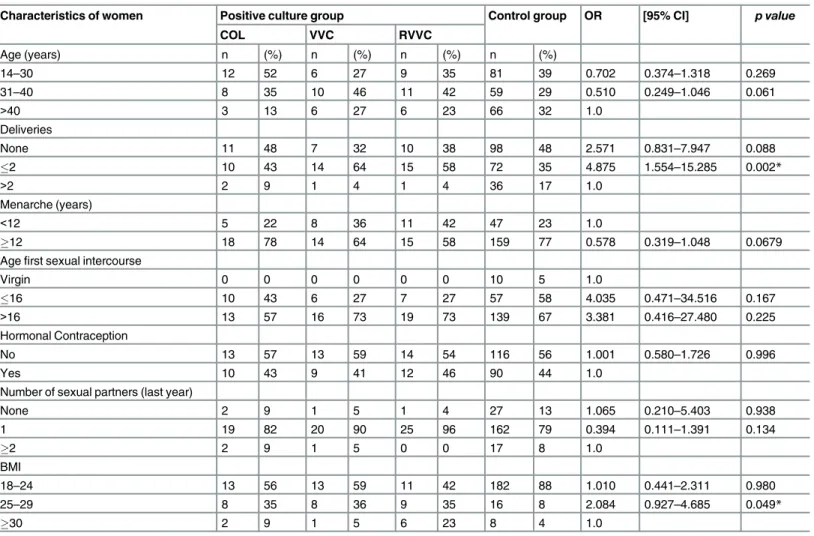
Table 1shows the epidemiological characteristics of the 277 women (mean age 33.5±8.95 years) included in the study. Women who had two or fewer births and overweight women were significantly more associated with a positive culture forCandidaspp. (OR 2.084; 95% CI 0.927–4.685;p= 0.049) and (OR 4.875; 95% CI 1.554–15.285;p= 0.002, respectively).
Vaginal yeasts were isolated in 71 (25.6%) of the 277 women. Women with a negative cul-ture composed the control group (n = 206; mean age 34±10.04 years). With respect to clini-cal profiles, 23 (32.4%) of the women with a positive culture were classified as COL (mean age 31±8.87 years), 22 (31.0%) as VVC (mean age 35±7.66 years), and 26 (36.6%) as RVVC (mean age 34±9.24 years). The mean ages were similar among women of all clinical profiles (p>0.05). Complete data on the isolation and identification of the yeasts were obtained
from all 71 women with a positive culture, as follows:C.albicans(n = 66/71; 93%) and Can-didanon-C.albicansspecies (n = 5/71; 7%).C.parapsilosiswas the most frequentCandida
non-C.albicansspecies isolated (n = 3/71, 4.2%), followed byC.tropicalisandC.glabrata
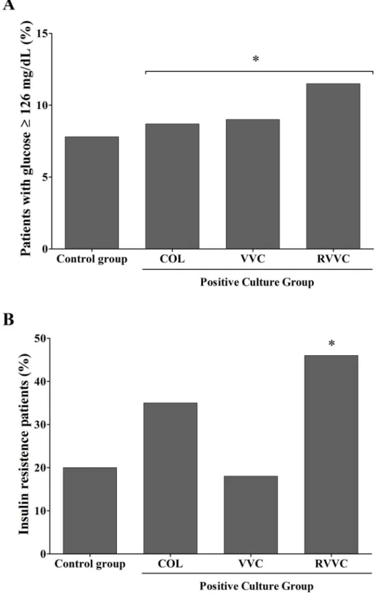
The mean values for glycemia were similar in all groups: control (83.40 ±12.47 mg/dL), COL (82±10.45 mg/dL), VVC (89.7±33.61 mg/dL), and RVVC (88.65± 15.9 mg/dL).
Abnor-mal glycemia (100 mg/dL) was detected in 16 women (7.8%) of the control group, in 2
(8.7%) of the COL, in 2 (9%) of VVC, and in 3 (11.5%) of RVVC. The HOMA Index (HI) indi-cating insulin resistance was detected in 42 women (20%) of the control group, in 8 (34.8%) of the COL group, in 4 (18%) of VVC, and in 12 (46%) of RVVC. Compared to the control group, women with a positive culture (COL, VVC and RVVC combined) were associated with an abnormal glucose metabolism (OR 4.6; 95% CI 2.0–10.9;p= 0.0002;Fig 2A) and insulin resis-tance (OR 2.2; 95% CI 1.05–4.84;p= 0.00317;Fig 2B).
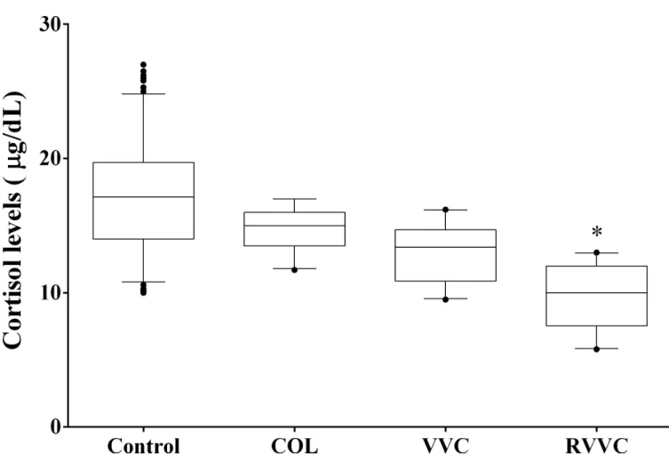
For early-morning cortisol levels, the median (25th-75th percentile) observed were: 17.15μg/dL (14.00–19.73μg/dL) in women of the control group, 15.00μg/dL (13.50–16.00μg/
dL) of the COL group, 13.40μg/dL (10.88–14.70μg/dL) of the VVC group, and 10.00μg/dL
(7.55–12.00μg/dL) of RVVC. Women of the RVVC group showed lower mean cortisol level
than the control and COL groups (p<0.001;Fig 3).
Fig 1. Flow chart of the study.
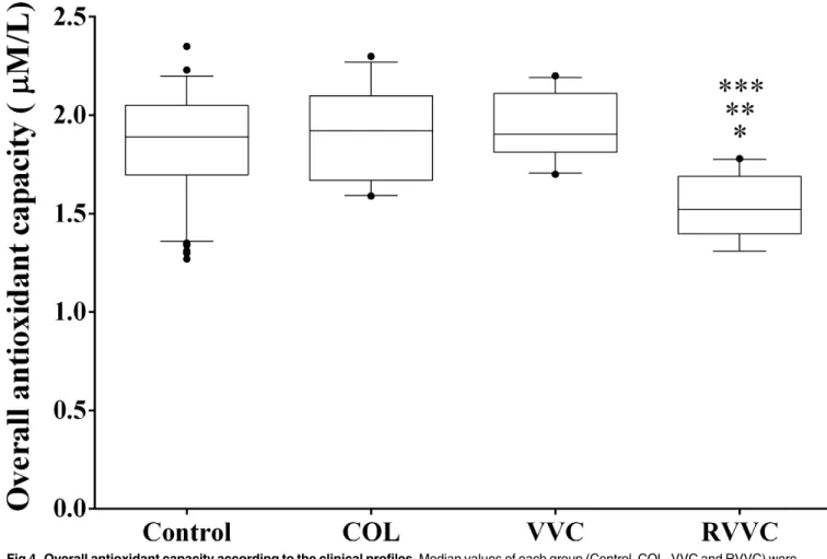
The overall median (25th-75th percentile) levels of antioxidant capacity were: 1.89 mM/L (1.69–2.05 mM/L) in the control group, 1.92 mM/L (1.67–2.10 mM/L) in the COL group, 1.90 mM/L (1.81–2.11 mM/L) in VVC, and 1.52 mM/L (1.39–1.69 mM/L) in RVVC. Women of the Table 1. Selected epidemiologic characteristics of 277 women who participated of study.
Characteristics of women Positive culture group Control group OR [95% CI] p value
COL VVC RVVC
Age (years) n (%) n (%) n (%) n (%)
14–30 12 52 6 27 9 35 81 39 0.702 0.374–1.318 0.269
31–40 8 35 10 46 11 42 59 29 0.510 0.249–1.046 0.061
>40 3 13 6 27 6 23 66 32 1.0
Deliveries
None 11 48 7 32 10 38 98 48 2.571 0.831–7.947 0.088
2 10 43 14 64 15 58 72 35 4.875 1.554–15.285 0.002*
>2 2 9 1 4 1 4 36 17 1.0
Menarche (years)
<12 5 22 8 36 11 42 47 23 1.0
12 18 78 14 64 15 58 159 77 0.578 0.319–1.048 0.0679
Agefirst sexual intercourse
Virgin 0 0 0 0 0 0 10 5 1.0
16 10 43 6 27 7 27 57 58 4.035 0.471–34.516 0.167
>16 13 57 16 73 19 73 139 67 3.381 0.416–27.480 0.225
Hormonal Contraception
No 13 57 13 59 14 54 116 56 1.001 0.580–1.726 0.996
Yes 10 43 9 41 12 46 90 44 1.0
Number of sexual partners (last year)
None 2 9 1 5 1 4 27 13 1.065 0.210–5.403 0.938
1 19 82 20 90 25 96 162 79 0.394 0.111–1.391 0.134
2 2 9 1 5 0 0 17 8 1.0
BMI
18–24 13 56 13 59 11 42 182 88 1.010 0.441–2.311 0.980
25–29 8 35 8 36 9 35 16 8 2.084 0.927–4.685 0.049*
30 2 9 1 5 6 23 8 4 1.0
COL, vaginal colonization; VVC, vulvovaginal candidiasis; RVVC, recurrent vulvovaginal candidiasis; BMI, body mass index; OR, Odds Ratio; 95% CI, confidence interval.
*p<0 .05 was considered significant
doi:10.1371/journal.pone.0158870.t001
Table 2. Relative distribution ofCandida albicansandCandidanon-C.albicansspecies according to the clinical profile.
Species Total (N = 71) COL (N = 23) VVC (N = 22) RVVC (N = 26) p value
n (%) n (%) n (%) n (%)
C.albicans 66 93 19 82.7 21 95.5 26 100 0.049*
CNCA C.parapsilosis 5 3 7 60 4 2 17.3 8.7 1 1 4.5 4.5 -
-C.glabrata 1 20 1 4.3 - - -
-C.tropicalis 1 20 1 4.3 - - -
-Total 71 100 23 100 22 100 26 100
COL, vaginal colonization; VVC, vulvovaginal candidiasis; RVVC, recurrent vulvovaginal candidiasis. CNCA,Candidanon-C.albicans.
*p<0.05 was considered significant
Fig 2. Abnormal glycemia and insulin resistance in 277 women studied. A.Percentage of women with abnormal glycemia (126 mg/dL) in control group compared to positive vaginal yeast culture group
RVVC group had a significantly lower overall median level of antioxidant capacity than the control group (p<0.0001), the COL group (p<0.0001), and also VVC (p<0.0001;Fig 4).
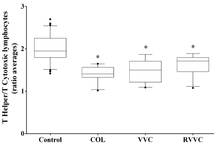
The median (25th-75th percentile) T helper/T cytotoxic lymphocyte ratios were: 1.95 (1.79– 2.25) in the control group, 1.41 (1.33–1.56) in COL, 1.50 (1.21–1.71) in VVC; and 1.71 (1.46– 1.79) in RVVC. The COL, VVC and RVVC group had lower T helper/T cytotoxic lymphocyte ratios than the control group (p<0.0001 for all;Fig 5).
Cytology of Pap showed a similar vaginal inflammatory process in women of the RVVC
and control groups (OR 1.96; 95% CI 0.83–3.87;p= 0.188) while women of the VVC and COL
groups showed a more-intense inflammatory process than the control group (OR 9.47; 95% CI 4.03–16.44;p= 0.0001) and (OR 5.22; 95% CI 1.97–8.69;p= 0.0003;Fig 6A).
Bacterioscopy showed that women in the COL and RVVC groups had similar normal
Lacto-bacillusspp. vaginal microbiota to the control group (OR 1.429; 95% CI 0.593–3.44;P= .5223)
and (OR 0.5896; 95% CI 0.2346–1.482;p= 0.2815, respectively). Women of the VVC group
group compared to control group. Positive culture group had more women with insulin resistance than control group (*p= 0.0002).
doi:10.1371/journal.pone.0158870.g002
Fig 3. Morning cortisol levels according to the women clinical profiles.Median values of each group (Control, COL, VVC and RVVC) were calculated and compared using a Kruskal-Wallis and subsequently Dunn’s post-hoc test with all pairwise comparisons identified which groups
differed. Box plots show the median, 25th and 75th percentiles of the cortisol levels. Whiskers extend to the 5th and 95th percentiles; filled circles represent outliers. Cortisol levels were determined in triplicate employing chemiluminescent microparticle immunoassay in the Architect-i1000SR Immunoassay Analyzer (Abbott, Illinois, USA).*p =0.001, shows that women of the RVVC group has a lower mean cortisol level than the control and COL groups.
showed fewer vaginal lactobacilli compared to women of the COL and RVVC groups (OR 2.914; 95% IC 0.8634–9.836;p= 0.0404, and OR 2.712; 95% CI 0.8275–8.810;p= 0.0480, respectively;Fig 6B).
Discussion
In this study, the early-morning cortisol level and overall antioxidant capacity were lower in women with RVVC than in women of the control and COL groups. This suggests that chronic stress and reduced antioxidant capacity may be specific predisposing factors related to the pathogenesis of RVVC. In addition, other factors such as diabetes mellitus and insulin resis-tance were related to positive vaginal yeast culture but not related to a specific clinical profile (COL, VVC and RVVC). These data support our hypothesis concerning the importance of chronic stress and reduced antioxidant capacity as specific predisposing factors for the devel-opment of RVVC.
Attenuated levels of morning cortisol and hyporesponsiveness of the hypothalamus-pitui-tary-adrenal axis have been shown to be indicative of chronic stress [13–15]. Also, stress may increase the risk for impaired function of the immune processes [16]. Clinical observations and Fig 4. Overall antioxidant capacity according to the clinical profiles.Median values of each group (Control, COL, VVC and RVVC) were calculated and compared using a Kruskal-Wallis and subsequently Dunn’s post-hoc test with all pairwise comparisons identified which groups differed. Box plots show the median, 25th and 75th percentiles of the overall median level of antioxidant capacity. Whiskers extend to the 5th and 95th percentiles; filled circles represent outliers. The overall antioxidant capacity was determined in triplicate by Trolox equivalent antioxidant activity (TEAC), to evaluate plasma non-enzymatic antioxidants. Women with RVVC had significantly lower mean levels of antioxidant capacity than control (*p<0.0001), COL (**p<0.0001) and VVC (***p<0.0001) groups.
experimental findings have further emphasized the role of chronic stress as an important trig-gering factor of many pathologies including atopic dermatitis,systemic lupus erythematosus, asthma, allergic rhinitis andCandidainfections [13–17]. These studies are in agreement with our results, which indicated a relationship between chronic stress and vulvovaginal candidiasis (RVVC and VVC). Despite these results being in line with earlier findings that correlate lower levels of early-morning cortisol with RVVC [17,18], it is possible that stress may be a predis-posing factor for RVVC as well as a response to repeated infections.
A reduced overall antioxidant capacity has also been associated with immune deficiency, since its mechanism of action seems to be the modulation of signal transduction factors involv-ing transcribinvolv-ing cells and immune-mediated cytokine production [19,20]. Additionally, it is important to consider that the elimination ofCandidais a cell-mediated immune responses by the recruitment and activation of neutrophils, which respond to a proinflammatory cytokine [19–21]. The reduced overall antioxidant capacity, and the altered immune status in the RVVC group compared to all other groups, suggest that impaired immunity plays a role in the patho-genesis of RVVC.
Fig 5. T helper/T cytotoxic lymphocytes ratio according to the clinical profiles.Median values of each group (Control, COL, VVC and RVVC) were calculated and compared using a Kruskal-Wallis and subsequently Dunn’s post-hoc test with all pairwise comparisons identified which groups differed. Box plots show the median, 25th and 75th percentiles of the overall median level of antioxidant capacity. Whiskers extend to the 5th and 95th percentiles; filled circles represent outliers. T helper/T cytotoxic lymphocytes ratio was determined in triplicate performed on a BD-FACS Calibur flow cytometer equipped with an argon laser and BD Multiset software for data analysis (Becton Dickinson TriTEST
immunofluorescence, Becton Dickinson, San José, California, USA).*p<0.0001, the COL, VVC and RVVC group had lower T helper/T cytotoxic lymphocyte ratios than the control group.
Health benefits have also been attributed to antioxidants, including a reduced risk of coro-nary vascular diseases and some types of cancer [22]. In their etiology, these chronic diseases seem to result from cellular oxidative damage, caused by pro-oxidative agents such as free radi-cals, affecting lipids, proteins and DNA [7,8]. Dietary antioxidants may protect against these oxidative events in the body in cases where insufficient levels of endogenous antioxidants can-not counteract the reactive species [22]. It is possible that an increase in antioxidant defense provided by dietary vitamins and minerals may prevent RVVC.
In this study, we assessed the immune status by determining the lymphocyte T helper/T cytotoxic ratio in blood. Our results showed that women with RVVC had reduction in systemic cell-mediated immunity (CMI) responsiveness and the cytological findings indicated that women with RVVC had a lower local inflammatory response than the women in other groups with a positive culture (Fig 6). These data are in accordance with the findings of Fidel et al. [23] who showed that a local rather than a systemic CMI represents important host-defense mecha-nisms in the vaginal mucosa. Thereby, additional efforts and studies with the local T-cell count, besides other strategies such as interleukin genotyping, will be needed to assess the CMI responsiveness of patients with RVVC.
Diabetes mellitus has long been considered as one of the factors causingCandidavaginitis, mainly due to the changes induced by hyperglycemia, including decreased random motion of neutrophils, chemotaxis, phagocytosis, and microbial death [24–26]. High blood glucose lev-els promote yeast attachment and growth, and also interfere with immune responses in the host [27]. However, in many cases there is a lack of reliable and clinically relevant informa-tion proving diabetes mellitus as a risk factor to RVVC [28]. We recently showed that diabe-tes mellitus type 2 in Brazilian women was associated with vaginal yeast colonization, VVC and RVVC [29]. Donders et al [26] found impaired glucose tolerance in women with RVVC, compared to controls. However, we found no relationship between RVVC and diabetes mel-litus. One hypothesis that partly explains this is that the increased glucose at the vaginal level could be more important than hyperglycemia in the pathogenesis of RVVC, because it would increase the adhesion and growth of yeasts as previously showed [26]. Still,Nyirjesyet al. [24] proposed that women with type 2 diabetes mellitus are at increased risk for vaginal Can-didacolonization because of glucosuria, which may also to justify our results. However, as we did not evaluate the vaginal and urinary glucose levels, additional studies are needed to examine such hypothesis.
The findings of studies on the impact of vaginal lactobacilar microbiota on the control of VVC and RVVC are contradictory [30,31]. Our results suggest that the vaginal communities of women with RVVC did not have reduced proportions of lactobacilli. These findings are consis-tent with others that also showed, using culture-dependent methods, that the development of RVVC was not correlated with an abnormal vaginal microbiota [31].
Fig 6. Cervical-vaginal cytology and bacterioscopy findings related to inflammation process and vaginal microbiota, respectively.A. Percentage of women with inflammatory processes demonstrated in microscopic analysis (400X) of vaginal cytology stained by Papanicolaou, according to clinical profiles. Samples were considered positive for inflammatory processes when smears showed leukocyte and cellular-inflammation characteristics of moderate and/or intense grade, in at least 20 different fields under optical microscopy at 400x magnification. Women with RVVC had a similar rates of vaginal inflammatory process in relation to women of control group (p= 0.188; OR = 1.96; 95% CI, 0.83–0.87%), while more women with VVC and COL had a vaginal inflammatory process than the control ones (*p= 0.0001; OR = 9.47; 95% CI, 4.03–16.44) and (*p= 0.0003; OR = 5.22; 95% CI, 1.97–8.69). B. Percentage of women with abundant vaginalLactobacillusspp. by bacterioscopy, according to clinical profiles. Women with RVVC had no reduction in the quantities of
Lactobacillusspp. compared to control group (p= 0.5223; OR = 1.429; 95% CI, 0.593–3.44).
Finally, we are making a substantial assumption that both chronic stress (decreased early-morning cortisol levels) and decreased overall antioxidant capacity are present in women with RVVC. In addition, we have considered these data as host predisposing factors for developing recurrent status. In fact, our findings provided important information to better understand this pathology and lead to improved care for women with RVVC. However, this speculation was made based in a cross-sectional study, and this kind of design implies a limitation regarding to statements made. Despite these data alone are unable to reach results that would qualify as definitive, they supply a strong possibility. For a definitive conclusion, a longitudinal study, where women’s cortisol levels be tested during an infection process, following the treatment, and in periods of remission, must be carried on.
Financial disclosure: The authors did not report any potential conflict of interest.
Author Contributions
Conceived and designed the experiments: LAG PSBM MELC. Performed the experiments: PSBM GT SM MMTI. Analyzed the data: MELC LAG PSBM TIES. Contributed reagents/ materials/analysis tools: MELC TIES LAG. Wrote the paper: TIES MELC PSBM LAG.
References
1. Cassone A. VulvovaginalCandida albicansinfections: pathogenesis, immunity and vaccine prospects.
BJOG 2015; 122:785–94. doi:10.1111/1471-0528.12994PMID:25052208
2. Sobel JD. Recurrent vulvovaginal candidiasis. Am J Obstet Gynecol 2015; 15:716–24.
3. Sobel JD. Vulvovaginal candidosis. Lancet 2007; 369:1961–71. PMID:17560449
4. Sandini S, La Valle R, Deaglio S, Malavasi F, Cassone A, De Bernardis F. A highly immunogenic recombinant and truncated protein of the secreted aspartic proteases family (rSap2t) ofCandida albi-cansas a mucosal anticandidal vaccine. FEMS Immunol Med Microbiol 2011; 62:215–24. doi:10.1111/ j.1574-695X.2011.00802.xPMID:21535228
5. Muzny CA, Schwebke JR. Biofilms: an underappreciated mechanism of treatment failure and recur-rence in vaginal infections. Clin Infect Dis 2015; 61:601–6. doi:10.1093/cid/civ353PMID:25935553
6. Bassyouni RH, Wegdan AA, Abdelmoneim A, Said W, AboElnaga F. Phospholipase and aspartyl pro-teinase activities ofCandidaspecies causing vulvovaginal candidiasis in patients with type 2 diabetes
mellitus. J Microbiol Biotechnol 2015; 25:01–22.
7. Ratti BA, Godoy JS, de Souza Bonfim-Mendonça P, Bidóia DL, Nakamura TU, Nakamura CV, et al. Microbicidal activity of neutrophils is inhibited by isolates from recurrent vaginal candidiasis(RVVC) caused byCandida albicansthrough fungal thioredoxin reductase. Cell Immunol 2015; 293:22–9. doi: 10.1016/j.cellimm.2014.11.004PMID:25497972
8. Bonfim-Mendonça PS, Ratti BA, Godoy JS, Negri M, Lima NC, Fiorini A, et al.β-glucan induces reac-tive oxygen species production in human neutrophils to improve the killing ofCandida albicansand
Candida glabrataisolates from vulvovaginal candidiasis. Plos One 2014; 9:e107805. doi:10.1371/
journal.pone.0107805PMID:25229476
9. Odds FC, Webster CE, Mayuranathan P, Simmons PD.Candidaconcentrations in the vagina and their association with signs and symptoms of vaginal candidosis. J Med Vet Mycol 1988; 26: 277–83. PMID: 3236147
10. Kurtzman CP, Fell JW. The yeast, a taxonomic study. New York (NY): Elsevier; 1998.
11. Nayar R, Wilbur CD. The Bethesda System for reporting cervical cytology: definitions, criteria and explanatory notes. Cham (Switzerland): Springer International Publishing; 2015.
12. American Diabetes Association (ADA). Standards of medical care in diabetes-2015: summary of revi-sions. Diabetes Care 2015; 38(suppl 1):S4.
13. Wallace TM, Levy JC, Mathews DR. Use and abuse of HOMA modeling. Diab Care 2004; 27:1487–95.
14. Re R, Pellegrini R, Proteggente A, Pannala A, Yang M, Rice-Evans C. Antioxidant activity applying an improved ABTS radical cation decolorization assay. Free Rad Biol Med 1999; 26:1231–7. PMID:
10381194
16. Straub RH. Neuroendocrine immunology: new pathogenetic aspects and clinical application. Z Rheu-matol 2011; 70:767–74. doi:10.1007/s00393-011-0784-8PMID:21956825
17. Ehrstrom SM, Kornfeld D, Thuresson J, Rylander E. Signs of chronic stress in women with recurrent candida vulvovaginitis. Am J Obstet Gynecol 2005; 4:1376–81.
18. Meyer H, Goettlicher S, Mendling W. Stress as a cause of chronic recurrent vulvovaginal candidosis and the effectiveness of the conventional antimycotic therapy. Mycoses 2006; 49:202–9. PMID: 16681811
19. Richardson M, Rautemaa R. How the host fights againstCandidainfections. Front Biosci 2009; 14:4363–75.
20. Fidel PL Jr. History and update on host defense against vaginal candidiasis. Am J Reprod Immunol 2007; 57:2–12. PMID:17156186
21. Amouri I, Hadrich I, Abbes S, Sellami H, Ayadi A. Local humoral immunity in vulvovaginal candidiasis. Ann Biol Clin 2013; 71:151–5.
22. Smith RA, Adlam VJ, Blaikie FH, Manas AR, Porteous CM, James AM, et al. Mitochondria-targeted antioxidants in the treatment of disease. Ann N Y Acad Sci 2008; 1147:105–11. doi:10.1196/annals. 1427.003PMID:19076435
23. Fidel PLJ, Lynch ME and Sobel JD. Circulating CD4 and CD8 T cells have little impact on host defense against experimental vaginal candidiasis. Infect Immun 1995; 63:2403–8. PMID:7790050
24. Nyirjesy P, Zhao Y, Ways K, Usiskin K. Evaluation of vulvovaginal symptoms andCandidacolonization in women with type 2 diabetes mellitus treated with canagliflozin, a sodium glucose co-transporter 2 inhibitor. Curr Med Res Opin 2012; 28:1173–8. doi:10.1185/03007995.2012.697053PMID:22632452
25. Carrara MA, Bazotte RB, Donatti L, Svidzinski TI, Consolaro ME, Patussi EV, et al. Effect of experimen-tal diabetes on the development and maintenance of vulvovaginal candidiasis in female rats. Am J Obstet Gynecol 2009; 200:659.e1-4.
26. Donders GG, Prenen H, Verbeke G, Reybrouck R. Impaired tolerance for glucose in women with recur-rent vaginal candidiasis. Am J Obstet Gynecol 2002; 187:989–93. PMID:12388993
27. Nyirjesy P, Sobel JD. Genital mycotic infections in patients with diabetes. Postgrad Med 2013; 125:33–
46. doi:10.3810/pgm.2013.05.2650PMID:23748505
28. Buchta V, Matula V, Kestřánek J, Vejsová M, Křivčíková L, Spaček J. Is diabetes mellitus a risk factor in
genital yeast infections?. Ceska Gynekol 2013; 78:537–44. PMID:24372432
29. Gunther LS, Martins HP, Gimenes F, Abreu AL, Consolaro ME, Svidzinski TI. Prevalence ofCandida
albicansand non-albicans isolates from vaginal secretions: comparative evaluation of colonization,
vaginal candidiasis and recurrent vaginal candidiasis in diabetic and non-diabetic women. São Paulo Med J 2014; 132:116–20. PMID:24714993
30. Liu M- B, Xu S-R, He Y, Deng G-H, Sheng H-F, Huang XM, et al. Diverse vaginal microbiomes in repro-ductive-age women with vulvovaginal candidiasis. Plos One 2013; 8:e79812. doi:10.1371/journal. pone.0079812PMID:24265786
31. Zhou X, Westman R, Hickey R, Hansmann MA, Kennedy C, Osborn TW, et al. Vaginal microbiota of women with frequent vulvovaginal candidiasis. Infect Immun. 2009; 77:4130–5. doi:10.1128/IAI.