Submitted 30 April 2015
Accepted 12 July 2015
Published13 August 2015
Corresponding author Sergios-Orestis Kolokotronis, [email protected]
Academic editor Keith Crandall
Additional Information and Declarations can be found on page 11
DOI10.7717/peerj.1147
Copyright
2015 Ammazzalorso et al.
Distributed under
Creative Commons CC-BY 4.0
OPEN ACCESS
To beat or not to beat a tick: comparison
of DNA extraction methods for ticks
(
Ixodes scapularis
)
Alyssa D. Ammazzalorso1,*, Christine P. Zolnik1,2,*, Thomas J. Daniels2 and Sergios-Orestis Kolokotronis1
1Department of Biological Sciences, Fordham University, Bronx, NY, USA
2Vector Ecology Laboratory, Louis Calder Center–Biological Field Station, Fordham University, Armonk, NY, USA
∗These authors contributed equally to this work.
ABSTRACT
Background.Blacklegged ticks (Ixodes scapularis) are important disease vectors in the United States, known to transmit a variety of pathogens to humans, including bacteria, protozoa, and viruses. Their importance as a disease vector necessitates reliable and comparable methods for extracting microbial DNA from ticks. Furthermore, to explore the population genetics or genomics of this tick, appropriate DNA extraction techniques are needed for both the vector and its microbes. Although a few studies have investigated different methods of DNA isolation from ticks, they are limited in the number and types of DNA extraction and lack species-specific quantification of DNA yield.
Methods.Here we determined the most efficient and consistent method of DNA extraction from two different developmental stages ofI. scapularis—nymph and adult—that are the most important for disease transmission. We used various methods of physical disruption of the hard, chitinous exoskeleton, as well as commercial and non-commercial DNA isolation kits. To gauge the effectiveness of these methods, we quantified the DNA yield and confirmed the DNA quality via PCR of both tick and microbial genetic material.
Results.DNA extraction using the Thermo GeneJET Genomic DNA Purification Kit resulted in the highest DNA yields and the most consistent PCR amplification when combined with either cutting or bead beating with select matrices across life stages. DNA isolation methods using ammonium hydroxide as well as the MoBio PowerSoil kit also produced strong and successful PCR amplification, but only for females. Discussion.We contrasted a variety of readily available methods of DNA extraction from single individual blacklegged ticks and presented the results through a quantitative and qualitative assessment.
Subjects Ecology, Entomology, Evolutionary Studies, Molecular Biology, Parasitology
Keywords Arthropod, Vector-borne, Blacklegged tick, Tick, DNA extraction, Nucleic acids, DNA quantification
INTRODUCTION
life cycle, the tick acquires a bloodmeal at each developmental stage (i.e., larva, nymph, and adult) prior to molting or egg-laying, in the case of adult females. These ticks are of great public health importance as pathogen vectors because they carry and transmit a variety of human disease agents, such asBorrelia burdgorferi, the causative agent of Lyme disease,Anaplasma phagocytophilumwhich causes human granulocytic anaplasmosis, and
Babesia microti, a protozoan responsible for the malaria-like illness, babesiosis (Speilman, 1976;Steere, Broderick & Malawista, 1978;Pancholi et al., 1995). Recently,I. scapularishas been found to transmitBorrelia miyamotoi(Scoles et al., 2001) and Powassan virus lineage 2 (a.k.a., Deer Tick Virus) (Telford et al., 1997). Their importance as human pathogen vectors necessitates research that involves successful isolation of genetic material needed in investigations of both the vector itself and of the wide range of pathogens that they carry. However, DNA isolation in ticks is challenging due to the hard chitinous exoskeleton and the small amount of microbial nucleic acids present (Halos et al., 2004). Furthermore, tick DNA is suseptible to degradation (Hubbard, Cann & Wright, 1995;Hill & Gutierrez, 2003; Halos et al., 2004) and PCR can be challenged by inhibitors (Halos et al., 2004).
Although DNA extraction from ticks for both pathogen isolation and tick genetic and genomic research is performed routinely by researchers, there is no consensus regarding the most effective method of DNA isolation from any tick species. A few such studies and reviews have been conducted (Hill & Gutierrez, 2003;Halos et al., 2004;Crowder et al., 2010;Mtambo et al., 2006;Sparagano et al., 1999); however, they are limited to a handful of extraction techniques, and quantitative data on DNA concentration is lacking. In this study we aim to identify the optimal DNA isolation procedure for both tick and microbial DNA from an important tick pathogen vector, the blacklegged tick.
MATERIALS & METHODS
Tick collection
Nymphal and adult female blacklegged ticks are important life stages in the transmission of disease agents compared to larvae, which are rarely infected with human pathogens, and adult males, whose brief feeding bouts minimize the risk of pathogen transmission (Piesman et al., 1986;Falco & Fish, 1988;Falco et al., 1999). Thus, the ability to dependably extract DNA, and in particular microbial DNA, from nymphal and adult female blacklegged ticks is of importance to tick-borne disease research and constitutes the focus of this study.
DNA isolation
We contrasted the efficiency of extracting DNA from ticks stored in 70% v/v ethanol using five different DNA isolation procedures coupled with either cross-sectional division or bead-based physical disruption of the tick body. These procedures included four commercially available DNA extraction kits: DNeasy Blood & Tissue Kit (QIAGEN, Valencia, California, USA), GeneJET Genomic DNA Purification Kit (Thermo Scientific, Waltham, Massachusetts, USA), Tissue & Insect DNA MicroPrep (Zymo Research, Irvine, California, USA), PowerSoil DNA Isolation Kit (MoBio, Carlsbad, California, USA); and one noncommercial DNA extraction method using ammonium hydroxide(NH4OH)and heat, which has been primarily used for DNA isolation from the European sheep tick,
Ixodes ricinus(Guy & Stanek, 1991;Rijpkema et al., 1996;Pichon et al., 2003;Humair et al., 2007;Pangr´acov´a et al., 2013).
All commercial kits examined here use a silica-based column procedure and have either been used in previous studies for DNA isolation in ticks or are marketed for efficient microbial DNA recovery or insect DNA isolation (Table 1). All kits also include filtering through a silica gel membrane and a variety of associated buffers. The Zymo kit and the NH4OH treatment did not include a protein digestion step. DNA extraction kits were used according to the manufacturers’ recommended protocols with few exceptions (Table 1), including a final elution completed following a 5-min room-temperature incubation in 100µl of deionized, sterilized, distilled water(sdH2O)for consistency. Each tick was air-dried to evaporate the ethanol prior to DNA extraction, as ethanol may inhibit PCR (Hubbard, Cann & Wright, 1995;Bessetti, 2007;Schrader et al., 2012). Due to the components similarity of the QIAGEN and Thermo kits, we used the same Proteinase-K incubation duration (overnight).
The NH4OH method included adding 150µl of a 0.7-M NH4OH solution to the tick sample in a 1.5-ml snap cap tube, and heating to 100◦C for 15 min. The solution was briefly centrifuged to concentrate fluid at the bottom and then was evaporated to 70–100µl by opening the tubes and heating at 100◦C for an additional 15 min. The solution was then centrifuged for 10 min at 10,000×gand the supernatant was collected and respun for 2 min at 10,000×g. The total supernatant was collected and stored at−20◦C.
Table 1 Methods of DNA isolation and physical disruption of tick samples.Only samples treated with the MoBio kit were processed on the GeneMate vortex mixer (BioExpress), while all remaining bead beating took place on the BeadBlaster 24 (Benchmark Scientific).
DNA extraction method Alterations to manufacturer protocols Physical disruption Bead beating
—speed and duration
Overnight incubation at 56◦C in lysis
buffer/proteinase K
Bisection (Nymphs), Quadrisection (Females)
N/A
QIAGEN DNeasy Blood & Tissue Kit
(cat. no. 69506) Elution in 100µl dsH20 with 5 min room temperature incubation
MP Bio Lysing Matrices 4 m/s 1.5 & 4.0 min Overnight incubation at 56◦C in lysis
buffer/proteinase K
Bisection (Nymphs), Quadrisection (Females)
N/A
Thermo GeneJET Genomic DNA Purification Kit
(cat. no. K0722) Elution in 100µl dsH20 with 5 min room temperature incubation
MP Bio Lysing Matrices 4 m/s 1.5 & 4.0 min Elution in 100µl dsH20 with 5 min
room temperature incubation
Bisection (Nymphs), Quadrisection (Females) followed by beating with Zymo beads
4 m/s 10 min
Zymo Research Tissue & Insect DNA MicroPrep
(cat. no. D6015) Zymo beads 4 m/s
10 min Elution in 100µl of dsH20 with 5 min
room temperature incubation
Bisection (Nymphs), Quadrisection (Females) followed by vortexing with MoBio garnet beads
3,200 rpm 10 min
MoBio PowerSoil DNA Isolation Kit
(cat. no. 12888) MoBio provided beads 3,200 rpm
10 min Initial volume of 150µl NH4OH
Final volume of 70–100µl dsH20
Bisection (Nymphs), Quadrisection (Females)
N/A
NH4OH
(Guy & Stanek, 1991;Pichon et al., 2003) Second centrifugation for 2 min at 10,000×g
MP Bio Lysing Matrices 4 m/s 1.5 & 4.0 min
Table 2 Bead matrices and their attributes.The composition, characteristics, and recommended uses for the different bead matrices tested are adapted from the manufacturers’ websites. (MP Bio:http://www.mpbio.com/index.php?cPath=2 77 425&country=223, MoBio:http://www.mobio. com/soil-dna-isolation/powersoil-dna-isolation-kit.html, Zymo: http://www.zymoresearch.com/dna/genomic-dna/solid-ffpe-tissue-dna/zr-tissue-insect-dna-miniprep).
Matrix Manufacturer Material Suggested use
G MP Bio 1.6 mm silicon carbide particles Samples with tough, hard, or brittle cell membranes
H MP Bio 2 mm glass beads & 2 mm zirconium oxide beads Tough, hard cells and organisms within dense exterior matrices, e.g., whole insects and ticks
I MP Bio 2 mm zirconium beads & one 4 mm ceramic sphere Very tough, hard samples including chitin exoskeletons, e.g., whole insects and ticks
M MP Bio Two 6.35 mm zirconium oxide-coated ceramic grinding spheres
Tough tissues, seeds, spores
S MP Bio 3.175 mm stainless steel beads Tough tissues, seeds, spores
Z MP Bio 2.0 mm yttria-stabilized zirconium oxide spheres Tough plant and animal samples
PowerBeads MoBio Garnet Environmental samples
BashingBead Lysis Matrix
Zymo Ceramic Ticks, mosquitoes, bees, lice, andDrosophila
marketed for tough samples or, as in the case of Matrices H and I, were marketed specifically for ticks (Table 2). We beat the ticks with the MP Bio Lysing Matrices for 1.5 and 4 min at 4 m/s (Table 1).
The variables that we maintained constant across DNA extraction methods were: bead beating speed (4 m/s), water volume for final DNA elution of 100µl, and a 5-min duration of incubation in sdH2O during DNA elution.
DNA was extracted from three nymphs and three females for all combinations of DNA isolation and physical disruption procedures, including the different bead beating durations and MP Bio Lysing Matrices. DNA was extracted using the MoBio and Zymo kits from six nymphs and six females each. Half were bisected or quadrisected and half remained whole prior to DNA extraction with one of these two kits. The QIAGEN, Thermo, and NH4OH methods were used to extract DNA from 39 nymphs and 39 females each. For every one of these three procedures, three ticks from each life stage were bisected or quadrisected and 36 ticks were bead-beaten. Among the 36 bead-beaten ticks processed per method, six nymphs and six females were beaten with one of the six MP Bio Lysing Matrices, half for 1.5 min and half for 4 min. Overall, DNA was isolated from 129 nymphs and 129 females for a total of 258 tick DNA extractions.
DNA quantification
The resulting DNA yields were quantified via double-stranded DNA (dsDNA) fluoro-metric quantitation on a Qubit 2.0 flurometer (Life Technologies, Norwalk, Connecticut, USA) using 10µl of extracted DNA template in 190µl of the High Sensitivty (HS) dsDNA assay.
PCR validation
The isolated DNA was validated using PCR amplification of tick mitochondrial and nuclear loci. We amplified the tick DNA using (1) the cytochromecoxidase subunit 1 (Cox1) DNA barcode region (∼650 bp) located on the mitochondrial genome with the HCO/LCO primers (Folmer et al., 1994), and (2) a dinucleotide (CA)nmicrosatellite repeat located on the nuclear genome with the bac7ea/bac7eb primer pair (139–197 bp) (Chan, 2012).
We targeted the genusRickettsiausing PCR to validate the successful extraction of microbial DNA from inside the tick, which is necessary for studies involving PCR detection of human pathogens transmitted by this tick species. Members of this genus are obligate intracellular bacteria and are abundant in blacklegged ticks (Benson et al., 2004;Moreno et al., 2006;Noda, Munderloh & Kurtti, 1997;Steiner et al., 2008). We targeted a 532-bp fragment of theompAgene usingRickettsia-specific primers (Vitorino et al., 2007).
72◦C for 30 s; 30 cycles of 95◦C for 20 s, 50◦C for 25 s, and 72◦C for 30 s; and a final extension for 5 min at 72◦C (Chan, 2012). TheRickettsia ompAlocus was amplified with an initial denaturation at 95◦C for 5 min, 35 cycles of 95◦C for 30 s, 52◦C for 30 s, and 72◦C for 30 s, and a final extension for 7 min at 72◦C (Vitorino et al., 2007). All PCR reactions were performed using a Techne Prime Elite Thermal Cycler (Bibby Scientific, Burlington, New Jersey, USA).
Each PCR was performed in a final 25-µl volume with 6.25µl 2×MyTaq HS Mix (Bioline, Taunton, Massachusetts, USA) and 0.2µM of each forward and reverse primer. The PCRs targeting nuclear and mitochondrial tick DNA were caried out using 16.25 µl sdH2O and 1.5µl DNA template. To account for the lower DNA concentrations of microbial DNA within this tick, and specifically for nymphs, the PCRs targeting the
Rickettsia ompAfragment were carried out with 15.75µl sdH2O and 2µl DNA template
from nymphs, and 16.75µl sdH2O and 1µl DNA template for adult females. PCRs were confirmed by agarose gel electrophoresis on a 1.5% w/v gel based on amplicon length.
Statistical analysis
Given the substantial differences in size and DNA yield between nymphs and adult females, their DNA concentrations were not comparable. Therefore, statistical analyses of the DNA concentration data from the two developmental stages were performed separately. Following a one-way ANOVA, Tukey’s HSD test was used for post-hoc analysis of the average DNA quantification values resulting from nymph bisection or female quadrisection across the five DNA isolation methods (QIAGEN, Thermo, MoBio, Zymo, and NH4OH). The Bonferroni correction was used to account for multiple comparisons. The same procedures were also used to compare the DNA yields resulting from the different MP Bio Lysing Matrices (G, H, I, M, S, and Z) and nymph bisection or female quadrisection methods using data from the highest-yielding DNA extraction method. The effect of bead beating duration was assessed through a two-tailed Student’st-test.
RESULTS AND DISCUSSION
Comparison of DNA yield based on bisection and quadrisection
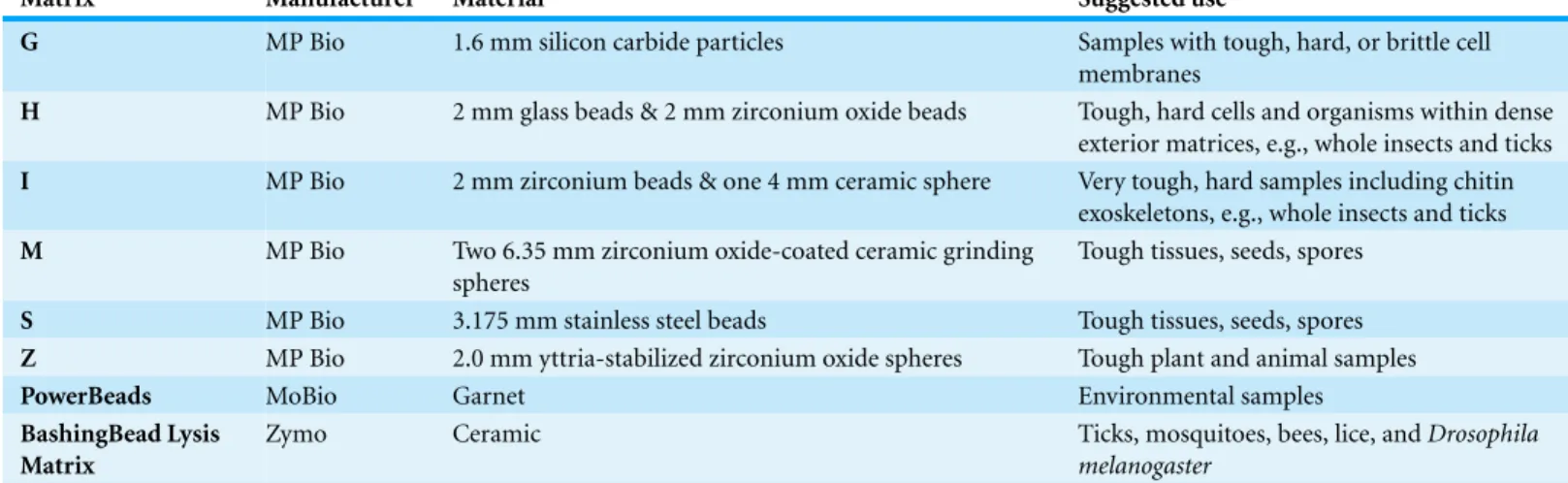
The comparison of DNA concentrations resulting from nymph bisection and female quadrisection across the five DNA extraction methods yielded no apparent differences between the QIAGEN DNEasy Blood & Tissue Kit and the Thermo GeneJET Genomic DNA Purification Kit (Fig. 1). For both nymphs and adult females, the QIAGEN and Thermo methods resulted in markedly higher DNA yields than all other methods (Fig. 1,
Table 3). The Zymo Research Tissue & Insect DNA MicroPrep Kit produced DNA yields that were too low to be quantifiable by the Qubit fluorometer in the case of nymphs and were extremely low in the case of adult females (average=0.02 ng/µl, SD=0.01).
Comparison of DNA yield based on bead beating
Figure 1 DNA concentrations (ng/µl) resulting from the five DNA extraction methods following
nymph bisection and female quadrisection as determined using the Qubit HS dsDNA Assay.Each sample set consisted of three individual tick DNA extractions. Note the difference in scale between the life stages. (A) Nymphs (B) Adult Females.
Table 3 Average DNA concentration (ng/µl) of whole and cut nymphal and adult female blacklegged
ticks.Average and standard deviation of the DNA concentration values determined using the Qubit HS dsDNA Assay. Unless otherwise indicated, samples were stored in 70% v/v ethanol. Three single-tick measurements were included in each treatment. All values listed as<0.0005 ng/µl indicate a reading of “too low” from the Qubit fluorometer.
Method Life stage Nymphs Adult females
Bisected Whole Quadrisected Whole
Average 1.70 9.41
QIAGEN
SD 0.640 Table S1 1.63 Table S1
Average 1.68 9.77
Thermo
SD 0.11 Table S1 2.35 Table S1
Average 0.070 0.0373 4.51 0.119
MoBio
SD 0.0585 0.0457 0.492 0.0133
Average <0.0005 <0.0005 3.14 0.0146
Zymo
SD 0.00 0.00 1.44 0.00341
Average 0.677 0.0240
NH4OH
SD 0.086 Table S1 0.0143 Table S1
lysing matrix bead beating results for whole nymphs and adult females treated with the QIAGEN, Thermo and NH4OH methods are detailled inTable S1.
PCR validation
The Thermo method exhibited the strongest and most consistent gel electrophoresis PCR product bands across physical disruption methods in the case of both nymphs and females for mtDNA (Cox1), nuDNA (microsatellite), and bacterial DNA (Rickettsia ompA). The DNA extractions resulting from the NH4OH protocol yielded strong and mostly consistent amplification of the tick mitochondrial and nuclear loci, and the bacterial gene for adult females. Although the NH4OH method yielded lower DNA concentration results than the QIAGEN and Thermo kits when using bead-beaten or quadrisected adult female ticks, this did not affect PCR success. However, PCR amplification was consistently poor for nymphs treated with the NH4OH protocol. Consistent with the very low DNA yields we measured, the DNA extracted using the Zymo kit did not result in any successful PCRs for ticks (either nymphs or adults) and only produced amplicons forRickettsia ompAfrom quadrisected females.
DNA extraction from adult female ticks with the MoBio kit resulted in the consistent amplification of the three targeted loci, with quadrisection substantially enhancing the amplicon gel band quality in contrast to whole females (beaten with garnet on a vortex). Those loci were not successfully amplified in the case of bisected and whole nymphs—concordant with low DNA yields.
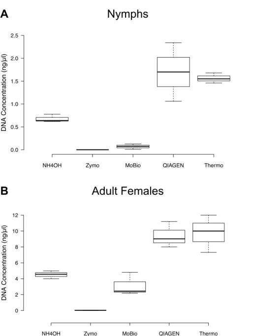
Figure 2 DNA concentrations (ng/µl) resulting from the Thermo DNA extraction method following
the bead beating of whole ticks.Bead beating was carried out with each of the MP Bio lysing matrices (G, H, I, M, S, and Z). Nymphs were bisected and adult females were quadrisected. The DNA concentration was determined using the Qubit HS dsDNA Assay. Six nymphs and six adults were used in each bead beating treatment, while three nymphs and three adults were used in each cutting treatment. (A) Nymphs (B) Adult Females.
In terms of rapidly quantifying PCR success, we can report the proportion of positive PCRs, while acknowledging that this is merely based on PCR product band presence on an agarose gel, rather than accounting for band intensity. The latter characteristic can be discussed on case-by-case basis, rather than being summarized in subjective fluorescence categories, e.g., faint, medium, strong. In terms of DNA extraction methods, the Thermo kit yielded the most “amplifiable” DNA (201 positive PCRs out of 234, i.e., 85.9% success rate) with comparable performance across developmental stages (87.18% of adult females vs. 84.62% of nymphs). The two other methods that used bead beating (QIAGEN and NH4OH) did not perform as well overall (61.11% and 58.55%, respectively) and further-more exhibited a skewed behavior across developmental stage with PCRs from female adult ticks working more consistently (72.65% of females vs. 49.57% of nymphs for QIAGEN; 85.47% of females vs. 31.62% of nymphs in NH4OH). PCRs using the DNA extracted with the MoBio kit worked well with female adults (88.89%) but not with nymphs (11.11%). The Zymo kit was the most poorly performing in terms of PCR with only three successful reactions out of 36 (8.33%) with none of the nymphal samples producing amplified PCR products. Those DNA isolation and physical disruption methods that were most successful for the nuclear and mitochondrial tick gene PCRs were also most effective for theRickettsia ompAgene amplification. All of this information is detailed inTable S2, yet we raise cau-tion against the strict interpretacau-tion of those results, as part of a full assessment should be the PCR amplicon yield (i.e., number of PCR fragments) that, during gel electrophoresis, is manifested by fluorescence intensity. Quantitative PCR can be used for such purposes.
CONCLUSIONS
Successful DNA extraction from tick species is important for both genetic and genomic studies of the tick vector itself, as well as for studies aimed at detecting pathogen presence in these tick vectors. This study was designed to determine the most reliable and efficient method of DNA extraction, including physical disruption of the tick exoskeleton.
Among the tested tick DNA extraction procedures, we recommend several different procedures depending on budget, time, contamination concerns, and study goals. The Thermo kit is recommended for its high DNA yields coupled with high-quality PCR amplification with both bead beating and nymph bisection or adult quadrisection, as well as for its lower cost in comparison with the QIAGEN kit—a similarly structured kit. The QIAGEN kit may be used if already available when cutting ticks, but is not recommended if bead beating is the chosen physical disruption strategy.
The NH4OH extraction method is an inexpensive alternative to commercially available kits and produced high-quality PCR products for adult females, although the DNA yield was generally lower than that of commercial kits. However, this method was not useful for DNA extraction from nymphs, resulting in low DNA yield and poor to non-existent PCR amplification, despite the frequent use of this method in studies on nymphalIxodes ricinus
The MP Bio H and I matrices, which MP Bio recommends for ticks, as well as the S matrix were best for bead beating in conjunction with the Thermo kit for nymphs and females or NH4OH for females. These results confirm MP Bio’s recommendation of the H and I matrices for ticks, and we would add to that list matrix S. Benefits of bead beating include less processing time and reduced direct sample handling, which may decrease the likelihood of contamination. However, cutting ticks requires less expensive equipment than bead beating.
The Zymo kit was a poor choice for extracting DNA from nymphs and adult female ticks, although it is marketed for DNA extraction from stored arthropods, including ticks (as per the manufacturer’s websitehttp://www.zymoresearch.com/dna/genomic-dna/ solid-ffpe-tissue-dna/zr-tissue-insect-dna-kits/zr-tissue-insect-dna-microprep). The MoBio kit also performed poorly with nymphs but showed suceess with adult females.
Admittedly, the sample size of three nymph and three female replicates for each combination of DNA extraction variables is limited. Additional replicates may better capture the potential variability in DNA concentration and quality resulting from each extraction procedure. Future studies with larger sample sizes would be useful to further assess the efficacy of these different methods with perhaps greater precision, although there is no reason to expect that increasing the number of replicates would necessarily yield globally different results. Our results clearly demonstrate that DNA yield quality varies among different extraction kits and methods, which can have an important impact on the success of PCR-based studies.
Our study expands on previous work that determined DNA extraction success from ticks based on PCR amplification alone, without a DNA quantification assessment (Halos et al., 2004). While a recent study (Crowder et al., 2010) quantified DNA yield, the reported values were averaged across multiple tick species and focused only on one developmental stage—adults. In order to test the efficiency of the DNA extraction techniques, we kept certain variables constant, such as the long-term storage method, bead beating speed, elution volume, and incubation time prior to elution. Alteration of these variables may result in increased DNA yield and should be considered when DNA concentration is important in downstream applications, such as high-throughput sequencing, pathogen surveillance, and microbial community profiling.
ACKNOWLEDGEMENTS
We thank Rachel Engstrand for assistance with R. We thank the reviewers and the editor for their insightful comments and suggestions that helped us shape the final version of this paper.
ADDITIONAL INFORMATION AND DECLARATIONS
Funding
Grant Disclosures
The following grant information was disclosed by the authors: Fordham University Summer Science Internship.
Competing Interests
The authors declare there are no competing interests.
Author Contributions
• Alyssa D. Ammazzalorso performed the experiments, analyzed the data, wrote the paper, prepared figures and/or tables, reviewed drafts of the paper.
• Christine P. Zolnik conceived and designed the experiments, performed the experi-ments, analyzed the data, wrote the paper, prepared figures and/or tables, reviewed drafts of the paper.
• Thomas J. Daniels contributed reagents/materials/analysis tools, wrote the paper, reviewed drafts of the paper.
• Sergios-Orestis Kolokotronis conceived and designed the experiments, analyzed the data, contributed reagents/materials/analysis tools, wrote the paper, prepared figures and/or tables, reviewed drafts of the paper.
Supplemental Information
Supplemental information for this article can be found online athttp://dx.doi.org/ 10.7717/peerj.1147#supplemental-information.
REFERENCES
Benson MJ, Gawronski JD, Eveleigh DE, Benson DR. 2004.Intracellular symbionts and other bacteria associated with deer ticks (Ixodes scapularis) from Nantucket and Wellfleet, Cape Cod, Massachusetts.Applied and Environmental Microbiology70:616–620 DOI 10.1128/AEM.70.1.616-620.2004.
Bessetti J. 2007.An introduction to PCR inhibitors.Profiles in DNA10:9–10.
Chan CTW. 2012.Comparative analysis of microsatellite and mitochondrial genetic variations in
Ixodes scapularis. MS Thesis, Georgia Southern University.
Crowder CD, Rounds MA, Phillipson CA, Picuri JM, Matthews HE, Halverson J, Schutzer SE, Ecker DJ, Eshoo MW. 2010. Extraction of total nuclec acids from ticks for the
detection of bacterial and viral pathogens. Journal of Medical Entomology47:89–94 DOI 10.1093/jmedent/47.1.89.
Falco RC, Fish D. 1988.Ticks parasitizing humans in a Lyme disease endemic area of southern New York State.American Journal of Epidemiology128:1146–1152.
Falco RC, McKenna DF, Daniels TJ, Nadelman RB, Nowakowski J, Fish D, Wormser GP. 1999.Temporal relation betweenIxodes scapularisabundance and risk for Lyme disease associated with erythema migrans. American Journal of Epidemiology149:771–776 DOI 10.1093/oxfordjournals.aje.a009886.
Guy EC, Stanek G. 1991.Detection ofBorrelia burgdorferiin patients with Lyme disease by the polymerase chain reaction.Journal of Clinical Pathology44:610–611DOI 10.1136/jcp.44.7.610. Halos L, Jamal T, Vial L, Maillard R, Suau A, Le Menach A, Boulouis HJ, Vayssier-Taussat M.
2004.Determination of an efficient and reliable method for DNA extraction from ticks.
Veterinary Research35:709–713DOI 10.1051/vetres:2004038.
Hill CA, Gutierrez JA. 2003. A method for extraction and analysis of high quality genomic DNA from ixodid ticks. Medical and Veterinary Entomology 17:224–227 DOI 10.1046/j.1365-2915.2003.00425.x.
Hubbard MJ, Cann KJ, Wright DJ. 1995.Validation and rapid extraction of nucleic acids from alcohol-preserved ticks.Experimental and Applied Acarology19:473–478.
Humair PF, Douet V, Mor´an Cadenas F, Schouls LM, Van De Pol I, Gern L. 2007.Molecular identification of bloodmeal source inIxodes ricinusticks using 12S rDNA as a genetic marker.
Journal of Medical Entomology44:869–880DOI 10.1093/jmedent/44.5.869.
Moreno CX, Moy F, Daniels TJ, Godfrey HP, Cabello FC. 2006.Molecular analysis of microbial communities identified in different developmental stages ofIxodes scapularisticks from Westchester and Dutchess counties, New York.Environmental Microbiology8:761–772 DOI 10.1111/j.1462-2920.2005.00955.x.
Mtambo J, Van Bortel W, Madder M, Roelants P, Backeljau T. 2006.Comparison of preservation methods ofRhipicephalus appendiculatus(Acari: Ixodidae) for reliable DNA amplification by PCR.Experimental and Applied Acarology38:189–199DOI 10.1007/s10493-006-0004-4. Noda H, Munderloh UG, Kurtti TJ. 1997.Endosymbionts of ticks and their relationship to
Wolbachiaspp. and tick-borne pathogens of human and animals.Applied and Environmental Microbiology63:3926–3932.
Pancholi P, Kolbert CP, Mitchell PD, Reed Jr KD, Dumler JS, Bakken JS, Telford 3rd SR, Persing DH. 1995.Ixodes damminias a potential vector of human granulocytic ehrlichiosis.
Journal of Infectious Diseases172:1007–1012DOI 10.1093/infdis/172.4.1007.
Pangr´acov´a L, Derd´akov´a M, Pek´arik L, Hviˇsˇcov´a I, V´ıchov´a B, Stanko M, Hlavat´a H, Peˇtko B. 2013.Ixodes ricinusabundance and its infection with the tick-borne pathogens in urban and suburban areas of Eastern Slovakia.Parasites & Vectors6:238DOI 10.1186/1756-3305-6-238. Pichon B, Egan D, Rogers M, Gray J. 2003.Detection and identification of pathogens and
host DNA in unfed host-seekingIxodes ricinus.Journal of Medical Entomology40:723–731 DOI 10.1603/0022-2585-40.5.723.
Piesman J, Donahue JG, Mather TN, Spielman A. 1986.Transovarially acquired Lyme disease spirochetes (Borrelia burgdorferi) in field-collected larvalIxodes dammini(Acari: Ixodidae).
Journal of Medical Entomology23:219–219DOI 10.1093/jmedent/23.2.219.
Rijpkema S, Golub´c D, Molkenboer M, Verbeek-De Kruif N, Schellekens J. 1996.Identification of four genomic groups ofBorrelia burgdorferi sensu latoinIxodes ricinusticks collected in a Lyme borreliosis endemic region of northern Croatia.Experimental and Applied Acarology 20:23–30DOI 10.1007/BF00130550.
Schrader C, Schielke A, Ellerbroek L, Johne R. 2012.PCR inhibitors–occurrence, properties and removal. Journal of Applied Microbiology 113:1014–1026 DOI 10.1111/j.1365-2672.2012.05384.x.
Sparagano OA, Allsopp MT, Mank RA, Rijpkema SG, Figueroa JV, Jongejan F. 1999.Molecular detection of pathogen DNA in ticks (Acari: Ixodidae): a review.Experimental and Applied Acarology23:929–960DOI 10.1023/A:1006313803979.
Speilman A. 1976.Human babesiosis on Nantucket Island: transmission by nymphalIxodesticks.
American Journal of Tropical Medicine and Hygiene25:784–787.
Steere AC, Broderick TF, Malawista SE. 1978.Erythema chronicum migrans and Lyme arthritis: epidemiologic evidence for a tick vector.American Journal of Epidemiology108:312–321. Steiner FE, Pinger RR, Vann CN, Grindle N, Civitello D, Clay K, Fuqua C. 2008.Infection
and co-infection rates ofAnaplasma phagocytophilumvariants,Babesiaspp.,Borrelia burgdorferi, and the rickettsial endosymbiont inIxodes scapularis(Acari: Ixodidae) from sites in Indiana, Maine, Pennsylvania, and Wisconsin.Journal of Medical Entomology45:289–297 DOI 10.1093/jmedent/45.2.289.
Telford 3rd SR, Armstrong PM, Katavolos P, Foppa I, Garcia AS, Wilson ML, Spielman A. 1997. A new tick-borne encephalitis-like virus infecting New England deer ticks,Ixodes dammini.
Emerging Infectious Diseases3:165–170DOI 10.3201/eid0302.970209.
Vitorino L, Chelo IM, Bacellar F, Z´e-Z´e L. 2007.Rickettsiae phylogeny: a multigenic approach.