RESEARCH ARTICLE
Leishmania
(
L.
)
mexicana
Infected Bats in
Mexico: Novel Potential Reservoirs
Miriam Berzunza-Cruz1‡, Ángel Rodríguez-Moreno2‡, Gabriel Gutiérrez-Granados2,
Constantino González-Salazar3,4, Christopher R. Stephens3,5, Mircea Hidalgo-Mihart6,
Carlos F. Marina7, Eduardo A. Rebollar-Téllez8, Dulce Bailón-Martínez1, Cristina
Domingo Balcells2, Carlos N. Ibarra-Cerde
ña9, Víctor Sánchez-Cordero2*,
Ingeborg Becker1 *
1Unidad de Investigación en Medicina Experimental, Facultad de Medicina, Universidad Nacional Autónoma de México, Mexico City, México,2Instituto de Biología, Universidad Nacional Autónoma de México, México D.F., México,3C3—Centro de Ciencias de la Complejidad, Universidad Nacional Autónoma de México, México D.F., México,4Comisión Nacional para el Conocimiento y Uso de la Biodiversidad, México D.F., 14010, México,5Instituto de Ciencias Nucleares, Universidad Nacional Autónoma de México, México D.F., México,6División Académica de Ciencias Biológicas, Universidad Juárez Autónoma de Tabasco, Villahermosa, México,7Centro Regional de Investigación en Salud Pública —INSP, Tapachula, Chiapas, México,8Laboratorio de Entomología Médica, Departamento de Zoología de Invertebrados, Facultad de Ciencias Biológicas, Universidad Autónoma de Nuevo León, San Nicolás de Los Garza, México,9Departamento de Ecología Humana, Centro de Investigación y de Estudios Avanzados del Instituto Politécnico Nacional (CINVESTAV), Unidad Mérida, Mérida, Yucatán, México
‡These authors contributed equally to this work. *victor@ib.unam.mx(VSC);becker@unam.mx(IB)
Abstract
Leishmania (Leishmania) mexicanacauses cutaneous leishmaniasis, an endemic zoonosis
affecting a growing number of patients in the southeastern states of Mexico. Some foci are found in shade-grown cocoa and coffee plantations, or near perennial forests that provide rich breeding grounds for the sand fly vectors, but also harbor a variety of bat species that live off the abundant fruits provided by these shade-giving trees. The close proximity be-tween sand flies and bats makes their interaction feasible, yet bats infected withLeishmania (L.) mexicanahave not been reported. Here we analyzed 420 bats from six states of Mexico
that had reported patients with leishmaniasis. Tissues of bats, including skin, heart, liver and/or spleen were screened by PCR forLeishmania (L.) mexicanaDNA. We found that 41
bats (9.77%), belonging to 13 species, showed positive PCR results in various tissues. The infected tissues showed no evidence of macroscopic lesions. Of the infected bats, 12 spe-cies were frugivorous, insectivorous or nectarivorous, and only one spespe-cies was sanguivor-ous (Desmodus rotundus), and most of them belonged to the family Phyllostomidae. The eco-region where most of the infected bats were caught is the Gulf Coastal Plain of Chiapas and Tabasco. Through experimental infections of twoTadarida brasiliensisbats in captivity,
we show that this species can harbor viable, infectiveLeishmania (L.) mexicanaparasites
that are capable of infecting BALB/c mice. We conclude that various species of bats belong-ing to the family Phyllostomidae are possible reservoir hosts forLeishmania (L.) mexicana, if
it can be shown that such bats are infective for the sand fly vector. Further studies are need-ed to determine how these bats become infectneed-ed, how long the parasite remains viable
PLOS Neglected Tropical Diseases | DOI:10.1371/journal.pntd.0003438 January 28, 2015 1 / 15 OPEN ACCESS
Citation:Berzunza-Cruz M, Rodríguez-Moreno Á, Gutiérrez-Granados G, González-Salazar C, Stephens CR, Hidalgo-Mihart M, et al. (2015)
Leishmania(L.)mexicanaInfected Bats in Mexico: Novel Potential Reservoirs. PLoS Negl Trop Dis 9(1): e0003438. doi:10.1371/journal.pntd.0003438
Editor:Paul Andrew Bates, Lancaster University, UNITED KINGDOM
Received:July 18, 2014
Accepted:November 25, 2014
Published:January 28, 2015
Copyright:© 2015 Berzunza-Cruz et al. This is an open access article distributed under the terms of the
Creative Commons Attribution License, which permits unrestricted use, distribution, and reproduction in any medium, provided the original author and source are credited.
Data Availability Statement:All relevant data are within the paper and its Supporting Information Files.
inside these potential hosts and whether they are infective to sand flies to fully evaluate their impact on disease epidemiology.
Author Summary
Leishmaniasis is endemic in southeastern Mexico, whereLeishmania(L.)mexicanais the principle parasite species causing disease in humans. Previous studies using methodology for inferring potential biotic interactions between species had shown a high prevalence of bat species as potential hosts forLeishmania, yet bats have not been identified as hosts in Mexico. Motivated by these predictions, we currently analyze whether bats are infected withLeishmania (L.) mexicanaand to what extent they serve as hosts for this parasite. We analyzed 420 bats from six states, and through PCR analysis of skin, heart, liver and/or spleen we found that 41 bats (9.77%) of 13 different bat species were infected. Most of the infected bats were netted in an eco-region characterized by plantations grown beneath shade-providing fruit trees that favors breeding conditions for the sand fly vectors and brings them in close proximity with frugivorous, insectivorous or nectarivorous bats. None of the infected bat tissues showed macroscopic lesions. Through artificial infections, we were also able to show thatLeishmania (L.) mexicanaremains viable and infective after passage through one species of bat. We conclude that various species of bats belonging to the family Phyllostomidae are possible reservoir hosts ofLeishmania(L.)mexicana.
Introduction
Many infectious diseases are zoonoses [1], where both vector and host play a crucial role in the transmission cycle of the pathogen. However, although many pathogens can have multiple hosts, there have been few systematic experimental studies conducted to identify host ranges and the relative importance of the hosts within them, as well as no systematic methods for pre-dicting them. A systematic approach is important, as many pathogens have a potentially broad host range [2]. Additionally, the characteristics of the set of hosts of a zoonosis will play a fun-damental role in determining the optimal interventions for combating the zoonosis.
Recently, a general methodology was presented [3] for inferring potential biotic interactions between species that can be used for identifying potential disease hosts and their relative im-portance. The method was used to predict the most important potential hosts forLeishmania (L.) mexicanain Mexico. Notable among the predictions was the high prevalence of bat species identified as important potential hosts, especially given that bats have not been previously iden-tified as hosts in Mexico.
Leishmania (L.) mexicanais widely distributed in Mexico and the highest prevalence is found in the southeastern states of Campeche, Chiapas, Quintana Roo, Tabasco and Veracruz [4]. The disease is transmitted by the bite of phlebotomine sand flies and can infect a variety of wild and domestic mammals, whereas humans are accidental hosts [5,6]. Cutaneous leishmani-asis caused byLeishmania (L.) mexicanawas first described in chicle collectors of the Yucatan peninsula in 1912 by Seidelin, hence its name“Chiclero´s Ulcer”[7]. The parasite is transmit-ted by various sand fly species (genusLutzomyia). Known hosts include dogs [8,9] as well as various small mammals, such asHeteromys gaumeri,Heteromys desmarestianus, Reithrodont-omys gracilis,Marmosa mexicana,Sigmodon hispidusandOryzomys melanotis. Furthermore,
Ototylomys phyllotisandPeromyscus yucatanicus, endemic rodents in southeast Mexico, have
Bats Infected withLeishmania mexicanain Mexico
PLOS Neglected Tropical Diseases | DOI:10.1371/journal.pntd.0003438 January 28, 2015 2 / 15
been incriminated as potential reservoirs based on their geographic and temporal distributions that overlap with those of vectors and patients as well as their life expectancy. The infection caused byLeishmania (L.) mexicanain these rodents is relatively benign. Although the preva-lence of infected hosts can vary greatly throughout the season, the proportion of animals that become infected during their lifetime can exceed 20% [10–12].
Mexico is ranked among the top countries worldwide with the highest mammalian species richness, holding approximately 525 species, 137 of which belong to the order Chiroptera (bats). Since bat can be a potential feeding source for sand flies, it has been suspected that they might also be potentialLeishmaniahosts [13–15]. The first evidence of direct interactions between the parasite and bats were reported forLeishmania chagasiisolated fromCarollia perspicillata(9.09% prevalence) [16]. Most of the previous studies onLeishmaniaspp. Infec-tions in bats revealed low prevalence: Savaniet al. Studied 659 bats belonging to 28 species in Brazil and reported 3.2% prevalence of infections withLeishmania (L.) amazonensisand
Leishmania (L.) infantum chagasi(in the studies done by PCR), Mutinga reported 3% preva-lence and Millánet al. Found no evidence ofLeishmaniainfections in cave bats of an endemic area in Spain. These data contrast with studies by Shapiroet al. That report 40% prevalence in an endemic area in Brazil [13,17,18,19].
In Mexico bats have not been studied as potential hosts forLeishmaniainfections. In this paper we present the results of an experimental study, motivated by the predictions of Stephens
et al. (2009), to determine the extent to which bats can and do serve as hosts forLeishmania (L.) mexicanain Mexico [3].
Methods
Study area
The study was conducted in 22 localities including the States of Tabasco, Chiapas, Jalisco, Mi-choacán, Nuevo León and Veracruz. As part of a project on the dynamics of national emerging zoonosis [3], grids with cells corresponding to 25 x 25 km were analyzed. Cells were stratified according to altitudes (<2000 m or>2000 m), aiming to assign at least 80% of the cells for the
analysis from the category<2000 m. The cells were also categorized into 4 types by land use
and vegetation types (bodies of water, natural or modified vegetation, rural or urban settle-ments) and excluding cells with>50% bodies of water or urban population. Fieldwork was
conducted within each 25 x 25 km in randomly selected cells.
Bat sampling
Bats were captured using 13 mist nets of 3, 6 and 12 m, opened from 18:00 h to 24:00 h during 34 nights between February 2009 and October 2010, totaling 2,448 nights/mist-net. Mist nets were set in a diversity of habitats, including small streams, pools, along vegetation borders, vegetation clearings, cattle-rearing and crops production lands, roads and other sites where bat flying was observed. In the State of Chiapas, 276 bats were netted (65.71%), 252 of which were collected in the eco-region of the Gulf Coastal Plain, whereas 24 were netted in the mountains of the northern part of the state. In Tabasco, 104 bats (24.76%) were netted in the Gulf Coastal Plain, whereas 17 (4%) were netted in Veracruz, 16 (3.8%) in Jalisco, 2 (0.48%) in Michoacán and 5 (1.19%) in Nuevo León. Widely distributed bat species such asArtibeus jamaicensisand
Sturnira liliumwere netted in four states, whereasDesmodus rotundus,Carollia sowelliand
Sturnira ludoviciwere only collected in three states.Artibeus lituratus,Dermanura phaeotis,
C.perspicillata,Choeroniscus godmani,Glossophaga soricina,Pteronotus parnelli,Pteronotus personatusandMyotis nigricansare also widely distributed species, however, they were only netted in two of the collection sites and an additional 22 species were netted only in one of the
Bats Infected withLeishmania mexicanain Mexico
six states. All captured bats were taxonomically identified to species, and sex was determined. Standard body measurements and weights were recorded. Captured bats were sacrificed ac-cording to the published guidelines and protocols of the American Society of Mammalogists [20]. All pregnant females were released at site of capture. The extracted tissues from bats (heart, liver, spleen and skin) were fixed in 90% ethanol or frozen in liquid nitrogen for PCR analysis.
DNA purification
DNA from tissues was purified from approximately 25 mg of starting material. The DNA extractions were done with a DNeasy1
Blood and Tissue kit (QIAGEN, Germany), following the manufacturer’s instructions. The genomic DNA was used for PCR-based amplification.
Oligonucleotides
Leishmaniainfections were analyzed with two sets of primers: the first set of primers was used to detect the presence of the genusLeishmaniaand a second set of primers was used to deter-mine theLeishmaniaspecies. The primers to detectLeishmaniagenus were 1S / L.MC-1R based onLeishmaniaminicircle kinetoplast DNA sequences, which is conserved among species (L.MC-1S:5´-CTRGGGGTTGGTGTAAAATAG-3´; and L.MC-1R:5´-TWTGAACGG GRTTTCTG-3´) [21]. For the identification ofLeishmania (L.) mexicanaspecies, the primer IR1, which corresponds to the 32 final nucleotides of the conserved sequences from the 3’ re-gion of the small subunit of the 18S ribosomal gene (5’- GCT GTA GGT GAA CCT GCA GCA GCT GGA TCA TT-3’), was used as forward primer [22]. The reverse primer used was LM17 (50-CCC CTC TCC TCCTCC CC-30) [23].
Polymerase chain reaction amplification
The PCR was performed using 50μl of the following reaction mixture:TaqPCR Master Mix (QIAGEN, Germany) (containing a premixed solution ofTaqDNA Polymerase, PCR buffer, MgCl2and dNTPs), 100 ng of the corresponding oligonucleotides, and DNA from tissues (we used 1μl of tissue extract corresponding to 100 ng of DNA). The amplification was performed in a Perkin Elmer 2720 thermocycler using various conditions, which depended on the oligo-nucleotides used.Leishmaniagenus was determined with primers L.MC-1S/L.MC-1R and PCR amplification was performed using 30 cycles of denaturation (95°C for one minute), an-nealing (55°C for one minute), and polymerization (72°C for one minute). The sensitivity anal-ysis for the primers L.MC-1S/L.MC-1R was made with DNA from culturedLeishmania (L.) mexicanapromastigotes using: 10 ng, 1 ng, 100 pg, 10 pg, 1 pg, 100 fg, 10 fg and 1 fg DNA, and evidenced by an amplification band of 700 bp. The sensitivity forLeishmaniaDNA in tissues permitted the detection of 100 fg DNA (corresponding to 1 parasite). All the tissues that tested positive forLeishmaniagenus were additionally analyzed with primers specific forLeishmania (L.) mexicanaspecies (with an amplification band of 790 bp), to determine whether bats were infected with the sameLeishmania (L.) mexicanaspecies as that found in patients of these en-demic regions.Leishmania (L.) mexicanaspecies was determined with primers IR1/LM17 using 35 cycles of 1 min at 94°C, 1 min at 65°C and 1 min at 72°C. In all cases, the cycles were preceded by another cycle at 94°C for 5 min and a final extension cycle of 72°C for 7 min. PCR products were analyzed by electrophoresis in 1.5% agarose gels in TAE 1X at 80 V. Gels were stained with 0.5μg/ml ethidium bromide and photographed under a UV light source.
For additional confirmation ofLeishmania (L.) mexicanainfections, the amplifications ob-tained with primers IR1/LM17 in 2 infected bats were sequenced. For this, the PCR products were purified using the QIAquickTMPCR purification kit (QIAGEN, Germany) and
Bats Infected withLeishmania mexicanain Mexico
sequencing was done at the Molecular Biology Unit of the Instituto de Fisiología Celular, UNAM. The Automated DNA sequencing was carried out on capillary-based electrophoresis sequencers. The Unit uses an ABI Prism 310 (1-capillar) Genetic Analyzer from Applied Bio-systems, with Big Dye Terminator Cycle Sequencing chemistry. The sequences were aligned with those from the National Center for Biotechnology Information, U.S. National Library of Medicine, Basic Local Alignment Search Tool (BLAST) and results confirmed that the bats were infected withLeishmania(L.)mexicana.
To analyze the permissiveness of bats toLeishmania (L.) mexicanainfections and to ascertain whether the parasites retained their infectivity after being exposed to the immune system of the bats, we artificially infected two individuals ofTadarida brasiliensis, which had been netted in Metztitlan, Hidalgo and kept in captivity. These animals were infected with 1x10⁶Leishmania (L.) mexicanapromastigotes (MHOM/MX/84/SET GS, isolated from a pa-tient with diffuse cutaneous leishmaniasis in Tabasco, Mexico) in the foot and kept in the animal facilities of the School of Medicine at UNAM, in the care of veterinary personnel ac-cording to the guidelines established by the American Society of Mammalogists [20]. After 30 days, the bats were sacrificed and fragments of the heart, liver, spleen and skin were placed in biphasic Novy-MacNeal-Nicolle (NNN) and RPMI-1640 (Gibco, Grand Island, NY) culture medium, supplemented with 10% fetal bovine serum. Cultures were analyzed daily for presence of promastigotes. Once the promastigotes appeared in the tissue cultures, they were analyzed by PCR to establish whether they wereLeishmania (L.) mexicana. To confirm that these pro-mastigotes conserved their infectivity, they were inoculated by intradermal injection into ear-lobes of male BALB/c mice aged to 6–8 weeks. After six weeks of infection, PCR analysis was done with the infected earlobes of BALB/c mice.
Ethics statement
The collection of specimens was performed according to the guidelines of the American Society for the Use of Mammalogists of Wild Mammals in Research and under a collecting permit is-sued by the General Direction of Wildlife of Mexico (permission number SGPA/DGVS/04631/ 14). The infections in mice were carried out following the National Ethical Guidelines for labo-ratory animals NOM-062-ZOO-1999. The project was approved by the Institutional Ethics Committee of the Medical Faculty of the National Autonomous University of Mexico (UNAM) with the registration number FMED/CI/RGG/013/01/2008.
Statistical analysis
We use a G-test to evaluate possible differences between the number of captured males and fe-males as well as of bats that tested positive forLeshmania (L.) mexicana, assuming a hypothesis of sexual ratio of 1:1 [24]. We additionally calculated the 95% confidence intervals based on the Wilson score [25].
Accession numbers
The gene sequences found in our study were aligned with genes reported in Genebank under the following accession numbers:Leishmania mexicanastrain MHOM/MX/94/INDRE NBO (AF466381.1);Leishmania mexicanastrain MHOM/MX/85/SOLIS (AJ000313.1);Leishmania mexicanaisolate 169 clone 1 (FJ948434.1);Leishmania mexicanastrain MHOM/MX/84/SET GS (AF466380.1);Leishmania mexicanastrain MHOM/MX/98/UNAM RR (AF466382.1);
Leishmania mexicanastrain MHOM/GT/86/GO22 (AJ000312.1);Leishmania mexicanaisolate 7 clone 1 (FJ948433.1);Leishmania mexicanastrain MNYC/BZ/62/M379 (AF466383.1); Leish-mania mexicanaisolate 169 clone 4 (FJ948436.1).
Bats Infected withLeishmania mexicanain Mexico
Results
We collected a total of 420 bats from 22 localities of six states, including 35 species of six fami-lies of Chiroptera. There were no statistical differences between the number of captured male (220) and female bats (200;χ2= 1.38;P= 0.23)
Polymerase chain reaction amplification
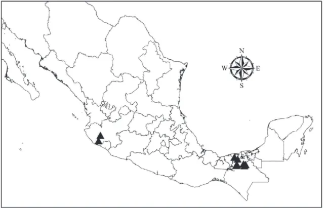
The PCR analysis of tissues taken from the heart, liver, spleen and skin of the 420 bats showed that 41 (9.77%) were positiveLeishmania. All the tissue samples that tested positive for the genusLeishmaniaalso showed positive results with specific primers forLeishmania (L.) mexi-canaspecies. Of the infected bats, 25 were males and 16 females (χ2= 1.97; P = 0.15), belonging to 13 species of six taxonomical families. There were no significant differences in the number of infected bats between the different families (Table 1). All the infected bats were caught in the Chiapas–Tabasco eco-region and Jalisco (Table 2,Fig. 1). 12 of the species were frugivorous, insectivorous or nectarivorous and only one species was sanguivorous (D.rotundus). There were significantly more infected bats than would be expected by random distribution in the fruit-feeding and nectar-feeding guilds (Table 1). Most of the infected bats from the eco-region of the Gulf Coastal Plain of Chiapas and Tabasco belonged to the family Phyllostomidae:Ch.
godmani(23.07% infected),Glossophaga commissarisi(75%),G.soricina(26.92%) andS.lilium
(11.11%) (Table 2). In Jalisco, only one individual of the speciesLeptonycteris curasoaeand one individual ofS.liliumwere infected withLeishmania(L.)mexicana. None of the bats netted in Michoacán, Nuevo León or Veracruz were infected.
None of the infected bats showed any dermal lesions and no apparent macroscopical lesions were found in the organs of the infected bats that tested positive forLeishmania(L.)mexicana. Only one of the bats from Tabasco (A.lituratus), which was found dead, showed dermal lesions suggestive of aLeishmaniainfection (Fig. 2). The PCR analysis of the dermal lesion of this ani-mal was positive forLeishmania(L.)mexicana, and although none of the additional tissues (heart, liver, spleen) of this animal showed lesions, they tested positive forLeishmania(L.)
mexicanaby PCR.
Table 1. 95% confidence intervals for number of species infected in different taxonomical families and trophic guilds.
Family Num. Species Num. Individuals Num. Infected Prevalence (Wilson Score) CI (95%)
Antrozoidae 1 1 0 N/A N/A
Emballonuridae 1 1 0 N/A N/A
Molossidae 2 2 0 N/A N/A
Mormoopidae 2 9 1 11 20–44
Phyllostomidae 26 402 40 10 7–13
Vespertilionidae 3 5 0 N/A N/A
Guild
Fruit-feeding 15 325 21 7* 4–10
Blood-feeding 2 14 1 7 1–32
Insect-feeding 14 26 8 30 6–48
Nectar-feeding 4 55 11 2* 12–32
N/A: not applicable,*Significant differences
doi:10.1371/journal.pntd.0003438.t001
Bats Infected withLeishmania mexicanain Mexico
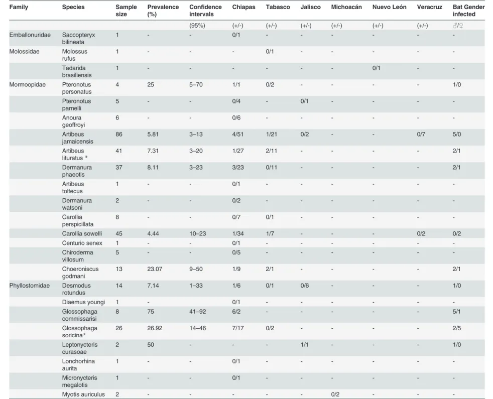
Table 2. Species and family of bats netted in different Mexican states.
Family Species Sample
size
Prevalence (%)
Confidence
intervals
Chiapas Tabasco Jalisco Michoacán Nuevo León Veracruz Bat Gender infected (95%) (+/-) (+/-) (+/-) (+/-) (+/-) (+/-) ♂/♀ Emballonuridae Saccopteryx
bilineata 1 - - 0/1 - - -
-Molossidae Molossus
rufus 1 - - - 0/1 - - - -
-Tadarida
brasiliensis 1 - - - 0/1 -
-Mormoopidae Pteronotus
personatus 4 25 5–70 1/1 0/2 - - - - 1/0
Pteronotus
parnelli 5 - - 0/4 - 0/1 - - -
-Anoura
geoffroyi 6 - - 0/6 - - -
-Artibeus
jamaicensis 86 5.81 3–13 4/51 1/21 0/2 - - 0/7 5/0
Artibeus lituratus*
41 7.31 3–20 1/27 2/11 - - - - 2/1
Dermanura
phaeotis 37 8.11 3–23 3/23 0/11 - - - - 2/1
Artibeus
toltecus 1 - - 0/1 - - -
-Dermanura
watsoni 2 - - 0/2 - - -
-Carollia
perspicillata 8 - - 0/7 0/1 - - - -
-Carollia sowelli 45 4.44 10–23 1/34 1/7 - - - 0/2 0/2
Centurio senex 1 - - 0/1 - - -
-Chiroderma
villosum 5 - - 0/5 - - -
-Choeroniscus
godmani 13 23.07 9
–50 1/9 2/1 - - - - 2/1
Phyllostomidae Desmodus
rotundus 14 7.14 1
–33 1/6 0/1 0/6 - - - 1/0
Diaemus youngi 1 - 0/1 - - -
-Glossophaga commissarisi
8 75 41–92 6/2 - - - 5/1
Glossophaga soricina*
26 26.92 14–46 7/17 0/2 - - - - 2/5
Leptonycteris curasoae
2 50 - - - 1/1 - - - 1/0
Lonchorhina aurita
1 - - 0/1 - - -
-Micronycteris megalotis
1 - - 0/1 - - -
-Myotis auriculus 2 - - - 0/2 - -
Table 2. (Continued)
Family Species Sample
size
Prevalence (%)
Confidence
intervals
Chiapas Tabasco Jalisco Michoacán Nuevo León Veracruz Bat Gender infected
Myotis velifer 3 - - - 0/3 -
-Phyllostomus
discolor 1 100 - 1/0 - - - 1/0
Platyrrhinus
helleri 5 - - 0/5 - - - -
-Sturnira lilium* 63 11.11 6–21 4/20 2/28 1/0 - - 0/8 3/4
Sturnira ludovici 25 4.0 1–20 0/14 1/6 0/4 - - - 0/1
Uroderma bilobatum
4 - - 0/4 - - -
-Vampyrodes caraccioli
1 - - 0/1 - - -
-Antrozoidae Antrozus pallidus
1 - - - 0/1 -
-Eptesicus fuscus
1 - - 0/1 - - -
-Vespertilionidae Myotis keaysi 2 - - - 0/2 - - - -
-Myotis nigricans 2 - - 0/1 0/1 - - - -
-Total # per state 420 - - 30/246 9/95 2/14 0/2 0/5 0/17 25/16
List shows the numbers ofLeishmania-infected and non-infected bats, sample size, prevalence, 95% confidence intervals as well as the percentage of infected bats according to sex.
*Species that have been reported in the literature to be infected withLeishmania.
doi:10.1371/journal.pntd.0003438.t002
Bats
Infected
with
Leishman
ia
me
xicana
in
Mexico
PLOS
Neglected
Tropical
Diseas
es
|DOI:10.13
71/journa
l.pntd.000343
8
January
28,
2015
8
Permissiveness of bats to parasites
The permissiveness of bats to harborLeishmania (L.) mexicanawas shown in twoT.
brasiliensisindividuals, which were artificially infected in captivity with 1x10⁶Leishmania (L.)
mexicanapromastigotes. No visible damage was observed in the tissues, yet promastigotes were recovered from heart and liver tissue fragments of these infected bats and culturedin
Figure 1. Black triangles show geographic localization of the sites in which infected bat species were collected.
doi:10.1371/journal.pntd.0003438.g001
Figure 2. Dermal lesions in bats suggestive ofLeishmaniainfection.A.lituratusfound dead in Tabasco, Mexico, showing dermal lesions on the edge of the wing membrane, suggestive of a Leishmaniainfection.
doi:10.1371/journal.pntd.0003438.g002
Bats Infected withLeishmania mexicanain Mexico
vitro. No parasites were recovered from the skin or spleen of these animals. The PCR analysis confirmed the presence ofLeishmania (L.) mexicanain the infected tissues. The parasites re-covered from the bat tissues were able to infect BALB/c mice, showing dermal lesions in the earlobes after six weeks of infection. PCR analysis confirmed the presence ofLeishmania (L.) mexicanain the infected mice. We were thus able to prove that this bat species is able to harbor
Leishmania(L.)mexicanaparasites without showing evidence of disease, and without loss of parasite virulence, thereby rendering them potential candidates for natural
Leishmaniareservoirs.
Discussion
In the American continent, 52 (non-flying) mammal species have been found infected by differentLeishmaniaspecies, eight of which have been reported in Mexico [5,26–30]. Usually, there is one principal reservoir host for a givenLeishmaniaspecies in a particular focus, whereas other mammals in the same geographic area possibly play a lesser role in disease trans-mission and are therefore considered minor or incidental hosts [5]. Finding the most impor-tant hosts ofLeishmaniais crucial to determine the natural transmission cycle of the parasite and to understand the epidemiology of the disease. A reservoir forLeishmaniamust ensure the subsistence and transmission of the parasite. Extensive ecological studies are needed to define a reservoir, which generally depends on the accumulation of evidence based on five criteria: (i) overlap between geographical and temporal distribution of vectors and hosts since intense host-sand fly contact is necessary; (ii) survival of the reservoir host long enough to permit transmission; (iii) infection prevalence higher than 20%, although it can vary greatly with season; (iv) parasites should be available in the skin or the blood in sufficient numbers to be infective for the sandfly vector and (v) the sameLeishmaniaspecies that infects humans should be present in the reservoir [5,31,32]. It is noteworthy that these criteria have been questioned on the basis of the ecological impact of each reservoir host [33].
Our study showed that, according to these criteria, some of the bats fulfill at least one of the criteria to be considered potential reservoirs forLeishmania(L.)mexicana. In particular, a high infection rate was observed in species of Phyllostomidae, such asCh.godmani(23.07%),G.
soricina(26.92%) andG.commissarisi(75%). Additionally, seven species of Phyllostomidae showed infection rates below 12%, which suggests that these species could be considered inci-dental hosts (A.jamaicensis,A.lituratus,C.sowelli,D.phaeotis,D.rotundus,S.liliumandS.
ludovici). Although our study found no differences in infected bat species between taxonomical families, there were more infected frugivorous and nectarivorous bats, which possibly indicates an overlapping of microhabitats while bats are feeding and roosting [34]. Interestingly, all the infected bats were netted in the eco-region of the Gulf Coastal Plain shared by Chiapas and Tabasco, a region that is considered a“hotspot”for leishmaniasis, as well as for other neglected tropical diseases [35]. Part of this region is known as“La Chontalpa”and is rich in cocoa plan-tations that require a shaded environment for their growth. Some of the larger trees that provide shade also produce fruits, which in turn nourish and attract bats (one such fruit is
“chico zapote”that is harvested from the tree whose latex was originally used to produce chew-ing gum or“chicle”that gave the disease its name). It is therefore not surprising that most of the infected bat species found in this region were frugivores, insectivores, nectarivores and only one species was a sanguivore. These environmental conditions are also optimal for sand fly breeding. The humid, shaded areas, rich in organic detritus from the cocoa shells, are optimal feeding sites forLutzomyialarvae. Thus, sand flies and bats co-inhabit the same nesting and feeding areas. VariousLutzomyiaspecies have been found to fly up to a height of 10 m [36], which coincides with that of bats. Additionally, both bats and sand flies have their highest
Bats Infected withLeishmania mexicanain Mexico
activity after dusk. Bats can return to the same tree up to 15 times during one night. Further-more, sand flies can inhabit caves and crevices that are also used by bats, enabling prolonged contact between bats and sand flies, which suggests that bats may provide an alternative blood source for blood sucking femaleLutzomyiaspecies in such niche environments [15,37,38].
Interestingly, the“Chontalpa”area of Tabasco is also the region with the highest incidence of patients with cutaneous leishmaniasis infected withLeishmania(L.)mexicana[4,23,39]. Thus, this ecological niche seems optimal forLeishmania(L.)mexicanatransmission, and where bats may play a role as potential reservoir hosts. The abundance of bats in this area, to-gether with their longevity, is consistent with the criteria for considering the bat as a potential host, allowing the persistence and dispersal ofLeishmania (L.) mexicanain this endemic area. Many bat species exhibit differential activity depending on sex [40,41]. For instance, male bats fly less and spend more time in roosts than females. Therefore, we would expect a differ-ence in their exposure toLeishmaniainfections, which could shed some information on mech-anisms of infection in bats. Yet our study showed no statistical differences between both genders, although there was a tendency for more infected male bats. The relatively small num-ber of infected male and female bats of our study could be the reason for not detecting any pref-erential infection pattern.
Bats presumably become infected via the bites of infected sand flies. SomeLutzomyia spe-cies, such asLutzomyia verspertilionis, feed exclusively on bats and have a distinct preference for the speciesC.perspicillata, whereasLu.panamensis,Lu.olmeca bicolorandLu.sanguinaria
are attracted to bats to a much lesser degree. Although someLutzomyiaspecies may become infected during blood feeding on infected bats, disease transmission to other mammals or hu-mans is probably related to sand fly vectors that are not specifically attracted to bats [42–44]. None of the knownLeishmania(L.)mexicanatransmitting sand flies (Lu.cruciataandLu.
olmeca olmeca) of the eco-region of the Gulf Coastal Plain is known to have bat-feeding prefer-ences. Thus, it remains to be determined how these bats become infected.
Interestingly,Leishmania(L.)mexicanaDNA could not only be detected in the skin, but also in the heart and liver of most infected bats, due the high sensitivity of the primers LMC1S/1R that detect 100 fg DNA, corresponding to oneLeishmaniaparasite. Yet none of the PCR positive tissues showed any macroscopical evidence of tissue damage. The PCR analyses also showed that the infected organs can vary between bats and that not all infected bats showed the same infected organs, which highlights the importance of analyzing different tissues in each individual.
These data seem to indicate that bats are not greatly affected by the parasites and possibly harborLeishmaniaundamaged. The only exception was one bat (A.lituratus) that showed skin ulcers. This bat was found dead and PCR analysis proved positive for all organs tested. It is tempting to speculate that this particular animal possibly had additional diseases, making it more susceptible to theLeishmaniaparasite. Unfortunately, the advanced decomposed state of this individual did not allow further analysis regarding its possible cause of death.
These observations seem to indicate that bats are possible reservoirs ofLeishmania(L.)
mexicana, harboring the parasite without suffering disease consequences. Yet the permissive-ness ofLeishmaniasurvival in the bat had to be determined. Since natural infection was not feasible in captivity, we therefore artificially infected two bats held in captivity for one month in order to establish if the parasite was able to survive the bat immune system. The recovery of live parasites from both bats, which could be cultured in NNN/RPMI1640 culture medium and thereafter inoculated into BALB/c mice, leading to their disease development, provided evidence thatLeishmania(L.)mexicanaremains viable and retains its infectivity after being ex-posed to theT.brasiliensisimmune system. The recovery of viable and infectious parasites from tissues of these bats after a one-month infection, suggests the feasibility of transmission cycles involving bats.
Bats Infected withLeishmania mexicanain Mexico
Our study provides the first evidence that these bats are a possible natural host reservoir for
Leishmania(L.)mexicana. The impact of this finding is that bats can provide a much greater dispersion capacity ofLeishmaniaparasites, in comparison to ground dwelling rodents or the low dispersal of sand fly vectors and thus represent a greater threat for disease spread.
To our knowledge, this is the first report of the natural infection of bats withLeishmania (L.) mexicana. Previous reports on infected wildlife have focused mainly on naturally infected rodents, although wild carnivores and rabbits have been proposed as possible sylvatic reser-voirs forLeishmania infantum[45,46,47]. So far, bats have received little attention as possible reservoirs in endemic areas (18), although they have been shown to be a blood source for some sand fly species such asLutzomyia longipalpis, that is capable of feeding on at least 4 species of bats from 3 different families:P.parnellii(Mormoopidae),Glossophaga longirostris (Phyllosto-midae),C.perspicillata(Phyllostomidae), andMyotis oxyotus(Vespertilionidae) [15]. We also show novel data of 10 new bat species that had not been previously shown to become infected withLeishmania, since infections had only been reported in three (A.lituratus,G.soricinaand
S.lilium) of the 13 infected bat species found in our study [17,19]. Additionally, we propose that three bat species of the family Phyllostomidae:Ch.godmani,G.commissarisiandG.
soricinaare potential host reservoirs ofLeishmania(L.)mexicanain the eco-region of the Gulf Coastal Plain (Chiapas—Tabasco).
Our findings that some species of bats are naturally infected withLeishmania(L.)mexicana
and are likely hosts to this parasite has important consequences for the geography of this dis-ease epidemiology. Species ofLutzomyiasand flies are usually habitat specialists, restricting their activities at the landscape level, and having limited dispersal capabilities. As previously discussed, small non-volant mammals, as rodents and marsupials, are the only incriminated hosts of this parasite so far. These non-volant small mammals have limited dispersal capabili-ties, despite that some species show wide distributions. However, their activities are restricted at a landscape level. Conversely, bats have high dispersal capabilities, even though some species are habitat specialists. All species of infected bats can fly several dozens of km per night, allowing movements of this parasite at a broader geographical scale. Since almost 62% of the bat species that tested positive in our study have a wide geographic distribution and belong to different trophic guilds, a nation-wide sampling seems warranted to understand the ecology of disease transmission more thoroughly [48]. Additional studies are needed to determine how these bats become infected and how long the parasite remains viable inside these potential hosts to better evaluate their impact on the geography of disease epidemiology.
Acknowledgments
We are indebted to Roberto Suárez Álvarez for the intense caring of the 2 captured bats in the animal facility of the Medical School.
Author Contributions
Conceived and designed the experiments: IB CRS VSC. Performed the experiments: MBC DBM ARM GGG MHM CFM EART CDB CNIC. Analyzed the data: IB VSC CRS EART MBC ARM GGG CGS. Contributed reagents/materials/analysis tools: IB VSC CRS MBC GGG ARM. Wrote the paper: IB CRS VSC MBC EART ARM GGG.
References
1. Jones KE, Patel NG, Levy MA, Storeygard A, Balk D, et al. (2008) Global trends in emerging infectious diseases. Nature 451: 990–993. doi:10.1038/nature06536PMID:18288193
Bats Infected withLeishmania mexicanain Mexico
2. Woolhouse ME, Gowtage-Sequeria S (2005) Host range and emerging and reemerging pathogens. Emerging Infect Dis 11: 1842–1847. PMID:16485468
3. Stephens CR, Heau JG, González-Rosas C, Ibarra-Cerdeña CN, Sánchez-Cordero V, et al. (2009) Using Biotic Interaction Networks for Prediction in Biodiversity and Emerging Diseases. PLoS One 4 (5): e5725. doi:10.1371/journal.pone.0005725PMID:19478956
4. Centro Nacional de Vigilancia Epidemiológica y Control de Enfermedades(CENAVECE) (2013). Anuarios de morbilidad, 1984–2013. Mexico City, Available:http://www.epidemiologia.salud.gob.mx/ anuario/html/anuarios.html. Accessed 25 March 2013. doi:10.1017/S0950268813001908PMID: 23924442
5. WHO (2010) Control of the Leishmaniasis: Report of the WHO Expert Committee on the Control of Leishmaniases. Geneva, 22–26 March 2010. WHO Technical Report Series 949.
6. Bray RS (1989) Zoonoses and leishmaniasis. In DT Hart (ed.), Leishmaniasis: the Current Status and New Strategies for Control, Plenum Press, New York 171: 57–60.
7. Seidelin H (1912) Leishmaniasis and babesiasis in Yucatán. Ann Trop Med Parasitol 6: 295–299. 8. Velasco-Castrejón O, Rivas-Sánchez B, Munguía-Saldaña A, Hobart O (2009) Leishmaniasis cutánea
de perros en México. Enf Inf Microbiol 29: 135–140.
9. Rosete-Ortíz D, Berzunza-Cruz M, Salaiza-Suazo NL, González C, Treviño-Garza N, et al. (2011) Ca-nine leishmaniasis in Mexico: the detection of a new focus of caCa-nine leishmaniasis in the state of Guer-rero correlates with an increase of human cases. Bol Med Hosp Infant Mex 68(2): 88–93.
10. Canto-Lara SB, Van Wynsberghe NR, Vargas-González A, Ojeda-Farfán FF, Andrade-Narváez FJ (1999) Use of monoclonal antibodies for the identification of Leishmania spp. isolated from humans and wild rodents in the State of Campeche, Mexico. Mem Inst Oswaldo Cruz 94(3): 305–309. PMID: 10348978
11. Van Wynsberghe NR, Canto-Lara SB, Damián-Centeno AG, Itzá-Ortiz MF, Andrade-Narváez FJ (2000) Retention of Leishmania (Leishmania) mexicana in naturally infected rodents from the State of Campeche, Mexico. Mem Inst Oswaldo Cruz 95(5): 595–600. PMID:10998205
12. Van Wynsberghe NR, Canto-Lara SB, Sosa-Bibiano EI, Rivero-Cárdenas NA, Andrade-Narváez FJ (2009) Comparison of small mammal prevalence of Leishmania (Leishmania) mexicana in five foci of cutaneous leishmaniasis in the State of Campeche, México. Rev Inst Med Trop 51: 87–94. PMID:
19390737
13. Multinga MJ (1975) The animal reservoir of cutaneous leishmaniasis on Mount Elgon, Kenya. East Afr Med 52: 142–151. PMID:1140141
14. Morsy TA, Salama MM, Abdel Hamid MY (1987) Detection of Leishmania antibodies in bats. J Egypt Soc Parasitol 17(2): 797–798. PMID:3693970
15. Lampo M, Feliciangeli MD, Márquez LM, Bastidas C, Lau P (2000) A possible role of bats as a blood source for the Leishmania vector Lutzomyia longipalpis (Diptera: Psychodidae). Am J Trop Med Hyg 62(6): 718–719. PMID:11304062
16. De Lima H, Rodríguez N, Barrios MA, Ávila A, Cañizales I, Gutiérrez S (2008) Isolation and molecular identification of Leishmania chagasi from a bat (Carollia perspicillata) in northeastern Venezuela. Mem Inst Oswaldo Cruz 103(4): 412–414. PMID:18661000
17. Savani ES, de Almeida MF, de Oliveira Camargo MC, D'Auria SR, Silva MM, et al. (2010) Detection of Leishmania (Leishmania) amazonensis and Leishmania (Leishmania) infantum chagasi in Brazilian bats. Vet Parasitol 26;168(1–2): 5–10.
18. Millán J, López-Roig M, Cabezón O, Serra-Cobo J (2014) Absence of Leishmania infantum in cave bats in an endemic area in Spain. Parasitol Res 113: 1993–1995. doi:10.1007/s00436-014-3855-3 PMID:24623348
19. Shapiro JT, da Costa Lima MS Junior, Dorval ME, de Oliveira França A, et al. (2013) First record of Leishmania braziliensis presence detected in bats, Mato Grosso do Sul, southwest Brazil. Acta Trop 128(1):171–174. doi:10.1016/j.actatropica.2013.07.004PMID:23886850
20. Sikes RS, Gannon WL, The Animal Care and Use Committee of the American Society of Mammalogists (2011) Guidelines of the American Society of Mammalogists for the use of wild mammals in research. Journal of Mammalogy 92: 235–253.
21. Kato H, Uezato H, Katakura K, Calvopina ML, Marco JD, et al. (2005) Detection and identification of Leishmania species within naturally infected sand flies in the Andean areas of Ecuador by a polymer-ase chain reaction. Am J Trop Med Hyg 72(1): 87–93. PMID:15728872
22. Cupolillo E, Grimaldi G Júnior, Momen H, Beverley SM (1995) Intergenic region typing (IRT): a rapid molecular approach to the characterization and evolution of Leishmania. Mol Biochem Parasitol 73 (1–2): 145–155. PMID:8577346
Bats Infected withLeishmania mexicanain Mexico
23. Berzunza-Cruz M, Bricaire G, Salaiza Suazo N, Pérez-Montfort Becker I (2009) PCR for identification of species causing American cutaneous Leishmaniasis. Parasitol Res 104(3): 691–699. doi:10.1007/
s00436-008-1247-2PMID:19002715
24. Sokal RR, Rohlf FJ (1995) Biometry: the principles and practice of statistics in biological research. W. H. Freeman, New York.
25. Newcombe R G (1998) Two‐sided confidence intervals for the single proportion: comparison of seven methods. Statistics in medicine 17(8): 857–872. PMID:9595616
26. Malta MC, Tinoco HP, Xavier MN, Vieira AL, Costa EA, et al. (2010) Naturally acquired visceral leish-maniasis in non-human primates in Brazil. Vet Parasitol 19;169(1–2): 193–197.
27. Marcelino AP, Ferreira EC, Avendanha JS, Costa CF, Chiarelli D, et al. (2011) Molecular detection of Leishmania braziliensis in Rattus norvegicus in an area endemic for cutaneous leishmaniasis in Brazil. Vet Parasitol 29;183(1–2): 54–58.
28. Quintal AP, Ribeiro Ede S, Rodrigues FP, Rocha FS, Floeter-Winter LM, et al. (2011) Leishmania spp. in Didelphis albiventris and Micoureus paraguayanus (Didelphimorphia: Didelphidae) of Brazil. Vet Parasitol 10;176(2–3): 112–119.
29. Carreira JC, da Silva AV, de Pita Pereira D, Brazil RP (2012) Natural infection of Didelphis aurita (Mammalia: Marsupialia) with Leishmania infantum in Brazil. Parasit Vectors 7; 5: 111.
30. Rovirosa-Hernández MJ, Cortes-Ortíz L, García-Orduña F, Guzmán-Gómez D López-Monteon A, et al. (2013) Seroprevalence of Trypanosoma cruzi and Leishmania mexicana in free-ranging howler monkeys in southeastern Mexico. Am J Primatol 75(2): 161–169. doi:10.1002/ajp.22094PMID:
23165742
31. Chable-Santos JB, Van Wynsberghe NR, Canto-Lara SB, Andrade-Narvaez FJ (1995) Isolation of Leishmania (L.) mexicana from wild rodents and their possible role in the transmission of localized cutaneous leishmaniasis in the state of Campeche, Mexico. Am J Trop Med Hyg 53(2): 141–145. PMID:7677214
32. Silva ES, Gontijo CM, Melo MN (2005) Contribution of molecular techniques to the epidemiology of neo-tropical Leishmania species. Trends in Parasitol 21(12): 550–552. PMID:16226490
33. Chaves LF, Hernandez MJ, Dobson AP, Pascual M (2007) Sources and sinks: revisiting the criteria for identifying reservoirs for American cutaneous leishmaniasis. Trends in Parasitol 23(7): 311–316. 34. Roque ALR, Jansen AM (2014) Wild and synanthropic reservoirs ofLeishmaniaspecies in the
Americas. International Journal for Parasitology: Parasites and Wildlife 3(3): 251–262. doi:10.1016/j. ijppaw.2014.08.004PMID:25426421
35. Hotez PJ (2014) Ten Global“Hotspots”for the Neglected Tropical Diseases. 8(5): e2496. doi:10.1371/ journal.pntd.0002496PMID:24873825
36. Rotureau B, Gaborit P, Issaly J, Carinci R, Fouque F, et al. (2006) Diversity and ecology of sand flies (Diptera: Psychodidae: Phlebotominae) in coastal French Guiana. Am J Trop Med Hyg 75(1): 62–69. PMID:16837710
37. Killick-Kendrick R, Leaney AJ, Ready PD (1977) The establishment,maintenance and productivity of a laboratory colony of Lutzomyia longipalpis (Diptera: Psychodidae). J Med Entomol 13: 429–440. PMID:845881
38. Ribeiro Alves V, Alves de Freitas R, Lima Santos F, Vincent Barrett T (2011) Diversity of sandflies (Psy-chodidae: Phlebotominae) captured in sandstone caves from Central Amazonia, Brazil. Mem Inst Oswaldo Cruz 106(3): 353–359. PMID:21655825
39. Berzunza-Cruz M, Cabrera N, Crippa-Rossi M, Sosa T, Pérez-Montfort R, et al. (2002) Polymorphism analysis of the internal transcribed spacer and small subunit of ribosomal RNA genes of Leishmania mexicana. Parasitol Res 88: 918–925. PMID:12209333
40. Fellers GM, Pierson ED (2002) Habitat use and foraging behavior of Townsend's big-eared bat (Cory-norhinus townsendii) in coastal California. Journal of Mammalogy 83(1): 167–177.
41. Thies W, Kalko EK, Schnitzler HU (2006) Influence of environment and resource availability on activity patterns of Carollia castanea (Phyllostomidae) in Panama. Journal of Mammalogy 87(2): 331–338.
42. Tesh RB, Chaniotis BN, Aronson MD, Johnson KM (1971) Natural host preferences of Panamanian phlebotomine sandflies as determined by precipitin test. Am J Trop Med Hyg 20(1): 150–156. PMID:
5567741
43. Tesh RB, Chaniotis BN, Carrera BR, Johnson KM (1972) Further studies on the natural host prefer-ences of Panamanian phlebotomine sandflies. Am J Epidemiol 95(1): 88–93. PMID:5007366 44. Christensen HA, Herrer A (1980) Panamanian Lutzomyia (Diptera: Psychodidae) host attraction
pro-files. J Med Entomol 17(6): 522–528. PMID:7218267
Bats Infected withLeishmania mexicanain Mexico
45. Sobrino R, Ferroglio E, Oleaga A, Romano A, Millán J, et al. (2008) Characterization of widespread ca-nine leishmaniasis among wild carnivores from Spain. Vet Parasitol 155(3–4): 198–203. doi:10.1016/j.
vetpar.2008.05.010PMID:18586404
46. Millán J, Zanet S, Gomis M, Trisciuoglio A, Negre N, et al. (2011) An investigation into alternative reser-voirs of canine leishmaniasis on the endemic island of Mallorca (Spain). Transbound Emerg Dis 58 (4):352–357. doi:10.1111/j.1865-1682.2011.01212.xPMID:21733133
47. Molina R, Jiménez MI, Cruz I, Iriso A, Martín-Martín I, et al. (2012) The hare (Lepus granatensis) as po-tential sylvatic reservoir of Leishmania infantum in Spain. Veterinary Parasitology 190: 268–271. doi: 10.1016/j.vetpar.2012.05.006PMID:22677135
48. Ostfeld RS, Glass GE, Keesing F (2005) Spatial epidemiology: an emerging (or re-emerging) discipline. Trends in ecology and evolution 20(6): 328–336. PMID:16701389
Bats Infected withLeishmania mexicanain Mexico