www.biogeosciences.net/8/1465/2011/ doi:10.5194/bg-8-1465-2011
© Author(s) 2011. CC Attribution 3.0 License.
Biogeosciences
Experimental fossilisation of viruses from extremophilic Archaea
F. Orange1,2, A. Chabin1, A. Gorlas3, S. Lucas-Staat4, C. Geslin3, M. Le Romancer3, D. Prangishvili4, P. Forterre4, and F. Westall1,2
1Centre de Biophysique Mol´eculaire, UPR4301, CNRS, Rue Charles Sadron, 45071 Orl´eans Cedex 2, France
2Observatoire des Sciences de l’Univers en r´egion Centre, UMS3116, 1A Rue de la F´erollerie, 45071 Orl´eans Cedex 2, France 3Universit´e de Bretagne Occidentale, UMR 6539, CNRS – Institut Universitaire Europ´een de la Mer,
Technopˆole Brest-Iroise, Rue Dumont d’Urville, 29280 Plouzan´e, France
4Molecular Biology of the Gene in Extremophiles Unit, Institut Pasteur, 25 rue du Docteur Roux, 75724 Paris Cedex 15, France
Received: 16 February 2011 – Published in Biogeosciences Discuss.: 4 March 2011 Revised: 20 May 2011 – Accepted: 26 May 2011 – Published: 9 June 2011
Abstract.The role of viruses at different stages of the origin of life has recently been reconsidered. It appears that viruses may have accompanied the earliest forms of life, allowing the transition from an RNA to a DNA world and possibly being involved in the shaping of tree of life in the three do-mains that we know presently. In addition, a large variety of viruses has been recently identified in extreme environ-ments, hosted by extremophilic microorganisms, in ecosys-tems considered as analogues to those of the early Earth. Traces of life on the early Earth were preserved by the pre-cipitation of silica on the organic structures. We present the results of the first experimental fossilisation by silica of viruses from extremophilic Archaea (SIRV2 –Sulfolobus is-landicusrod-shaped virus 2, TPV1 –Thermococcus prieurii virus 1, and PAV1 –Pyrococcus abyssivirus 1). Our results confirm that viruses can be fossilised, with silica precipitat-ing on the different viral structures (proteins, envelope) over several months in a manner similar to that of other experi-mentally and naturally fossilised microorganisms. This study thus suggests that viral remains or traces could be preserved in the rock record although their identification may be chal-lenging due to the small size of the viral particles.
Correspondence to:F. Orange ([email protected])
1 Introduction
virus infecting Amoeba (La Scola et al., 2003), permits a virus genome to be included in the universal tree of life. This suggests that giant viruses could be intermediates between cells and previously known viruses and could form a new domain of life (Raoult et al., 2004).
A major development in recent years in virology has been the discovery of very diverse new viruses in extreme environ-ments. Such environments are particularly interesting as they are often considered as analogues of the environments on the early Earth in which life could have originated. Microorgan-isms found in these environments are also considered to be possible analogues to the earliest forms of life on Earth and possibly on Mars (Nisbet and Sleep, 2001; Konhauser et al., 2003). Recognition of the ancient nature of viruses suggests that early life forms could have hosted viruses. The study of viruses of extremophilic microorganisms is a relatively re-cent field. Nevertheless, it has already allowed the identifi-cation of several thousands of viruses having different mor-phologies and characteristics, and also of new virus families (see review in Le Romancer et al., 2007). Viruses have been identified in all known extreme environments: hypersaline (Oren et al., 1997; Dyall-Smith et al., 2003; Pagaling et al., 2007; Sime-Ngando et al., 2010), alkaline lakes (Jiang et al., 2004), deserts (Prigent et al., 2005), polar regions (Maranger et al., 1994; Kepner et al., 1998; Borriss et al., 2003; Gow-ing, 2003), acid mine drainages (Kyle et al., 2008a), deep subsurface rocks (Bird et al., 2001; Kyle et al., 2008b), and in hydrothermal environments. The search for new viruses in the latter environment has been especially fruitful, follow-ing the pioneerfollow-ing work of Wolfram Zillig on the viruses of hyperthermophilic Archaea (Martin et al., 1984; Rice et al., 2001; Rachel et al., 2002). Many new viral families that infect hyperthermophilic Archaea in terrestrial and ma-rine hot springs have been identified (Geslin et al., 2003b; Ortmann and Suttle, 2005; Geslin et al., 2005; Prangishvili et al., 2006a; Ortmann et al., 2006; Le Romancer et al., 2007). Although these viruses all have double-stranded DNA genomes, they produce virions with very diverse morpholo-gies (e.g. rod-shaped, filamentous, spindle-shaped, ellipsoid, head and tail) and most proteins encoded in their genomes have no homologues, except sometimes in other viral lin-eages (Prangishvili et al, 2006b).
Although structural and comparative genomics point to the antiquity of viruses, their fossil remains have yet to be de-tected in the rock record. Although challenging, consider-ing the size of viral particles, detection of possible fossilised viral remains in the vicinity of putative fossilised microor-ganisms could be a direct proof of the antiquity of viruses and an additional clue to assessing the biogenicity of the ob-served structures. The oldest known fossil microorganisms, dating back to almost 3.5 billion years (Ga) ago, were pre-served as silicified remains (see review in Westall, 2010). The preservation by silica was due to the fact that the early Earth’s oceans were silica enriched compared to the under-saturated oceans of the present Earth, where siliceous
organ-isms are a sink for available silica. An additional source of silica in the early oceans came from the very active hy-drothermal processes cycling silica and other elements from the crust back into the ocean (see review in Westall and Southam, 2006). In situ and experimental silicification of microorganisms in these hydrothermally-influenced environ-ments has been studied in depth over the past two decades (Westall et al., 1995; Toporski et al., 2002; Konhauser et al., 2004; Orange et al., 2009 and references therein). These investigations have provided precious information regarding the processes involved and helped the identification of sili-cified traces of life in ancient rocks (see review in Westall, 2010). Recent studies have started to explore the ability of viruses to be mineralised and the possibility that they could be preserved in the fossil record. Several studies have studied interactions between different viruses and iron (Daughney et al., 2004; You et al., 2005; Templeton et al., 2006; Kyle et al., 2008b). Daughney et al. (2004) have shown the abil-ity of a marine bacteriophage to interact with dissolved pro-tons, and to act as a site for iron binding, due to the presence of negatively-charged functional groups in its capsid. Kyle et al. (2008b) observed the iron mineralisation of viruses in the acid waters of Rio Tinto (Spain) and discussed the influ-ence of this mineralisation on the biogeochemical processes and on the possibility for viruses to be preserved in the rock record. Poinar and Poinar (2005) reported the possible pres-ence of preserved viruses in 15 to 100 Ma insect preserved in amber. Laidler and Stedman (2010) monitored the experi-mental fossilisation of bacteriophage T4 for a few days under simulated hot spring silicifying conditions and showed that silica could precipitate around viral structures and preserve them.
Our study presents the results of the first long-term ex-perimental fossilisation of three viruses and the first experi-mental fossilisation of viruses hosted by hyperthermophilic Archaea. Among the great variety of viruses in hyperther-mophilic Archaea, we chose for this study viruses produc-ing virions (viral particles) with very different structures and morphologies: the rod-shaped virus SIRV2 (Sulfolobus is-landicusrod-shaped virus 2) and the spindle-shaped TPV1 (Thermococcus prieurii virus 1) and PAV1 (Pyrococcus abyssi virus 1) viruses. These morphologies appear to be specific for viruses infecting organisms living in extreme en-vironments, either at hot temperatures (rod-shaped, spindle-shaped) or with high salt concentrations (spindle-spindle-shaped), with the exception of some plant viruses which also produce rod-shaped particles.
2 Materials and methods
2.1 Description of the viruses used for this study
SIRV2 (Sulfolobus islandicus rod-shaped virus 2) belongs to theRudiviridaefamily (Prangishvili et al., 1999; Bize et al., 2009) and is a lytic virus (i.e. which causes the death of the host cell) that infects a strain of the Crenarchaeota Sul-folobus islandicus, an acidophilic and hyperthermophilic Ar-chaea that was originally isolated from samples taken from solfataric fields in Iceland (Zillig et al., 1994). SIRV2 par-ticles are stiff rods up to 900 nm long and ∼20 nm wide
(Fig. 1a; Prangishvili et al., 1999) and consist of a super-helix including a protein with double-stranded linear DNA. In contrast to other viruses, such as rod-shaped members of theLipothrixviridae, SIRV2 virions are not enveloped.
TPV1 (Thermococcus prieuriivirus 1) and PAV1 ( Pyro-coccus abyssivirus 1) were isolated from the Euryarchaeota Thermococcus prieuriistrain Bio-pl-0405IT2 and Pyrococ-cus abyssi strain GE23, respectively (Geslin et al., 2003a; Gorlas et al., 2009). The latter are two neutrophilic and hy-perthermophilic Archaea of the Thermococcales order (Er-auso et al., 1993; Gorlas et al., 2011). TPV1 and PAV1 are non-lytic, spindle-shaped viruses (TPV1: 140×80 nm; PAV1: 120×80 nm), characterised by double-stranded circu-lar DNA, and are found either isolated or in groups (Fig. 1c and d). TPV1 and PAV1 have an envelope composed of viral proteins and lipids from the host. These viruses morpho-logically resemble members of theFuselloviridaefamily but have not yet been classified.
2.2 Virus isolation
The viruses used in this study were harvested from fresh cul-tures of the host strains.
For the production of SIRV2, the Sulfolobus islandicus strain LAL14/1 was grown until the late exponential phase was reached, as described by Zillig et al. (1994). Cells were removed by low-speed centrifugation (3500 g in a Sorvall GS3 rotor). The viruses were precipitated from the super-natant by the addition of 1 M NaCl and 10 % polyethylene glycol 6000 (PEG 6000) and incubated overnight at 4◦
C. A pellet of viruses was collected by centrifugation in a Sorvall GSA rotor at 23 000 g for 30 min and suspended in TA buffer (20 mM Tris-acecate, pH 6). SIRV2 were purified by cen-trifugation in a CsCl buoyant density gradient (0.45 g ml−1) in a Beckman SW41 rotor centrifuge at 250 000 g for 48 h. Fractions containing the viral particles were collected with a syringe then dialyzed against TA buffer for further analysis (Bettstetter et al., 2003). The viruses were stored at 4◦
C in a 20 mM Tris-acetate buffer (pH 6) until used.
Thermococcus prieurii strain Bio-pl-0405IT2 (Gorlas et al., 2011) and Pyrococcus abyssi strain GE23 (Er-auso et al., 1993; Marteinsson et al., 1995) were grown in the medium described by Geslin et al. (2003a), at
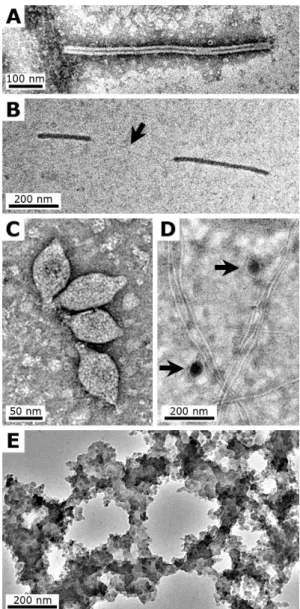
Fig. 1. (A–D): TEM micrographs showing examples of the viruses used for this study. (A)SIRV2 particle; note the central cavity, sometime discontinued.(B)Fragments of SIRV2 particle on an un-stained grid; note the absence of visible features, and a viral DNA-protein filament that links the two fragments (arrow). (C) Aggre-gate of TPV1 particles.(D)PAV1 particles (arrows); note the pres-ence of remnantP. abyssi flagellas in the sample. (E)TEM mi-crograph showing example of the silica precipitate formed sponta-neously in the control sample, which contained no viruses, observed after 4 days; unstained grid. All TEM micrographs were made at 200 kV on grids negatively stained with uranyl acetate, unless oth-erwise stated.
80◦
C and 85◦
C, respectively, up to the late exponential phase. The cells were pelleted by low-speed centrifuga-tion at 6000 g for 15 min. TPV1 and PAV1 were precip-itated from the supernatant in 1 M NaCl with 10 % PEG 6000 overnight at 4◦
for each virus (TPV1: TPV1-buffer – 10 mM Tris-HCL, 100 mM NaCl, 5 mM CaCl2; PAV1: TE buffer – 10 mM Tris-HCl and 1 mM EDTA, pH 8). A second precipitation with PEG 6000 (10 %) and 1 M NaCl was made during 1.5 h and the precipitate was collected as described above. After cen-trifugation (5000 g for 10 min), the supernatant was kept and the pellet was extracted two more times under the same con-ditions with reduced volumes of TPV1 or TE buffer. The virus-containing supernatants were pooled and stored for one night at 4◦
C. After centrifugation (5000 g for 15 min) to re-move residual cell debris, the supernatant was concentrated by ultracentrifugation at 33 000 g for 1 h 45 min (Beckman Optima LE-80K 70.1Ti rotor) and the pellet was resuspended in TPV1 or TE buffer. Viruses were purified from these sus-pensions by centrifugation in a CsCl buoyant density gradi-ent (TPV1: 1.32 g ml−1; PAV1: 1.298 g ml−1) in a Beckman Optima LE-80K 70.1Ti centrifuge rotor (TPV1: 180 000 g for 6 h; PAV1: 220 000 g for 24 h). Fractions containing the nucleic acids were detected at 254 nm and collected using a density gradient fractionator (model 185, ISCO). These frac-tions were then dialyzed against a large volume of TPV1 or TE buffer and were stored at 4◦
C until used.
2.3 Silicification procedure
The fossilisation experiments were made directly in the buffering media of the viruses (SIRV2: Tris-acetate buffer – 20 mM acetate, pH6; TPV1: TPV1 buffer – 10 mM Tris-HCl, 100 mM, 5 mM CaCl2, pH 8; PAV1: TE buffer – 10 mM Tris-HCl, 1 mM EDTA, pH 8). This was done for practical reasons as well as to maintain the viruses in a favourable en-vironment so that their evolution could be followed during fossilisation over several months.
A stock silica solution (3200 ppm Si) was prepared from a pure sodium silicate solution (Riedel de Ha¨en) containing ∼27 % SiO2 and ∼10 % NaOH (Na2Si3O7, M=242 g mol−1). Its pH was adjusted to 8 before injection into the virus suspension.
19 µl of a suspension of purified SIRV2 were mixed with 1 µl of the stock silica solution in a sealed glass vial to ob-tain a final concentration of ∼160 ppm Si. For TPV1 and PAV1, 180 µl of suspensions of purified viruses were mixed with 20 µl of the stock silica solution to obtain a final concen-tration of∼320 ppm Si. The SIRV2 fossilisation experiment vials were stored at room temperature (due to the small vol-ume, high temperatures led to evaporation). The TPV1 and PAV1 fossilisation experiment vials were placed in an oven at 60◦
C. A control sample without viruses, consisting of a
∼160 ppm Si silica solution in distilled water, was also
pre-pared and left at room temperature.
The small volumes involved in the experiments meant that we were not able to make precise monitoring of the pH and silica concentration over time. We assumed that the injection of the silica solution at pH 8 only slightly increased the pH
in the SIRV2 medium, while it did not change the pH of the TPV1 and PAV1 media.
2.4 Electron microscopy
The vials were sampled at different times (between 2 and 60 days for the SIRV2 experiment; between 1 and 180 days for the TPV1 and PAV1 experiments; 4 days for the control sample) by collecting∼1 µl of the sample and immediately preparing it for negatively-stained transmission electron mi-croscopy (Geslin et al., 2003a).
For negative staining, a droplet of sample (either unsili-cified or silicificed) was placed on a carbon-coated copper TEM grid. The sample was allowed to absorb to the carbon layer for 2 min before removing the excess liquid with a piece of filter paper. Some samples were stained to increased con-trast by placing a droplet of saturated uranyl acetate ethanolic solution for 40 s and then removing the excess liquid. Some grids were also observed unstained to better distinguish sil-ica deposition on the viral structures. The prepared samples were then air dried. They were observed and analysed with a Philips CM20 Transmission Electron Microscope (Centre de Microscopie Electronique, University of Orl´eans), equipped with an EDX detector (Oxford Instruments).
3 Results
3.1 Experimental fossilisation of SIRV2
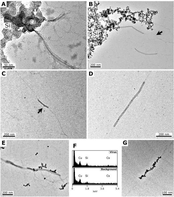
Fig. 2. TEM micrographs showing progressive steps of the experimental fossilisation of SIRV2 at a∼160 ppm Si silica concentration.
(A)48 h, SIRV2 particle partially trapped inside the silica precipitate (Si); the arrows underline the shape of the virion inside the precipitate.
(B)7 days, fragments of SIRV2 particles in the vicinity of a silica precipitate (Si); the two separated filaments of viral DNA-protein are visible (arrow).(C)7 days, unstained grid, fragment of SIRV2 particle; a dark∼10 nm particle is seen attached on the virion (arrow).(D)30 days, SIRV2 particle; note the numerous dark silica particles in the central cavity.(E)60 days, unstained grid, fragments of SIRV2 particle; note the dark silica particles attached on the outer surface of the virion, and filling the central cavity. (F)60 days, EDX spectra made on the SIRV2 particles of(E), showing a Si signal slightly higher than on the background; the Cs signal comes from the cesium chloride used for virus purification and the Cu signal comes from the copper grid.(G)60 days, unstrained grid, viral DNA-protein filament covered by silica particles. All TEM micrographs were made at 200 kV on grids negatively stained with uranyl acetate, unless otherwise stated.
dark particles had formed in the central cavity of some of the virions (Fig. 2d). This phenomenon was more evident after 60 days of fossilisation with virions showing, on un-stained grids, a strongly contrasted central cavity (Fig. 2e), suggesting that the individual particles observed previously had continued their growth and eventually merged. More pronounced silica nucleation and binding on the outside of
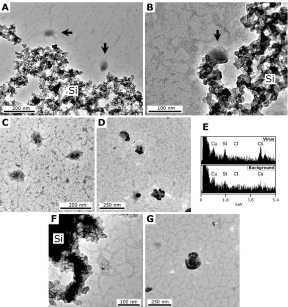
Fig. 3. TEM micrographs showing progressive steps of the experimental fossilisation of TPV1 at a ∼320 ppm Si silica concentration.
(A)1 week, two TPV1 particles (arrows) near a silica precipitate (Si). (B)1 month, TPV1 particle (arrow) attached to the silica precipitate (Si). (C)75 days, TPV1 particles with dark nanometric particles within. (D)75 days, unstained grid, TPV1 particles on which a dark precipitate has formed. (E)75 days, EDX spectra obtained on the virions of(D)and on the background, showing Si and Cs signals on the precipitate; the Cl signal comes from buffering medium and the Cu signal comes from the copper grid. (F)180 days, deformed TPV1 particle near a silica precipitate (Si); note the dark particle within.(G)180 days, unstained grid, TPV1 particle covered and filled with a dark precipitate. All TEM micrographs were made at 200 kV on grids negatively stained with uranyl acetate, unless otherwise stated.
3.2 Experimental fossilisation of TPV1 and PAV1
Although a granular, alveolar silica precipitate formed spon-taneously within one day of the experiment, similar to that observed in the SIRV2 and control experiments (Fig. 3a), it was only after 30 days that the TPV1 particles were ob-served in direct contact with the precipitate (Fig. 3b). No virions trapped in the silica precipitate were observed. The first signs of possible silica precipitation on the viral struc-tures occurred only after 75 days when nanometer sized, dark particles were observed within (Fig. 3c) and on the envelope
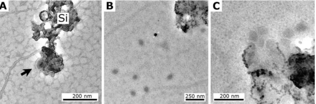
Fig. 4. TEM micrographs showing progressive steps of the experimental fossilisation of PAV1 at a∼320 ppm Si silica concentration.
(A)1 day, PAV1 particle (arrow) attached to the silica precipitate (Si).(B)60 days, unstained grid, numerous PAV1 particles near the silica precipitate.(C)60 days, unstained grid, aggregate of PAV1 particles trapped in a finely grained silica precipitate. All TEM micrographs were made at 200 kV on grids negatively stained with uranyl acetate, unless otherwise stated.
Monitoring of the PAV1 fossilisation experiment was com-plicated by the presence of numerous fragments ofP. abyssi flagella as well as numerous artefacts in the preparation of purified PAV1 (Fig. 1d). PAV1 particles could be observed in direct contact with the silica precipitate after only 1 day (Fig. 4a). This precipitate was different from that formed in the SIRV2 and TPV1 experiments and consisted of very fine particles (compare Figs. 4c and 3b). The reason for this is unknown, although it possibly could be a consequence of the different composition of the PAV1 buffering medium (which includes EDTA, a chelating and binding agent). No precip-itates formed inside or on free PAV1 particles after 60 days (Fig. 4b), although several virus particles were seen at the edge of the silica precipitate (Fig. 4c), suggesting that an im-portant number of PAV1 particles had been trapped in it.
4 Discussion
The viruses used in this experiment belong to different virus families having completely different morphologies and struc-tures. This may partly explain why the results of the fossil-isation experiments were different. In the case of SIRV2, a non-enveloped rod-shaped virus, silica binding on the viral particles was significant and progressive whereas only lim-ited silica binding occurred on TPV1 and PAV1, enveloped spindle-shaped viruses.
4.1 Fossilisation of the viruses
We assumed (but could not verify, due to the small volumes of the samples) that the silica behaviour during the experi-ments was similar to that described in previous experimental fossilisation studies (review in Konhauser et al., 2004). Upon injection into the vials, silica was in monomeric (Si(OH)4) or slightly polymeric form and quickly spontaneously poly-merised as a colloidal amorphous silica precipitate. Dis-solved silica must have remained in the medium after this
initial polymerisation at a concentration close to the satura-tion concentrasatura-tion (62 ppm Si in distilled water; Gunnarsson and Arn´orsson, 2000; Lalonde et al., 2005). In support of this, the silica precipitate formed in the SIRV2, TPV1 and control experiments was similar to that observed in previous fossilisation experiments (Orange et al., 2009).
particles and the hydroxyl and/or carboxyl functional groups contained in the protein of the helix (see review of the fossil-isation processes in Konhauser et al., 2004).
Fossilisation of the viruses TPV1 and PAV1 differed from that of SIRV2. The former have an envelope that contains lipids that derive from the hosts (the ArchaeaP. abyssiand T. prieurii, respectively) (Geslin et al., 2003a; Gorlas et al., 2011). Previous results from the experimental fossilisation of Archaea showed that the simple cell wall of these mi-croorganisms (S-Layer + plasma membrane) has only a lim-ited ability to bind silica (Orange et al., 2009). It was there-fore to be expected that, having a similar composition, the envelopes of TPV1 and PAV1 would not bind significant amounts of silica. This was indeed observed for TPV1, al-though some virions occasionally occurred in direct contact with silica (Fig. 3b), and, after 75 days of exposure to the silica solution, nanometer-sized particles were ubiquitously observed inside TPV1 (Fig. 3c, f). Only at this advanced stage in the fossilisation procedure (75 days), were nanopar-ticles of a SiO2-Cs precipitate first observed on the virion en-velopes (Fig. 3d). The source of the Cs was the cesium chlo-ride used for harvesting purified TPV1. Chelated metal ions may act as bridges in the fixation of mineral ions to organic materials. This has been demonstrated with Fe(III) chelated to Bacteria and Archaea (Beveridge and Murray, 1976; Bev-eridge and Koval, 1981; Orange et al., 2011). However, as a monovalent cation, Cs+
cannot act as a direct intermediate between silica and the virion envelope or internal materials. Silica binding to the TPV1 particles most likely occurred in the same way as with the SIRV2 particles, namely through covalent or hydrogen bonding of the silica. Similar results for TPV1 and PAV1 were expected, since these two viruses have similar structures. Instead, no silica precipitation was observed on free PAV1 particles (Fig. 4b) while numerous virions were trapped in the silica precipitate (Fig. 4c). The unknown factors that led to the formation of a different silica precipitate may also be responsible of these differences in the silicification process.
4.2 Implication for the preservation and identification of viral remains in rocks
The three viruses used in this artificial fossilisation study were well preserved, apart from a slight deformation pos-sibly due to the length of the experiment rather than to the exposure to silica. The fossilisation was made directly on virions stored in buffered solutions, which is obviously not a natural condition. The rationale for making the experi-ments on the microorganisms in a buffered solution is the previously noted importance for the cell to remain alive, or at least not to lyse, during the time necessary for silica to form a deposit around the cells thick enough to allow for fos-silisation and further preservation (Orange et al., 2009). This period of time varies depends on the strain or the type of mi-croorganism. Viral particles obviously do not lyse but their
physical integrity can quickly be affected due to their small size and constitution. Our results show that, if viral particles can be conserved over several months, they can theoretically become fossilised. In addition, our results suggest that viral particles may have been trapped in the silica precipitate, with viral remains being possibly preserved, although the quality of the preservation could not be precisely ascertained in this case.
The fact that viruses can be fossilised suggests that natu-rally fossilised viruses may occur in rocks in the same way as fossilised microbial cells. The question is, how could we identify them? Morphology is a criterion for their identifi-cation but their simple nanometric-sized structures may be confused with minerals or other artifacts of the same size (of the order of∼100 nm). On the other hand, it may be possible to identify filamentous or rod-shaped structures, such as the about 500×20 nm-sized SIRV2 particles, in ancient rocks. These viruses are about the same size and shape as the so-called “nanobacteria” identified in 3.9 Ga old carbonate con-cretions in the Martian meteorite ALH 84001 (McKay et al., 1996 – note that it is now accepted that the latter are simply mineral precipitations; Gibson Jr. et al., 2001). Although sil-ica precipitated in the central cavity of SIRV2 (Fig. 2e) as a structure of less than 10 nm in width and several hundreds of nanometers long, it would be difficult to distinguish such a feature from polymeric fibrils or filaments of other biologi-cal origin that had been silicified. Viral organic compounds, such as lipids and proteins, could also be preserved in very small amounts but they would rapidly be degraded, hence rendering their detection and identification as viral biomark-ers difficult. Thus, even though it is theoretically feasible, detection of virus remains in rocks will be highly challeng-ing.
5 Conclusions
Acknowledgements. This research was funded by the CNRS – PID Origines des Plan`etes et de la Vie (2007–2009). Aurore Gorlas was funded through a Ph.D. grant from the Minist`ere de l’Enseignement Sup´erieur et de la Recherche. Dominique Jalabert is thanked for his aid with the transmission electron microscope.
Edited by: J. Toporski
The publication of this article is financed by CNRS-INSU.
References
Bamford, D. H.: Do viruses form lineages across different domains of life?, Res. Microbiol. 154, 231–236, doi:10.1016/S0923-2508(03)00065-2, 2003.
Bamford, D. H., Grimes, J. M., and Stuart, D. I.: What does struc-ture tell us about virus evolution?, Curr. Opin. Struc. Biol., 15, 655–663, doi:10.1016/j.sbi.2005.10.012, 2005.
Bettstetter, M., Peng, X., Garrett, R. A., and Prangishvili, D.: AFV1, a novel virus infecting hyperthermophilic archaea of the genusAcidianus, Virology, 315, 68–79, 2003.
Beveridge, T. J. and Koval, S. F.: Binding metals to cell envelopes ofEscherichia coli, Appl. Environ. Microb., 42, 325–335, 1981. Beveridge, T. J. and Murray, R. G. E.: Uptake and retention of met-als by cell walls ofBacillus subtilis, J. Bacteriol., 127, 1502– 1518, 1976.
Bird, D. F., Juniper, S. K., Ricciardi-Rigault, M., Martineu, P., Prairie, Y. T., and Calvert, S. E.: Subsurface viruses and bac-teria in Holocene/Late Pleistocene sediments of Saanich Inlet, BC: ODP Holes 1033B and 1034B, Leg 169S, Mar. Geol., 174, 227–239, doi:10.1016/S0025-3227(00)00152-3, 2001.
Birnbaum, S. J., Wireman, J. W., and Borowski, R.: Silica precipi-tation by the anaerobic sulphate reducing bacterium Desulfovib-rio desulfuricans: effects upon cellmorphology and implications for preservation, in: Origin, Evolution, and Modern Aspects of Biomineralization in Plants and Animals, Crick, R.E., Plenum Press, New York, USA, 507–516, 1989.
Bize, A., Karlsson, E. A., Ekefj¨ard, K., Quax, T. E. F., Pina, M., Prevost, M. C., Forterre, P., Tenaillon, O., Bernan-der, R., and Prangishvili, D.: A unique virus release in the Archaea, P. Natl. Acad. Sci. USA, 106, 11306–11311, doi:10.1073/pnas.0901238106, 2009.
Borriss, M., Helmke, E., Hanschke, R., and Schweder, T.: Iso-lation and characterization of marine psychrophilic phage– host systems from Arctic sea ice, Extremophiles, 7, 377–384, doi:10.1007/s00792-003-0334-7, 2003.
Daughney, C. J., Chˆatellier, X., Chan, A., Kenward, P., Fortin, D., Suttle, C. A., and Fowle, D. A.: Adsorption and precipitation of iron from seawater on a marine bacteriophage (PWH3A-P1), Mar. Chem., 91, 101–115, doi:10.1016/j.marchem.2004.06.003, 2004.
Dyall-Smith, M., Tang., S. L., and Bath, C.: Haloarchaeal viruses: how diverse are they?, Res. Microbiol., 154, 309–313, doi:10.1016/S0923-2508(03)00076-7, 2003.
Erauso, G., Reysenbach, A. L., Godfroy, A., Meunier, J. R., Crump, B., Partensky, F., Baross, J. A., Marteinsson, V., Barbier, G., Pace, N. R., and Prieur, D.: Pyrococcus abyssisp. nov., a new hyperthermophilic archaeon isolated from a deep-sea hydrother-mal vent, Arch. Microbiol., 160, 338–349, 1993.
Forterre, P.: A hot story from comparative genomics: reverse gyrase is the only hyperthermophile-specific protein, Trends Genet., 18, 236–238, doi:10.1016/S0168-9525(02)02650-1, 2002.
Forterre, P.: Three RNA cells for ribosomal lineages and three DNA viruses to replicate their genomes : A hypothesis for the origin of cellular domain, P. Natl. Acad. Sci. USA, 103, 3669–3674, doi:10.1073/pnas.0510333103, 2006.
Forterre, P.: Manipulation of cellular syntheses and the nature of viruses: The virocell concept, C.R. Chim., 14, 392–399, doi:10.1016/j.crci.2010.06.007, 2011.
Forterre, P. and Prangishvili, D.: The great billion-year war between ribosome- and capsid-encoding organisms (cells and viruses) as the major source of evolutionary novelties, Ann. NY. Acad. Sci., 1178, 65–77, doi:10.1111/j.1749-6632.2009.04993.x, 2009a. Forterre, P. and Prangishvili, D.: The origin of viruses, Res.
Micro-biol., 160, 466–472, doi:10.1016/j.resmic.2009.07.008, 2009b. Geslin, C., Le Romancer, M., Erauso, G., Gaillard, M., Perrot, G.,
and Prieur, D.: PAV1, the first virus-like particle isolated from a hyperthermophilic Euryarchaeote,Pyrococcus abyssi, J. Bacte-riol., 185, 3888–3894, doi:10.1128/JB.185.13.3888-3894.2003, 2003a.
Geslin, C., Le Romancer, M., Gaillard, M., Erauso, G., and Prieur, P.: Observation of virus-like particles in high temperature enrich-ment cultures from deep-sea hydrothermal vents, Res Microbiol., 154, 303–307, doi:10.1016/S0923-2508(03)00075-5, 2003b. Geslin, C., Le Romancer, M., Gaillard, M., and Prieur, D.: Diversit´e
virale associ´ee aux ´ecosyst`emes hydrothermaux oc´eaniques pro-fonds et aux sources chaudes terrestres, Virologie, 9, 357–366, 2005.
Gibson Jr., E. K., McKay, D. S., Thomas-Keprta, K. L., Wentworth, S. J., Westall, F., Steele, A., Romanek, C. S., Bell, M. S., and Toporski, J.: Life on Mars: evaluation of the evidence within Martian meteorites ALH 84001, Nakhla and Shergotty, Precam-brian Res., 106, 15–34, doi:10.1016/S0301-9268(00)00122-4, 2001.
Gorlas, A., Geslin, C., and Prieur, D.: TV1, the first virus-like par-ticle ofThermococcus, a hyperthermophilic Archaea genus, Ori-gins Life Evol. B., 39, p. 62, 2009.
Gorlas, A., Alain, K., Bienvenu, N., Isaac, S., and Geslin, C.: Ther-mococcus prieuriisp. nov., a novel hyperthermophilic archaeon isolated from a deep-sea hydrothermal vent at the East Pacific Rise, Int. J. Syst. Evol. Micr., submitted, 2011.
Gowing, M. M.: Large viruses and infected microeukaryotes in Ross Sea summer pack ice habitats, Mar. Biol., 142, 1029–1040, doi:10.1007/s00227-003-1015-x, 2003.
Gunnarsson, I. and Arn´orsson, S.: Amorphous silica solubility and the thermodynamic properties of H4SiO◦4 in the range of 0◦
to 350◦C at Psat, Geochim. Cosmochim. Ac., 64, 2295–2307,
doi:10.1016/S0016-7037(99)00426-3, 2000.
hypersaline Mono Lake, California, Microb. Ecol., 47, 9–17, doi:10.1007/s00248-003-1023-x, 2004.
Kepner, R. L., Wharton Jr., R. A., and Suttle, C. A.: Viruses in Antarctic lakes, Limnol. Oceanogr., 43, 1754–1761, 1998. Konhauser, K. O., Jones, B., Reysenbach, A. L., and Renaut, R.
W.: Hot spring sinters: Keys to understanding Earth’s earliest life forms, Can. J. Earth Sci., 40, 1713–1724, 2003.
Konhauser, K. O., Jones, B., Phoenix, V. R., Ferris, G., and Renaut, R. W.: The microbial role in hot spring silicification, Ambio, 33, 552–558, 2004.
Koonin, E. V. and Dolja, V. V.: Evolution of complexity in the viral world: the dawn of a new vision. Virus Res., 117, 1–4, doi:10.1016/j.virusres.2006.01.018, 2006.
Koonin, E. V., Senkevich, T. G., and Dolja, V. V.: The an-cient virus world and evolution of cells, Biol. Direct, 1, 29, doi:10.1186/1745-6150-1-29, 2006.
Krupovic, M. and Bamford, D. H.: Virus evolution: how far does the double beta-barrel viral lineage extend?, Nat. Rev. Micro-biol., 6, 941–9488, doi:10.1038/nrmicro2033, 2008.
Kyle, J. E., Eydal, H. S. C., Ferris, F. G., and Pedersen, K.: Viruses in granitic groundwater from 69 to 450 m depth of the ¨Asp¨o hard rock laboratory, Sweden, ISME J., 2, 571–574, doi:10.1038/ismej.2008.18, 2008a.
Kyle, J. E., Pedersen, K., and Ferris, F. G.: Virus mineralization at low pH in the Rio Tinto, Spain, Geomicrobiol. J., 25, 338–345, doi:10.1080/01490450802402703, 2008b.
Laidler, J. R. and Stedman, K. M.: Virus silicification under simulated hot spring conditions, Astrobiology, 10, 569–576, doi:10.1089/ast.2010.0463, 2010.
Lalonde, S. V., Konhauser, K. O., Reysenbach, A. L., and Ferris, F. G.: The experimental silicification of Aquificales and their role in hot spring formation, Geobiology, 3, 41–52, doi:10.1111/j.1472-4669.2005.00042.x, 2005.
La Scola, B., Audic, S., Robert, C., Jungang, L., de Lambal-lerie, X., Drancourt, M., Birtles, R., Claverie, J. M., and Raoult, D.: A giant virus in Amoebae, Science, 299, p. 2033, doi:10.1126/science.1081867, 2003.
Le Romancer, M., Gaillard, M., Geslin, C., and Prieur, D.: Viruses in extremes environments, Reviews in Environmental Science and Biotechnology, 6, 17–31, doi:10.1007/s11157-006-0011-2, 2007.
Maranger, R., Bird, D. F., and Juniper, S. K.: Viral and bacterial dynamics in Arctic sea ice during the spring algal bloom near Resolute, N.W.T., Canada, Mar. Ecol.-Prog. Ser., 111, 121–127, 1994.
Marteinsson, V. T., Watrin, L., Prieur, D., Caprais, J. C., Ragu´en`es, G., and Erauso G.: Phenotypic characterization, DNA simi-larities, and protein profiles of twenty sulfur-metabolizing hy-perthermophilic anaerobic Archaea isolated from hydrothermal vents in the southwestern Pacific Ocean, Int. J. Syst. Bacteriol., 45, 623–632, doi:10.1099/00207713-45-4-623, 1995.
Martin, A., Yeats, S., Janekovic, D., Reiter, W. D., Aicher, W., and Zillig, W.: SAV1, a temperate u.v.-inducible DNA virus-like par-ticle from the archarbacteriumSulfolobus acidocaldariusisolate B12, EMBO J., 3, 2165–2168, 1984.
McKay, D. S., Gibson Jr., E. K., Thomas-Keprta, K. L., Vali, H., Romanek, C. S., Clemett, S. J., Chillier, X. D. F., Maechling, C. R., and Zare, R. N.: Search for Past Life on Mars: Possible Relic Biogenic Activity in Martian Meteorite ALH84001,
Sci-ence, 273, 924–930, doi:10.1126/science.273.5277.924, 1996. Moreira, D. and L´opez-Garc´ıa, P.: Ten reasons to exclude
viruses from the tree of life, Nat. Rev. Microbiol., 7, 306–311, doi:10.1038/nrmicro2108, 2009.
Nisbet, E. G. and Sleep, N. H.: The habitat and nature of early life, Nature, 409, 1083–1091, doi:10.1038/35059210, 2001. Orange, F., Westall, F., Disnar, J. R., Prieur, D., Bienvenu, N., Le
Romancer, M., and D´efarge, C.: Experimental silicification of the extremophilic ArchaeaPyrococcus abyssiand Methanocal-dococcus jannaschii. Applications in the search for evidence of life in early Earth and extraterrestrial rocks, Geobiology, 7, 403– 418, doi:10.1111/j.1472-4669.2009.00212.x, 2009.
Orange, F., Disnar, J. R., Westall, F., Prieur, D., and Baillif, P.: Metal cation binding by the hyperthermophilic microorganism, ArchaeaMethanocaldococcus jannaschii, and its effects on sili-cification, Palaeontology, accepted, 2011.
Oren, A., Bratbak, G., and Heldal, M.: Occurrence of virus like particles in the Dead Sea, Extremophiles, 1, 143–149, doi:10.1007/s007920050027, 1997.
Ortmann, A. C. and Suttle, C. A.: High abundances of viruses in deep-sea hydrothermal vent system indicate viral medi-ated microbial mortality, Deep-Sea Res. Pt. I, 52, 1515–1527, doi:10.1016/j.dsr.2005.04.002, 2005.
Ortmann, A. C., Wiedenheft, B., Douglas, T., and Young, M.: Hot crenarchaeal viruses reveal deep evolutionary connections, Nat. Rev. Microbiol., 4, 520–528, doi:10.1038/nrmicro1444, 2006. Pagaling, E., Haigh, R. D., Grant, W. D., Cowan, D. A., Jones,
B. E., Ma, Y., Ventosa, A., and Heaphy, S.: Sequence analysis of an Archaeal virus isolated from a hypersaline lake in Inner Mongolia, China, BMC Genomics, 8, 410, doi:10.1186/1471-2164-8-410, 2007.
Poinar, G. P. and Poinar, R.: Fossil evidence of insect pathogens, J. Invertebr. Pathol., 89, 243–250, doi:10.1016/j.jip.2005.05.007, 2005.
Prangishvili, D., Arnold, H. P., G¨otz, D., Ziese, U., Holz, I., Krist-jansson, J. K., and Zillig, W.: A novel virus family, the Rudiviri-dae: Structure, virus-host like interactions and genome variabil-ity of the Sulfolobus viruses SIRV1 and SIRV2, Genetics, 152, 1387–1396, 1999.
Prangishvili, D., Forterre, P., and Garrett, R. A.: Viruses of the Archaea: a unifying view, Nat. Rev. Microbiol., 4, 837–848, doi:10.1038/nrmicro1527, 2006a.
Prangishvili, D., Garrett, R. A., and Koonin, E. V.: Evolu-tionary genomics of archaeal viruses: unique viral genomes in the third domain of life, Virus Res., 117, 52–67, doi:10.1016/j.virusres.2006.01.007, 2006b.
Prigent, M., Leroy, M., Confalonieri, F., Dutertre, M., and DuBow M. S.: A diversity of bacteriophage forms and genomes can be isolated from the surface sands of the Sahara Desert, Ex-tremophiles, 9, 289–296, doi:10.1007/s00792-005-0444-5, 2005. Rachel, R., Bettstetter, M., Hedlund, B. P., H¨aring, M., Kessler, A., Stetter, K. O., and Prangishvili, D.: Remarkable morphological diversity of viruses and virus-like particles in hot terrestrial en-vironments, Arch. Virol., 147, 2419–2429, doi:10.1007/s00705-002-0895-2, 2002.
Rice, G., Stedman, K., Snyder, J., Wiedenheft, B., Willits, D., Brumfield, S., and McDermott, T.: Viruses from extreme ther-mal environments, P. Natl. Acad. Sci. USA, 98, 13341–13345, doi:10.1073/pnas.231170198, 2001.
Schelble, R. T., Hall, J. A., Nealson, K. H., and Steele, A.: DNA perseverance of microorganisms exposed to silica: an ex-perimental study, Geobiology, 6, 503–511, doi:10.1111/j.1472-4669.2008.00177.x, 2008.
Schultze-Lam, S., Ferris, F. G., Kohnauser, K. O., and Wiese, R. G.: In situ silicification of an Icelandic microbial mat: impli-cations for microfossil formation, Can. J. Earth Sci., 32, 2021– 2026, 1995.
Sime-Ngando, T., Lucas, S., Robin, A., Pause Tucker, K., Colom-bet, J., Bettarel, Y., Desmond, E., Gribaldo, S., Forterre, P., Breitbart, M., and Prangishvili, D.: Diversity of virus–host sys-tems in hypersaline Lake Retba, Senegal, Environ. Microbiol., doi:10.1111/j.1462-2920.2010.02323.x, in press, 2010. Templeton, M. R., Andrews, R. C., and Hofmann, R.: Impact of
iron particles in groundwater on the UV inactivation of bac-teriophages MS2 and T4, J. Appl. Microbiol., 101, 732–741, doi:10.1111/j.1365-2672.2006.02980.x, 2006.
Toporski, J. K. W., Steele, A., Westall, F., Thomas-Keprta, K. L., and McKay, D. S.: The simulated silicification of bacteria – new clues to the modes and timing of bacterial preservation and im-plications for the search for extraterrestrial microfossils, Astro-biology, 2, 1–26, 2002.
Vestergaard, G., Shah, S. A., Bize, A., Reitberger, W., Reuteur, M., Phan, H., Brigel, A., Rachel, R., Garrett, R. A., and Prangishvili, D.: StygiolobusRod-Shaped Virus and the interplay of Crenar-chaeal Rudiviruses with the CRISPR Antiviral System, J. Bacte-riol., 190, 6837–6845, doi:10.1128/JB.00795-08, 2008. Westall, F.: Early life: nature, distribution and evolution, in:
Ori-gins and evolution of life, an astrobiological perspective, edited by: Gargaud, M., L´opez-Garc´ıa, P., and Martin, H., Cambridge University Press, Cambridge, United-Kingdom, 391–413, 2010. Westall, F. and Southam, G.: The early record of life, Geoph.
Monog. Series, 164, 283–304, 2006.
Westall, F., Boni, L., and Guerzoni, E.: The experimental silicifica-tion of microorganisms, Palaeontology, 38, 495–528, 1995. You, Y., Han, J., Chiu, P. C., and Jin, Y.: Removal and
inactiva-tion of waterborne viruses using zerovalent iron, Environ. Sci. Technol., 39, 9263–9269, doi:10.1021/es050829j, 2005. Zillig, W., Kletzin, A., Schleper, C., Holz, I., Janekovic, D., Hain,