Universidade Nova de Lisboa
FLAVIVIRUSES IN MOSQUITOES FROM SOUTHERN
PORTUGAL, 2009-2010
Sónia Cristina Fernandes Da Costa
DISSERTAÇÃO PARA A OBTENÇÃO DO GRAU DE MESTRE EM PARASITOLOGIA MÉDICA
Universidade Nova de Lisboa
Instituto de Higiene e Medicina Tropical
FLAVIVIRUSES IN MOSQUITOES FROM SOUTHERN
PORTUGAL, 2009-2010
Sónia Cristina Fernandes Da Costa
Dissertação apresentada para cumprimento dos requisitos necessários à obtenção do grau de Mestre em Parasitologia Médica realizada sob a orientação científica de:
Orientador: Professor Doutor Paulo Almeida
iii
I dedicate this thesis to my family, especially
to João and Daniela who have given up
iv
ACKNOWLEDGEMENTS
Firstly and foremost, I want to thank everyone that directly or indirectly contributed to the making of this thesis, especially my dear colleague, Dr. Ferdinando Freitas, who made this work possible.
I wish to thank my supervisors, Professor Dr. Paulo Almeida and Professor Dr. Ricardo Parreira for the continuous support, encouragement, and friendship. I thank them for the invaluable teachings; unending patience, good advice and critique that helped me develop my scientific knowledge, in both Medical Entomology and Virology.
To Dr. Ferdinando Freitas, I want to thank for his great support, encouragement and friendship. Foremost I am grateful for his teachings and mentoring in conducting research in the Virology and Entomology laboratories, and for allowing me personal freedom to learn through the trials and tribulations presented throughout the course of this research. I thank him for performing tasks essential for this project to go forward, such as field mosquito collection and specimen identification. I am thankful for the time, dedication and expertise devoted to this project; his enthusiasm was contagious and the experience gained while working with him has strengthen my determination in pursuing a career in research.
I want to give thanks to all the members of staff of the Medical Parasitology and Microbiology Unit of the Institute of Hygiene and Tropical Medicine who have been extremely helpful from the very beginning.
To Professor Carla Sousa, Professor Teresa Novo and Dr. Bruno Gomes I want to thank for performing tasks, such as fieldwork and specimen identification. To Dr. Cristina Branco and Dr. Sandra Castro for their support and encouragement. To Dr. José Luís Vicente for his good advice and willingness to help. To Dr. Vera Benavente for the companionship, encouragement and good moments. To my colleagues Dr. Gonçalo Seixas, Dr. Sofia Branco, Dr. Manuel Roque and Dr. Eliane Arez for the incentives, companionship and good moments shared.
v
vi
ABSTRACT
Flaviviruses are viruses belonging to the Flaviviridae family, genus Flavivirus. They
comprise a large group of widely spread and genetically diverse arthropod-borne viruses including human and animal pathogens that can potentially cause large-scale epidemics and high mortality and morbidity. In the past few years, flaviviruses have largely expanded their geographical distribution and host range. West Nile virus has been continuously detected throughout Europe lately and has been isolated from mosquitoes in Southern Portugal, where human and animal cases have been reported.
The main aim of this work was to search for flaviviruses in mosquitoes collected from two areas in Southern Portugal where West Nile virus and other flaviviruses have previously been detected.
Mosquito surveys were carried out in 24 locations in the wetlands of the Faro and Setúbal districts, by CDC-CO2 light-traps and indoors resting collections. Pools
containing approximately 50 mosquitoes were screened for flaviviruses by heminested RT-PCR, directed at the amplification of a small fragment of the viral NS5 gene, using degenerated flavivirus-specific primers.
A total of 36273 mosquitoes were collected during 2009 and 2010 from April through October, from the following species: Anopheles algeriensis, An.atroparvus, Aedes
berlandi, Ae. caspius, Ae. detritus, Coquillettidia richiardii, Culex laticinctus, Cx.
pipiens, Cx. theileri, Cx. univittatus, Culiseta annulata, Cs. longiareolata, Cs.
subochrea, and Uranotaenia unguiculata. Most abundant species were Ae. caspius Cx.
theileri and Cx. pipiens, respectively. However, mosquito densities varied according to
collection method and sampling area. A fourfold increase in mosquito density was registered in 2010 compared to 2009. A total of 745 pools were analysed of which 31% tested positive for flaviviral sequences.
The species with higher positivity rates were An. algeriensis with Minimum infection
rate (MIR) of 56/1000 in the Algarve 2009, Cs. annulata MIR =22/1000 in the Algarve
2010, Cx.theileri and Cx.pipiens in Setúbal 2010, MIR =20/1000. An. atroparvus, Ae.
caspius, Ae. detritus and Cx. univittatus also yielded positive pools. Overall, positivity
was higher in the Algarve.
Viral sequences obtained from positive pools showed homology with insect-specific flavivirus (ISF) sequences deposited in free access public databases. Phylogenetic analysis reflected the genetic variability of flaviviruses and revealed the relatedness of our sequences with other known flaviviruses, especially the insect-specific.
vii
RESUMO
Os flavivírus são vírus pertencentes à família Flaviviridae, género Flavivirus. Estes
formam um grande grupo caraterizado pela sua ampla distribuição e diversidade genética. Os flavivírus são, na sua maioria, transmitidos por artrópodes vectores incluíndo agentes patogénicos para humanos e animais que podem potencialmente provocar grandes epidemias e causar elevadas taxas de mortalidade e morbidade. Nos últimos anos, tem-se registado uma grande expansão a nível da distribuição geográfica dos flavivírus e diversidade dos seus hospedeiros. O vírus do Nilo Ocidental tem sido continuamente detectado em toda a Europa recentemente, e também isolado de mosquitos colhidos no Sul de Portugal, onde já foram registados casos humanos e animais.
O principal objectivo deste trabalho é o rastreio de flavivírus em mosquitos colhidos em duas regiões do Sul de Portugal, onde os mesmos foram anteriormente detectados.
As colheitas de mosquitos foram realizadas em 24 locais em zonas húmidas nos districtos de Faro e Setúbal, através de armadilhas luminosas tipo CDC com CO2 e
aspiradores mecânicos manuais para colheita de mosquitos em repouso em abrigos de animais.
Os mosquitos colhidos foram agrupados por lotes contendo aproximadamente 50 espécimens cada, e rastreados para a presença de flavivírus por heminested RT-PCR,
direccionado à amplificação de um pequeno fragmento do gene NS5 usando oligonucleótidos degenerados específicos para flavivírus.
Entre Abril e Outubro de 2009 e 2010 foram colhidos no total 36273 mosquitos pertencentes às seguintes espécies: Anopheles algeriensis, An.atroparvus, Aedes
berlandi, Ae.caspius, Ae.detritus, Coquillettidia richiardii, Culex laticinctus,
Cx.pipiens, Cx.theileri, Cx.univittatus, Culiseta annulata, Cs.longiareolata,
Cs.subochrea, e Uranotaenia unguiculata. As espécies mais abundantes foram
Ae.caspius, Cx.theileri e Cx.pipiens, respectivamente. Contudo, as densidades de
mosquitos foram variáveis de acordo com o método de colheita e área de amostragem. As densidades de mosquitos colhidos em 2010 foram quatro vezes superior às registadas no ano anterior. No total foram analisados 745 lotes dos quais 31% testaram positivos para a presença de sequências de flavivírus.
As espécies que apresentaram taxas de positividade mais elevadas foram: An.algeriensis
com uma Taxa Mínima de Infecção (TMI) de 56/1000 no Algarve em 2009,
Cs.annulata TMI =22/1000 no Algarve em 2010, Cx.theileri e Cx.pipiens em Setúbal
em 2010, TMI =20/1000. An. atroparvus, Ae. caspius, Ae. detritus e Cx. univittatus
também produziram lotes positives. No geral, a positividade foi maior no Algarve.
viii
Tendo em consideração os anteriores isolamentos do vírus do Nilo Ocidental, o aumento acentuado nas densidades de mosquitos, o aumento de temperaturas que se tem vindo a registar, os casos recentes de transmissão de flavivírus por toda a Europa e o padrão desconhecido e imprevisível dos surtos destes vírus, os programas contínuos de vigilância epidemiológica têm-se revelado uma ferramenta indispensável para a Saúde Pública.
FLAVIVIRUSES IN MOSQUITOES FROM SOUTHERN PORTUGAL,
2009-2010
Sónia Cristina Fernandes Da Costa
Keywords: West Nile virus, flaviviruses, arboviruses, Algarve, flavivirus detection,
insect-specific flaviviruses.
Palavras Chave: Vírus do Nilo Ocidental, flavivírus, arbovírus, Algarve, detecção de
ix
TABLE OF CONTENTS
ACKNOWLEDGEMENTS ...iv
ABSTRACT ...vi
RESUMO ... vii
TABLE OF CONTENTS ...ix
LIST OF ABBREVIATIONS ... 1
1. Introduction ... 5
1.1. Arboviruses ... 5
1.1.2. Medical importance of mosquitoes (Diptera: Culicidae) ... 8
1.2. The Flaviviridae family ... 10
1.3. The importance of Flaviviruses ... 11
1.3.1. Flavivirus: genome structure and morphology of the viral particle ... 11
1.3.2. Replication cycle ... 14
1.3.3. Flavivirus classification: phylogeny ... 16
1.4. Medically Important Pathogenic Flaviviruses ... 20
1.4.1. Dengue and Yellow Fever ... 21
1.4.2. West Nile Virus ... 25
1.5. Insect-specific flaviviruses ... 31
1.6. Europe: current situation ... 40
1.7. Portugal: the imminent risk ... 46
2. Objectives ... 52
2.1 Specific objectives ... 52
3. Materials and methods ... 54
3.1. Mosquito surveys ... 54
3.1.1. Characterisation of the study areas ... 55
x
3.1.3. Specimen identification ... 56
3.1.4. Mosquito density and seasonal dynamics study ... 56
3.1.5. Statistical analysis ... 56
3.2. Viral screening ... 57
3.2.1. Mechanical maceration of mosquitoes... 57
3.2.2. RNA extraction ... 57
3.2.3. cDNA synthesis ... 58
3.2.4. Preliminary tests ... 59
3.2.5. Amplification of viral sequences by PCR ... 60
3.2.6. Purification and sequencing of PCR products ... 62
3.2.7. Phylogenetic analysis of viral sequences ... 63
3.2.8 Extraction controls ... 64
4. Results ... 66
4.1. Mosquito collections ... 66
4.2. Mosquito seasonal dynamics in the Algarve, 2009-2010 ... 68
4.3. Mosquito relative species distribution in the Algarve, 2009-2010... 73
4.4. Mosquito screening for flaviviral sequences ... 78
4.4.1. RNA extracted from mosquito homogenates ... 79
4.4.2. Amplification reactions preliminary tests ... 79
4.5. Phylogenetic analysis of flaviviral sequences obtained ... 86
5. Discussion and conclusions ... 90
5.1. Mosquito abundance and seasonal dynamics... 90
5.2. Mosquito screening for flaviviral sequences and respective phylogeny ... 98
5.3. Final conclusions ... 105
6. Bibliography ... 108
7. List of figures ... 123
1
LIST OF ABBREVIATIONS
µl Micro litre
AeFv Aedes flavivirus
bp Base pair
BSA Bovine serum albumin
C Capsid
CDC Centres for Disease Control and Prevention cDNA Complementary deoxyribonucleic acid CFAV Cell-fusing agent virus
CO2 Carbon dioxide CPE Cytopathic effect CSA Cell-silent agent CSAV Cell silent agent virus CxFv Culex flavivirus
DENV Dengue virus
DEPC Diethylpyrocarbonate DF Degrees of freedom
DHF Dengue haemorrhagic fever DNA Deoxyribonucleic acid DSS Dengue shock syndrome
E Envelope
ER Endoplasmic reticulum
g Grams
2
ISF Insect-specific flavivirus JEV Japanese encephalitis virus KDFV Kyasanur Forest Disease virus KRV Kamiti River virus
LAMV Lammi virus
LIV Louping ill virus
M Membrane
MBV Mosquito-borne viruses MIR Minimum infection rate
ml Millilitre
MVEV Murray Valley encephalitis virus
NAKV Nakiwogo virus
NCR Non-coding region
NKV No-known vector
nm Nano metre
NOUV Nounané virus
NS Non-structural
nt Nucleotide
ºC Degrees Celsius
OHFV Omsk haemorrhagic fever virus ORF Open reading frame
PBS Phosphate buffered saline PCR Polymerase Chain Reaction pmol Pico moles
3
prM Precursor M protein QBV Quang Binh virus RBV Rio Bravo virus RFV Royal Farm virus RNA Ribonucleic acid
spp Species
TABV Tamana bat virus
TBEV Tick-borne encephalitis virus TBV Tick-borne viruses
TGN Trans-Golgi network USA United States of America UTR Untranslated region
UV Ultraviolet
WHO World Health Organisation WNV West Nile virus
4
1.
5
1. Introduction
1.1. Arboviruses
The term arbovirus originated in the 1940’s as the result of the abbreviations made to describe the viruses transmitted by arthropods (arthropod-borne viruses) (Kuno and Chang, 2005). Arboviruses are recognised as an extremely diverse group that harbours many medically important viruses (table 1), which can cause serious disease such as yellow fever, dengue and several encephalitis (Pabbaraju et al., 2009). They are
included mainly into three viral families: Flaviviridae, Togaviridae and Bunyaviridae
(Pabbaraju et al., 2009).
Table 1: Medically important arboviruses belonging to families Flaviviridae, Togaviridae and Bunyaviridae (adapted from Gubler, 2002).
Virus Human disease Reservoir host Arthropod vector Geographic distribution
Flaviviridae Yellow fever virus
(YFV)
Yellow fever –
haemorrhagic fever
Primates, humans
Mosquito Africa, America Dengue virus 1-4
(DENV) Dengue haemorrhagic fever/ shock syndrome Humans, primates
Mosquito Africa, America, Asia Japanese encephalitis
virus (JEV)
Encephalitis Birds Mosquito Asia
Saint Louis encephalitis virus
(SLEV)
Encephalitis Birds Mosquito North America
West Nile virus (WNV)
FAR syndrome, encephalitis
Birds Mosquito Worldwide
Murray Valley encephalitis virus
(MVEV)
Encephalitis Birds Mosquito Australia
Tick-borne encephalitis virus
(TBEV)
Encephalitis Small mammals, rodents, birds
Ticks Europe, Asia
Togaviridae
Chikungunya virus FAR syndrome Primates, humans
Mosquito Africa, Asia, Europe Ross River virus FAR syndrome Marsupials Mosquito Australasia
Sindbis virus Fever/Rash Birds Mosquito Africa, Asia,
Australia, Europe
O’Nyong nyong Fever Unknown Mosquito Africa
Equine encephalitis
viruses (EEV,WEV) encephalitis Passerine birds Mosquito America Bunyaviridae
Bunyamwera Fever Rodents Mosquito Global
California encephalitis virus
Encephalitis Mammals Mosquito North America
La Cross virus Encephalitis Mammals Mosquito North America Tahyna virus Fever, respiratory
disease, encephalitis
Mammals Mosquito Asia, Europe
6 These viruses are, thus, transmitted by arthropod vectors via a biological process, which can occur vertically or horizontally (Kuno and Chang, 2005; Weaver and Reisen, 2010). For viruses to be biologically transmitted they must replicate in the arthropod vector prior to transmission (Kuno and Chang, 2005; Goddard, 2008; Weaver and Reisen, 2010). In vertical transmission, the virus is passed on from the female to both male and female offsprings by trans-ovarial or trans-stadial transmission (Kuno and Chang, 2005; Weaver and Reisen, 2010). In turn, horizontal transmission can occur venereally (where the virus is passed on from infected males directly onto females, when mating), or orally. This is the most typical arboviral transmission mode, which involves the infection of a susceptible arthropod vector after ingestion of viruses during feeding or, from maternal origin. Viruses are subsequently disseminated within the arthropod, replicate in the salivary glands, ensuring that transmission might occur during the following bloodmeal, through injection of contaminated saliva in a susceptible host (Kuno and Chang, 2005; Weaver and Riesen, 2010).
7
Figure 1: Arbovirus transmission cycle (http://www.cdc.gov/ncidod/dvbid/arbor/schemat.pdf - accessed on the 4th of November 2011).
Transmission of arboviruses appears to be seasonal, depending on vector and reservoir densities, as well as on climatic variables that affect the former (Hollidge et al., 2010).
For example, despite the fact that in tropical or endemic regions the transmission occurs all year round, there are still seasonal peaks of activity that frequently coincide with the rainy seasons, and leads to high mosquito densities (Hollidge et al., 2010).
In temperate regions, a higher arboviral activity is usually observed during the summer months, since most vectors are not able to survive the cold winter months or do so in hibernation (Hollidge et al., 2010). However, due to the raise in temperatures resultant
8
1.1.2. Medical importance of mosquitoes (Diptera: Culicidae)
The Culicidae family is very important from a human and veterinary medical
perspective since it harbours a large number of species, including some of the most important hematophagous arthropods capable of transmitting infectious agents (Eiras, 2004; Eldridge, 2005). This family comprises over 3,500 mosquito species and subspecies, belonging to two medical important subfamilies: the Anophelinae and
Culicinae. (Eldridge, 2005). Mosquitoes included in these subfamilies are capable of
transmitting arboviruses such as the dengue and yellow fever viruses as is the case of
Aedes aegypti and Ae. albopictus; moreover, Culex species mosquitoes that can transmit
West Nile virus, Saint Louis encephalitis and Rift Valley fever viruses are also part of
the Culicinae subfamily (Manson-Bahr and Bell, 1987; Eldridge, 2005). Apart from
viruses, mosquitoes can also transmit nematode worms and protozoa (Rutledge, 2008).
The mosquito life cycle comprises four distinct stages: the egg, the larvae, pupae and adult (figure 2), as they go through complete metamorphosis (Eldridge, 2005; Rutledge, 2008)
9 The immature stages of mosquitoes, egg, larvae and pupae, are aquatic while the adult stage is the only that is terrestrial (Rutledge, 2008). Mosquito biology is highly dependent on climatic conditions, particularly temperature variations, and in warmer temperatures, the life cycle may take 10 days or less to complete (Eldridge, 2005). Eggs may be deposited in water surface or moist ground, in groups or individually, and hatching occurs either in a day or so or when flooding occurs (Eldridge, 2005; Rutledge, 2008). A larva must undergo four molts to become a pupa; though their time of development has no direct relation to water temperatures (Eldridge, 2005). Adult emergence usually happens 1-3 days after pupa formation (Eldridge, 2005; Rutledge, 2008). Mosquito distribution is determined by the climatic conditions, hence their permanent existence in tropical, warm and humid climates, temperate countries and in cooler climate countries (e.g. northern Europe) (Rutledge, 2008). Therefore, changes in these climatic conditions may forcibly change their bioecology.
Mosquito vectors of flaviviruses, including arboviruses, are autochthonous in many European countries, including Portugal, where due to beneficial climatic and ecological conditions there is production of very high-density populations in some areas, especially in the Mediterranean Basin (Almeida et al., 2008). This is the case of Aedes and Culex
species mosquitoes, in wetlands, estuarine regions and manmade and natural farming lands all across Europe. Many mosquitoes established in Portugal include potential vectors of WNV (e.g. Aedes caspius, Cx.theileri, Cx. pipiens and Cx. univitattus),
Sindbis and Rift Valley fever viruses (e.g. Cx.theileri and Aedes caspius), Chikungunya
and Tahyna viruses (Aedes caspius), and it is only a matter of time and viral pathogen
seasonality, until arboviral activity is detected (Jupp et al., 1972; McIntosh et al., 1980;
Jupp et al., 1985; Turell et al., 1996; Hubalek and Halouzka, 1999; Lundstrom, 1999;
10
1.2. The Flaviviridae family
Flaviviruses are viruses belonging to the Flaviviridae family, genus Flavivirus. The
Flaviviridae is a large, widely spread and genetically diverse family of viral agents, that
includes human and animal pathogens that can potentially cause large-scale epidemics and tens of thousands of deaths annually (Cook et al., 2003; Mukhopadhyay et al.,
2005). This family comprises four genera:
The Pestivirus genus (derives from the Latin word “pestis” which means
“plague”) that includes four viruses, namely border disease virus, bovine viral diarrhoea viruses 1 and 2, and classical swine fever virus (Lindenbach et al.,
2007);
The Hepacivirus genus (derives from the Greek words “hepar” and “hepatos”,
which mean “liver”) the sole member of which is the Hepatitis C virus (Lindenbach et al., 2007);
The Flavivirus genus (derives from the Latin word “flavus”, that means
“yellow”) which is the largest genus and contains more than 70 RNA viruses (Mukhopadhyay et al., 2005; Cook and Holmes, 2006; Lindenbach et al., 2007;
Cook et al., 2009);
The Pegivirus genus (“pe” from the word persistent and “g” from GB or G)
which includes the GB viruses (variants A, B, C and D) and Hepatitis G virus (HGV) (Lindenbach et al., 2007; Stapleton et al., 2011).
The worldwide spread flaviviruses comprises over seventy recognised RNA viruses including many that are responsible for epidemics and high mortality rates among humans (Cook et al., 2006; Cook et al., 2009; Junglen et al., 2009; Huhtamo et al.,
2009; Monini et al., 2010). At least 30 members of the genus Flavivirus are regarded as
medically important since they can cause serious human disease, including haemorrhagic fever and encephalitis (Sanchéz-Seco et al., 2005; Hoshino et al., 2009).
Nonetheless, the clinical condition of the infected individual is not always life threatening since it may present itself as a mild febrile illness or be completely asymptomatic, as most infections are (Sanchéz-Seco et al., 2005; Hoshino et al., 2009).
11 therefore, considered to be insect-specific (Morales-Betoulle et al., 2008; Blitvich et al.,
2009). Furthermore, there are also some members of the genus Flavivirus for which no
vector is known, designated by no-known vector (NKV) viruses (Cook et al., 2006;
Blitvich et al., 2009).
1.3. The importance of Flaviviruses
Viruses belonging to genus Flavivirus represent some of the most important emerging
or re-emerging pathogenic agents that cause disease in humans (Solomon and Mallewa, 2001). The expanding distribution of these viruses is directly related to the spread and extension of vector distribution within an environment that provides beneficial conditions for vector maintenance and establishment, as well as the presence of vertebrate hosts (Weaver and Reisen, 2010).
The major factors that contribute to geographical dispersal of arboviral diseases include human activity, genetic and environmental changes. These include (Petersen and Marfin, 2005; Gould and Solomon, 2008; Weaver and Reisen, 2010):
The ability of the RNA viruses to undergo rapid genetic alterations that allow them to adapt more easily to virtually any hosts, vertebrate or invertebrate, under changing climate conditions;
Population growth and urbanisation;
Increased travel and commercial transportation around the world;
The receptivity of an area to viral emergence;
Invasion of vector natural habitats;
Lack of vaccination and/or effective vector control programmes in endemic areas possibly due to economic or political issues.
1.3.1. Flavivirus: genome structure and morphology of the viral particle
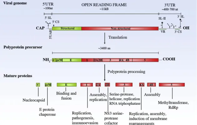
12 [M] and envelope [E]) and seven non-structural proteins (NS1, NS2a, NS2b, NS3, NS4a, NS4b and NS5), as shown on figure 3 (Sanchéz-Seco et al., 2005; Harris et al.,
2006; Hoshino et al., 2009).
Figure 3: Flavivirus genome structure and functions of viral proteins (Fernandez-Garcia et al., 2009).
Flavivirus genomes encode a large polyprotein and contain a single open reading frame (ORF) flanked by two non-coding regions (NCR) - the NCR of 5’ end is approximately
100 nucleotides (nt) in length, whereas the NCR at the 3’ end is between 400 and 700 nucleotides long (Lindenbach et al., 2007). Viruses within this genus are small (around
13 complex structure that contains a nucleocapsid, which is hexahedral/icosahedral in symmetry as shown in figure 4 (Carter et al., 2008).
Figure 4: The flavivirus virion (Petersen et al., 2001).
Flavivirus virions contain two structural proteins at its surface: the E protein (the envelope glycoprotein that surrounds the nucleocapsid) and the M protein (Barrett, 2001; Lindenbach et al., 2007). In addition, surrounding the single stranded viral RNA
genome is a small capsid protein that has a basic charge in order to interact with the genome (Barrett, 2001).
The envelope glycoprotein E is the major antigenic determinant for the production of neutralizing antibodies that relate to a protective immune response (McMinn, 1997). Furthermore, it mediates receptor binding and membrane fusion, through its cellular receptor binding sites and fusion peptides (Lindenbach et al., 2007).
14
Figure 5: Flavivirus particle structure. A- Envelope proteins of immature and mature virions;
B- Immature dengue virus type-2 virion; C- Mature dengue virus type-2 virion
(adapted from Lindenbach et al., 2007).
1.3.2. Replication cycle
The molecular biology of flavivirus has been actively studied, as the Flaviviridae family
comprises many viruses with major medical importance. A schematic representation of the viral replication cycle (Lindenbach and Rice, 2003) is shown in figure 6.
Flavivirus virions adhere to the surface of the host cell and enter it by means of receptor-mediated endocytosis via attachment to high affinity cellular receptors specific for viral envelope proteins (unknown for most viruses) (Mukhopadhyay et al., 2005;
Harris et al., 2006; Lindenbach et al., 2007). The low pH of the endosomal vesicles
triggers the particles to undergo conformational changes and induces viral fusion with host cell membranes and virus disassembly, causing the uncoating and release of the virus nucleocapsid into the cytoplasm (Lindenbach and Rice, 2003; Mukhopadhyay et
al., 2005; Harris et al., 2006; Lindenbach et al., 2007). It is thought that viral genomes
15 polyprotein that is cleaved by viral and host proteases (Mukhopadhyay et al., 2005;
Lindenbach et al., 2007).
Receptor binding and endocytosis
Acid catalysed fusion and uncoating
Translation and polyprotein processing
Viral genome replication
Virus assembly Virus maturation
Exocytosis
Figure 6: Flavivirus replication cycle (adapted from Fernandez-Garcia et al., 2009).
The translation process yields proteins that play an important role in the replication of the viral genome and the formation of new virus particles (Lindenbach and Rice, 2003). Genome replication takes place on intracellular membranes, more specifically on cytoplasm replication complexes associated with perinuclear membranes (Lindenbach and Rice, 2003; Lindenbach et al., 2007). Virions are thought to assemble by budding
into an intracellular membrane compartment, in the lumen of the endoplasmic reticulum (ER), resulting in the formation of immature non-infectious viral particles containing E and prM proteins, nucleocapsid and lipid membrane thus making them unable to induce host-cell fusion (Mukhopadhyay et al., 2005; Lindenbach et al., 2007). These immature
16 mature infectious virions (Mukhopadhyay et al., 2005). Finally, the mature virions are
ready to be released from the host cell by exocytosis (Mukhopadhyay et al., 2005).
1.3.3. Flavivirus classification: phylogeny
The genus Flavivirus is unique in the Flaviviridae family since, contrarily to genus
Hepacivirus and Pestivirus, its members present the ability to infect and replicate in
vertebrate and invertebrate host cells and display genetic, epidemiological and ecological characteristics that are distinct from the other two genera (Gould et al.,
2003). Even though flaviviruses are known to be related, based on phylogenetic and antigenic analysis, the members of this genus characteristically present high genetic divergence thus emphasizing that these correlations are not simple, nor are they always clear (Kuno et al., 1998; Gaunt et al., 2001; Gould et al., 2003; Cook and Holmes,
2006). The evolutionary process may have contributed greatly to the genetic diversity and divergence among these viruses, through factors such as the gradual adaptation to new hosts, new geographical areas and genetic alterations, which might still be occurring these days (Cook and Holmes, 2006).
The genus Flavivirus is considered to be an invaluable model for the evolutionary
investigation of vector-borne diseases and their modes of transmission, since it has been confirmed by many publications that the viral transmission mode is strongly related to virus phylogeny (Gaunt et al., 2001; Cook and Holmes, 2006). In order to understand
the origin, and spread patterns of emerging and re-emerging diseases, it is essential to gather valuable knowledge by the analysis of the evolutionary history of flaviviruses (Kuno et al., 1998; Gaunt et al., 2001). Genetic characteristics were typically
investigated based on antigenic cross reactivity, haemagglutination and complement fixation tests (Gaunt et al., 2001). More recently, investigators have implemented the
use of other tools such as molecular sequencing, and phylogenetic data analysis, in order to understand and solve taxonomic issues, thus allowing viruses to be correctly assigned positions within their respective genus (Gaunt et al., 2001).
17 vector viruses (NKV) (figure 7). Moreover, these groups of viruses can be subdivided into several clades determined by their epidemiology, clinical condition caused, type of preferred host and their geographic distribution (Kuno et al., 1998; Gaunt et al., 2001;
Gould et al., 2003; Cook and Holmes, 2006).
Figure 7: Phylogeny of the Flavivirus genus based on analysis of the NS5 gene
(adapted from Gaunt et al., 2001).
Many previous studies have found their phylogenetic results to be questionable due to inconsistency in the positions occupied by the three main transmission groups; this is thought to originate mainly from taxonomic and virus nomenclature errors (Kuno et al.,
18 virus and many others that are not incorporated into any of the serological complexes that flaviviruses are divided into (Kuno et al., 1998). For instance, in the study carried
out by Kuno et al (1998), phylogenetic analysis of sequences obtained from the E and
NS5 genes yielded conflicting results. Despite the similarities in the overall division and positioning of viruses, there were some exceptions, as was the case for some bat-related viruses that were included in the mosquito-transmitted clade, grouped together with yellow fever and Sepik viruses (figure 7, red box) (Kuno et al., 1998; Gaunt et al.,
2001).
To the contrary, there are also viruses that do not display clear relationships to other members and are placed within this genus (Kuno et al., 1998). Tamana bat virus and
cell-fusing agent are good examples of “candidate” viruses occupying uncertain positions within the genus Flavivirus (Gould et al., 2003). Despite many attempts, the
doubts and uncertainty surrounding the flaviviruses phylogenetic relationships still linger on (Cook and Holmes, 2006).
Flavivirus classification through molecular genetics has been undertaken using different methods and targeting different viral genes (Gould et al., 2003; Cook and Holmes,
2006). Viral sequences obtained from the envelope (E) gene, NS3, NS4, and NS5 have been used for phylogenetic inference. However, recent work has been mainly directed at the analysis of the most conserved region of the flavivirus genome (the NS5 coding sequence), as well as their complete sequence (Cook and Holmes, 2006).
The analysis of sequences gathered from distinct genes indicates an important separation between the mosquito-borne viruses from those transmitted by ticks (Cook and Holmes, 2006). According to the study carried out by Kuno et al (1998), the
non-vectored and vector-borne virus groups were the first to have emerged, assuming cell-fusing agent virus (CFAV) as sharing a common ancestor with which other members of the genus Flavivirus. Following these events, the vector-borne group was split into
tick-borne and mosquito-tick-borne clades (Kuno et al., 1998; Cook and Holmes, 2006). Based
19 Furthermore, other associations have resulted from this type of analysis, as is the case of the mosquito-borne clade that was subdivided into a further two groups according to the mosquito vector species, their vertebrate hosts and the nature of the disease caused (Kuno et al., 1998; Gaunt et al., 2001). As shown on figure 7, the mosquito-borne
viruses were clearly separated into two clades: the viruses isolated from Aedes species
and those from Culex spp. (Gaunt et al., 2001). Besides, when other characteristics such
as the sort of disease caused were included in the analysis, the generated tree revealed a solid association between the Culex clade and the neurotropic viruses causing
encephalitic disease; and between the Aedes clade and the non-neurotropic viruses that
tend to produce hemorrhagic disease (Gaunt et al., 2001; Gould et al., 2003). In
addition, correlations were established based on host preference, linking Culex species
mosquitoes with birds, and Aedes mosquitoes with primates (Gaunt et al, 2001; Gould
et al., 2003).
Although no-known vector (NKV) viruses have not been thoroughly examined, the results from many studies tend to support the hypothesis that they have evolved away from the vector-borne group of viruses (Kuno et al., 1998; Gaunt et al., 2001; Gould et
al., 2003; Cook and Holmes, 2006). Sequence analysis of the NS5 gene produces a
phylogenetic tree that separates the NKV group into three, the rodent and bat clades and
a “basal lineage” composed of an individual virus – the APOI virus that is linked to rodents and therefore would later be added to the rodent-borne clade (Kuno et al., 1998;
Gaunt et al., 2001). However, as mentioned before, some members of this group were
included into the mosquito-borne clade, which suggests that during the flavivirus evolution, and following the divergence of the NKV viruses from the vector-borne viruses, there was a consequent loss of vector-borne transmission (Gould et al., 2003;
Cook and Holmes, 2006). Phylogenetic analysis was not conclusive as to which group was the most divergent (Gould et al., 2003). Contrarily, more recently it was concluded
that the NKV viruses group is indeed the most divergent of the three main flavivirus groups (Cook and Holmes, 2006).
20 Holmes, 2006). The latter is said to be linked to rodent hosts and Ixodes spp. ticks in
woodland areas (Gaunt et al., 2001; Gould et al., 2003). With the exception of Omsk
haemorrhagic fever virus (OHFV) and Kyasanur Forest disease virus (KFDV) that tend to cause haemorrhagic disease, infections by flaviviruses within this group primarily produces encephalitic disease (Gaunt et al., 2001).
Figure 8: Phlylogenetic tree of tick-borne flaviviruses based on the analysis of NS3 gene (Mansfield et al., 2009).
1.4. Medically Important Pathogenic Flaviviruses
Included into the Flavivirus genus are some of the most virulent and medically
important viruses known to medical science including, amongst others, the Dengue viruses (DENV- 1, DENV- 2, DENV- 3 and DENV- 4), Yellow Fever virus (YFV) and West Nile virus (WNV) (Harris et al., 2006; Junglen et al. 2009; Calzolari et al., 2010).
21 Hypothetically, in case of continued Ae. albopictus geographic expansion (especially
northwards) and Aedes aegypti reestablishment, combined with changes to ecological
conditions as a consequence of climate change, there may be a risk of eventual pathogenic flaviviruses transmission in Europe (Reiter, 2010). This may be the case of DENV and YFV since the historical records of these diseases in the continent confirm that the conditions existent are suitable for their transmission (Reiter, 2010).
1.4.1. Dengue and Yellow Fever
1.4.1.1. Epidemiology
Viruses of the genus Flavivirus are the causative agents of both dengue and yellow
fever, which are associated with Aedes spp mosquitoes and cause haemorrhagic disease
in humans and simians (Endy et al., 2010; Reiter, 2010). These viruses are considered
the most important arboviral pathogens that, for a long time, have been causing disease around the world (Endy et al., 2010; Weaver et al., 2010). Infection by DENV and YFV
can be manifested as a mild or potentially fatal disease, and it can take a mere seven to ten days of disease before death occurs (Gould and Solomon, 2008; Endy et al., 2010;
Weaver and Reisen, 2010).
The main factors that contributed to the great geographical dispersal of Aedes aegypti
and Aedes albopictus, the principal vectors of dengue and yellow fever, include the
commercial transportation of goods and people during the slave trade, particularly by ship (Endy et al., 2010). This resulted in the evolution and adaptation of these vectors to
urban environments and, consequently, in epidemics affecting particularly port cities (Reiter, 2010; Weaver and Reisen, 2010). Northern cities such as Dublin (Ireland) and Boston (North America) were unexpectedly hit by these epidemics (Reiter, 2010).
22
Figure 9: Dengue risk areas, 2010(http://www.wpro.who.int/health_topics/dengue/– accessed on the 5th July 2011).
Dengue is an emerging disease that has shown dramatic geographic and incidence increase in the past few decades, and has caused large epidemics in the Americas in 2007 (890,000 cases) and in Greece in 1927-28 (over one million cases) (Endy et al.,
23
Figure 10: Geographic distribution of yellow fever.
(http://www.cdc.gov/mmwr/preview/mmwrhtml/rr5907a1.htm – accessed on the 7th of July 2011).
Reports state that the true incidence of this haemorrhagic disease is unknown, and the real figure is thought to be far higher (Petersen and Marfin, 2005). Despite the number of people affected, this virus is the only flavivirus for which a vaccine is available internationally, and nevertheless, it may be of too high a cost for endemic countries to create a standard vaccination programme against the disease (Reiter, 2010).
1.4.1.2. Transmission cycle
Dengue and yellow fever viruses are both transmitted by Aedes spp mosquitoes that are
characterised as anthropophilic1 and live in close association with domestic and peridomestic environments (Reiter, 2010). Originally, dengue and yellow fever shared an enzootic/sylvatic cycle that consisted in the transmission of viruses between sylvatic
Aedes mosquitoes and primates (Endy et al., 2010; Reiter, 2010). Nowadays, dengue is
mainly transmitted via the direct/urban transmission cycle between mosquitoes and humans (figure 11), perhaps because of the adaptation of Aedes aegypti to urban
environments (Endy et al., 2010; Reiter, 2010).
24
Figure 11: Dengue and yellow fever transmission cycle.
(http://www.stanford.edu/group/parasites/ParaSites2008/Nkem_Cristina%20Valdoinos/ugonabon_ valdovinosc_dengueproposal.htm – accessed on the 6th July 2011).
Yellow fever can be transmitted via three different cycles (figure 12). In the sylvatic cycle YFV is transmitted between primates and forest-dwelling mosquitoes, humans become infected when in contact with the sylvatic cycle through forest invasion and the virus can then be transmitted via the urban cycle between infected humans and Ae.
aegypti (CDC, 2010b). The intermediate cycle which occurs only in Africa, is
maintained between primates/humans and Aedes spp mosquitoes which bite humans
who live or work in the forest periphery (CDC, 2010b).
Figure 12: Yellow fever transmission cycles.
25
1.4.2. West Nile Virus
1.4.2.1. Epidemiology
West Nile virus (WNV) is a mosquito-transmitted flavivirus that belongs to the Japanese encephalitis antigenic complex (Hubalek et al., 1999; Petersen and Roehrig,
2001). This virus was isolated in 1937 for the first time in the West Nile Province in Uganda, from the blood of a woman presenting with classical systemic febrile illness (Hubalek et al., 1999; Monini et al., 2010). The virus was later recognized and isolated
from mosquitoes, birds and humans in the 1950’s, in Egypt, and also in France (1962-63). During the 1970s-1990s WNV caused isolated outbreaks in countries such as South Africa (1974), India (1980-81) and Romania (1996) (Hubalek et al., 1999; Zeller and
Schuffenecker, 2004; Monini et al., 2010). Since its first isolation, WNV has been
considered one of the most widely spread flaviviruses thus becoming an increasing public health and veterinarian problem all over the globe (Zeller and Schuffenecker, 2004; Monini et al., 2010). The virus already affects several European countries,
predominantly in the Mediterranean region, whereas in the Americas its transmission has been recorded from Canada to Argentina, after its recent introduction in the United States (figure 13) (Zeller and Schuffenecker, 2004; Monini et al., 2010).
Figure 13: West Nile distribution in North America, as of 15th of November 2011.
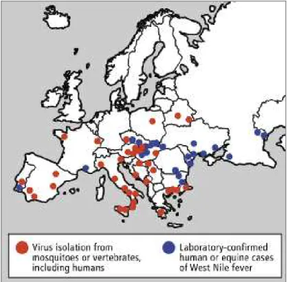
26 The virus is thought to have entered the United States through viraemic migratory birds or imported domestic birds, however the exact means of entry are unclear (Epstein and Defilippo, 2001; Solomon and Mallewa, 2001). WNV is the causative agent of West Nile fever and has been spreading globally (figure 14) using migratory birds as dispersal vehicles since it was first documented in Africa in the 1930’s (Zeller and Schuffenecker, 2004; Gould and Solomon, 2008).
Figure 14: Approximate West Nile virus distribution map, 2006 (Gubler, 2007).
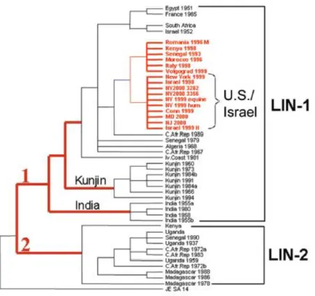
Most known viral strains have been distributed into two main genetic lineages (figure 15), both clearly correlating to the virus geographic distribution (Zeller and Schuffnecker, 2004; Monini et al., 2010).
Viruses in lineage 1 are classified as more virulent and broadly distributed through Africa, Australia, India, Asia and Europe, mainly in Mediterranean countries (Monini et
al., 2010) In the other hand, WNV strains composing lineage 2 have kept a more strict
distribution in Sub-Saharan Africa (Petersen and Roehrig, 2001; Zeller and Schuffenecker, 2004; Petersen and Marfin, 2005; Weaver and Reisen, 2010). However, they have recently been found circulating in Hungary and Greece (Zeller et al., 2010;
27 associated with severe human disease, including cases of clinical human encephalitis (Petersen and Marfin, 2005; Petersen and Roehrig, 2001).
Figure 15: Phylogenetic tree of West Nile viruses based on the analysis of the envelope (E) gene
(Gubler, 2007).
West Nile fever outbreaks affecting humans and/or animals have occurred all over the globe, in both tropical and temperate regions (Almeida et al., 2008). In 1996-1997,
Romania had the largest outbreak of arboviral disease seen in Europe, where more than 600 people presented neurological complications of which nearly 10% died (Hubalek, 1999; Solomon and Mallewa, 2001).
Other outbreaks have occurred in Israel in the 1950’s and in 2000, South Africa in the
28 causing 60 clinical cases of encephalitis leading to 7 human deaths with a dramatically higher equine and bird mortality rate (figure 16) (Hubalek, 1999; Zeller and Schuffenecker, 2004; Gould and Solomon, 2008).
Figure 16: West Nile virus epidemics between 1937 and 2006 (Gubler, 2007).
Red stars represent epidemics associated with severe neurological disease in both humans and animals.
More recently, nine cases of WNV infection were reported in Italy in 2010, six of which were symptomatic (Barzon et al., 2011). Three of the patients presenting symptoms
were confirmed to have developed neurological disease (Barzon et al., 2011). In Greece
35 deaths from 262 human cases were reported in that same year (Chaskopoulou et al.,
29
1.4.2.2. Transmission cycle
WNV is mainly transmitted by mosquito species belonging to the Culex genus via two
possible modes of transmission, the rural and the urban cycles (figure 17) (Hubalek, 1999; Monini et al., 2010).
Figure 17: West Nile virus transmission cycle and possible modes of dispersal (Pfeffer and Dobler, 2010).
In a rural setting, the primary transmission cycle occurs involving competent ornitophilic2 Culex species mosquitoes and migratory and local bird species that act as
reservoirs as well as amplifying hosts whilst humans and horses represent accidental
“dead-end” hosts, since they do not reach the level of viraemia needed for mosquito
30 infection (Solomon and Mallewa, 2001; Petersen and Marfin, 2005; Monini et al., 2010;
Weaver and Reisen, 2010).
The urban cycle occurs in a similar way, however it does involve different vector and bird species; for example, in this transmission mode, the principal vectors are Culex
pipiens and Cx. molestus seen as both species feed on synanthropic3, domestic birds and
humans (Hubalek, 1999).
Moreover, the main WNV vectors vary according to their geographic distribution, for example, the predominant vector species in Europe are Culex pipiens, Cx. molestus and
Coquillettidia richiardii (Hubalek, 1999). In Africa and The Middle East, the most
important vector is Culex univittatus, whereas in Asia, Culex quinquefasciatus, Cx.
tritaeniorhyncus and Cx. vishnui are the main vectors (Hubalek, 1999).
Although the rural cycle is of utmost importance for WNV transmission, this flavivirus can also be transmitted via organ transplants, blood transfusions and via infectious maternal milk (Zeller and Schuffenecker, 2004; Gould and Solomon, 2008). Additionally, reports show that transovarial transmission occurs in some species, namely Cx. univittatus, Cx. tritaeniorhyncus, Aedes aegypti and Ae. albopictus
(Hubalek,1999).
1.4.2.3. Clinical features
West Nile disease was at first considered a minor health concern, as the majority of infections were mild or completely asymptomatic; however, after the major outbreaks that occurred in Europe and especially in North America from 1999, WN disease became a public health priority (Zeller and Schuffenecker, 2004; Gould and Solomon, 2008).
After an incubation period of 3 to 14 days, between 15-20% of humans present with a mild febrile illness accompanied by flu-like symptoms, a rapid onset of moderate to high fever, headache, malaise, myalgia, anorexia, nausea, backache and retro-orbital
31 pain (Hubalek, 1999; Zeller and Schuffenecker, 2004; Gould and Solomon, 2008). Other disease manifestations include lymphadenopathy, conjunctivitis, maculopapular or roseolar rash, which is normally observed in 50% of patients (Hubalek, 1999; Gould and Solomon, 2008). Around 1% of all patients tend to develop neurological signs such as acute aseptic meningitis, encephalitis or myelitis4 (Hubalek, 1999; Zeller and Schuffenecker, 2004). Additionally, severe infections may also provoke hepato- and splenomegaly, hepatitis, pancreatitis and myocarditis (Zeller and Schuffenecker, 2004; Gould and Solomon, 2008). A mortality rate of 5 to 10% usually results from all patients presenting with neurological symptoms (Gould and Solomon, 2008).
During the outbreaks in North America, Romania, Russia and Israel mortality rates of 5% to 14% among neurologically affected patients were registered, as well as other symptoms such as serious motor complications, skin rash and lymphadenopathy (Petersen and Roehrig, 2001). In the Romania outbreak alone, 17 fatalities resulted from 352 neurological affected patients, from whom 44% presented with meningoencephalitis, a further 40% had meningitis and the remaining 16% suffered from encephalitis (Zeller and Schuffenecker, 2004). In accordance with most studies, fatalities are more likely to arise among patients over 50 years of age (Zeller and Schuffenecker, 2004; Gould and Solomon, 2008).
1.5. Insect-specific flaviviruses
This group of flaviviruses have had many designations throughout the years, from “non
-classical” to “mosquito-only” flaviviruses. However, as the latter designation is somehow conflicting since insect-specific flaviviruses have been detected in field caught phlebotomine sand flies and ticks it completely invalidates the continued use of that designation, hence in this study it is referred to as insect-specific flaviviruses.
As previously mentioned, most viruses within the genus Flavivirus are arthropod-borne,
and only a few have no-known vectors (Crabtree et al., 2003; Cook et al., 2003; Sang et
al., 2003; Mukhopadhyay et al., 2005). However, the increasing surveillance and
investigations into these pathogens have lead to a better understanding of their genetics,
32 classification and phylogenetic relationships (Sang et al., 2003). It is now common
knowledge that pathogenic flaviviruses do not exhaust the Flaviviridae family, as it also
harbours another type of viruses that are, so far, not known to be medically important (Blitvitch et al., 2009). On the contrary, they are thought to be insect-specific since they
replicate in mosquitoes but have not yet been detected in a vertebrate host. They are, thus, designated by insect-specific flaviviruses (ISFs) (Blitvitch et al., 2009). ISFs are a
heterogeneous group of highly diverse and widely geographically dispersed viruses.
Even though most ISFs have only been described and classified over the past decade, it is certain that they have existed for much longer. For example, the first one was found many years ago and was referred to as “an agent in the Ae. aegypti cell line that causes
fusion of Ae. albopictus cells” by Stollar and Thomas (1975), and was, as a result,
named cell-fusing agent virus (CFAV). This insect-specific flavivirus causes distinguishable massive syncytia formation, an effect that had only been observed with JEV, WNV and DENV (Stollar and Thomas, 1975; Igarashi et al., 1976). However, it
was stated that syncytium formation was not observed immediately after infection, instead, it became evident after 60 hours of cell infection and, after 72 hours more than 90% of the cells in culture would be fused having formed large syncytia (Stollar and Thomas, 1975). Infection of mammalian cells with CFAV resulted in no cytopathic effect (CPE), including cell fusion. Indeed, the virus does not seem to replicate at all, thus confirming that it is insect-specific (Stollar and Thomas, 1975; Sang et al., 2003).
In addition, replication in Ae. aegypti cells is not cytopathic (Stollar and Thomas, 1975).
Further analysis revealed that CFAV did not cross react with other known flaviviruses (Igarashi et al., 1976). CFAV was described as similar to togaviruses in size and
morphology, whereas other characteristics indicated strong similitude to flaviviruses (Igarashi et al., 1976). However, despite all evidence and tests, the classification of
CFAV remains unresolved.
The isolation of a new flavivirus described as CFA-like from mosquitoes collected in
33 This virus, named Kamiti River virus (KRV), was isolated from Ae. macintoshi
immature mosquitoes and was temporarily classified as a flavivirus (Crabtree et al.,
2003). The fact that KRV was isolated from adult male and female mosquitoes that were collected, in nature, in their immature forms, is indicative of the maintenance of this virus via transovarial transmission between generations. Nonetheless, there also remains the possibility that the virus may have been acquired by larval ingestion (Crabtree et al., 2003).
Like CFAV, only replicated in mosquito cells and presented no antigenic cross-reaction with other arboviruses (Sang et al., 2003). However, contrarily to CFAV, there was no
cell fusion observed in culture, despite the morphologic and genomic similarities (Crabtree et al., 2003). The genomic organisation of KRV was found to be similar to
that of other flaviviruses, though its nucleotide sequence was considerably longer (11,375 nt) (Crabtree et al., 2003). Two viral strains were isolated and their RNA
sequences compared which revealed they were virtually identical, thus suggesting they represent the same virus; however, when comparison was extended to other flaviviruses a low sequence identity was observed (Crabtree et al., 2003; Sang et al., 2003). KRV
showed maximum identity to CFAV based on nucleotide and amino acid sequence analysis of the NS3 and NS5 genes (Crabtree et al., 2003; Sang et al., 2003). Both KRV
isolates produced cytopathic effect5 (CPE) in Ae. albopictus cells (C6/36) in culture.
However, the same was not observed with Vero cells (monkey kidney cell line) or in baby hamster kidney cells (BHK-21), as expected (Crabtree et al., 2003; Sang et al.,
2003).
Some years later, the discovery of 40 CFA-like viral sequences from adult mosquitoes collected in Puerto Rico after the rainy season of 2002 was reported (Cook et al., 2006).
These were obtained from male and female mosquitoes of different species, namely Ae.
aegypti, Ae. albopictus and Culex sp. The sequences analysed were found to represent
different strains of CFAV and, therefore have been referred to as CFAV Culebra stains (Cook et al., 2006).
34 In addition to CFAV and KRV, other insect-specific flaviviruses have been detected and isolated. However, the new isolate seems to be associated only to Culex sp. mosquitoes,
especially Cx. pipiens, hence their name Culex flavivirus (CxFV) (Hoshino et al., 2007).
The genome of this new flavivirus is 10,834 nucleotides long, also flanked by two untranslated (UTR) regions, as described for other flaviviruses (Hoshino et al., 2007).
Infection of mosquito C6/36 cells in culture resulted in moderate CPE visible 4 days post-infection. In addition, no viral replication was detected in mammalian cell lines (Hoshino et al., 2007). Phylogenetically, CxFV showed greater similarities with CFAV
and KRV than with other mosquito-transmitted flaviviruses (Hoshino et al., 2007).
Alike the CFA-like viruses and KRV, CxFV have been detected in healthy wild-caught mosquitoes, thus suggesting once again that vertical transmission occurs in the wild, and despite causing moderate CPE in culture, it does not seem to negatively affect their hosts. (Hoshino et al., 2007).
A virus similar to CxFV found in Japan was identified in Guatemala and was later named CxFV Izabal 2006; it was isolated from Culex quinquefasciatus collected from
urban and rural areas between March and October 2006 (Morales-Betoulle et al., 2008).
Analysis of the envelope and NS5 gene sequences were the basis for characterisation of the detected viral strains which, after phylogenetic assessment were classified as strains of the CxFV species following a result of 89% nucleotide homology with the Japanese virus (Morales-Betoulle et al., 2008). The isolates were cultured into C6/36 and Vero
cells, with no CPE observed for either (Morales-Betoulle et al., 2008).
In addition to the diversity of this heterogeneous group of flaviviruses previously identified, Crabtree et al (2009) have also found a new insect-only flavivirus from Cx.
tritaeniorhyncus captured in Vietnam in August 2002. These virus, denominated Quang
Binh virus (QBV) was described as genetically different from pre-existing flaviviruses and though its complete genome had similar size (10,865 nt) and genomic organisation (Crabtree et al., 2009). Sequence analysis revealed that Quang Binh virus had over 60%
35 Meanwhile, a new ISF of Asian origin was discovered and designated Aedes flavivirus
(AEFV) (Hoshino et al., 2009). Contrarily to most pre-existing flaviviruses, AEFV was
isolated from Aedes and not Culex mosquitoes, namely Ae. albopictus and Ae.
flavopictus collected from Japan and Indonesia (Hoshino et al., 2009). However, other
flavivirus-like sequences were also obtained (Hoshino et al., 2009). Infection of C6/36
produced moderate CPE after 4 days, whereas, as expected for ISFs, mammalian cells showed no such effects (Hoshino et al., 2009). AEFV was therefore classified a
flavivirus with similar replication and translation mechanisms; furthermore, it was stated that it showed high virus-host adaptation to its invertebrate host, an Aedes
mosquito (Hoshino et al., 2009).
AEFV’s genome mirrors that of flaviviruses and its polyprotein is thought to be divided into 3 structural (C, prM and E) and seven non-structural proteins (NS1, NS2A, NS2B, NS3, NS4A, NS4B and NS5) (Hoshino et al., 2009). AEFV was classified as a new
species of insect flavivirus, particularly similar to KRV (Hoshino et al., 2009).
Insect-only flaviviruses previously characterised have been isolated from Aedes and
Culex mosquitoes; however, a study carried out by Cook and others (2009) reports the
first isolations of a ISF from a Mansonia species mosquito and an African strain of
CxFV. Both new strains, namely CxFV Uganda and Nakiwogo virus (NAKV) were obtained from mosquitoes collected in the same trap (CDC-type, with CO2) on the 24th
of February 2008 in Entebbe, Uganda (Cook et al., 2009). The NAKV and CxFV
Uganda strain were isolated from Mansonia africana nigerrima and Cx.
quinquefasciatus, respectively (Cook et al., 2009). The genomes of both viruses were
found to be 10,092 nt long for CxFV, and 10,122 nt long for NAKV (Cook et al., 2009).
These viruses were morphologically identical to flaviviruses, and phylogenetic analysis revealed that CxFV Uganda to be more closely related to the Mexico strain, while NAKV was inserted into the ISF clade, but as a sister group (figure 18) (Cook et al.,
2009).
36
Figure 18: Maximum-likelihood tree showing relative positions of CxFV Uganda and NAKV within the
Flavivirus genus, based on NS5 gene analysis (Cook et al., 2009).
A study conducted by Junglen et al. (2009) revealed the discovery of a novel flavivirus
particle isolated from Uranotaenia mashonaensis from the tropical rain forest, in Côte
37 CFAV and KRV (Junglen et al., 2009). As observed for ISFs, viral replication was
observed only in insect-cell culture (Junglen et al., 2009).
Further away, in North America many other flavivirus-like sequences have been found. For example, as a result of a surveillance programme in Alberta (Canada), some flaviviruses were detected, the majority were isolated from Culex tarsalis mosquitoes
while a lower number was obtained from non-Culex mosquitoes; these sequences
showed higher similarities to KRV (Pabbaraju et al., 2009). In Iowa, again as part of the
surveillance programme, mosquitoes were collected between May and October 2007, of which Cx. pipiens and Cx. tarsalis pools turned out positive for flaviviruses (Blitvich et
al., 2009). Analysis of the sequences obtained revealed that the isolates had 98%
homology with CxFV isolated in Japan but also shows close relation to the Texas and Mexico strains (Huhtamo et al., 2009). It was characterised as having a 10,089 long
genomic sequence and production of moderate CPE and cell clumping were observed in culture with insect cell lines only (Huhtamo etal., 2009).
Once again, it was due to arboviral surveillance programmes that flavivirus strains have been found and isolated from mosquitoes collected from January to December 2007, in the Yucatan Peninsula of Mexico (Farfan-Ale et al., 2009). Several of the analysed
pools tested positive for flaviviruses, the majority of them showing identical sequences to that of CxFV (Farfan-Ale et al., 2009). A new strain of CxFV closely related to the
Japanese strain (figure 19) was isolated from Cx. quinquefasciatus, and as some pools
consisted only of male specimens, this suggested, again, vertical transmission in nature (Farfan-Ale et al., 2009). However, contrarily to previously isolated strains of CxFV,
this virus did not produce CPE on C6/36 cells (Farfan-Ale et al., 2009).
It is noteworthy that CxFV presented a high prevalence, as it was found in 145 of 146 pools from which flavivirus RNA was detected (Farfan-Ale et al., 2009; Kim et al.,
38
Figure 19: Phylogenetic tree showing the position of the newly isolated CxFV and T’Ho virus, based on
the analysis of a fragment of the NS5 gene (Farfan-Ale et al., 2009).
Collections made during February and March 2008 in Texas (USA) and Trinidad yielded many pools positive for flaviviruses that had been obtained from Cx. restuans
and Cx. quinquefasciatus females (Kim et al., 2009). From these positive pools, it was
possible to isolate nine strains from both locations, however only one, designated by TX24518, was selected for further analysis (Kim et al., 2009). Inoculation of some of
39 replication or CPE, thus suggesting that they belong to the insect-only flavivirus group (Kim et al., 2009).
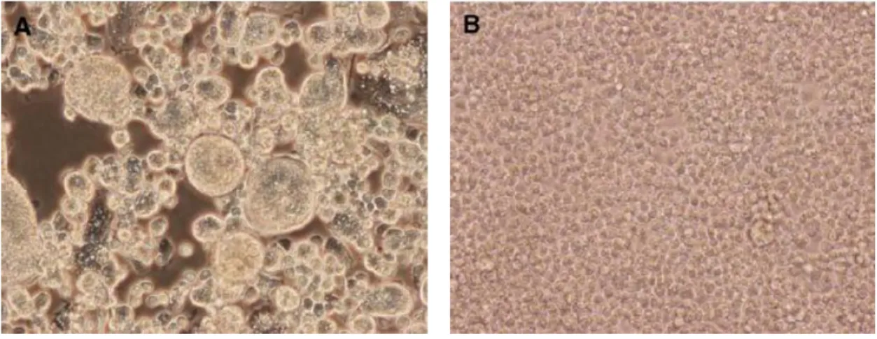
TX24518 was inoculated into C6/36 cells, which resulted in the formation of large syncytia as observed for CFA (figure 20).
Figure 20: A) Cell fusion and large syncytia present in C6/36 cells, 7 days post-infection with TX24518; B) Uninfected C6/36 cells (control) (Kim et al., 2009).
A polyprotein similar to those of the Flavivirus members was identified, consisting of
three structural and seven non-structural proteins, and its genome was found to be 10,089nt long (Kim et al., 2009). Sequence identity values revealed TX24518 to have
higher similarities to CxFV-Tokyo than to CFAV or KRV, based on protein analysis; the other isolates from Houston were more similar to Japanese CxFV strains, while the Trinidad isolates showed higher identity to the Izabal virus isolated in Guatemala (Kim
et al., 2009).
Kim and others (2009) concluded that the isolates from both locations represented new genetic variants of CxFV and were genetically distinct from CFA and KRV; moreover, it was suggested that the activity of these viruses in their arthropod vectors is seasonal, as continued testing did not detect any more flavivirus-positive pools.
After the isolation of cell-fusing agent and Kamiti river viruses, many other insect-specific flaviviruses have been described, classified and incorporated into the Flavivirus