Fat Content and Nitrite-Curing Influence the Formation
of Oxidation Products and NOC-Specific DNA Adducts
during
In Vitro
Digestion of Meat
Thomas Van Hecke1, Els Vossen1, Julie Vanden Bussche2, Katleen Raes3, Lynn Vanhaecke2, Stefaan De Smet1*
1Laboratory for Animal Nutrition and Animal Product Quality, Department of Animal Production, Faculty of Bioscience Engineering, Ghent University, Melle, Belgium, 2Laboratory of Chemical Analysis, Department of Veterinary Public Health and Food Safety, Faculty of Veterinary Medicine, Ghent University, Merelbeke, Belgium, 3Laboratory for Food Microbiology and Biotechnology, Department of Industrial Biological Sciences, Faculty of Bioscience Engineering, Ghent University – Campus Kortrijk, Kortrijk, Belgium
Abstract
The effects of fat content and nitrite-curing of pork were investigated on the formation of cytotoxic and genotoxic lipid oxidation products (malondialdehyde, 4-hydroxy-2-nonenal, volatile simple aldehydes), protein oxidation products (protein carbonyl compounds) and NOC-specific DNA adducts (O6-carboxy-methylguanine) duringin vitrodigestion. The formation of these products during digestion is suggested to be responsible for the association between red meat and processed meat consumption and colorectal cancer risk. Digestion of uncured pork to which fat was added (total fat content 5 or 20%), resulted in significantly higher lipid and protein oxidation in the mimicked duodenal and colonic fluids, compared to digestion of pork without added fat (1% fat). A higher fat content also significantly favored the formation of O6 -carboxy-methylguanine in the colon. Nitrite-curing of meat resulted in significantly lower lipid and protein oxidation before and after digestion, while an inconsistent effect on the formation of O6-carboxy-methylguanine was observed. The presented results demonstrate that haem-Fe is not solely responsible for oxidation and nitrosation reactions throughout anin vitrodigestion approach but its effect is promoted by a higher fat content in meat.
Citation:Van Hecke T, Vossen E, Vanden Bussche J, Raes K, Vanhaecke L, et al. (2014) Fat Content and Nitrite-Curing Influence the Formation of Oxidation Products and NOC-Specific DNA Adducts duringIn VitroDigestion of Meat. PLoS ONE 9(6): e101122. doi:10.1371/journal.pone.0101122
Editor:Roger A. Coulombe, Utah State University, United States of America ReceivedFebruary 5, 2014;AcceptedJune 3, 2014;PublishedJune 30, 2014
Copyright:ß2014 Van Hecke et al. This is an open-access article distributed under the terms of the Creative Commons Attribution License, which permits unrestricted use, distribution, and reproduction in any medium, provided the original author and source are credited.
Funding:This study was financed by the Federal Public Service of Health, Food Chain Safety and Environment, Belgium (grant RF-11/625 MEATNOX) (http:// www.health.belgium.be/eportal). L. Vanhaecke is supported by a postdoctoral fellowship from the Research Foundation - Flanders (FWO) (http://www.fwo.be/ Postdoctorale-mandaten.aspx). The funders had no role in study design, data collection and analysis, decision to publish, or preparation of the manuscript. Competing Interests:The authors have declared that no competing interests exist.
* Email: [email protected]
Introduction
Several meta-analyses have reported a significant epidemiolog-ical association between colorectal cancer (CRC) and high intake of red and processed meat [1,2]. However, the biochemical mechanisms underlying this association have not been completely elucidated yet. To date, the formation of cyto- and genotoxic oxidation products and genotoxic N-nitroso-compounds (NOCs) during digestion are considered the most plausible factors contributing to the increased risk of developing CRC when consuming large amounts of red and in particular processed meat [3]. Meat contains proteins, unsaturated lipids, haem-Fe and in case of processed meat also nitrite, which are all compounds involved in oxidation and nitrosation processes.
Major cyto- and genotoxic aldehydes such as malondialdehyde (MDA) and 4-hydroxy-2-nonenal (4-HNE), and less cytotoxic simple aldehydes, e.g. hexanal, are formed by free radical-induced damage to polyunsaturated fatty acids (PUFA). Lipid oxidation products are formed during processing, storage and preparation of meat, as well as during digestion in the gastro-intestinal tract. Next to fat, also proteins can be oxidized, resulting in the formation of protein carbonyl compounds (PCC). Lipid and protein oxidation products may cause oxidative stress and induce DNA damage due
to their highly reactive nature. MDA and 4-HNE are prevailing in higher concentrations in colonic mucosae of CRC patients compared to a healthy control group [4]. Also PCC were described to be higher in plasma of CRC patients compared to a control group [5].
Previously, it was shown that volunteers on a high red meat diet had higher endogenous formation of genotoxic NOCs in feces compared to vegetarians, measured as ‘apparent total nitroso-compounds’ (ATNC) [6]. Furthermore, colonic exfoliated cells in the red meat-eating group of this study displayed higher concentrations of the NOC-specific DNA adduct O6 -carboxy-methylguanine (O6-C-MeG), which were positively correlated with ATNC concentrations in feces [6]. Measurement of O6-C-MeG is far more specific than ATNC since the latter also include nitrosothiols and nitrosyl-heme in addition to the genotoxic NOCs.
Haem-Fe in meat has been shown to be the main promoter of the peroxidation and nitrosation pathway [3]. However, when rats were fed a diet rich in haem-Fe, the effect of haem on colonic epithelial proliferation and cytolytic activity of fecal water was lower in a low fat diet (4.2%), compared to diets containing more fat (11.5 and 20.3%) [7]. A lean beef diet (5.0% fat) high in
Fe did not promote colon carcinogenesis in cancer-induced rats [8]. Hence, we hypothesized that the fat content in meat might modulate the effect of haem on peroxidation and nitrosation.
Nitrite salt is widely used as a curing agent in meat products for color formation, to inhibit outgrowth ofClostridium botulinum, and to delay oxidative rancidity. However, nitrite-curing has also been suggested to increase NOC formation in meat products. Several studies have reported on the influence of nitrite-cured meats on the promotion of colorectal carcinogenesis. Rats consuming nitrite-cured hot-dogs (4.7% fat) had 3.7 to 5.0 fold higher NOC levels in feces compared to a control group, while consumption of beef (3.6% fat) resulted in a 2.0 to 2.9 fold increase [9]. A nitrite-cured ham diet increased cytotoxicity, lipid peroxidation and amount of ‘mucin depleted foci’ (MDF) in the colon of rats, compared to a control group [10]. However, fresh meat and processed meats differ in more properties than only nitrite-curing. Variation in haem-Fe content, fat content, and different processing procedures such as mincing and heating do not allow to draw conclusions on the isolated effect of nitrite-curing in previously mentioned studies. In a controlled study, Santarelli et al. [11] showed that consumption of nitrite-cured meat and oxidized meat increased ATNC and MDF in the colon of rats compared to consumption of similar non-cured and non-oxidized meat. Moreover, a human intervention study showed increased fecal ATNC when a high proportion of red meat (21.4% fat) or nitrite-cured processed meat (24.0% fat) was consumed, compared to a vegetarian diet, but no significant difference was seen between the red meat and processed meat diets [12].
Therefore, we aimed at elucidating the likely modulating effects of fat content and nitrite-curing of meat on lipid and protein oxidation markers, and the formation of NOC-specific DNA adducts during digestion. For this purpose, we used an in vitro
digestion protocol adapted from Versantfoortet al.[13], followed by a colonic fermentation step according to Van de Wieleet al.
[14].
Materials and Methods
1. Experimental set-up
Uncured and nitrite-cured heated pork samples differing in fat content (targeted at 1, 5 or 20% fat) were compared before and after mimicked duodenal and colonic digestion. Each incubation run included all treatments in quadruplicate from which 2 duplicates underwent in vitro digestion until the duodenum and the remaining 2 until the colon. Each incubation run was performed three times with fecal inoculum originating from three different persons. In total, 2 meat samples before digestion, 6 duodenal digests and 6 colonic digests were obtained for each of the 6 prepared meat products.
2. Preparation of the meat samples
Commercially available lean meat samples from the m. Longissimus dorsi of pig were purchased in a local supermarket. The loin was manually chopped into cubes of approximately 1– 2 cm3. Subcutaneous pork fat from one batch was added to the chopped meat to obtain a targeted total fat content of 1 (no fat added), 5 and 20%. Meat samples with added fat were first minced in a grinder equipped with a 10 mm plate, followed by grinding through a 3.5 mm plate. After grinding, nitrite-curing was applied by adding 20 g 0.6% nitrite salt/kg meat, corresponding to an added concentration of 120 mg nitrite/kg meat. All meat samples were heated in a warm water bath for 15 min after the core temperature had reached 65uC. After manufacturing, all meat samples were homogenized in three 5 s bursts using a food
processor, vacuum packed and stored at220uC until the start of the incubation.
3. Digestive simulations
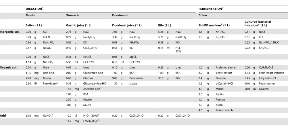
The in vitro digestions consisted of an enzymatic digestion simulating the mouth, stomach, and duodenum, followed by a simulation of the colonic fermentation. For the enzymatic digestion, the protocol described by Versantfoort et al. [13] was adapted by adding oxidants and antioxidants that are normally present in digestive juices (Table 1). Hence, peroxidase [15] and NaNO2[16] were added to the saliva juice, and ascorbic acid [17],
H2O2[18] and ferrous iron [18] were added to the gastric juice.
Meat samples (4.5 g) were sequentially incubated for 5 minutes with 6 ml saliva, 2 hours with 12 ml gastric juice, and 2 hours with 2 ml bicarbonate buffer (1 M, pH 8.0), 12 ml duodenal juice and 6 ml bile. These enzymatic incubations were performed in quadruplicate. After completion, 2 of the 4 replicates were diluted with 44 ml H2O to obtain the same solid/liquid ratio as in the
colon (see below), homogenized with an ultraturrax (9500 rpm) and stored at 220uC and 280uC pending analysis. The 2 remaining replicates underwent the additional colonic fermenta-tion stage according to Van deWiele et al.[14]. SHIME (simulator of the human intestinal microbial ecosystem) medium (22 ml) [19] and a human fecal inoculum (22 ml) (for preparation cfr. 2.4) were added to the digesta. The vessels were continuously flushed with N2for 30 minutes to obtain an anaerobic environment. Anaerobic
conditions of the flasks containing digesta at the colonic digestion stage were confirmed, using resazurin saturated test strips. Subsequently, the vessels were incubated for 72 hours while stirring at 37uC. To evaluate the rate of bacterial fermentation, total anaerobic bacteria were counted after 72 h of fermentation. One ml digestive fluid was serially diluted (10-fold) using a sterile peptone solution (1 g/l peptone, 0.4 g/l agar, 8.5 g/l NaCl and 0.5 g/l cystein), after which 0.1 ml was added on an RCM-plate (Reinforced Clostridial Medium). After 48 hours of incubation at 37uC, colony forming units (CFU) were counted and expressed as log10 CFU/ml digesta. As for the duodenal samples, colonic
digestive fluids were homogenized by ultraturrax at 9500 rpm and subdivided in 1.3 ml aliquots while stirring on a magnetic field in dark and stored at220uC and 280uC till analyses. Undigested control samples were obtained in duplicate by homogenizing 4.5 g meat in 82 ml H2O, mimicking the liquid/solid ratio in the
digested samples.
4. Preparation of human fecal inoculum
Fresh fecal material was collected from 3 volunteers without known gastro-intestinal diseases and without intake of antibiotics for at least 6 months. The 3 human donors of fecal material were recruited among the laboratory personnel through informal announcement and volunteers have given their written informed consent. The research was approved by the Federal Public Service of Health, Food Chain Safety and Environment, Belgium, but was not submitted to an ethical committee for approval, nor a waiver was received. The data and volunteer information were analyzed anonymously and de-identified. All volunteers were male, meat-eaters on a Western diet, and aged 49, 26 and 38 years, respectively. Fresh fecal material was diluted in pre-cooked PBS solution (1/4; w/v), to which sodium thioglycolate (1 g/l) was added as a reducing agent. The fecal slurry was filtered by a 1 mm metal sieve to remove the particulate matter. Afterwards, the inocula were stored at280uC on glycerol stock (20%) in different aliquots. Before use in the colonic fermentation stage, the bacterial inoculum was cultured during 24 hours at 37uC to obtain the necessary microbiotic culture. For this purpose, fecal inoculum
In VitroGenotoxicity of Fat and Nitrite-Cured Pork
Table 1.Composition of digestive juices used for thein vitrodigestion of meat samples.
DIGESTION1 FERMENTATION7
Mouth Stomach Duodenum Colon
Saliva (1 L) Gastric juice (1 L) Duodenal juice (1 L) Bile (1 L) SHIME medium8(1 L) Cultured bacterialinoculum* (1 L)
Inorganic sol. 0.90 g KCl 2.75 g NaCl 7.01 g NaCl 5.26 g NaCl 6.8 g KH2PO4 0.51 g NaCl
0.20 g KSCN 0.27 g NaH2PO4 3.39 g NaHCO3 5.79 g NaHCO3 8.8 g K2HPO4 0.01 g KCl
0.90 g NaH2PO4 0.82 g KCl 0.08 g KH2PO4 0.38 g KCl 0.23 g Na2HPO4.12H2O
0.57 g NaSO4 0.40 g CaCl2,2H2O 0.56 g KCl 0.15 ml HCl 37%
0.02 g KH2PO4
0.30 g NaCl 0.31 g NH4Cl 0.05 g MgCl2
1.69 g NaHCO3 6.50 ml HCl 37% 0.18 ml HCl 37%
Organic sol. 0.20 g Urea 0.09 g Urea 0.10 g Urea 0.25 g Urea 1.0 g Arabinogalactan 0.06 g C2H3NaO2S
11.5 mg Uric acid 0.02 g Glucuronic acid 1.00 g BSA 1.80 g BSA 3.0 g Yeast extract 33.3 g Brain heart infusion
25.0 mg Mucin 0.65 g Glucose 9.00 g Pancreatin 30.0 g Bile 0.4 g Glucose 0.45 g L-Cystein-HCl
2.50 IU Peroxidase2 0.33 g Glucoseamine-HCl 1.50 g Lipase 0.5 g L-Cystein-HCl 16.0 g Fecal matter
17.6 mg Ascorbic acid4 4.0 g Mucin 20.0 ml Glycerol
1.00 g BSA 2.0 g Pectin
2.50 g Pepsin 1.0 g Pepton
3.00 g Mucin 1.0 g Xylan
4.0 g Potato starch
Add 6.90 mg NaNO23 10.0 ml H2O2(30%)5 0.20 g CaCl2.2H2O 0.22 g CaCl2.2H2O
11.2 mg FeSO4.7H2O6
DIGESTION1: based on Versantfoortet al.(13) unless otherwise indicated; Peroxidase2: Gu¨venet al.(15), NaNO
23: Takahamaet al.(16), Ascorbic acid4: Dabrowska-Ufniarzet al.(17), H2O25: Naliniet al.(18), FeSO46: Naliniet al.(18), FERMENTATION7: Based on Van de Wielleet al.(14), SHIME MEDIUM8: Mollyet al.(19), *Bacterial inoculum was cultured under anaerobic conditions for 24 hours at 37uC and used immediately in the fermentation procedure. doi:10.1371/journal.pone.0101122.t001
In
Vitro
Genotoxicity
of
Fat
and
Nitrite-Cured
Pork
PLOS
ONE
|
www.ploson
e.org
3
June
2014
|
Volume
9
|
Issue
6
|
was diluted with BHI broth (37 g/l Brain Heart Infusion and 0.5 g/l cysteine) at a 1/9 ratio. Subsequently, anaerobic conditions in the flask were reached by continuously flushing the headspace with N2during 1 hour.
5. Chemical composition of the meat samples
The meat samples were analyzed for dry matter, crude protein and crude fat content according to the ISO 1442–1973, ISO 937– 1978 and ISO 1444–1973 methods, respectively. Lipids were extracted using chloroform/methanol (2/1; v/v) [20], whereas fatty acids (FA) were analyzed as described by Raes et al. [21]. Briefly, FA were methylated and analyzed by gas chromatography (HP6890, Brussels, Belgium) using a CP-Sil88 column for fatty acid methyl esters (FAME; 100 m60.25 mm60.25mm;
Chrom-pack, the Netherlands). Peaks were identified, based on their retention times corresponding with standards (NuChek Prep. Inc., Sigma, Bornem, Belgium). Nonadecanoic acid (C19:0) was used as an internal standard to quantify the individual and total FA content. The fatty acid profiles were expressed in g/100 g FAME. Residual nitrite concentrations were measured colorimetrically at 538 nm after diazotization with sulfanilamide and N-(1-naphtyl)-ethylenediamine dihydrochloride (ISO 2918–1975). Nitrite con-centrations were calculated based on a standard curve obtained with SN and expressed as mg nitrite/kg meat. Hematin was determined colorimetrically by the method of Hornseyet al. [22], converted to haem-Fe using the formula haem-Fe = hematin *atomic weight Fe/molecular weight hematin and expressed as mg haem-Fe/100 g meat. Total Fe was determined by ICP-AES (Iris Intrepid II XSP, Thermo Electron corporation) following destruction by Bunsen burner and dry incineration at 550uC for 4 h, followed by dissolving in 3 ml concentrated HNO3, diluting
to 10 ml bidistilled water and filtration. Total Fe was calculated based on a standard curve and expressed as mg Fe/100 g meat.
6. Oxidation products
MDA concentrations in digesta (220uC) were measured by a modified method in accordance with Grottoet al. [23]. TBARS were formed from the reaction of MDA with 2-thiobarbituric acid in an acid environment. After extraction in 1-butanol, the absorbance of the colored complex was measured colorimetrically at 532 nm. A standard curve with 1,1,3,3-tetramethoxypropane was used and the concentration was expressed as nmol MDA/ml solution.
Levels of 4-HNE, pentanal, hexanal, heptanal, octanal and nonanal were analyzed in digesta (280uC) through HPLC (Agilent 1200 series, provided with a degasser, autosampler, quaternary pump, column oven and fluorescence detector) using an adapted method of Holley et al. [24]. All solutions were purified with activated carbon and filtrated. For preparation of the cyclohex-anedione (CHD) reagent, 10 g ammonium sulfate and 0.29 ml acetic acid were resolved in 100 ml, adjusted to pH 5, and purified with activated carbon. After filtration, 0.25 g CHD, dissolved in 2 ml activated carbon purified MeOH, was added to the mixture. All glassware was cleaned with ethanol and H2O and dried in a
100uC oven to remove contaminating aldehydes. One ml digest was mixed with 4 ml CHCl3:MeOH (2:1, v/v) and 0.4 ml NaCl
(9%). After 30 s vortex, the mixture was centrifuged (5 min, 1100 g) and the CHCl3 phase was collected. The remaining
aqueous solution was mixed again with 2 ml CHCl3:MeOH and
the CHCl3phase was collected as described before. The combined
CHCl3 phases were dried under gentle N2 stream. The dried
residue was resolved in 0.1 ml MeOH, 0.4 ml H2O and 1 ml
CHD reagent. After 1 h of heating at 60uC in a warm water bath, samples were filter sterilized (0.2mm cellulose membrane filter) in
dark HPLC vials. The injection volume was 80ml and flow rate 1 ml/min. Separation was done on a Supelcosil LC-18 column (25 cm64.6 mm, 5mm, cat. 58295 Supelco), using stepwise
elution (50% tetrahydrofuran from 0–40 min). The derivatized aldehydes were detected by a fluorescence detector at an excitation wavelength of 380 nm and an emission wavelength of 446 nm. Aldehydes were quantified using a standard curve and expressed as pmol aldehyde/ml digest.
The measurement of PCC following their covalent reaction with 2,4-dinitrophenylhydrazine (DNPH) was done according to Ganha˜o et al. [25]. This reaction leads to the formation of a stable 2,4-dinitrophenyl hydrazone product. Total carbonyl content was quantified colorimetrically at 370 nm, using a molar absorption coefficient of 21.0/(mM?cm) and expressed as nmol DNPH/mg protein. Protein concentrations were measured at 280 nm after reaction with 2 M HCl instead of DNPH, quantified using a standard curve with BSA and expressed as mg/ml solution.
7. O6-Carboxy-methylguanine and O6- methylguanine The NOC-specific DNA adduct O6-C-MeG was quantified using U-HPLC-MS/MS [26]. In brief, 182ml of filter sterilized sample was incubated for 18 hours at 37uC with 100mg calf
thymus (CT-)DNA. After addition of the internal standard (50mL,
20 ng/ml O6-D3-MeG), the mixture was dissolved in 2 ml of 0.1 M formic acid and hydrolyzed by heating (80uC for 30 min). The hydrolysate was cooled on ice and then applied to an Oasis HLB cartridge (SPE) (30 mg, 1 ml), which was conditioned with 2 ml of 100% MeOH and equilibrated with 2 ml deionized water. After loading the hydrolysate, a vacuum suction was applied on the SPE cartridge, followed by the elution step with 2 ml of 100% MeOH. The collected fraction was evaporated to dryness (90 min, 20uC) using a SpeedVac Plus (Savant, Holbrook, NY, USA). Finally, the dried residue was dissolved in a total volume of 100ml of mobile phase consisting of 95:5 0.05% aqueous acetic acid:H2O.
Chromatographic separation of the DNA-adducts was achieved by reversed phase chromatography and gradient elution. Separa-tion of the DNA-adducts was carried out on a Nucleodur C18 ISIS column (5mm, 25064 mm, Machery Nagel, Du¨ren, Germany), kept at 30uC. Analysis was performed on a triple quadrupole mass analyzer (TSQ Vantage, Thermo Electron, San Jose, USA), fitted with a heated electrospray ionization (HESI II) source operating in the positive ion mode. A standard curve of O6-C-MeG was made by derivatization of O6-carboxymethyl-deoxyguanosine (O6 -C-MedG) with 0, 1 M formic acid at 70uC for 1 hour [27]. O6 -C-MeG was quantified using the standard curve and the area ratio of internal standard O6-D3-MeG and expressed as ng O6-C-MeG/ ml digesta.
8. Short chain fatty acids and NH3
Five ml of 10% formic acid containing internal standard (1 mg 2-ethyl butyric acid) was added to 1 ml digest. After 15 min centrifugation at 4uC and 22.000g, supernatant was filtered and an aliquot was transferred into a 1.5 ml glass vial. Samples were stored at 4uC until analysis using gas chromatography (Shimadzu Corporation, ‘s-Hertogenbosch, The Netherlands) equipped with a Nukol column (30 m60.25 mm60.25mm, Supelco) with a
flame ionization detector. Briefly, 0.5ml of sample was injected with the carrier gas N2, the injector temperature was 250uC and
the inlet pressure 52.7 kPa. The temperature program was 90uC at the start of the injection, increasing 20uC/min until 160uC (kept for 8.5 min), increasing 10uC/min until 170uC (kept for 2 min). The detection temperature was 250uC. Ammoniac concentrations were measured by a colorimetrical method described by Chaney and Marbach [28].
In VitroGenotoxicity of Fat and Nitrite-Cured Pork
9. Statistical analysis
Data were analyzed using SAS enterprise guide 5. For the data on the meat samples prior to digestion, a linear model with the fixed effects of fat content, nitrite-curing and fat content6nitrite was used. For the duodenal and colonic digestion samples, a mixed model was used with the same fixed effects and the random effect of incubation run. Data on O6-C-MeG were analyzed per incubation run separately due to very high variation between runs related to the use of different human fecal inocula. Tukey-adjustedpost hoctests were performed for all pairwise comparisons. P,0.05 was considered significant.
Results
1. Meat characteristics
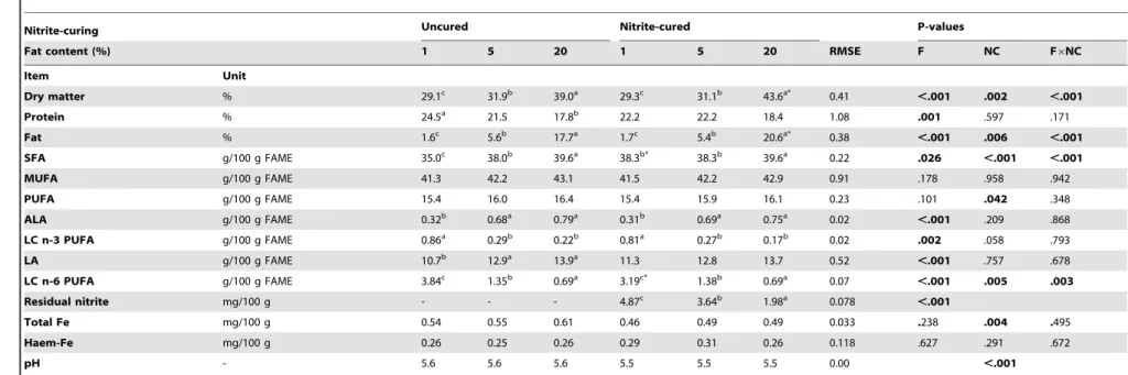
The characteristics of the prepared meats prior to digestion are shown in Table 2. Dry matter content and fatty acid concentra-tions increased, whereas protein content decreased with increasing fat content. There was an unexpected effect of nitrite and fat content6nitrite interaction for the fat content due to a lower than targeted fat content of the 20% fat uncured samples. Addition of subcutaneous pork fat to the lean meat resulted in a significantly lower proportion of LC n-3 and n-6 PUFA and higher ALA, LA and SFA proportions in the fatty acid profile of the prepared meat samples. Residual nitrite levels in the cured meat samples amounted to approximately 41%, 30% and 17% of the added amount (120 mg/kg meat), hence there was a significant effect of fat content on the residual nitrite level. Even though originating from the same pork batch, nitrite-cured meats contained significantly less Fe than the corresponding uncured meats. However, no effect was observed on haem-Fe. The pH was slightly lower in the nitrite-cured meats while no effect of fat content was observed.
2. Total anaerobic bacterial count
Colonic digestive samples counted 7.460.6 log10CFU/ml and
was not influenced by fat content (P = .442) or nitrite-curing (P = .241) (data not shown). However, colonic digestive fluids incubated with fecal inoculum 1 contained significantly less log10
CFU/ml (6.860.4) than with inoculum 2 (7.660.2) and 3 (7.960.2).
3. Lipid oxidation
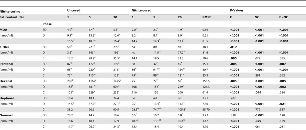
In the meat samples prior to digestion, MDA, pentanal and hexanal were significantly affected by fat content and nitrite-curing, the interaction term appeared also significant (Table 3). MDA was by far the most abundant aldehyde. Octanal was not detected at all, whereas 4-HNE and heptanal were detected in uncured but not in nitrite-cured meats. The concentration of nonanal was not affected by fat content, but nitrite-curing had a significant effect. MDA and volatile aldehyde concentrations were at least 2-fold up to 20-fold lower in the nitrite-cured versus the uncured meat samples. Aldehyde concentrations generally in-creased with increasing fat content in the uncured samples, but no clear effect of fat content on the aldehyde concentrations in the nitrite-cured samples was observed.
After digestion, MDA was by far the most abundant aldehyde, followed by hexanal. The effects of fat content, nitrite-curing and their interaction term were significant for most aldehydes. Nitrite-curing had no significant effect on pentanal and heptanal concentrations after colonic digestion. Higher concentrations of MDA were measured in both uncured and nitrite-cured meats compared to undigested meat. In contrast, concentrations of 4-HNE and volatile aldehydes were lower or similar after digestion
of uncured meats, but higher for cured meats compared to before digestion. However, nitrite-cured meats had significantly lower concentrations of all measured aldehydes compared to uncured meats after the duodenal digestion and significantly lower MDA and hexanal with trends of lower 4-HNE and nonanal after colonic digestion. After duodenal digestion, nitrite-curing resulted in equal MDA independent of the fat content, while 4-HNE and pentanal were significantly higher in the cured 20% fat meat digest. Compared to their uncured equivalents, 4-HNE formation was only 2.5-fold lower in the 20% fat meat digest, while 12-fold lower concentrations were found in the 5% fat meat digest and no 4-HNE was detected in the 1% fat meat digest.
Because MDA was the only aldehyde that was present in higher concentrations in the colonic as compared to the duodenal digestion, it was hypothesized that MDA might be present in the human fecal inocula. To investigate this, blank incubations were performed without meat for the duodenal and colonic digestive simulation in a separate run. No aldehydes were detected in the blank duodenal digest, while MDA concentrations after 72 h of incubation were 5.160.1; 17.060.2 and 18.361.1 nmol/ml digest for the three fecal inocula respectively, indicating a significant contribution of the fecal inoculum to the MDA levels in the colonic meat digests. One might assume similar effects for the other aldehydes, but these were not measured in the present experimental set up.
4. Protein oxidation
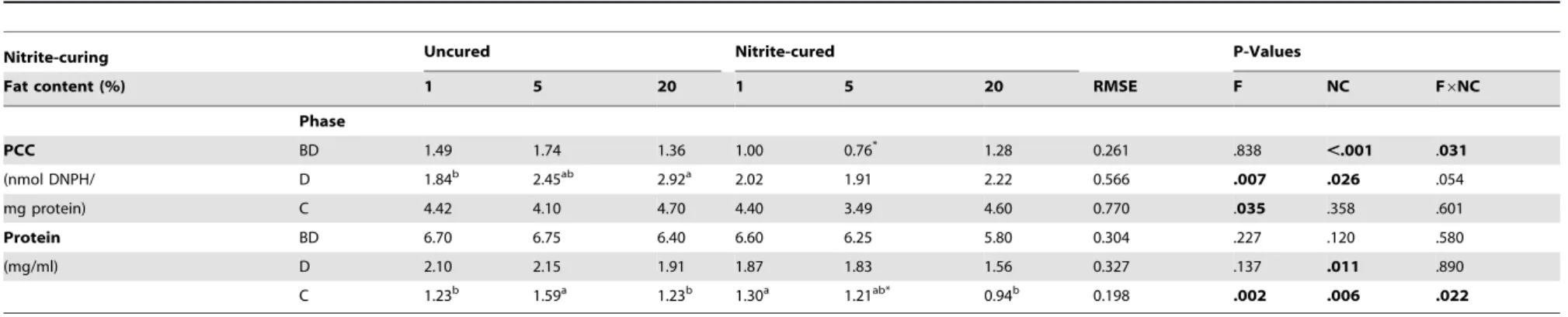
Increasing PCC concentrations were observed in the following order: undigested, duodenal ,colonic digests independent of nitrite-curing (Table 4). Fat content did not affect the concentra-tion of PCC before digesconcentra-tion, but nitrite-cured products had lower PCC concentrations than its uncured equivalents before digestion. In the duodenal digests, uncured samples with added fat showed significantly higher PCC concentrations than the ones without added fat. Nitrite-curing led to significantly lower PCC concen-trations for the duodenal digests with no significant differences between digests with different fat content. After 72 h of colonic fermentation, a marginal effect of fat content was observed with lower PCC concentrations in the 5% fat meat digests as compared to the other samples.
Due to digestion, lower protein concentrations were measured in the duodenal and colonic digestive fluids compared to the meat before digestion (Table 4). Cured meats had significantly lower protein levels after duodenal and colonic simulation compared to uncured equivalent meats. From the blank incubations, it was estimated that the fecal inocula and SHIME medium contributed for 30–40% of the protein in the colonic fluid. Fecal proteins contained between 5.0 and 8.5 nmol DNPH/mg protein and hence largely contributed to the higher PCC concentrations in the meat colonic digests.
5. O6-Carboxy-methylguanine and O6-methylguanine No O6-C-MeG was detected in meats prior to digestion or at the end of the duodenal digestive simulation (Table 5). After mimicking colonic digestion, detection was highly dependent on the applied fecal inoculum. Colonic incubation with the inoculum from volunteer 1 did not result in detectable O6-C-MeG. The fecal inocula from volunteers 2 and 3 resulted however in low and very high levels of O6-C-MeG, respectively. A significant influence of fat content was observed, with higher concentrations of O6 -C-MeG in both cured and uncured samples containing 20% fat compared to samples lower in fat. The effect of nitrite-curing on the O6-C-MeG content was not consistent with significantly lower O6-C-MeG when using inoculum 2 and higher O6-C-MeG when
In VitroGenotoxicity of Fat and Nitrite-Cured Pork
Table 2.Composition of the pork model products used in thein vitrodigestion.
Nitrite-curing Uncured Nitrite-cured P-values
Fat content (%) 1 5 20 1 5 20 RMSE F NC F6NC
Item Unit
Dry matter % 29.1c 31.9b 39.0a 29.3c 31.1b 43.6a* 0.41 ,.001 .002 ,.001
Protein % 24.5a 21.5 17.8b 22.2 22.2 18.4 1.08 .001 .597 .171
Fat % 1.6c 5.6b 17.7a 1.7c 5.4b 20.6a* 0.38
,.001 .006 ,.001
SFA g/100 g FAME 35.0c 38.0b 39.6a 38.3b* 38.3b 39.6a 0.22 .026 ,.001 ,.001
MUFA g/100 g FAME 41.3 42.2 43.1 41.5 42.2 42.9 0.91 .178 .958 .942
PUFA g/100 g FAME 15.4 16.0 16.4 15.4 15.9 16.1 0.23 .101 .042 .348
ALA g/100 g FAME 0.32b 0.68a 0.79a 0.31b 0.69a 0.75a 0.02
,.001 .209 .868
LC n-3 PUFA g/100 g FAME 0.86a 0.29b 0.22b 0.81a 0.27b 0.17b 0.02 .002 .058 .793
LA g/100 g FAME 10.7b 12.9a 13.9a 11.3 12.8 13.7 0.52
,.001 .757 .678
LC n-6 PUFA g/100 g FAME 3.84c 1.35b 0.69a 3.19c* 1.38b 0.69a 0.07
,.001 .005 .003
Residual nitrite mg/100 g - - - 4.87c 3.64b 1.98a 0.078 ,.001
Total Fe mg/100 g 0.54 0.55 0.61 0.46 0.49 0.49 0.033 .238 .004 .495
Haem-Fe mg/100 g 0.26 0.25 0.26 0.29 0.31 0.26 0.118 .627 .291 .672
pH - 5.6 5.6 5.6 5.5 5.5 5.5 0.00 ,.001
Data (total n = 12) were analyzed using a linear model with the fixed effects of fat content (F), nitrite-curing (NC) and their interaction term (F6NC); RMSE = root mean square error; a, b, c = means for different fat content (within
curing treatment) with different superscripts are significantly different (P,0.05); * = significantly different from uncured equivalent (P,0.05); SFA = saturated fatty acids; MUFA = monounsaturated fatty acids;
PUFA = polyunsaturated fatty acids; ALA =a-linolenic acid (C18:3, n-3); LC n-3 PUFA = long chain omega-3 polyunsaturated fatty acids (C20:5, n-3; C22:5, n-3; C22–6, n-3); LA = linoleic acid (C18:2, n-6); LC n-6 PUFA = Long chain
omega-6 polyunsaturated fatty acids (C20:4, n-6; C22:4, n-6; C22:5, n-6); FAME = fatty acid methyl esters. doi:10.1371/journal.pone.0101122.t002
In
Vitro
Genotoxicity
of
Fat
and
Nitrite-Cured
Pork
PLOS
ONE
|
www.ploson
e.org
6
June
2014
|
Volume
9
|
Issue
6
|
Table 3.Lipid aldehyde concentrations in uncured and nitrite-cured pork containing different amounts of fat (1, 5, 20%) before and afterin vitrodigestion.
Nitrite-curing Uncured Nitrite-cured P-Values
Fat content (%) 1 5 20 1 5 20 RMSE F NC F6NC
Phase
MDA BD 4.0b 5.4a 5.3a 2.0* 2.0* 1.9* 0.19 ,.001 ,.001 ,.001
(nmol/ml) D 9.1b 13.3a 12.6a 8.2* 8.4* 8.0* 0.51 ,.001 ,.001 ,.001
C 15.5b 18.0a 16.4b 14.7 14.3* 13.4* 0.85
,.001 ,.001 ,.001
4-HNE BD 58b 221a 290a nd nd nd 38.1 .019
(pmol/ml) D 3.2c 143b 192a nd 11.5b* 77.2a* 31.6 ,.001 ,.001 ,.001
C 13.2b 28.2a 35.5a 14.1 19.5 23.3 10.6
.005 .079 .335
Pentanal BD 87b 172a 192a 38 32* 45* 15.1 .005
,.001 .007
(pmol/ml) D 65b 220a 211a 56b 91ab* 124a* 30.7 ,.001 ,.001 ,.001
C 73b 115ab 123a 75b 89ab 121a 25.3
,.001 .291 .352
Hexanal BD 286b 1162a 1425a 75 77* 68* 155.5 .005
,.001 .005
(pmol/ml) D 158b 581a 604a 106 154* 219* 126.3 ,.001 ,.001 .002
C 121b 229a 252a 116 156 200 61.4
,.001 .044 .384
Heptanal BD 18.4 18.4 24.4 nd nd nd 2.91 .202
(pmol/ml) D 14.5b 37.7a 27.1x 9.7 13.4* 11.3* 7.86 ,.001 ,.001 .021
C 38.2 90.6 90.5 28.5b 74.7ab 105.8a 35.70 ,.001 .779 .537
Nonanal BD 20.2 14.9 18.6 6.5* 10.2 5.8* 2.92 .838
,.001 .128
(pmol/ml) D 18.6 18.4 12.9 18.6a 13.1ab 10.4b 3.42 ,.001 .029 .179
C 11.7b 20.2a 24.3a 12.4 15.4 19.4 4.70 ,.001 .064 .261
Malondialdehyde (MDA) was measured colorimetrically and expressed as nmol/ml digest. 4-hydroxy-nonenal (4-HNE) and the simple aldehydes were measured through HPLC-fluorescence and expressed as pmol/ml. Aldehydes in meat before digestion (BD) (total n = 12) were analyzed using a linear model with the fixed effects of fat content (F), nitrite-curing (NC) and their interaction term (F6NC). Aldehydes in duodenal (D) digests (total n = 36) and colonic
(C) digests (total n = 36) were analyzed using a mixed model with the fixed effects of F, NC and F6NC and the random effect of incubation run. RMSE = root mean square error; a, b, c = means for different fat content (within curing
treatment) with different superscripts are significantly different (P,0.05); * = significantly different from uncured equivalent (P,0.05);nd= not detected. doi:10.1371/journal.pone.0101122.t003
In
Vitro
Genotoxicity
of
Fat
and
Nitrite-Cured
Pork
PLOS
ONE
|
www.ploson
e.org
7
June
2014
|
Volume
9
|
Issue
6
|
Table 4.Protein oxidation in uncured and nitrite-cured pork containing different amounts of fat (1, 5, 20%) before and afterin vitrodigestion.
Nitrite-curing Uncured Nitrite-cured P-Values
Fat content (%) 1 5 20 1 5 20 RMSE F NC F6NC
Phase
PCC BD 1.49 1.74 1.36 1.00 0.76* 1.28 0.261 .838 ,.001 .031
(nmol DNPH/ D 1.84b 2.45ab 2.92a 2.02 1.91 2.22 0.566 .007 .026 .054
mg protein) C 4.42 4.10 4.70 4.40 3.49 4.60 0.770 .035 .358 .601
Protein BD 6.70 6.75 6.40 6.60 6.25 5.80 0.304 .227 .120 .580
(mg/ml) D 2.10 2.15 1.91 1.87 1.83 1.56 0.327 .137 .011 .890
C 1.23b 1.59a 1.23b 1.30a 1.21ab* 0.94b 0.198
.002 .006 .022
Protein carbonyl compounds (PCC) and protein in meat before digestion (BD) (total n = 12) were analyzed using a linear model with the fixed effects of fat content (F), nitrite-curing (NC) and their interaction term (F6NC). Aldehydes in duodenal (D) digests (total n = 36) and colonic (C) digests (total n = 36) were analyzed using a mixed model with the fixed effects of F, NC and F6NC and the random effect of incubation run. RMSE = root mean square
error; a, b = means for different fat content (within curing treatment) with different superscripts are significantly different (P,0.05); * = significantly different from uncured equivalent (P,0.05). doi:10.1371/journal.pone.0101122.t004
Table 5.NOC-induced DNA adduct formation by uncured and nitrite-cured pork containing different amounts of fat (1, 5, 20%) before and afterin vitrodigestion.
Nitrite-curing Uncured Nitrite-cured P-Values
Fat content (%) 1 5 20 1 5 20 RMSE F NC F6NC
Phase
O6-C-MeG BD nd nd nd nd nd nd
-(ng/ml) D nd nd nd nd nd nd
-C1 nd nd nd nd nd nd
-C2 46.8c 50.6b 57.8a 48.9a 44.6b* 50.8a* 0.90 ,.001 ,.001 ,.001
C3 441b 486b 697a 544b 541b 783a 55.2
,.001 .043 .824
O6-carboxy-methylguanine (O6-C-MeG) was not detected (nd) in meat before digestion (BD) (total n = 12) and after duodenal (D) digestion (total n = 36). O6-C-MeG in colonic (C1, C2, C3) digests were analyzed separately (total n = 12 per fecal inoculum), due to the very high variation between fecal inocula, using a linear model with the fixed effects of fat content (F), nitrite-curing (NC) and their interaction term (F6NC). RMSE = root mean square error; a,
b, c = means for different fat content (within curing treatment) with different superscripts are significantly different (P,0.05); * = significantly different from uncured equivalent (P,0.05). doi:10.1371/journal.pone.0101122.t005
In
Vitro
Genotoxicity
of
Fat
and
Nitrite-Cured
Pork
PLOS
ONE
|
www.ploson
e.org
8
June
2014
|
Volume
9
|
Issue
6
|
using inoculum 3. The DNA adduct O6-MeG was not detected in any digestive fluid.
In the blank (no meat) incubations, no O6-C-MeG was detected when incubated with fecal inoculum 1 but high DNA adduct concentrations were measured when fecal inoculum 2 (101.0610.6 ng/ml) and 3 (82.8626.1 ng/ml) were applied. It should be noted that these blank incubations were performed in a separate run and are therefore not fully comparable to the test incubations.
6. Short chain fatty acids and NH3
Since the blank (no meat) incubations resulted in higher O6 -C-MeG formation compared to the meat digests when the second fecal inoculum was used, we wondered if this might be caused by differences in the fermentation process. Therefore, we repeated 3 blank incubations and 3 digestions of uncured pork containing 5% fat, using fecal inoculum 2. There was no detection of O6-C-MeG in the blank or meat digests just after addition of the fecal inoculum. After 72 h of fermentation, the blank incubations contained 118.6626.2 ng O6-C-MeG/ml whereas the meat digests contained only 56.564.9 ng O6-C-MeG/ml, in line with our previous findings. Significantly higher concentrations of acetate (56.769.0mmol/ml vs. 33.160.6mmol/ml), propionate (24.760.7 vs. 14.960.3mmol/ml), butyrate (10.661.0 vs. 3.460.4mmol/ml) and NH3 (34.263.1 vs. 18.760.8mmol/ml)
were found in the meat digests, compared to the blank digests. Repeated incubations with fecal inoculum 3 confirmed these observations (data not shown).
Discussion
The presentin vitro digestion study using pork model products allowed to investigate the effects of crucial factors that reflect the variability in composition and properties of meat products, i.e. nitrite-curing and fat content, on oxidation and nitrosation processes during digestion. By adding known pro-oxidants and antioxidants to the applied digestive juices, thein vivosituation was mimicked as close as possible. As expected, higher fat contents enhanced the formation of oxidation products and the NOC-specific DNA adduct O6-C-MeG during digestion. Nitrite-curing on the other hand resulted in clearly lower concentrations of oxidation products before, during and at the end of the in vitro
digestion, whereas the effect on the formation of O6-C-MeG proved subjective for inter-individual variation. Previously, Corpet [3] suggested that the formation of oxidation products and NOCs are amongst those that could explain for the association between high red meat consumption and CRC.
In accordance to Hur et al. [29] who used a similar in vitro
digestion model for beef patties, we observed a significant increase in MDA concentrations throughout the duodenal digestive simulation. Previously, it was reported that MDA in the small intestine is absorbed into the blood stream [30] after which it can reach several organs. Free MDA could react with DNA, resulting in the formation of the MDA-specific DNA adduct pyrimido
[1,2-a] purine-10(3H)-one-29-deoxyribose (M1dG) [31]. In contrast to
MDA, highest concentrations of 4-HNE were observed in meat before digestion, after which they decreased throughout the digestion. After subtraction of the MDA values present in the applied fecal inocula, also MDA concentrations decreased from the duodenal to the colonic step. This decrease of MDA and 4-HNE during the colonic simulation could be due to reaction with proteins leading to the formation of Schiff bases or with bacterial DNA leading to DNA-adducts [31].
The antioxidative properties of nitrite were already very clear before digestion since MDA, 4-HNE, volatile simple aldehydes and PCC were clearly lower or even undetected in cured meats compared to uncured meats. Nitrite is a known antioxidant trough oxygen sequestration and may act as a precursor of the heat-stable NO-myoglobin by which the release of Fe2+ during heating is inhibited [32]. Consequently, less Fe2+is available to catalyze the Fenton reaction, which is responsible for the initiation of oxidation processes. Since all meats were sampled fresh and oxidation in fresh meat is generally negligible, the higher lipid aldehyde and PCC concentrations in uncured meats must originate from processing (mincing, adding fat, heating). The clearly lower residual nitrite, found in the nitrite-cured meat samples when higher amounts of subcutaneous pork fat were added, could be explained by interaction of nitrite with unsaturated FA [33]. Addition of subcutaneous pork fat to meat samples resulted in an expected higher concentration of lipid aldehydes in meat, since more substrate is available for oxidation. Except for 4-HNE, the expected differences in lipid aldehydes between the 5 and 20% fat meat samples throughout digestion were not observed. Fat content of grounded beef varying between 10, 15 and 20% also had little effect on lipid oxidation parameters in the meat [34].
The lower values of oxidation products of nitrite-cured meats compared to uncured equivalents in duodenal and colonic fluids are in agreement with recently published work by Chenniet al.
[35] who found that intake of nitrite through drinking water (1 g/l) reduced (25%) haem-induced lipid peroxidation in the colon of rats. This effect was not observed to be significant at lower doses (0.17 g/l nitrite and 0.23 g/l nitrate). Nitric oxide (NO) is able to react with ROS generating peroxynitrite, which can induce lipid peroxidation. However, NO can also react with lipophilic peroxyl radicals, generating far more stable alkyl peroxynitrates. Conse-quently, it was concluded that a 1:1 ratio of NO to ROS enhances lipid peroxidation, while an excess of NO results in inhibition [36]. The reaction between nitrite and unsaturated FA leads to lower residual nitrite levels in the 20% fat meat samples and hence could modify the NO:ROS ratio and induce lipid oxidation. This could explain the higher HNE, pentanal and hexanal concentrations in the cured 20% fat meat sample compared to the cured 1 and 5% fat meat samples.
Also increased protein oxidation was observed throughout the digestion of the meat samples. The higher PCC concentrations after the colonic digestive simulation could be largely attributed to proteins in the fecal inoculum and SHIME medium. No significant differences in PCC concentrations according to fat content were noticed before digestion. It was previously described that lipid radicals may damage proteins by the binding of lipid components such as MDA and 4-HNE to the protein [37]. This could explain the higher PCC concentrations in the uncured 20% fat treatment after duodenum digestion. The lower PCC concentrations in cured meat samples before and after duodenal digestive simulations correspond with the associated lower concentrations of lipid oxidation products.
Next to higher PCC formation, we also observed a significantly higher amount of residual protein in uncured samples, indicating lower protein digestion. These findings agree with Sante´-Lhoutellier et al. [38] who found a highly significant negative correlation between PCC concentrations and proteolytic suscep-tibility to pepsin. As mentioned before, MDA and 4-HNE can also react with proteins, leading to the formation of Schiff bases, which are known to play a role in protein aggregation. Previously, it was shown that a higher amount of proteins reaching the colonic fermentation, resulted in a higher formation of potentially toxic protein fermentation products such as ammonium, phenol,
p-In VitroGenotoxicity of Fat and Nitrite-Cured Pork
cresol and indol. However, higher formation of these products was not associated with enhanced colon cancer promotion in rats [39]. Previously, it was reported that haem-Fe is responsible for endogenous intestinalN-nitrosation [40]. Our results demonstrate that the fat content in meat products enhances the haem-induced intestinalN-nitrosation by detecting higher concentrations of the NOC-specific DNA adduct O6-C-MeG when a higher fat content was present in the digested meat. Salivary nitrite or nitrite-cured meats are sources for NO formation. Both NO and O2
preferentially diffuse in a lipid environment resulting in accumu-lation of both reactants in the lipid fraction to form nitrosating species. The rate of this reaction was described to be 300 times faster in a lipid compared to an aqueous environment [41], which could explain the facilitating role of fat in the formation of NOCs. However, the effect of nitrite-curing of meat on O6-C-MeG
formation was inconsistent in our results, being either promoting or protective depending on the applied fecal inoculum. Possibly, the effect of nitrite-curing depends on the incubation conditions (e.g. O2tension), the bacterial composition of the fecal inoculum
and interactions with other dietary compounds or metabolites formed during fermentation. This certainly warrants further investigation. However, the effect of nitrite-curing was perceived to be minor compared to the promoting effect of fat content for the meat products examined in the present study.
The formation of the NOC-specific DNA adduct O6-C-MeG was also highly dependent on the applied fecal inoculum. A difference in total anaerobic bacteria in the colonic fluids, fermentation rate or bacterial composition of the used inocula could explain the differences in the magnitude of detection. More research is required to elucidate the underlying mechanisms causing these differences.
When O6-C-MeG was measured in the colonic digests, blank incubations without meat displayed high values after 72 h of fermentation. Since no O6-C-MeG was detected just after addition of the fecal inoculum, its formation must have occurred during the fermentation. The sometimes higher values for the blank incubations than the meat digests is surprising since one expects a higher fermentation rate and more bacterial growth in the meat digests due to the higher supply of nutrients (protein, glycogen, B vitamins,…). Indeed, meat digests contained approximately 2-fold higher concentrations of NH3, acetate and propionate, and 3-fold
higher concentrations of butyrate compared to the blank digests. However, it has been shown in rats on a high protein diet that high butyrate concentrations in the colon decreased DNA damage in the colonocytes [42]. Butyrate was also able to decrease H2O2
-induced DNA damage in human colonocytes [43]. Therefore, it seems likely that the 3-fold higher butyrate production during fermentation of the meat digests interfered in the genotoxic activity of compounds. The values of O6-C-MeG in the blank digests can thus not be simply subtracted from the values of the meat digests.
In conclusion, our results indicate that addition of fat favors the formation of oxidation products and the NOC-specific DNA adduct O6-C-MeG during the digestion of pork. Nitrite-curing of meat inhibited lipid and protein oxidation but this inhibition was less pronounced throughout digestion when meat in the incuba-tion fluids contained 20% fat. The effect of nitrite-curing on O6 -C-MeG formation was contradictory in our results and requires more research. The measurement of O6-C-MeG was highly variable between fecal inocula originating from different volunteers. In this research, we simulated the passage of meat through the gastro-intestinal tract as close as possible to the in vivo situation using static in vitro incubations. Several in vivo experiments have been conducted towards this subject but these studies do not always gain insights in some mechanistic pathways as the presented in vitro
work does. However, since the in vivo situation is always more complex thanin vitroconditions, the outcomes in this study should be confirmedin vivo.
Acknowledgments
We would like to thank S. Lescouhier, D. Baeyens and E. Claeys for the preparation and characterization of meat samples. C. Boterdaele is thanked for her assistance in performing the incubations. L. Hemeryck is thanked for her assistance in the analysis of the NOC-specific DNA adducts.
Author Contributions
Conceived and designed the experiments: TVH KR LV SDS. Performed the experiments: TVH EV. Analyzed the data: TVH. Contributed reagents/materials/analysis tools: JVB LV. Wrote the paper: TVH SDS. Read and commented on the draft manuscript: EV JVB KR LV.
References
1. Chan DS, Lau R, Aune D, Vieira R, Greenwood DC, et al. (2011). Red and processed meat and colorectal cancer incidence: meta-analysis of prospective studies. PLOS ONE 6: e20456.
2. Larsson SC, Wolk A (2006). Meat consumption and risk of colorectal cancer: A meta-analysis of prospective studies. Int J Cancer 119: 2657–2664.
3. Corpet DE (2011). Red meat and colon cancer: Should we become vegetarians, or can we make meat safer? Meat Sci 89: 310–316.
4. Skrzydlewska E, Sulkowski S, Koda M, Zalewski B, Kanczuga-Koda L, et al. (2005). Lipid peroxidation and antioxidant status in colorectal cancer. World J. Gastroenterol 11: 403–406.
5. Yeh CC, Lai CY, Hsieh LL, Tang R, Wu FY, et al. (2010). Protein carbonyl levels, glutathione S-transferase polymorphisms and risk of colorectal cancer. Carcinogenesis 31: 228–233.
6. Lewin MH, Bailey N, Bandaletova T, Bowman R, Cross AJ, et al. (2006). Red meat enhances the colonic formation of the DNA adduct O6-carboxymethyl guanine: implications for colorectal cancer risk. Cancer Res 66: 1859–1865. 7. Sesink AL, Termont DS, Kleibeuker JH, Van der Meer R (2000). Red meat and
colon cancer: dietary haem, but not fat, has cytotoxic and hyperproliferative effects on rat colonic epithelium. Carcinogenesis 21: 1909–1915.
8. Lai C, Dunn DM, Miller MF, Pence BC (1997). Non-promoting effects of iron from beef in the rat colon carcinogenesis model. Cancer Let 112: 87–91. 9. Mirvish SS, Haorah J, Zhou L, Clapper ML, Harrison KL, et al. (2002). Total
N-nitroso compounds and their precursors in hot dogs and in the gastrointestinal tract and feces of rats and mice: possible etiologic agents for colon cancer. J Nutr 132: 3526S–3529S.
10. Pierre FH, Santarelli RL, Allam O, Tache´ S, Naud N, et al. (2010). Freeze-dried ham promotes azoxymethane-induced mucin-depleted foci and aberrant crypt foci in rat colon. Nutr Cancer 62: 567–573.
11. Santarelli RL, Vendeuvre JL, Naud N, Tache´ S, Gue´raud F, et al. (2010). Meat processing and colon carcinogenesis: cooked, nitrite-treated, and oxidized high-heme cured meat promotes mucin-depleted foci in rats. Cancer Prev Res 3: 852– 864.
12. Joosen AM, Kuhnle GG, Aspinall SM, Barrow TM, Lecommandeur E, et al. (2009). Effect of processed and red meat on endogenous nitrosation and DNA damage. Carcinogenesis 30: 1402–1407.
13. Versantfoort CHM, Oomen AG, Van de Kamp E, Rompelberg CJM, Sips AJAM (2005). Applicability of an in vitro digestion model in assessing the bioaccessibility of mycotoxins from food. Food Chem Toxicol 43: 31–40. 14. Van de Wiele T, Vanhaecke L, Boeckaert C, Peru K, Headley J, et al. (2005).
Human colon microbiota transform polycyclic aromatic hydrocarbons to estrogenic metabolites. Environ Health Persp 113: 6.
15. Gu¨ven Y, Satman I, Dinc¸c¸ag N, Alptekin S (1996). Salivary peroxidase activity in whole saliva of patients with insulin-dependent (type-1) diabetes mellitus. J Clin Periodontol 23: 879–88.
16. Takahama U, Yamamoto A, Hirota S, Oniki T (2003). Quercetin-dependent reduction of salivary nitrite to nitric oxide under acidic conditions and interaction between quercetin and ascorbic acid during the reduction. J Agr Food Chem 51: 6014–6020.
17. Dabrowska-Ufniarz E, Dzieniszewski J, Jarosz M, Wartanowicz M (2002). Vitamin C concentration in gastric juice in patients with precancerous lesions of the stomach and gastric cancer. Med Sci Monitor 8: CR96–CR103.
In VitroGenotoxicity of Fat and Nitrite-Cured Pork
18. Nalini S, Ramakrishna BS, Mohanty A, Balasubramanian KA (1992). Hydroxyl radical formation in human gastric juice. J Gastroen Hepatol 7: 497–501. 19. Molly K, Woestyne MV, Smet ID, Verstraete W (1994). Validation of the
simulator of the human intestinal microbial ecosystem (SHIME) reactor using microorganism-associated activities. Microb Ecol Health D 7: 191–200. 20. Folch J, Lees M, Sloane-Stanley GH (1957). A simple method for the isolation
and purification of total lipids from animal tissues. J Biol Chem 226:497–509. 21. Raes K, De Smet S, Demeyer D (2001). Effect of double-muscling in Belgian Blue young bulls on the intramuscular fatty acid composition with emphasis on conjugated linoleic acid and polyunsaturated fatty acids. Anim Sci 73: 253–260. 22. Hornsey H (1956). The colour of cooked cured pork - Estimation of the Nitric
oxide-Haem Pigments. J Sci Food Agric 7: 534–547.
23. Grotto D, Santa Maria LD, Boeira S, Valentini J, Chara˜o MF, et al. (2007). Rapid quantification of malondialdehyde in plasma by high performance liquid chromatography–visible detection. J Pharmaceut Biomed 43: 619–624. 24. Holley AE, Walker MK, Cheeseman KH, Slater TF (1993). Measurement of
n-alkanals and hydroxyalkenals in biological samples. Free Radical Bio Med 15: 281–289.
25. Ganha˜o R, Morcuende D, Este´vez M (2010). Protein oxidation in emulsified cooked burger patties with added fruit extracts: Influence on colour and texture deterioration during chill storage. Meat Sci 85:402–409.
26. Vanden Bussche J, Moore SA, Pasmans F, Kuhnle GGC, Vanhaecke L (2012). An approach based on ultrahigh pressure liquid chromatography-tandem mass spectrometry to quantify O6
-methyl and O6
-carboxymethylguanine DNA adducts in intestinal cell lines. J Chromatogr A 1257: 25–33.
27. Moore SA, Xeniou O, Zeng ZT, Humphreys E, Burr S, et al. (2010). Optimizing immunoslot blot assays and application to low DNA adduct levels using an amplification approach. Anal Biochem 403: 67–73.
28. Chaney AL, Marbach EP (1962). Modified reagents for determination of urea and ammonia. Clin Chem 8: 130.
29. Hur SJ, Lim BO, Park GB, Joo ST (2009). Effects of various fiber additions on lipid digestion during in vitro digestion of beef patties. J Food Sci 74: C653– C657.
30. Gorelik S, Ligumsky M, Kohen R, Kanner J (2008). A novel function of red wine polyphenols in humans: prevention of absorption of cytotoxic lipid peroxidation products. FASEB J 22: 41–46.
31. Nair U, Bartsch H, Nair J (2007). Lipid peroxidation-induced DNA damage in cancer-prone inflammatory diseases: a review of published adduct types and levels in humans. Free Radical Biol Med 43: 1109–1120.
32. Honikel KO (2008). The use and control of nitrate and nitrite for the processing of meat products. Meat Sci 78: 68–76.
33. Trostchansky A, Rubbo H (2008). Nitrated fatty acids: mechanisms of formation, chemical characterization, and biological properties. Free Radical Biol Med 44: 1887–1896.
34. Ismail HA, Lee EJ, Ko KY, Ahn DU (2009). Fat content influences the color, lipid oxidation, and volatiles of irradiated ground beef. J Food Sci 74: C432– C440.
35. Chenni FZ, Tache´ S, Naud N, Gue´raud F, Hobbs DA, et al. (2013). Heme-Induced Biomarkers Associated with Red Meat Promotion of colon Cancer Are Not Modulated by the Intake of Nitrite. Nutr Cancer 65: 227–233. 36. Darley-Usmar V, Wiseman H, Halliwell B (1995). Nitric oxide and oxygen
radicals: a question of balance. FEBS Lett 369: 131–135.
37. Dean R, Fu S, Stocker R, Davies M (1997). Biochemistry and pathology of radical-mediated protein oxidation. Biochem J 324: 1–18.
38. Sante´-Lhoutellier V, Astruc T, Marinova P, Greve E, Gatellier P (2008). Effect of meat cooking on physicochemical state andin vitrodigestibility of myofibrillar proteins. J Agr Food Chem 56: 1488–1494.
39. Corpet DE, Yin Y, Zhang XM, Remesy C, Stamp D, et al. (1995). Colonic protein fermentation and promotion of colon carcinogenesis by thermolyzed casein. Nutr Cancer 23: 271–276.
40. Cross AJ, Pollock JR, Bingham SA (2003). Haem, not protein or inorganic iron, is responsible for endogenous intestinal N-nitrosation arising from red meat. Cancer Res 63: 2358–2360.
41. Liu X, Miller MJ, Joshi MS, Thomas DD, Lancaster JR (1998). Accelerated reaction of nitric oxide with O2 within the hydrophobic interior of biological membranes. P Natl Acad Sci USA 95: 2175–2179.
42. Bajka BH, Clarke JM, Cobiac L, Topping DL (2008). Butyrylated starch protects colonocyte DNA against dietary protein-induced damage in rats. Carcinogenesis 29: 2169–2174.
43. Rosignoli P, Fabiani R, De Bartolomeo A, Spinozzi F, Agea E, et al. (2001). Protective activity of butyrate on hydrogen peroxide-induced DNA damage in isolated human colonocytes and HT29 tumour cells. Carcinogenesis 22: 1675– 1680.
In VitroGenotoxicity of Fat and Nitrite-Cured Pork