Study carried out at Ophthalmology Department, Escola Paulista de Medicina, Universidade Federal de São Paulo - UNIFESP - São Paulo (SP), Brazil; Clínica Oftalmológica Renato Ambrósio - Rio de Janeiro (RJ), Brazil.
1Physician, Ophthalmology Department, Escola Paulista de Medicina, Universidade Federal de
São Paulo - UNIFESP - São Paulo (SP), Brazil.
2Physician, Sociedade Brasileira de Administração em Oftalmologia - SBAO - Rio de Janeiro
(RJ), Brazil.
3Physician, Clínica Oftalmológica Renato Ambrósio - Rio de Janeiro (RJ), Brazil. 4Statistician, Statistics Department, Universidade Federal Fluminense - UFF - Rio de Janeiro
(RJ), Brazil.
5Physician, Universidade Metropolitana de Santos - UNIMES - Santos (SP), Brazil. Correspondence address: Bruno M. Fontes. Rua Castro Alves, 10 - Rio de Janeiro (RJ) CEP 20775-040 - E-mail: [email protected]
Recebido para publicação em 30.07.2010 Última versão recebida em 07.05.2010 Aprovação em 12.07.2010
No financial support was available for this project.
Renato Ambrosio, MD is a consultant for Oculus and Reichert Companies.
Nota Editorial:Depois de concluída a análise do artigo sob sigilo editorial e com a anuência dos Drs. Luis Alberto Carvalho e Diane Ruschel Marinho sobre a divulgação de seus nomes como revisores, agradecemos sua participação neste processo.
Ability of corneal biomechanical metrics and anterior segment data in the
differentiation of keratoconus and healthy corneas
Estudo da performance diagnóstica de parâmetros biomecânicos e dados anatômicos
da câmara anterior na diferenciação de córneas saudáveis e com ceratocone
BRUNO MACHADO FONTES1, RENATO AMBRÓSIO JUNIOR2,DANIELA JARDIM3, GUILLERMO COCA VELARDE4, WALTON NOSÉ1,5
ABSTRACT
Purpose: To evaluate the sensitivity, specificity, and test accuracy of corneal biomechanical metrics and anterior segment data in differentiating keratoconus from healthy corneas.
Methods: Comparative case series. Patients with and without keratoconus (gender and age-matched) were submitted for complete eye examinations including corneal hysteresis (CH) and corneal resistance factor (CRF) as measured by the Ocular Response Analyzer and anterior segment data as gathered through Pentacam assessments. The anterior segment data measu-rement included average central keratometric readings (K-Ave), corneal astig-matism (CA), central corneal thickness (CCT), anterior chamber depth (AC depth) and corneal volume (CV). All parameters were assessed, compared and analyzed. A receiver operating characteristic (ROC) curve was used to identify the best cutoff point by which to maximize the sensitivity and specificity of discriminating keratoconus from normal corneas for each data category. Results: Seventy seven eyes from forty three patients (24 male, 19 female) with keratoconus and eighty six eyes from forty three (24 male, 19 female) healthy controls were enrolled. ROC curve analysis showed poor overall predictive accuracy for all studied parameters in differentiating keratoconus from normal corneas. The highest sensitivity (79.2%) was obtained for both AC depth and CH (cutoff points 3.22 mm and 9.39 mmHg respectively). The best specificity (89.5%) and test accuracy (80.34%) were obtained for CA (cutoff point of 2.2 D).
Conclusion: When considered together, studied parameters showed statistical differences between groups. However, when considered independently they presented low sensitivity, specificity and test accuracy in differentiating kera-toconus from healthy corneas.
Keywords: Cornea; Corneal diseases; Corneal topography; Biomechanics; Keratoconus.
RESUMO
Objetivo: Avaliar a sensibilidade, especificidade e acurácia de parâmetros biomecânicos e anatômicos do segmento anterior isolados na diferenciação de córneas saudáveis e com ceratocone.
Métodos: Estudo tipo série de casos comparativa. Pacientes com ceratocone e controles saudáveis foram pareados (idade e sexo) e submetidos a exame oftalmológico completo, incluindo avaliação biomecânica (ORA) e tomográfica (Pentacam). Ceratometria central média, astigmatismo corneano, espessura corneana central, profundidade da câmara anterior, volume corneano, CH e CRF foram estabelecidos, avaliados e comparados. Curvas ROC (Receiver operating characteristic) foram utilizadas para identificar o melhor valor de corte que apresentasse a maior sensibilidade e especificidade na discrimina-ção entre ceratocone e córneas saudáveis para cada dado estudado. Resultados: Setenta e sete olhos de 43 pacientes com ceratocone (24 homens e 19 mulheres) e 86 olhos de pacientes saudáveis (24 homens e 19 mulheres) foram incluídos no estudo. Curvas ROC mostraram baixa acurácia na predição do diagnóstico de ceratocone em todos os parâmetros isolados estudados. Maior sensibilidade encontrada foi 79,2% para profundidade da câmara ante-rior e CH (ponto de corte 3,22mm e 9,39mmHg respectivamente); maior especificidade e acurácia foram encontradas na análise do astigmatismo corneano (ponto de corte 2,2 D; 89,5% e 80,34% respectivamente). Conclusão: Todos os parâmetros estudados mostraram diferença estatistica-mente significativa entre os grupos. No entanto, quando considerados isolada-mente apresentaram baixas sensibilidade, especificidade e acurácia na dife-renciação entre ceratocone e córneas saudáveis.
Descritores: Córnea; Doenças da córnea; Topografia da córnea; Biomecânica; Ceratocone
INTRODUCTION
K
eratoconus is an ectatic disease of the cornea, with pro-gressive noninflammatory thinning and anterior protru-sion that leads to an irregular conical shape(1-4). It isusually a bilateral and asymmetric condition that manifests at puberty. Clinical (as corneal stromal thinning, conical protru-sion, Vogt striae and Fleischer ring) and topographic (as irre-gular astigmatism, inferior steepening and inferior-superior asymmetry) findings are habitually combined for diagnosing and staging the disease(5-6).
stored. The Pentacam assessed central keratometry (K-Ave), corneal astigmatism (CA), central corneal thickness (CCT), an-terior chamber depth (AC depth) and corneal volume (CV).
The Pentacam system was connected to a personal compu-ter, with automated software. The manufacturer performed calibration of the device. The system uses a rotating Scheimp-flug camera and a monochromatic slit light source (a blue light-emitting diode at 475 nm) that rotate together. After proper alignment of the patient’s face, a fixation target is shown, which guides the patient’s gaze. A real-time image of the patient’s eye is shown to the examiner on the computer screen, and the image is manually focused and centered. The rotating camera is set to take 25 slit images of the anterior eye segment in approximately 2 seconds with 500 true elevation points incorporated in each slit image. Minute eye movements are captured by a second camera and corrected simultaneously. Single point pachymetric measurements of the entire cornea are calculated from the anterior and posterior corneal surfaces. The ORA determines corneal biomechanical properties using an applied force-displacement relationship. Details have been extensively described in previous studies17. Briefly, a
pre-cisely-metered air pulse is delivered to the eye, causing the cornea to move inward, past a first applanation, and into a slight concavity. Milliseconds after the first applanation, the air pump is shut down and the pressure applied to the eye decreases in an inverse-time, symmetrical fashion. As the pressure decreases, the cornea passes through a second applanated state while returning from concavity to its normal convex curvature. Energy absorption during rapid corneal deformation delays the occurrence of the inward and outward applanation signal peaks, resulting in a difference between the applanation pres-sures. The difference between these inward and outward mo-tion applanamo-tion pressures is called corneal hysteresis (CH). Corneal hysteresis is an indication of viscous damping and elastic resistance, reflecting the capacity of corneal tissue to absorb and dissipate energy. Corneal resistance factor (CRF) was empirically derived to maximize correlation to CCT, and it can be considered to be weighted by elastic resistance since it has a stronger correlation to CCT than CH. Though CH and CRF are related, in some instances they can be significantly different, each providing distinct information about the cornea.
The Kolmogorov-Smirnov test was used to check for a nor-mal distribution of quantitative data, which are provided as the mean and standard deviation (SD). Differences between data were evaluated using the Welch modified Student’s two-sample t-test and Wilcoxon rank-sum test. The level of signifi-cance for each parameter was set at p<0.05. A receiver opera-ting characteristic (ROC) curve was used to identify the cutoff point of studied parameters to maximize sensitivity and spe-cificity in discriminating keratoconus from normal corneas. This curve is obtained by plotting sensitivity against 1- specifi-city, calculated for each value observed. An ideal area of 100% implies that the test perfectly discriminates between groups. Logistic regression was used to support the cutoff point iden-tified with the ROC curve analysis.
RESULTS
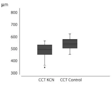
K-Ave was 47.03 ± 5.22 diopters (D) (range 40.4 to 74.15 D) in keratoconus and 43.31 ± 1.53 D (range 39.9 to 46.75 D) in the control group (p=0). CA was 3.46 ± 2.20 D (range 0.7 to 10.9 D) in keratoconus and 1.08 ± 0.81 D (range 0 to 4.9 D) in the control group (p=0). CCT was 493.17 ± 42.84 μm (range 349 to 568 μm) in keratoconus and 543.90 ± 34.87 μm (range from 457 to 627 μm) in the control group (p=0) (Figure 1). AC depth was 3.25 ± 0.38 mm (range 2.41 to 5.21 mm) in kerato-conus and 3.07 ± 0.42 mm (range 2.08 to 3.80 mm) in the provided by a variety of currently available equipment(5-14).
Ho-wever, accurate differentiation of keratoconus from healthy cor-neas is not yet sufficient, as there is a need to detect corcor-neas with a higher susceptibility to becoming ectatic after laser pho-toablative surgery(15-16).
In vivo corneal biomechanical evaluation was first descri-bed by Luce(17) in 2005, with the development of the Ocular
Response Analyzer (ORA, Reichert Ophthalmic Instruments, De-pew, New York, USA). A number of researchers published diverse and exciting new data regarding corneal hysteresis (CH) and corneal resistance factor (CRF) in healthy and pathological conditions(18-22). If ORA proves that “fragile” corneas are more
susceptible than “strong” corneas to developing ectasia in the future, then the best use for such data in refractive surgery would be in preoperative screening.
The present study compared the findings of biomechani-cal and anterior segment parameters in differentiating kera-toconus from healthy corneas, and evaluated the ability of each individual parameter to differentiate them.
METHODS
This was a comparative case series. The research followed the tenets of the Declaration of Helsinki and was approved by the ethics committee of the Federal University of São Paulo (protocol 0123/06). All subjects were informed about the purpose of the study and gave informed consent before inclu-sion. Patients were sequentially evaluated from October 2005 to December 2008. Demographic and clinical data were obtained, including date of birth, gender and self-reported race or ethnicity.
The keratoconus group consisted of 77 eyes from 43 pa-tients (24 male, 19 female) with a mean age of 34.95 ± 11.95 years (ranging from 18 to 73 years). The control group consis-ted of 86 eyes from 43 (24 male, 19 female) gender- and age-matched healthy patients, with a mean age of 35.02 ± 12.19 years (ranging from 18 to 72 years-old) (p=1).
Each subject underwent a comprehensive ophthalmolo-gic examination including review of medical history, best-corrected visual acuity, slit lamp biomicroscopy, fundoscopic examination, Placido disc topography (Humphrey ATLAS, Carl Zeiss Meditec Inc. Dublin, USA), Pentacam tomographic eva-luation and ORA measurements.
Diagnosis of keratoconus was made by clinical (corneal stromal thinning, Vogt’s striae, Fleischer ring, scissoring of the red reflex or oil droplet sign identified by retinoscopy) and topographic (an increased area of corneal power surrounded by concentric areas of decreasing power, inferior-superior power asymmetry, and skewing of the steepest radial axes above and below the horizontal meridian(2,5-6,10,23) evaluation.
Cases were gender- and age-matched with controls for data comparison21. Exclusion criteria were: less than 18
years-old, any previous corneal or ocular surgery, any eye disease that could possibly interfere with the readings/results (e.g., glau-coma, uveitis, corneal ectatic disease, Fuch’s dystrophy, diabe-tic retinopathy, etc,.) chronic and/or continuous use of topical medications, corneal scars and/or opacities, irregular astigma-tism, systemic collagen diseases and refusal to sign an infor-med consent agreement. Contact lenses were required to be removed at least 72 h before examination.
control group (p=0.012). CV was 57.01 ± 3.53 mm³ (range 49.5 to 66.9 mm³) in keratoconus and 60.19 ± 3.40 mm³ (range 53.7 to 68.5 mm³) in the control group (p=0).
CH was 8.23 ± 1.51 mmHg (range 4.60 to 11.80 mmHg) in keratoconus and 10.13 ± 1.75 mmHg (range 5.95 to 14.58 mmHg) in the control group (p=0) (Figure 2). CRF was 7.46 ± 1.76 mmHg (range 2.80 to 11.20 mmHg) in keratoconus and 10.06 ± 1.97 mmHg (range 5.45 to 15.10 mmHg) in the control group (p=0) (Figure 3). The results are summarized in table 1.
ROC curve analyses showed poor overall predictive accura-cy for all studied parameters in differentiating keratoconus from normal corneas. The results are summarized in table 2.
Higher sensitivity in differentiating keratoconus from healthy corneas was 79.2% for AC depth and CH (cutoff point 3.22 mm and 9.39 mmHg respectively); the best specificity and test accuracy for CA (cutoff point 2.2 D; 89.5% and 80.34% res-pectively). Lowest sensitivity was 62% for CV, with a specificity of 44.2% for AC depth and 69.93% test accuracy for K-Ave.
The cutoff point for K-Ave was 44.35 D with sensitivity of 74%, specificity of 66.3% and test accuracy of 69.93%. For CA, the cutoff point was 2.2 D with sensitivity of 70.1%, specificity of 89.5% and test accuracy of 80.34%. The cutoff point for CCT was 521 μm, with sensitivity of 77.9%, specificity of 80.2% and test accuracy of 79.11%. The cutoff point for AC depth was 3.22 mm, with sensitivity of 79.2%, specificity of 44.2% and test accuracy of 60.72%. The cutoff point for CV was 57.8 mm³, with sensitivity of 62%, specificity of 77.9% and test accuracy of 70.71%. The cutoff point was 9.39 mmHg for CH, with sensitivity of 79.2%, specificity of 70.9% and test accuracy of 74.82% (ROC curve for CH is seen in Figure 4). The cutoff point was 8.68 mmHg for CRF, with sensitivity of 77.9%, specificity of 75.6% and test accuracy of 76.69%.
DISCUSSION
Biomechanical study of the cornea is crucial for refractive surgery progress not only for better preoperative screening, but also for prediction of individual outcomes. As Ethier et al.(24)
stated, material properties of the cornea are heterogeneous, highly anisotropic, nonlinear, and viscoelastic. In a broad re-view, Torres et al.(25) described CCT and corneal collagen fiber
density as the most important intrinsic factors determining corneal biomechanics. We would include corneal hydration (and its control by the endothelium), corneal thickness regio-nal variation, collagen fibril orientation and distribution.
Kida et al.(26), and Laiquzzaman et al.(27) found that CH
remai-ned almost constant throughout the day, whereas CCT and intraocular pressure showed statistically significant variations (higher values during the nocturnal period) in young adults. The small number of patients in both studies might restrict their findings to these specific populations. Previous studies, inclu-ding ours(21), indicate a through relation between CRF and CH
with CCT and an inverse relation with age. The present data, in agreement with previous research(28-30), show that
biome-chanical metrics are statistically lower in keratoconus then in normal corneas. However, the big overlap of the results of both groups involves the issue of accuracy in discriminating normal from abnormal corneas. New data presented recently by David Luce (ASCRS 2009 meeting, San Francisco - CA) regarding waveform parameters provided from the ORA signal may turn out to be more sensitive than CH and CRF in discriminating abnormal corneas.
Anterior segment tomography has been the subject of several papers(5,7-8,31-32), and has shown its accuracy in corneal and
anterior segment mapping. New parameters, such as corneal volume, pachymetric progression and elevation maps are of
Figure 1. Central corneal thickness (CCT) distribution.
Figure 3. Corneal resistance factor (CRF) distribution.
KCN= keratoconus
KCN= keratoconus
Figure 2. Corneal hysteresis (CH) distribution.
Table 1. Summary of the anterior segment parameters and biomechanical metrics results of studied population
K-Ave (D) CA (D) CCT (µm) AC depth (mm) CV (mm3) CH (mmHg) CRF (mmHg)
Keratoconus
(mean ± SD) 47.03 ± 05.22 3.46 ±02.20 493.17 ± 42.84 3.25 ± 0.38 57.01 ± 3.53 08.23 ± 01.51 7.46 ±01.76 (min - max) 40.40 - 74.15 0.70 - 10.90 349.00 - 568.00 2.41 - 5.21 49.50 - 66.90 04.60 - 11.80 2.80 - 11.20 Controls
(mean ± SD) 43.31 ±01.53 1.08 ± 0.81 543.90 ± 034.87 3.07 ± 0.42 60.19 ± 03.40 10.13 ±01.75 10.06 ± 01.97 (min - max) 39.90 - 46.75 0.00 -04.90 457.00 - 627.00 2.08 - 3.80 53.70 - 68.50 05.95 - 14.58 05.45 - 15.10 Statistical
analysis Wilcoxon rank- Wilcoxon rank- Welch modified Wilcoxon rank- Welch modified Welch modified Welch modified sum test sum test two-sample t-test sum test two-sample t-test two-sample t-test two-sample t-test
P=0 P=0 P=0 P=0.012 P=0 P=0 P=0
(95% CI) (95% CI) (95% CI) (95% CI)
38.55 - 62.90 1.77 - 4.05 1.39 - 2.40 2.03 - 3.18
SD= standard deviation; min= minimum; max= maximum; D= diopters; μm= micrometers; mm= millimeters; mmHg= millimeters of mercury; K-Ave= central keratometry; CA= corneal astigmatism ; CCT= central corneal thickness; AC depth= anterior chamber depth; CV= corneal volume; CH= corneal hysteresis; CRF= corneal resistance factor
great utility in clinical practice(8-10,13-14,33-36). In the present study,
we were able to detect statistical difference in all anterior segment parameters given by the Pentacam rotating Scheimp-flug camera. But, as in CH and CRF, a big overlap was found. The corneal color maps given by the Pentacam, as well as auto-mated software for keratoconus screening and new indices such as the Belin/Ambrosio enhanced ectasia screening did not constitute a subject of our study. We studied only the isolated data given by the machine during anterior segment screening. In conclusion, although all studied parameters showed sta-tistical differences between the two groups, when conside-red individually they showed low sensitivity, specificity and test accuracy for keratoconus and healthy cornea differentia-tion. Corneal maps and automated software given by the Pentacam were not the subject of our study. New studies are warranted to expand the knowledge of corneal biomechani-cal metrics and anterior segment tomography.
REFERENCES
1. Shirayama-Suzuki M, Amano S, Honda N, Usui T, Yamagami S, Oshika T. Longitudi-nal aLongitudi-nalysis of corneal topography in suspected keratoconus. Br J Ophthalmol. 2009;93(6):815-9. Comment in: Br J Ophthalmol. 2009;93(7):845-7.
2. Reeves SW, Ellwein LB, Kim T, Constantine R, Lee PP. Keratoconus in the Medicare population. Cornea. 2009;28(1):40-2.
3. Ertan A, Muftuoglu O. Keratoconus clinical findings according to different age and gender groups. Cornea. 2008;27(10):1109-13. Comment in: Cornea. 2009;28(7):836; author reply 836.
4. Rabinowitz YS. Keratoconus. Surv Ophthalmol. 1998;42(4):297-319. Review. 5. Vinciguerra P, Albè E, Trazza S, Rosetta P, Vinciguerra R, Seiler T, Epstein D. Refractive,
topographic, tomographic, and aberrometric analysis of keratoconic eyes under-going corneal cross-linking. Ophthalmology. 2009;116(3):369-78. Comment in: Ophthalmology. 2009;116(10):2036-7; author reply 2037-8.
6. Wang M, editor. Corneal topography in the wavefront era: a guide for clinical application. Thorofare, NJ: Slack Inc.; 2006. p. xvi, 313.
7. Prospero Ponce CM, Rocha KM, Smith SD, Krueger RR. Central and peripheral corneal thickness measured with optical coherence tomography, Scheimpflug imaging, and ultrasound pachymetry in normal, keratoconus-suspect, and post-laser in situ keratomileusis eyes. J Cataract Refract Surg. 2009;35(6):1055-62. 8. Piñero DP, Alió JL, Alesón A, Escaf M, Miranda M. Pentacam posterior and anterior corneal
aberrations in normal and keratoconic eyes. Clin Exp Optom. 2009; 92(3):297-303. 9. Miranda MA, Radhakrishnan H, O’Donnell C. Repeatability of corneal thickness
measured using an Oculus Pentacam. Optom Vis Sci. 2009;86(3):266-72. 10. Emre S, Doganay S, Yologlu S. Evaluation of anterior segment parameters in keratoconic
eyes measured with the Pentacam system. J Cataract Refract Surg. 2007; 33(10):1708-12. 11. de Sanctis U, Missolungi A, Mutani B, Richiardi L, Grignolo FM. Reproducibility and
repeatability of central corneal thickness measurement in keratoconus using the rotating Scheimpflug camera and ultrasound pachymetry. Am J Ophthalmol. 2007; 144(5):712-8. 12. Uçakhan OO, Ozkan M, Kanpolat A. Corneal thickness measurements in normal and keratoconic eyes: Pentacam comprehensive eye scanner versus noncontact specular microscopy and ultrasound pachymetry. J Cataract Refract Surg. 2006;32(6):970-7.
Table 2. Receiver operating characteristic (ROC) identified the best cutoff point of studied parameters to maximize sensitivity and specificity in differentiating keratoconus and healthy corneas
Cutoff Sensitivity Specificity Test
point (%) (%) accuracy
K-Ave 44.35 D 74.0 66.3 69.93
CA 2.2 D 70.1 89.5 80.34
CCT 521 μm 77.9 80.2 79.11
AC depth 3.22 mm 79.2 44.2 60.72
CV 57.8 mm³ 62.0 77.9 70.71
CH 9.39 mmHg 79.2 70.9 74.82
CRF 8.68 mmHg 77.9 75.6 76.69
K-Ave= central keratometry; CA= corneal astigmatism; CCT= central corneal thickness; AC depth= anterior chamber depth; CV= corneal volume; CH= corneal hysteresis; CRF= corneal resistance factor
Figure 4 . Receiver operating characteristic (ROC) curve (graphical plot of the sensitivity vs. 1 - specificity) for corneal hysteresis (CH) data. Cutoff point was 9.39 mmHg, with sensitivity of 79.2%, specificity of 70.9% and test accuracy of 74.82%.
13. Luz A, Ursulio M, Castaneda D, Ambrosio R Jr. Progressão da espessura corneana do ponto mais fino em direção ao limbo: estudo de uma população normal e de portadores de ceratocone para criação de valores de referência. Arq Bras Oftalmol. 2006;69(4):579-83.
14. Ambrosio R Jr, Alonso RS, Luz A, Coca Velarde LG. Corneal-thickness spatial profile and corneal-volume distribution: tomographic indices to detect keratoconus. J Cataract Refract Surg. 2006;32(11):1851-9.
15. Randleman JB. Post-laser in-situ keratomileusis ectasia: current understanding and future directions. Curr Opin Ophthalmol. 2006;17(4):406-12.
16. Rabinowitz YS. Ectasia after laser in situ keratomileusis. Curr Opin Ophthalmol. 2006;17(5):421-6.
17. Luce DA. Determining in vivo biomechanical properties of the cornea with an ocular response analyzer. J Cataract Refract Surg. 2005;31(1):156-62.
18. Sun L, Shen M, Wang J, Fang A, Xu A, Fang H, Lu F. Recovery of corneal hysteresis after reduction of intraocular pressure in chronic primary angle-closure glaucoma. Am J Ophthalmol. 2009;147(6):1061-6, 1066 e1-2.
19. Shen M, Wang J, Qu J, Xu S, Wang X, Fang H, Lu F. Diurnal variation of ocular hysteresis, corneal thickness, and intraocular pressure. Optom Vis Sci. 2008;85(12): 1185-92.
20. Shah S, Laiquzzaman M, Mantry S, Cunliffe I. Ocular response analyser to assess hysteresis and corneal resistance factor in low tension, open angle glaucoma and ocular hypertension. Clin Experiment Ophthalmol. 2008;36(6):508-13. Comment in: Clin Experiment Ophthalmol. 2008;36(6):499-500.
21. Fontes BM, Ambrosio R Jr, Alonso RS, Jardim D, Velarde GC, Nosé W. Corneal biomechanical metrics in eyes with refraction of -19.00 to +9.00 D in healthy Brazilian patients. J Refract Surg. 2008;24(9):941-5.
22. Kotecha A. What biomechanical properties of the cornea are relevant for the clini-cian? Surv Ophthalmol. 2007;52 Suppl 2:S109-14.
23. McMahon TT, Szczotka-Flynn L, Barr JT, Anderson RJ, Slaughter ME, Lass JH, Iyengar SK; CLEK Study Group. A new method for grading the severity of keratoco-nus: the Keratoconus Severity Score (KSS). Cornea. 2006;25(7):794-800. 24. Ethier CR, Johnson M, Ruberti J. Ocular biomechanics and biotransport. Annu Rev
Biomed Eng. 2004;6:249-73.
25. Torres RM, Merayo-Lloves J, Jaramillo MA, Galvis V. [Corneal biomechanics]. Arch Soc Esp Oftalmol. 2005;80(4):215-23. Spanish.
26. Kida T, Liu JH, Weinreb RN. Effects of aging on corneal biomechanical properties and their impact on 24-hour measurement of intraocular pressure. Am J Ophthal-mol. 2008;146(4):567-72.
27. Laiquzzaman M, Bhojwani R, Cunliffe I, Shah S. Diurnal variation of ocular hysteresis in normal subjects: relevance in clinical context. Clin Experiment Ophthalmol. 2006;34(2):114-8. Comment in: Clin Experiment Ophthalmol. 2006; 34(2):100-1. 28. Saad A, Lteif Y, Azan E, Gatinel D. Biomechanical properties of keratoconus suspect
eyes. Invest Ophthalmol Vis Sci. 2010;51(6):2912-6.
29. Fontes BM, Ambrosio R Jr, Velarde GC, Nosé W. Ocular Response Analyzer Measu-rements in Keratoconus with Normal Central Corneal Thickness Compared with Matched Normal Control Eyes. J Refract Surg. 2010 May 19:1-7.
30. Fontes BM, Ambrosio R Jr, Jardim D, Velarde GC, Nosé W. Corneal biomechanical metrics and anterior segment parameters in mild keratoconus. Ophthalmology. 2010;117(4):673-9.
31. Swartz T, Marten L, Wang M. Measuring the cornea: the latest developments in corneal topography. Curr Opin Ophthalmol. 2007;18(4):325-33.
32. Konstantopoulos A, Hossain P, Anderson DF. Recent advances in ophthalmic an-terior segment imaging: a new era for ophthalmic diagnosis? Br J Ophthalmol. 2007; 91(4):551-7.
33. Lema I, Romero P, Mato JL, Feijóo ED. Corneal descriptive indices in the fellow eye of unilateral keratoconus. Eye Contact Lens. 2009;35(2):65-8.
34. Kawamorita T, Uozato H, Kamiya K, Bax L, Tsutsui K, Aizawa D, Shimizu K. Repeata-bility, reproduciRepeata-bility, and agreement characteristics of rotating Scheimpflug photography and scanning-slit corneal topography for corneal power measure-ment. J Cataract Refract Surg. 2009;35(1):127-33.
35. Belin MW, Khachikian SS. An introduction to understanding elevation-based topography: how elevation data are displayed - a review. Clin Experiment Ophthal-mol. 2009;37(1):14-29.