Negative Selection by an Endogenous Retrovirus
Promotes a Higher-Avidity CD4
+
T Cell Response to
Retroviral Infection
George R. Young1, Mickae¨l J.-Y. Ploquin1¤a, Urszula Eksmond1, Munisch Wadwa1¤b, Jonathan P. Stoye2, George Kassiotis1*
1Division of Immunoregulation, MRC National Institute for Medical Research, London, United Kingdom,2Division of Virology, MRC National Institute for Medical Research, London, United Kingdom
Abstract
Effective T cell responses can decisively influence the outcome of retroviral infection. However, what constitutes protective T cell responses or determines the ability of the host to mount such responses is incompletely understood. Here we studied the requirements for development and induction of CD4+T cells that were essential for immunity to Friend virus (FV) infection of mice, according to their TCR avidity for an FV-derived epitope. We showed that a self peptide, encoded by an endogenous retrovirus, negatively selected a significant fraction of polyclonal FV-specific CD4+T cells and diminished the response to FV infection. Surprisingly, however, CD4+ T cell-mediated antiviral activity was fully preserved. Detailed repertoire analysis revealed that clones with low avidity for FV-derived peptides were more cross-reactive with self peptides and were consequently preferentially deleted. Negative selection of low-avidity FV-reactive CD4+T cells was responsible for the dominance of high-avidity clones in the response to FV infection, suggesting that protection against the primary infecting virus was mediated exclusively by high-avidity CD4+T cells. Thus, although negative selection reduced the size and cross-reactivity of the available FV-reactive naı¨ve CD4+T cell repertoire, it increased the overall avidity of the repertoire that responded to infection. These findings demonstrate that self proteins expressed by replication-defective endogenous retroviruses can heavily influence the formation of the TCR repertoire reactive with exogenous retroviruses and determine the avidity of the response to retroviral infection. Given the overabundance of endogenous retroviruses in the human genome, these findings also suggest that endogenous retroviral proteins, presented by products of highly polymorphicHLA alleles, may shape the human TCR repertoire that reacts with exogenous retroviruses or other infecting pathogens, leading to interindividual heterogeneity.
Citation:Young GR, Ploquin MJ-Y, Eksmond U, Wadwa M, Stoye JP, et al. (2012) Negative Selection by an Endogenous Retrovirus Promotes a Higher-Avidity CD4+
T Cell Response to Retroviral Infection. PLoS Pathog 8(5): e1002709. doi:10.1371/journal.ppat.1002709
Editor:Susan R. Ross, University of Pennsylvania School of Medicine, United States of America
ReceivedJanuary 3, 2012;AcceptedApril 4, 2012;PublishedMay 10, 2012
Copyright:ß2012 Young et al. This is an open-access article distributed under the terms of the Creative Commons Attribution License, which permits
unrestricted use, distribution, and reproduction in any medium, provided the original author and source are credited.
Funding:This work was supported by the UK Medical Research Council (U117581330). The funders had no role in study design, data collection and analysis, decision to publish, or preparation of the manuscript.
Competing Interests:The authors have declared that no competing interests exist.
* E-mail: [email protected]
¤a Current address: Institut Pasteur, Unite´ de Re´gulation des Infections Re´trovirales, Paris, France.
¤b Current address: Institute of Medical Microbiology, University Hospital, University Duisburg-Essen, Essen, Germany.
Introduction
Adaptive immunity to viral infection relies on appropriate activation of T cells by complexes of viral peptides with MHC molecules. The host MHC haplotype, the nature of the viral peptide targeted and the T cell receptor (TCR) repertoire of responding T cells are three interconnected parameters that play a decisive role in the outcome of infection. Indeed, theMHCis the predominant genetic factor affecting susceptibility to many infectious diseases [1–4]. For example, theHLAlocus shows the strongest genetic association with control of HIV infection, with certain HLA alleles having been consistently found to confer a protective advantage [3,5,6]. Although the precise underlying mechanism is not completely understood, T cell responses restricted by protectiveHLA/MHCalleles often comprise narrow-er TCR repnarrow-ertoires, dominated by public TCR sequences, and exhibit higher magnitude, avidity or depth, and thus greater
contribution to HIV or SIV control, than those restricted by non-protectiveHLA/MHCalleles [7–9].
or Vbchains able to respond to a given antigen [10]. It can also be generated at the initiation of the immune response, where clones using particular Va or Vb chains may have a recruitment or proliferative advantage and can quickly dominate the response [10]. Lastly, bias can also be generated during chronic viral infection either due to preferential maintenance of certain T cell clones or differential margins for cross-reactivity with viral escape mutations [10] or by prior or concurrent infection with heterologous viruses, sharing cross-reactive epitopes [13].
We have previously described the TCRb-transgenic strain EF4.1, which generates increased frequencies of CD4+
T cells reactive with the H2-Ab-restricted env122-141epitope of Friend murine leukemia
virus (F-MLV) [14]. Virus-specific EF4.1 CD4+
T cells show bias in the use of endogenous Va2 chains in their response to infection with Friend virus (FV), a retroviral complex of F-MLV and spleen focus-forming virus (SFFV) [14,15]. Use of Va2 chains by virus-specific CD4+
T cells creates higher avidity for the same epitope than use of other Va chains, and although they represent a minority in the naı¨ve repertoire, high-avidity Va2 T cells become the dominant subset at the peak of the response [15]. Here we have examined the potential mechanisms underlying the formation of TCR repertoire diversity in this system, which might be responsible for the high-avidity response to FV infection. We have found that a thymic selection event was necessary for the dominance of Va2 virus-specific CD4+T cells during the response to FV infection. Selection
of virus-specific CD4+
T cells was mediated by a self peptide encoded by an endogenous retrovirus with substantial similarity to F-MLV. Unexpectedly, despite deleting a sizeable fraction of virus-specific CD4+ T cells, negative selection by this endogenous
retrovirus was necessary for a predominantly high-avidity response to FV infection.
Results
Higher functional avidity of Va2 F-MLV env122-141-specific
CD4+ T cells
On average, 4% of EF4.1 CD4+
T cells in virus-naı¨ve mice react with the env122-141 peptide, of which approximately 25%
stain positive with the anti-Va2 monoclonal antibody B20.1 [14,15]. Va2 env-specific CD4+
T cells were previously [14,15] found to be .30-fold more sensitive than non-Va2 T cells to stimulation with a 20-mer env122-141 peptide spanning the core
env128-134epitope [16]. This higher avidity of Va2 CD4+T cells
was not due to recognition of the core epitope-flanking residues by this family of Vachains, as has been described for other TCR – epitope combinations [17], since it was maintained against a series of N-terminal truncated peptide epitopes (Figure S1A). Thus, Va2 CD4+
T cells would recognize with higher avidity all the nested peptides of variable lengths likely to be generated duringin vivo processing of env [18].
To examine whether the polyclonal Va2 CD4+
T cell population displayed higher affinity for F-MLV env-derived epitopes even at the clonal level, we generated hybridoma cell lines from primary EF4.1 CD4+ T cells stimulated in vitro with
either a low (1027 M) or a high (1025 M) peptide dose. In agreement with our previous findings [14,15], 71% (20/28) and 30% (9/30) of hybridoma cell lines derived from CD4+
T cells stimulated with the low or high peptide dose, respectively, were Va2+. Similarly to primary EF4.1 CD4+T cells, randomly selected
Va2 T cell hybridomas were more sensitive to stimulation with all the peptides tested than non-Va2 ones, irrespective of whether a high or low peptide dose was used for their generation (Figure S1B). Thus, the higher avidity of Va2 CD4+T cells for F-MLV
env-derived epitopes was also observed at the level of individual clones.
To assess whether low-avidity F-MLV env-reactive CD4+ T
cells were characterized by expression of any particular family of endogenous Va chains, we screened env122-141-specific non-Va2
CD4+
T cells for expression ofTravtranscripts encoding different Va families. Although this analysis indicated enrichment for Trav9 expression (encoding Va3), only a small percentage of env122-141-reactive non-Va2 CD4+T cells stained positive with
the anti-Va3.2 monoclonal antibody RR3-16 (unpublished data), and only 2 out of 4 F-MLV env-reactive non-Va2 T cell hybridomas were positive for Va3.2 (Table S1). However, Va3.2 is used preferentially in CD8+
T cells in B6 mice, whereas the other three of the four expressed Va3 family members are preferentially expressed in CD4+
T cells [19]. It was therefore possible that env122-141-reactive non-Va2 CD4+T cells that did
not stain positive with the RR3-16 antibody were also expressing Va3. Indeed, cloning and sequencing of expressed endogenous Trav genes from theses hybridomas revealed that they were all members of the Trav9 family (Table S1). Thus, similarly to selective usage of Va2 chains in high-avidity cells, low-avidity F-MLV env122-141-reactive CD4+ T cells selectively used Va3
chains. However, in the absence of a Va3-specific antibody that can detect all Va3 family members, these cells were referred to here as non-Va2 cells.
Lastly, we tested whether biased use of Va2 chains also characterized the response of non-transgenic CD4+T cells to
F-MLV env. CD4+T cells from wild-type (wt) C57BL/6 (B6) mice 7
days post FV infection were stained with an env123-141-presenting
tetramer (Ab-env123-141). In comparison with a control tetramer,
staining with Ab-env123-141 tetramer identified a measurable
population of env122-141-specific CD4+T cells in all infected mice
(Figure 1A), in agreement with published data [20,21]. FV infection had no impact on the frequency of Va2 cells in naı¨ve (CD44lo) and total memory (CD44hi) CD4+
T cells (15% and 12%, respectively), with minimal variation between individual mice (Figure 1B). In contrast, the frequency of Va2 cells in Ab-env123-141
tetramer+
CD4+
T cells varied considerably between 4% and 23%. These results revealed substantial deviation in Va2 usage in Ab
-Author Summary
env123-141 tetramer+CD4+T cells from the same usage in total
CD4+
T cells, but also indicated substantial heterogeneity. However, this particular tetramer is known to bind only some env122-141-specific T cell clones but not others [14,22].
Further-more, at the peak of their response, env122-141-specific CD4+T cell
reversibly downregulate up to 70% of their surface TCR [15,23], which could prevent tetramer binding. Indeed, combining adoptive transfer of EF4.1 CD4+
T cells and tetramer staining revealed that Ab-env123-141 tetramer staining was restricted to
env122-141-reactive CD4+T cells with above-average TCR levels,
independently of Va usage, and TCR re-expression improved tetramer staining (Figure 1C–F). Thus, detection of env122-141
-reactive CD4+
T cells by Ab-env123-141 tetramer staining was
eclipsed byin vivoantigen-induced TCR downregulation. Collec-tively, these results both validated and necessitated the use of env122-141-reactive EF4.1 CD4+ T cells that can be indelibly
identified, independently of Ab-env123-141 tetramer binding, to
study the requirements for induction of a high-avidity CD4+
T cell response to F-MLV env.
Deletion of F-MLV env122-141-specific CD4+T cells by
Emv2-encoded env
F-MLV env122-141-reactive CD4+T cells in EF4.1 mice have a
naı¨ve phenotype [14], and it was therefore likely that the F-MLV env122-141-reactive TCR repertoire and associated avidity
differ-ences were the result of thymic selection events. We searched the mouse proteome for the presence of self-derived epitopes with homology to F-MLV env122-141. This approach identified the
single-copy endogenous ecotropic MLV at the Emv2locus [24]. Emv2shares 80% homology with F-MLV at the DNA sequence level, and although it represents a full-length provirus, it is unable to produce infectious particles due to a single inactivating point-mutation in thepolgene [24]. Nevertheless,Emv2has full potential for env expression, and, importantly, the env122-141 epitope is
almost identical betweenEmv2and F-MLV, with the exception of a Y instead of an L at position 128 (Figure S2A). For this reason, Emv2and F-MLV env-derived epitopes were referred to here as env122-141Y and env122-141L, respectively. Position 128, together
with 129 and 133, have been previously mapped as important
Figure 1. Detection of env-specific CD4+T cells by Ab-env
123-141tetramer eclipsed by antigen-induced TCR downregulation.(A) Ab
-hCLIP (control) or Ab-env123-141tetramer staining in total CD4+T cells isolated from the spleen of wild-type B6 mice 7 days post FV infection. Plots are
representative of 7 mice. (B) Frequency of Va2 cells in either bulk naı¨ve (CD44lo), bulk memory (CD44hi) or Ab- env123-141tetramer+
CD4+
T cells from the same FV infected mice. Horizontal short lines in naı¨ve and memory subsets denote the mean frequency of Va2 cells in the same populations from uninfected mice. Each symbol represents an individual mouse. (C–F) CD45.1+
EF4.1 CD4+
T cells were adoptively transferred into wild-type B6 recipients that were infected with FV the same day. (C) Ab-env123-141tetramer staining in host (CD45.12) or donor (CD45.1+) CD4+T cells according to
TCRaor TCRbstaining. Gates in donor CD4+
T cells are set around the median TCRaand TCRbstaining, respectively. (D) Percentage of Ab-env123-141 tetramer+
cells in donor CD4+
T cells with TCRb(left) or TCRa(right) staining below or above the median. (E) Ab-hCLIP or Ab-env123-141tetramer staining in host or donor CD4+T cells from the same recipients assessed directlyex vivo(top) or following 3-dayin vitroculture in the absence of
antigenic stimulation (bottom). (F) Percentage of Ab-env123-141tetramer+
cells in donor CD4+
T cells before and afterin vitroculture. In (D) and (F) each symbol represents an individual mouse from one of two experiments.
doi:10.1371/journal.ppat.1002709.g001
contact residues for the SB14-31 TCR (Figure S2B), which was the donor of the TCRbchain transgene used in EF4.1 mice [16].
We next investigated whether or notEmv2could be involved in T cell selection. In vitrostimulation with the env124-138Y epitope
activated a fraction of EF4.1 CD4+
T cells, which was however smaller than the fraction activated by the env124-138L epitope
(Figure 2A). As EF4.1 mice generate a polyclonal TCR repertoire, it was unclear whether the same CD4+
T cells could respond to both epitopes. However, analysis of env124-138L-reactive T cell
hybridomas revealed the same TCR could be activated by both env124-138L and env124-138Y epitopes, albeit less potently by the
latter peptide (Figure 2B). Thus, F-MLV env124-138-reactive TCRs
have the potential to recognize Emv2 env. This analysis also revealed thatEmv2was not causing complete tolerance of either env124-138L or env124-138Y epitopes. We then confirmed thatEmv2
was expressed in primary and secondary lymphoid organs. Using primers specific to the spliced envmRNA that could distinguish between genuine transcripts and contaminating genomic DNA, Emv2 was found to be expressed at low levels in the majority of mice tested (Figure 2C). This low level of expression was further confirmed by comparison with a newly-generated B6 congenic strain lackingEmv2 (Figure S3). To evaluate the extent ofEmv2 -mediated deletion of env-reactive CD4+T cells more directly, we
generated B6-Emv22/2 EF4.1 mice and compared them with Emv2-expressing EF4.1 mice. Emv2-deficient EF4.1 mice con-tained a significantly higher frequency of env124-138L-reactive
CD4+
T cells thanEmv2-sufficient EF4.1 mice, withEmv2, when present, being responsible for the deletion of approximately 35% of these cells (Figure 2D). Thus, albeit low, expression ofEmv2in B6 mice significantly impacted on the frequency of env124-138
L-reactive EF4.1 CD4+
T cells.
TheEmv2-selected CD4+ T cell repertoire retains full antiviral activity
Emv2-mediated deletion of a proportion of env124-138L-reactive
EF4.1 CD4+ T cells suggested that Emv2 may impinge on
resistance to FV infection. We therefore examined the possible effect ofEmv2expression on FV control. Firstly, we infected non-transgenic B6 andEmv2-deficient B6 mice and measured the levels
of infected cells in the spleen. B6 mice are relatively resistant to FV infection due to genetic resistance at the Fv2 locus and due to mounting a strong adaptive immune response [1,25], resulting in control of the infection by the third week. Percentages of FV-infected (glyco-Gag+) erythroid precursor (nucleated Ter119+) cells
were significantly lower in B6-Emv22/2 mice than in wt counterparts at day 7 of infection (Figure 3A). Nevertheless, wt B6 mice effectively controlled FV infection to levels comparable with those in B6-Emv22/2mice by the second week of infection (Figure 3A). Thus,Emv2deficiency did not extensively increase the natural resistance of B6 mice to FV infection.
The modest increase in resistance to FV infection in B6-Emv22/2 mice suggested that this low Emv2 expression was immunologically relevant, but did not indicate if any arm of the adaptive immune response was affected. We thus measured the FV-specific CD4+
T cell, CD8+
T cell and antibody responses in these mice. In contrast to the MHC class II-restricted env122-141L
epitope, the FV-derived MHC class I-restricted epitopes that have been described do not share extensive homology or cross-reactivity with those derived fromEmv2[26–28]. We examined the CD8+
T cell response to FV by measuring numbers of activated CD44hiCD43+CD8+T cells, irrespective of antigen specificity, in
the spleens of B6 and B6-Emv22/2mice 7 days post FV infection (Figure S4A). The two types of host showed comparable expansion of CD44hiCD43+
CD8+
T cells, suggesting they mounted a CD8+
T cell response of similar magnitude (Figure S4A). We further measured the CD8+T cell response to the immunodominant Db
-restricted epitope from the leader sequence gPr80gag85-93encoded
by the F-MuLV gag gene [28]. CD8+
T cells specific to the gPr80gag85-93epitope display strong bias for the use of Va3.2 and
Vb5.2 in combination, which allows their identification by flow cytometry [29]. Expectedly, FV infection led to an increase in the percentage of Va3.2+
Vb5.2+
cells in antigen-experienced (CD44hi), but not naı¨ve (CD44lo) CD8+
T cells (Figure S4B). However, this expansion of Va3.2+
Vb5.2+
CD44hiCD8+
T cells was comparable in B6 and B6-Emv22/2 mice 7 days post FV infection (Figure S4B).
We next examined the FV-specific antibody response of B6 and B6-Emv22/2hosts. As FV-neutralizing antibodies are not readily
Figure 2.Emv2selects against a fraction of env124-138L-specific CD4+T cells.(A) Dilution of prior CFSE label by primary EF4.1 CD4+T cells
incubated for three daysin vitroin the absence of peptide stimulation (-) or in the presence of 1025
M env124-138L or env124-138Y peptides. Numbers within the plots denote the percentage of CFSE2cells and are representative of 4 mice per condition. (B) IL-2 production by three env124-138L-specific hybridoma T cell lines in response toin vitrostimulation with the same peptides at 561026M. (C)Emv2transcription, relative toHprttranscription, in thymi (Th) and spleens (Sp) of wild-type B6 orEmv2-deficient B6 mice (B6-Emv22/2). Each symbol is an individual mouse. The dashed line represents the limit of detection. (D) Frequency of env124-138L-specific cells in primary CD4+T cells from B6 (B6-EF4.1) orEmv2-deficient B6 (B6-EF4.1Emv22/2) EF4.1 mice. Data are the means6SEM (n= 9) from 3 experiments.
detected in B6 mice on day 7 post FV infection [25], we measured titers of antibodies that were able to bind F-MLV-infected cells (Figure S4C). On day 7 post FV infection all mice produced both IgG and IgM F-MLV-infected cell-binding antibodies that could be measured by flow cytometry (unpublished data). However, titers of these antibodies were low at this early time-point and in a proportion of FV-infected mice they were below 50, a value that we set as the detection limit (Figure S4C). Importantly, serum titers of both IgG and IgM F-MLV-infected cell-binding antibodies were similar between B6 and B6-Emv22/2hosts (Figure S4C).
Lastly, the frequency of Ab-env123-141tetramer+CD4+T cells as
well as the frequency of Va2 cells within this population was highly variable between individual mice and as a result not statistically different between groups of B6 and B6-Emv22/2hosts on day 7 post FV infection (Figure S4D). However, staining with the Ab -env123-141 tetramer may have underestimated the frequency of
env122-141L-reactive CD4+ T cells (Figure 1) and it was also
possible that env122-141L-reactive CD4+ T cells selected in the
presence or absence of Emv2 expressed TCRs with distinct Ab
-env123-141tetramer-binding properties. Furthermore, virus-specific
CD4+
T cells can mediate both direct and indirect protection against FV infection [15,23,30], and env122-141-specific CD4+T
cells have been shown to mediate direct cytotoxic activity [31]. It was thus uncertain whether weakened immunity in Emv2 -expressing mice was directly linked to a potentially less effective CD4+
T cell response. We therefore examined the effect ofEmv2 expression on the FV-specific CD4+
T cell response functionally and directly. To this end, equal numbers ofEmv2selected or -nonselected EF4.1 CD4+
T cells were transferred into the same type of host. This approach ensured that only donor EF4.1 CD4+
T cells differed with respect to exposure toEmv2. Surprisingly, the two types of donor CD4+
T cells provided comparable and almost complete protection of wild-type B6 hosts, at the peak of FV replication on day 7 post infection (Figure 3B, C). To rule out that differences in antiviral activity between the two types of donor CD4+T cells were not missed because this activity was already
maximal, we have additionally used B6.A-Fv2s hosts, expressing the susceptibility allele at theFv2locus, which confers susceptibility to FV infection by enhancing proliferation of infected erythroid
Figure 3.Emv2-selected CD4+T cells retain full antiviral activity.
(A) Mean frequency (6SEM,n= 8–19) of FV-infected (glyco-Gag+
) Ter119+
cells in the spleens of FV-infected B6 orEmv2-deficient B6 mice (B6-Emv22/2). (B–C) CD4+T cells isolated from either B6 (B6-EF4.1) orEmv2-deficient
B6 (B6-EF4.1Emv22/2) EF4.1 mice were adoptively transferred into B6 or B6.A-Fv2s
recipients that were infected with FV the same day and analyzed 7 days later. (B) Flow cytometric example of FV-infected Ter119+cells from B6 recipients and (C) frequency of FV-infected cells in Ter119+cells from the
spleens of B6 or B6.A-Fv2srecipients of CD4+
T cells. Control B6 and B6.A-Fv2smice that received no CD4+
T cells (-) are also included. Each symbol is an individual mouse. (D) Spleen index (left) and RBC count (right) of B6-Rag12/2Fv2smice that were infected with FV and either received the same day CD4+
T cells isolated from either B6 (B6-EF4.1) orEmv2-deficient B6 (B6-EF4.1Emv22/2) EF4.1 mice or no cells (-). Each symbol is an individual mouse analyzed 3 weeks post infection. (E) Titers of FV-neutralizing antibodies during the course of FV infection (left) and titers of F-MLV-infected cell-binding IgG (middle) and IgM (right) 7 days post FV infection, in the sera of B6-Tcra2/2mice that either received CD4+
T cells isolated from either B6 (B6-EF4.1) orEmv2-deficient B6 (B6-EF4.1Emv22/2) EF4.1 mice or no cells (-) the day of the infection. Dashed lines represent the limit of detection. Data are the means6SEM (n= 11–12) from 2 experiments.
doi:10.1371/journal.ppat.1002709.g003
precursors [25]. The two types of donor CD4+
T cells provided significant, suboptimal and, importantly, comparable protection in B6.A-Fv2shosts, at the peak of FV replication and expansion of infected erythroid precursors on day 7 post infection in this strain (Figure 3C). Thus, Emv2-mediated selection did not impair the antiviral activity of CD4+
T cells exerted in wt hosts. To further examine direct CD4+
T cell-mediated protection we transferred equal numbers of Emv2-selected or -nonselected EF4.1 CD4+
T cells into T and B cell-deficientRag12/2Fv2s
hosts. Both types of donor CD4+T cells were similarly protective against severe
FV-induced splenomegaly (Figure 3D) that otherwise develops in these hosts [15]. In addition, the two types of donor CD4+T cells caused
comparable levels of anemia in these T and B cell -deficient hosts (Figure 3D), which results from bone marrow pathology [14]. Lastly, FV-neutralizing antibodies were similarly and efficiently induced in T cell-deficientTcra2/2hosts by transfer of either type of donor CD4+
T cells, although they were slightly, but not significantly higher in hosts ofEmv2-nonselected CD4+
T cells on day 7 post infection (Figure 3E). Nevertheless, at this time-point, the two types of donor CD4+
T cells induced comparable titers of IgG or IgM antibodies that were able to bind F-MLV-infected cells, which also included antibodies potentially mediating antibody-dependent cell-mediated cytotoxicity (Figure 3E). Col-lectively, these results demonstrated that despite selecting against a significant fraction of env124-138L-reactive CD4+ T cells, Emv2
expression did not compromise CD4+T cell function against FV
infection.
Selection byEmv2promotes a higher-avidity response to F-MLV
Retention of full CD4+
T cell-mediated antiviral activity, despite deletion of over a third of env124-138L-reactive CD4+T cells in
Emv2-expressing mice, suggested that the deleted cells were not contributing to immunity against FV infection. We therefore assessed the impact ofEmv2expression on both the magnitude and composition of the CD4+
T cell response to FV. Equal numbers of EF4.1 CD4+T cells from eitherEmv2-sufficient or -deficient donor
B6 mice, positive for CD45.2 (encoded by thePtprc2allele), were adoptively transferred into Ptprc1/2 syngeneic B6 recipients that were positive for both CD45.1 and CD45.2. Recipient mice were infected with FV on the day of T cell transfer and FV-responding donor CD4+
T cells were identified as CD44hiCD45.2+
CD45.12
cells (Figure S5). Consistent with increased precursor frequency in Emv2-deficient donor mice, significantly higher numbers of total responding CD4+
T cells could be recovered at the peak of the response from secondary recipients that receivedEmv2 -nonselect-ed than those that receiv-nonselect-ed Emv2-selected donor CD4+
T cells (Figure 4A). Notably, the two types of donor CD4+
T cells generated comparable numbers of high-avidity responding CD4+
T cells, and the numerical increase in total numbers of responding CD4+
T cells from Emv2-nonselected donors was due to significantly higher expansion of low-avidity non-Va2 responding CD4+T cells from these donors in comparison with the expansion
of non-Va2 responding CD4+T cells fromEmv2-selected donors
(Figure 4A). As a result, peak expansion ofEmv2-selected CD4+T
cells was dominated by high-avidity Va2 CD4+
T cells, whereas that ofEmv2-nonselected CD4+
T cells was dominated by low-avidity non-Va2 CD4+
T cells (Figure 4B, C). Thus, Emv2 expression converted a predominantly low-avidity response to FV to a predominantly high-avidity response.
Emv2-encoded env preferentially deletes non-Va2 CD4+T cells
The shift from a predominantly Va2 response ofEmv2-selected CD4+
T cells to a predominantly non-Va2 response of Emv2 -nonselected CD4+T cells could be the result of Emv2-induced
modulation of either the relative frequency in the naı¨ve repertoire of the two subsets of env124-138L-reactive CD4+T cells, or their
relative avidity for env124-138L (or both). We first measured the
overall functional avidity to env124-138L of EF4.1 CD4+ T cells
selected with or withoutEmv2as an indicator of potential avidity repertoire changes. Surprisingly, although the presence ofEmv2 reduced the precursor frequency of env124-138L-reactive CD4+T
cells, it had no effect on the avidity with which they responded to env124-138L stimulation (Figure 5A). This result suggested that
Emv2-mediated selection either affected high- and low-avidity cells similarly, or that potential loss of higher-avidity T cells was compensated by an increase in average avidity of the remaining T cells. To examine whether Emv2 preferentially selected against high-avidity env124-138L-reactive cells, we measured their
frequen-cy separately in either Va2 or non-Va2 CD4+T cells from EF4.1
mice. Notably,Emv2expression significantly reduced the frequen-cy of non-Va2, but not Va2 env124-138L-reactive cells in EF4.1
CD4+
T cells (Figure 5B), indicating that it only selected against
Figure 4.Emv2-selected CD4+T cells mount a predominantly high-avidity response.(A–C) CD45.2+(Ptprc2/2) CD4+T cells isolated from
either B6 (B6-EF4.1) orEmv2-deficient B6 (B6-EF4.1Emv22/2) EF4.1 donor mice were adoptively transferred intoPtprc1/2B6 recipients that were infected with FV the same day and analyzed 7 days later. (A) Absolute number of total, Va2 or non-Va2 FV-responding (CD44hi) donor (CD45.2+
CD45.12) CD4+
T cells isolated from the spleens of recipient mice according to donor type. (B) Flow cytometric example and (C) frequency of high-avidity Va2 cells in responding CD4+T cells according to donor type. In (A) and (C) each symbol is an individual mouse.
non-Va2 CD4+
T cells. Correspondingly, the avidity of Va2 CD4+
T cells to env124-138L was not altered byEmv2expression, whereas
the avidity of non-Va2 CD4+
T cells was 3-fold higher in the absence than in the presence of Emv2(Figure 5C). Nevertheless, non-Va2 CD4+
T cells fromEmv2-deficienct mice still displayed lower avidity than Va2 CD4+
T cells from eitherEmv2-deficienct or -sufficient mice (Figure 5C).
To test whetherEmv2-mediated changes in the frequency and avidity for env124-138L of non-Va2 CD4+T cells could account
for the dominance of this subset in thein vivoresponse to FV of Emv2-nonselected CD4+
T cells, we examined thein vitroresponse of Emv2-selected or -nonselected primary naı¨ve EF4.1 CD4+
T cells to env124-138L stimulation. As a result of differences in initial
frequency and functional avidity between virus-naı¨ve Va2 and non-Va2 env122-141-specific cells, the composition of the
respond-ing population varied accordrespond-ing to the amount of env122-141
presentation [14] and Va2 T cells dominated the response at doses lower than 1027 M (Figure 5D). Importantly, this percentage of Va2 cells was significantly lower at all peptide doses in CD4+
T cells selected in the absence than in the presence of Emv2 (Figure 5D), demonstrating that selection by this single provirus heavily influenced the clonal composition of env124-138
L-reactive CD4+
T cells, in favor of high-avidity cells. We further
confirmed that this effect of Emv2 expression of reducing the overall frequency of env124-138L-reactive cells, but significantly
increasing the percentage of high-avidity Va2 cells in the env124-138L-reactive population was already evident in
CD4+
CD82 thymocytes (Figure S6), consistent with a thymic, rather than peripheral event.
Preferential deletion byEmv2of non-Va2 CD4+
T cells, which had low avidity for F-MLV env124-138L raised the possibility that
these cells may have been cross-reactive with Emv2-encoded env124-138Y. Indeed, lack ofEmv2expression in EF4.1 mice had a
small, non-significant effect on env124-138Y-reactive Va2 CD4+T
cells, but caused a significant 3.5-fold increase in the frequency of env124-138Y-reactive non-Va2 CD4+T cells (Figure 5E).
Further-more, Va2 CD4+
T cells from eitherEmv2-deficient or -sufficient EF4.1 mice could only react with env124-138Y at the highest dose of
1025 M, whereas non-Va2 CD4+
T cells from Emv2- deficient mice were markedly more sensitive to env124-138Y than those from
Emv2-sufficient mice (and as sensitive as Va2 CD4+
T cells to env124-138L) (Figure 5F). Together, these findings indicated that
Emv2expression was not affecting env124-138L-reactive Va2 CD4+
T cells because they displayed low avidity for env124-138Y, but was
deleting a significant proportion of non-Va2 CD4+
T cells that could react with either env124-138L or env124-138Y.
Figure 5.Emv2preferentially selects against non-Va2 env-specific CD4+T cells.(A) Dose-response to env124-138L stimulation of CD4+T cells isolated from either B6 (B6-EF4.1) orEmv2-deficient B6 (B6-EF4.1Emv22/2) EF4.1 mice. (B) Frequency of env124-138L-specific cells in Va2 or non-Va2 primary CD4+T cells from the same donors. (C) Functional avidity ofEmv2-selected (B6-EF4.1) or -nonselected (B6-EF4.1Emv22/2) EF4.1 CD4+T cells
for env124-138L. (D) Frequency of Va2 cells in env124-138L-specific CD4+
T cells from the same donors as a function of peptide concentration. (E) Frequency of env124-138Y-specific cells in Va2 or non-Va2 primary CD4+T cells from the same donors. (F) Functional avidity ofEmv2-selected
(B6-EF4.1) or -nonselected (B6-EF4.1Emv22/2) EF4.1 CD4+
T cells for env124-138Y. Numbers in (C) and (F) represent the ED50. Data in (A–F) are the means6 SEM (n= 9–12) of 18-hr stimulations from 3 experiments.
doi:10.1371/journal.ppat.1002709.g005
Shaping of env-reactive CD4+T cell repertoire depth by
Emv2
Although EF4.1 CD4+
T cells selected byEmv2mounted high-avidity responses to the index env124-138L sequencein vitro, and to
FV infectionin vivo, and retained full antiviral activity, counter-selection of env124-138Y-reactive clones indicated that this
reper-toire would be less able to respond to viral escape mutations, and especially to an L128Y mutation. To extend these findings, we used another variant of env, which differed from F-MLV env in two of the three putative TCR-binding residues. This variant has Y and S in positions 128 and 129, respectively (referred to as env124-138YS) and is a naturally-occurring functional form of
ecotropic env, encoded by the Fv4 locus in certain strains and species of mouse, other than the B6 strain [32,33]. Again, a very small fraction of EF4.1 Va2 CD4+ T cells could react to
env124-138YS, regardless of the presence or absence of Emv2
(Figure 6A). In contrast, lack ofEmv2led to a 7-fold increase in the frequency of env124-138YS-reactive EF4.1 non-Va2 CD4+T cells,
which now made a sizable fraction (Figure 6A). Thus, non-Va2 CD4+T cells fromEmv2-deficient EF4.1 mice could react with the
index sequence and the two env variants and with high avidity to env124-138Y, suggesting thatEmv2-mediated selection significantly
reduced the ability of CD4+
T cells, at the population level, to recognize these env variants.
This analysis of polyclonal cells fromEmv2-deficient EF4.1 mice did not reveal whether the same T cell could react to all three env variants or if env124-138L-, env124-138Y- and env124-138YS-reactive
non-Va2 CD4+ T cells were distinct. We therefore tested the
reactivity of hybridoma cell lines generated from env124-138
L-reactive EF4.1 CD4+
T cells that developed either in the presence or the absence ofEmv2expression to other env variants. Similarly to non-Va2 CD4+
T cell hybridomas fromEmv2-sufficient donors, all 4 non-Va2 CD4+T cell hybridomas tested fromEmv2-deficient
donors used members of the TCRVa3 family (encoded by the Trav9gene family; Table S2). Notably, neitherEmv2selected nor -nonselected env124-138L-reactive non-Va2 CD4+ T cell
hybrid-omas responded to env124-138Y more potently than Va2 CD4+T
cell hybridomas from the same donor strain, and only 1 out of 4 had a measureable response to env124-138YS (Figure 6B, C). These
findings suggested that the env124-138L-reactive non-Va2 CD4+T
cells that developed in Emv2-deficient EF4.1 mice were largely distinct from env124-138Y- and env124-138YS-reactive T cells in the
same mice. They also indicated that env124-138L-reactive non-Va2
CD4+
T cells were not inherently more cross-reactive than env
124-138L-reactive Va2 CD4+T cells at the clonal level.
To gain a more detailed view of the depth, defined here as the ability to tolerate epitope mutations, of env124-138L-reactive Va2
or non-Va2 TCRs, we screened Emv2-selected or -nonselected env124-138L-reactive T cell hybridomas for reactivity against a
library of env126-138 peptide variants carrying all possible single
mutations in each of the amino acid residues in positions 128, 129 and 133 (Figure S7). Amino acids that elicited at least 40% of the maximal response were listed in the order they were preferred by the individual TCRs (Figure 7). All Va2 T cell hybridomas displayed strong preference for L at position 128 and also recognized similar amino acids with hydrophobic side chains, namely F, I, M and V, but not the less hydrophobic Y (Figure 7A). Va2 T cell hybridomas also showed strong preference and specificity for the amino acid residues of the index sequence against which they were derived, T or highly similar S at position 129, and N at position 133 (Figure 7B, C). Overall, the depth of Va2 T cell hybridomas was highly homogeneous and unaffected byEmv2expression. In contrast to Va2 T cell hybridomas, and as expected by their low avidity for the index env124-138L sequence,
none of the non-Va2 T cell hybridomas derived from Emv2 -deficient mice displayed strong preference for L at position 128 (Figure 7A). The latter hybridomas did, however, respond strongly to env variants with a different amino acid residue at this position, most frequently V or I, or in the case of clone E2H10 the unrelated S (Figure 7A). Non-Va2 T cell hybridomas selected by Emv2 were also heterogeneous, with two clones showing similar preference and specificity for V or I, and two other clones showing much wider reactivity to at least 10 different amino acid residues, including L (Figure 7A). Furthermore, the low reactivity to the index env124-138L sequence of two of the four non-Va2 T cell
hybridomas derived from Emv2-deficient mice, but not those derived from Emv2-sufficient mice, could be enhanced by substitutions at another position (C for clone E2H10, and S or T for clone E2L18, instead of N at position 133) (Figure 7C). Non-Va2 T cell hybridomas that could recognize L at position 128 also preferred the amino acid residue of the index env124-138L sequence
at the two other positions (T and N for positions 129 and 133, respectively) (Figure 7B, C). Collectively, these results confirmed the differential preference for L at position 128 between Va2 and
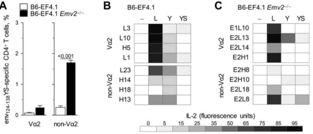
Figure 6. Cross-reactivity of individual Va2 or non-Va2 CD4+T cells.(A) Frequency of env124-138YS-reactive cells in Va2 or non-Va2 CD4+T cells isolated from either B6 (B6-EF4.1) orEmv2-deficient B6 (B6-EF4.1Emv22/2) EF4.1 mice. Data are the means
6SEM (n= 9) of 18-hr stimulations from 3 experiments. (B–C) IL-2 production in response to stimulation with 561026M env124-138L (L), env124-138Y (Y) or env124-138YS (YS) in comparison with the absence of peptide stimulation (-) of Va2 or non-Va2 env124-138L-reactive hybridoma T cell lines derived fromEmv2+/+
(B) orEmv22/2(C) EF4.1 mice.
non-Va2 T cell hybridomas and further suggested that selection byEmv2enriched the non-Va2 repertoire for clones with relative indifference for this position.
Genetic contribution to a high-avidity env124-138
L-reactive CD4+T cell repertoire
Analysis of the env-reactive CD4+
T cell repertoire in B6 mice revealed a clear effect of Emv2-mediated selection. However, in addition to Emv2, the presence of numerous other endogenous retroviruses could affect the formation of the env124-138L-reactive
CD4+T cell repertoire, even if their primary amino acid sequence
is not closely homologous with that of F-MLV env. Furthermore, the functional avidity of env124-138L-reactive CD4+T cells could
also be affected by additional genetic determinants other than endogenous retroviruses. To address this question we generated congenic EF4.1 mice on the 129S8 background. 129S8 mice share the same MHC class II allele with B6 mice (H2-Ab), thus allowing restriction of env124-138L-specific EF4.1 CD4+T cells. However,
they do differ substantially with respect to the composition of endogenous retroviruses and, importantly, 129S8 mice are naturally devoid of endogenous ecotropic MLVs [34,35]. Similar frequency of env124-138L-reactive Va2 CD4+T cells developed in
B6 and 129S8 EF4.1 mice (Figure 8A). In contrast, the frequency of env124-138L-reactive non-Va2 CD4+ T cells was significantly
higher on the 129S8 than on the B6 background (Figure 8A), and was comparable with that on theEmv2-deficient B6 background
Figure 7. Depth of Va2 or non-Va2 env-specific CD4+T cell repertoires.(A–C) Va2 or non-Vaenv124-138L-reactive hybridoma T cell lines were derived fromEmv2+/+
(B6-EF4.1) orEmv22/2(B6-EF4.1Emv22/2) EF4.1 mice and tested for reactivity against a library of env126-138peptide epitopes. The amino acid residues in positions 128 (A), 129 (B) and 133 (C) that elicited at least 40% of the maximal response are listed in the order of preference by the individual clones.
doi:10.1371/journal.ppat.1002709.g007
(Figure 5B), as was their functional avidity (Figure 8B). This finding indicated that deletion of env124-138L-reactive non-Va2
CD4+
T cells in B6, but not in B6-Emv22/2 or 129S8 mice was mediated primarily byEmv2. Surprisingly, however, the functional avidity of env124-138L-reactive Va2 CD4+T cells in 129S8 mice
was very much reduced in comparison with that of Va2 CD4+
T cells in B6 mice (Figure 8B), and was as low as that of low-avidity non-Va2 CD4+
T cells. As, a result of differences in frequency and functional avidity, the env124-138L-specific response of 129S8 mice
was dominated by non-Va2 CD4+
T cells at all peptide doses, in contrast to that of B6 mice, which was dominated by Va2 CD4+
T cells at low peptide doses (Figure 8C).
To further explore the origin of high-avidity env124-138
L-reactive Va2 CD4+
T cells in B6, but not in 129S8 mice, we tested the response of a series of B66129S8-EF4.1 F1 mice. In
comparison with B66129S8-EF4.1 F1mice, which inheritedEmv2
from the B6 parent, B6-Emv22/26129S8-EF4.1 F1 mice, which
lacked ecotropic MLVs, had elevated frequencies of env124-138
L-reactive non-Va2 CD4+
T cells, whereas frequencies of env
124-138L-reactive Va2 CD4+T cells were similar (Figure 8D). These
results confirmed that elevated frequencies of env124-138L-reactive
non-Va2 CD4+
T cells in 129S8 mice were indeed due to lack of
Emv2-mediated selection. Interestingly, both B66129S8-EF4.1 and B6-Emv22/2
6129S8-EF4.1 F1 mice generated env124-138
L-reactive Va2 CD4+
T cells with higher avidity than those of 129S8 mice (Figure 8E), suggesting that a genetic contribution of the B6 parent, other than Emv2, was necessary for the development of high-avidity env124-138L-reactive Va2 CD4+ T cells. To assess
whether this genetic contribution arose from polymorphisms in the Trav locus itself, we tested B6-Tcra2/2
6129S8-EF4.1 F1 mice,
which inherited Emv2 from the B6 parent, but could generate endogenous Va chains only from the locus inherited from the 129S8 parent. The presence ofEmv2in B6-Tcra2/2
6129S8-EF4.1 F1mice had the predicable effect on the frequency of env124-138
L-reactive non-Va2 CD4+
T cells (Figure 8D), which displayed comparably low avidity in all three F1strains tested (Figure 8E).
Surprisingly, however, env124-138L-reactive Va2 CD4+T cells that
had developed in B6-Tcra2/2
6129S8-EF4.1 F1 mice were also
low-avidity, which was comparable with that of Va2 CD4+T cells
in 129S8 mice (Figure 8E), suggesting that the ability of B6 mice to generate high-avidity env124-138L-reactive Va2 CD4+T cells was
germline-encoded. Consequently, the env124-138L-specific response
of B66129S8-EF4.1 F1 mice, but not of isogenic mice lacking
eitherEmv2or the B6-originTrav, was dominated by Va2 CD4+T Figure 8. Genetic contribution to a high-avidity env-reactive CD4+T cell repertoire.
(A) Frequency of env124-138L- reactive cells in Va2 or non-Va2 primary CD4+T cells isolated from either B6 (B6-EF4.1) or 129S8 (129S8-EF4.1) EF4.1 mice. (B) Functional avidity of env124-138L-reactive Va2 or
non-Va2 primary CD4+
T cells from the same donors in A. (C) Frequency of Va2 cells in env124-138L-reactive CD4+
T cells from the same donors in A as a function of peptide concentration. (D) Frequency of env124-138L- reactive cells in Va2 or non-Va2 primary CD4+
T cells isolated from either B66129S8-EF4.1 F1, B6-Emv22/26129S8-EF4.1 F1or B6-Tcra2/26129S8-EF4.1 F1, EF4.1 mice. (E) Functional avidity of env124-138L-reactive Va2 or non-Va2 primary CD4+
T cells from the same donors in D. (F) Frequency of Va2 cells in env124-138L-reactive CD4+
T cells from the same donors in D as a function of peptide concentration. Numbers in (B) and (E) represent the ED50. In (C) and (F) the CD4+T cell response elicited by the last peptide dose
(1028
M) was too small to allow accurate measurement of the frequency of Va2 cells and was therefore omitted. Data in (A–F) are the means6SEM (n= 4–8) of 18-hr stimulations from 3 experiments.
cells at low peptide doses (Figure 8F). The peak percentage of Va2 CD4+
T cells in the env124-138L-reactive population was lower in
B66129S8-EF4.1 F1 mice than in B6 mice, as the former were
expressing endogenous Va chains from both parentalTrav loci. Thus, the combined effect ofEmv2on the frequency of non-Va2 T cells and ofTravon the avidity of Va2 T cells was necessary for the dominance of high-avidity Va2 CD4+T cells in the response to
env124-138L.
Discussion
As a result of the combinatorial process that creates TCRs, their specificity is random and has to undergo selection. Thymic positive and negative selection of developing T cells ensures that mature T cells in the periphery have a functional TCR and minimal reactivity to self proteins, respectively [36]. Negative selection is thought to decrease the frequency, avidity and cross-reactivity of the developing TCR repertoire specific to foreign epitopes that may be similar to self-derived epitopes presented in the thymus [36] and promote peptide specificity [37]. Here we used a well-characterized molecular system to show that negative selection by a defined self peptide fromEmv2env indeed decreased the frequency in the naı¨ve CD4+
T cell repertoire of clones specific to a range of foreign env epitopes, thus reducing the magnitude of the CD4+
T cell response to all env epitope variants. However, negative selection counter-intuitively also promoted the avidity of the CD4+T cell response to F-MLV env by shifting the clonal
composition of responding CD4+T cells in favor of high-avidity
cells.
CD4+ T cells play a central coordinating role in the
orchestration of adaptive immunity to infection, and may also mediate direct antiviral activity. Recent studies in diverse systems have indicated an essential role for the CD4+
T cell response in the control of retroviral infection [15,38–42]. We have previously shown that protection of wt mice against acute FV infection is proportional to the frequency of virus-specific CD4+T cells [23].
Surprisingly, we found that although negative selection signifi-cantly reduced both the precursor frequency and peak expansion of F-MLV env-specific CD4+
T cells, it did not compromise CD4+
T cell-mediated antiviral activity. This finding suggested that not all virus-specific CD4+
T cells were equal in their ability to mediate antiviral functions. Indeed, negative selection byEmv2env affected CD4+T cells with low avidity for F-MLV env, but not
those with high avidity for the same epitope. Preservation of full antiviral activity in the Emv2-selected CD4+
T cell repertoire therefore indicated that this activity is primarily, if not exclusively, exerted by high-avidity CD4+
T cells. High-avidity virus-specific CD4+
T cells may be superior in certain direct antiviral or indirect helper functions than low-avidity ones, but there may also be important exceptions. High-avidity CD4+
T cells responding to FV infection have been reported to show enhancedex vivoproduction of IFN-cand IL-21 cytokines and reduced expression of PD-1 inhibitory receptor [15] than low-avidity counterparts, properties that may contribute to superior antiviral activity. However, T follicular helper (Tfh) differentiation and function were previously found to be similar between high-and low-avidity virus-specific CD4+
T cells [15], suggesting that provision of T cell help for the production of virus-neutralizing antibodies may be more sensitive to the frequency of virus-specific CD4+T cells, rather than their avidity. However, in addition to
the frequency of virus-specific CD4+ T cells, the virus-specific
antibody response is also proportional to the frequency of rare antigen-specific B cells. Thus, when availability of T cell help is abundant, the virus-specific antibody response may be limited by
the frequency of antigen-specific B cells and additional T cell help would not be expected to enhance antibody production. Consis-tent with this idea, adoptive transfer of virus-specific EF4.1 CD4+
T cells into wt B6 mice did not accelerate the virus-neutralizing antibody response [23]. In addition to an effect ofEmv2 on the availability of T cell help for the FV-specific antibody response, Emv2 could in principle also directly affect the development of virus-specific B cells [43]. Although we observed comparably low FV-specific antibody responses between B6 and B6-Emv22/2mice at the peak of FV infection, our results did not exclude a potential direct effect ofEmv2on FV-specific B cell and antibody responses at later time-points, when these responses are fully induced. Indeed,Emv2-encoded env shares 79% amino acid identity with F-MLV env and it is therefore possible that Emv2 expression, especially when upregulated, might affect the FV-specific antibody response.
As previously shown, high-avidity F-MLV env122-141L-specific
Va2 CD4+
T cells are a minority subset in the naı¨ve repertoire and only dominate the immune response to FV as a result of their preferential expansion during infection [15]. We have now found that for this ability of high-avidity F-MLV env122-141L-specific
Va2 CD4+
T cells to dominate the peak response, negative selection byEmv2of at least some of the competitor low-avidity F-MLV env122-141L-specific non-Va2 CD4+ T cells is necessary.
These findings indicate that even subtle thymic events can have profound effects on the induction of an effective T cell response to retroviral infection. Recently, a comprehensive theoretical study has indicated thatHLAclass I alleles that associated with control of HIV infection, such asHLA-B*5701, sample far fewer self peptides than other HLAalleles [5]. As a result of less stringent negative selection, a higher frequency of CD8+
T cells restricted by these protective alleles were predicted to recognize viral peptide epitopes and to cross-react with variants of the targeted epitopes [5].
Our results with a single self peptide provide further experi-mental confirmation of negative selection reducing both the precursor frequency and cross-reactivity of env-specific CD4+T
cells, although in this case the effect on cross-reactivity was more pronounced at the population, rather than the single-cell level. These results also suggest that from the thousands of self peptides that can mediate thymic selection of retrovirus-specific T cells, the main effects may be mediated by only a few self peptides. Moreover, self peptides with such strong influence may also be polymorphic between different individuals, which might contrib-ute to the partial association ofHLA polymorphisms with virus control [3,5,6].
In addition to polymorphisms at theMHC/HLAlocus or of self peptides mediating thymic selection, the Trav/TRAV and Trbv/ TRBV loci may also display allelic sequence variation. A polymorphism in the TRBV9 gene has been shown to affect TCR affinity for and functional recognition of an HLA-B*3501-restricted epitope from the EBNA-1 protein of Epstein-Barr virus (EBV), leading to a public T cell response dominated by the high-affinity variant [44]. Similarly, we found that the ability of Va2 chains to confer high avidity for env122-141L in EF4.1 mice seems
to be germline-encoded, as only Va2 chains encoded by the B6, but not the 129 Trav locus had this ability. It is tempting to speculate that amino acid residues unique to the B6-germline Trav14-encoded Va2 chains participate in recognition of the strongly interacting L (or a limited set of amino acids with similar properties) at env position 128. Notably, the CD8+
T cell response to an HLA-B8-restricted epitope from the latent antigen EBNA 3A of EBV uses almost exclusively identical Vaand Vb, as well as other TCR-region sequences, and comprehensive structural studies have shown that a unique amino acid residue in the
germline-encoded complementarity-determining region 2 (CDR2) of the preferred Va chain, encoded by TRAV26-2, is critically required for binding to a residue from the peptide epitope [45]. Despite the vast number of somatically-generated random TCRs that can arise during T cell development, these studies highlight the potential for germline-encoded residues to provide exquisite specificity and competitive advantage to the TCRs that carry them.
In addition to likely representing the best-fit for recognition of Ab -restricted env122-141L, the dominance of Va2 EF4.1 CD4+T cells
could also result from preferential pairing of the transgenic TCRb
chain with Va2 chains in general. This is unlikely to be the case as the usage of Va2 cells was not increased in either total or env122-141L-reactive EF4.1 CD4+T cells, and indeed in the env
122-141L-reactive preimmune repertoire clones using other Vachains
were at least 3 times more frequent than those using Va2. However, although non-Va2 env122-141L-reactive CD4+T cells were still the
majority inEmv2-expressing mice, their ability to participate in the response to FV and compete with env122-141L-reactive Va2 CD4+T
cells was severely compromised byEmv2. Thus, the dominance of Va2 CD4+T cells in the response to FV infection can be seen as a
combination of germline-encoded advantage in Ab-restricted env122-141L recognition conferred to Va2 CD4+ T cells and of
Emv2-mediated self-tolerance of other non-Va2 CD4+
T cells capable of recognizing Ab-restricted env122-141L.
One important novel insight of the current study is the proof of principle that negative selection is not necessarily always impairing high-avidity T cell responses. By counter-selecting some cross-reactive CD4+
T cells, negatively selecting self peptides have the ability to significantly enhance the avidity for the response to at least some epitope variants. Higher precursor frequency and cross-reactivity with emerging epitope variants seem to be the best correlates for an effective cytotoxic CD8+
T cell response [5]. Whether higher avidity for the primary infecting epitope, rather than cross-reactivity with epitope variants better describes an effective CD4+T cell response to retroviral infection needs to be
further addressed.
It should be noted that differences in avidity for antigen in this system were defined functionally. Indeed, Va2 env122-141L-specific
primary CD4+
T cells or hybridomas reacted to much lower concentrations of env122-141L peptide stimulationin vitrothan their
non-Va2 counterparts. Furthermore, this higher sensitivity trans-lated to higher in vivo expansion and increased potential for cytokine production [15]. It is currently unclear whether differences in functional avidity between Va2 and non-Va2 env122-141L-specific CD4+ T cells resulted from overall higher
affinity of individual TCRs of these polyclonal populations for the peptide-MHC class II complex. Although dissociation kinetics between TCRs and peptide-MHC class II tetramers are often informative with respect to the biochemical affinity of these TCRs, they may not be universally useful. For example, the available env123-141-A
b
tetramer (Ab-env) is known to bind only some env124-138L-specific CD4+T cell clones but not others, irrespective
of their functional avidity or Va usage [14,22]. Therefore, this reagent could not be used to access the biochemical affinity of all env124-138L-specific CD4+ T cells in the polyclonal repertoire.
Furthermore, identification of antigen-specific cells using a sensitive two-dimensional binding assay has recently demonstrated that the affinity of many CD4+
T cells that participate in the response to two separate antigens is below detection with peptide-MHC class II tetramers [46]. Thus, peptide-peptide-MHC class II tetramers may generally only detect some but not all antigen-specific CD4+
T cells. In addition, such detection is conditional on expression of sufficient TCR levels. Indeed, we have found that the
extensive, antigen-induced downregulation of their TCR in vivo, eclipses detection with the Ab-env123-141 tetramer of even the
env122-141L-reactive CD4+T cells that could otherwise bind this
reagent. Similar observations have been recently made with peptide-MHC class I tetramer staining of virus-specific effector CD8+
T cells [47], suggesting that the inability of peptide-MHC multimers to identify antigen-specific effector T cells that have downregulated their TCRs may be a general problem for T cells restricted by both classes of MHC molecules.
Negative selection ensures minimal reactivity of developing thymocytes to self proteins. However, endogenous retroviruses are a large constituent of mammalian genomes and thus represent a potentially large pool of self proteins able to mediate selection, both positive and negative. Self peptides encoded by endogenous MLVs have been shown to mediate positive selection of CD4+
T cells with specificity for an unrelated H2-Ek-restricted moth cytochrome C peptide, and to enhance the response of mature CD4+T cells with
this specificity in the periphery [48]. We found that Emv2 was expressed at very low levels in the thymus of B6 mice, in agreement with a previous report [49], and was undetectable by qRT-PCR in some of the mice. It should be noted, however, that the qRT-PCR method employed was specific only for the splicedenvmRNA that is transcribed byEmv2. This was chosen to eliminate the possibility of detecting contaminating genomic DNA or viral genomic RNA, but may underestimate the total amount of spliced and unspliced mRNA that leads to the production of other viral proteins. Nevertheless, as demonstrated by its effect on thymic development, this low level ofEmv2expression was clearly functional.
Endogenous retroviruses have been known for many years to cause a range of different diseases in mice, including cancer, immunodeficiency and autoimmunity, although a similar causal effect in humans has been questioned [50]. Immune reactivity to endogenous retroviruses has been amply demonstrated in mice where is has been strongly associated with the development of spontaneous autoimmune conditions [51,52]. Interestingly, immune reactivity to endogenous retroviruses has also been frequently observed in humans during infection, inflammation, autoimmunity and cancer [50,53–56]. Expression of human endogenous retrovi-ruses, as well as CD8+
T cell responses against their antigens, have been documented in HIV infection [57,58]. Furthermore, a whole-genome association study has suggested that part of the effect of the protective HLA-B*5701 allele during the asymptomatic period of HIV infection may be mediated by a linked human endogenous retrovirus at the same locus [59]. Human endogenous retroviral antigens have also been reported to serve as targets for CD8+
T cell-mediated rejection of cancer cells [60]. It might be evident from the studies in humans and the results of the current study that peptide epitopes encoded by endogenous retroviruses have a strong influence on T cell thymic selection and may also participate in the shaping of the peripheral T cell response. It is also clear that endogenous retroviruses do not always cause immunological tolerance, and although their activation in infected or transformed cells may provide a non-mutable target for immune attack, activation of endogenous retroviruses may also trigger inflammatory or autoim-mune phenomena frequently associated with infection and cancer. Further study of endogenous retrovirus regulation during infection, autoimmunity or cancer, and of the immune responsiveness to them should shed more light into their pathogenic potential.
Materials and Methods
Ethics statement
UK Home Office regulations under the Animals Scientific Procedures Act 1986 (ASPA).
Mice
Inbred C57BL/6J (B6), A/J and B6.SJL-Ptprca Pep3b/BoyJ (CD45.1+
B6) mice were originally obtained from The Jackson Laboratory (Bar Harbor, Maine, USA) and were subsequently maintained at NIMR animal facilities. Inbred 129S8/SvEvNimrJ (129S8) mice were developed from an 129/Sv substrain, maintained at NIMR animal facilities, and were subsequently deposited at The Jackson Laboratory. The B6 TCRb-transgenic strain EF4.1, expressing a transgenic TCRb chain from a T cell clone specific to F-MuLV env122-141 presented by H2-Ab, has been described
[14]. 129S8-congenic EF4.1 mice were generated by serial backcrossing of B6-EF4.1 mice for 10 nuclear generations onto the 129S8 genetic background. B6-backcrossed Rag1-deficient (Rag12/2) mice [61] and T cell receptora-deficient (Tcra2/2) mice [62] were also maintained at NIMR animal facilities.Fv2s-congenic B6 (Fv2s) andRag12/2(Fv2sRag12/2) mice have been previously described [25].Emv2-deficient (Emv22/2) B6 mice were created by introducing theEmv2integration site of chromosome 8 from the A/J strain, which lacks this proviral integration, by serial backcrossing for at least 12 nuclear generations onto the B6 genetic background. Lack of Emv2 was validated by PCR for both the D8Mit49 microsatellite marker close to the locus that detects polymorphisms in A/J (Emv22) and B6 (Emv2+
) strains of mice (D8Mit49forward 59 -TCTGTGCATGGCTGTGTATG-39 and D8Mit49 reverse 59 -TGGTGTGCTGCTGATGCT-39), and also for the actual inte-gration site using three primers, two of which were flanking the integration site (forward 59 -ACCCACTAAGTAACCCAG-GCTGCCTCAGCT-39 and reverse 59 -GACCAGAATAGAAA-GACGTTCAAGTGAGCT-39) and one located in theEmv2LTR (59-ATCAGCTCGCTTCTCGCTTCTGTACCCGCG-39) (Fig-ure S3).
In vitroT cell activation
Spleen or lymph node single-cell suspensions were prepared from EF4.1 mice and 56105cells per well were stimulated in 96-well plates with the indicated amount of env peptide variants. The frequency of env-reactive cells in stimulated CD4+
T cells was defined as the frequency of cells that responded to 18-hr stimulation, before cell division or death had occurred, by upregulating CD69 expression. Correct identification of env-reactive CD4+
T cells by CD69 upregulation was confirmed in control experiments by co-staining for CD154 (CD40L) expression in stimulated T cells. Both antibodies were obtained from eBiosciences. For assessment of T cell activation on day 3, cells were labeled with CFSE before stimulation and responding cells were identified by CFSE dilution.
Hybridoma cell line generation and stimulation
Single-cell suspensions were prepared from spleens and lymph nodes from Emv2-sufficient or -deficient EF4.1 mice and stimu-lated in vitro with 1027 M or 1025 M env122-141L peptide and
4 ng/ml recombinant human IL-2 for 4 days. CD4+
T cells were subsequently purified from stimulated cultures using immuno-magnetic positive selection (StemCell Technologies, Vancouver, BC, Canada) and fused to TCRab-negative BW5147 thymoma cells to produce hybridoma cell lines. Established hybridoma cell lines were stimulated with a range of env peptide variants presented by dendritic cells. Dendritic cells were obtained from cultures of bone marrow cells isolated from B6 mice and supplemented with granulocyte macrophage colony-stimulating factor (GM-CSF). GM-CSF was obtained from culture
superna-tant of663 cells transfected with mouseCsf2and was used at 1:10 dilution. Bone marrow cells were culture in these conditions for 7 days, at which point they consisted of 50–70% dendritic cells. These cells were then used to stimulate hybridoma cells at a ratio of 56104dendritic cells to 16105hybridoma cells, for 18 hrs, in the presence or absence of env peptide variants. Dendritic cell-hybridoma cell co-cultures were plated in flat-bottom 96-well plates in 200ml final volume. The concentration of peptides used
is indicated in individual figures and figure legends. In additional experiments peritoneal macrophages were also used as antigen-presenting cells with results comparable to the use of dendritic cells. Macrophages were isolated from B6 mice following plating of the peritoneal cavity exudate cells for 1 hr and washing off the non-adherent fraction. Env-specific responses were assessed by measuring the amount of IL-2 secreted in co-culture supernatants using an AlamarBlue (Invitrogen, Carlsbad, CA, USA)-based CTLL-2 assay.
Tragene usage
Trav and Traj usage by T cell hybridomas was probed by staining with an anti-Va2 (clone B20.1) or anti-Va3.2 (clone RR3-16) monoclonal antibodies, and by reverse transcription (RT)-PCR amplification and sequencing of expressed Trav genes, using previously described primers [63]. Trav and Traj segment identification and alignment, and confirmation of productive rearrangements were performed on the International Immunoge-netics Information System website (http://www.imgt.org).
Viruses and infections
The FV used in this study was a retroviral complex of a replication-competent B-tropic F-MuLV and a replication-defec-tive polycythemia-inducing spleen focus-forming virus (SFFVp). Stocks were propagated in vivo and prepared as 10% w/v homogenate from the spleen of 12-day infected BALB/c mice. Mice received an inoculum of,1,000 spleen focus-forming units
of FV. All viral stocks were free of Sendai virus, Murine hepatitis virus, Parvoviruses 1 and 2, Reovirus 3, Theiler’s murine encephalomyelitis virus, Murine rotavirus, Ectromelia virus, Murine cytomegalovirus, K virus, Polyomavirus, Hantaan virus, Murine norovirus, Lymphocytic choriomeningitis virus, Murine adenoviruses FL and K87, and Lactate dehydrogenase-elevating virus. Virus inocula were injected via the tail vein in 0.1 ml of phosphate-buffered saline. FV-infected cells were detected by flow cytometry using surface staining for the glycosylated product of the viral gag gene (glyco-Gag), using the matrix (MA)-specific monoclonal antibody 34 (mouse IgG2b), followed by an anti-mouse IgG2b-FITC secondary reagent (BD, San Jose, CA, USA). For the assessment of anemia, mice were bled by a small incision of the tail vein and blood was collected into heparinized capillary tubes. Complete blood counts were measured on a VetScan HMII hematology analyzer (Abaxis, CA, USA), following the manufac-turer’s instructions. RBC counts of uninfected mice were
,9.956106 per mm3 of blood. FV-induced splenomegaly in infected mice was expressed as spleen index, which is the ratio of the weight of the spleen (in mg) to the weight of the rest of the body (in g).
FV-neutralizing and F-MLV-infected cell-binding antibody assays
Serum titers of FV-neutralizing antibodies were measured as previously described [25]. The dilution of serum which resulted in 75% neutralization was taken as the neutralizing titer. Serum titers of F-MLV-infected cell-binding antibodies were determined by