Crystallographic Study of Peptidoglycan
Biosynthesis Enzyme MurD: Domain
Movement Revisited
RomanŠink1☯, Miha Kotnik2☯, Anamarija Zega1‡, Hélène Barreteau3‡, Stanislav Gobec1‡,
Didier Blanot3‡, Andréa Dessen4,5,6,7‡, Carlos Contreras-Martel4,5,6‡ *
1University of Ljubljana, Faculty of Pharmacy, Aškerčeva 7, Ljubljana, Slovenia,2Lek Pharmaceuticals d. d., Verovškova 57, Ljubljana, Slovenia,3Laboratoire des Enveloppes Bactériennes et Antibiotiques, Institut
de Biologie Intégrative de la Cellule (I2BC), CEA, CNRS, Université Paris-Sud, Gif-sur-Yvette, France, 4Univ. Grenoble Alpes, Institut de Biologie Structurale, Grenoble, France,5CNRS, IBS, Grenoble, France, 6CEA, IBS, Grenoble, France,7Brazilian National Laboratory for Biosciences (LNBio), CNPEM,
Campinas, São Paulo, Brazil
☯These authors contributed equally to this work. ‡These authors also contributed equally to this work.
Abstract
The biosynthetic pathway of peptidoglycan, an essential component of bacterial cell wall, is a well-recognized target for antibiotic development. Peptidoglycan precursors are synthe-sized in the bacterial cytosol by various enzymes including the ATP-hydrolyzing Mur ligases, which catalyze the stepwise addition of amino acids to a UDP-MurNAc precursor to yield UDP-MurNAc-pentapeptide. MurD catalyzes the addition of D-glutamic acid to UDP-MurNAc-L-Ala in the presence of ATP; structural and biochemical studies have suggested the binding of the substrates with an ordered kinetic mechanism in which ligand binding inevitably closes the active site. In this work, we challenge this assumption by reporting the crystal structures of intermediate forms of MurD either in the absence of ligands or in the presence of small molecules. A detailed analysis provides insight into the events that lead to the closure of MurD and reveals that minor structural modifications contribute to major overall conformation alterations. These novel insights will be instrumental in the develop-ment of new potential antibiotics designed to target the peptidoglycan biosynthetic pathway.
Introduction
The development of bacterial resistance to antibiotics worldwide is a phenomenon that urges the need to discover new antibacterial drugs. The biosynthetic pathway of peptidoglycan, a key component of the bacterial cell wall, is a tractable target since proteins that catalyze key reac-tions in the pathway can be targeted both by natural and synthetic antibiotics. [1–4] Peptido-glycan is a complex heteropolymer that consists of Peptido-glycan chains cross-linked by short peptides. [5] The glycan chains are made up of alternatingN-acetylglucosamine (GlcNAc) and
OPEN ACCESS
Citation:Šink R, Kotnik M, Zega A, Barreteau H, Gobec S, Blanot D, et al. (2016) Crystallographic Study of Peptidoglycan Biosynthesis Enzyme MurD: Domain Movement Revisited. PLoS ONE 11(3): e0152075. doi:10.1371/journal.pone.0152075
Editor:Ivo G. Boneca, Institut Pasteur Paris, FRANCE
Received:August 19, 2015
Accepted:March 8, 2016
Published:March 31, 2016
Copyright:© 2016Šink et al. This is an open access article distributed under the terms of theCreative Commons Attribution License, which permits unrestricted use, distribution, and reproduction in any medium, provided the original author and source are credited.
Data Availability Statement:All relevant data are within the paper and its Supporting Information files. All coordinates files as relevant data are available from the Protein Data Bank database (accession numbers: 5a5e, 5a5f).
Funding:This work was financially supported by the European Union FP6 Integrated Project
EURINTAFAR (project no. LSHM-CT-2004-512138;
N-acetylmuramic acid (MurNAc) residues linked byβ1!4 bonds. The lactoyl group of the
MurNAc residues is substituted by a peptide stem most commonly composed of L-Ala-γ -D-Glu-meso-A2pm (or L-Lys)-D-Ala-D-Ala (A2pm, 2,6-diaminopimelic acid). Cross-linking of the stem peptides generally occurs between the carboxyl group of D-Ala at position 4 and the amino group of the diaminoacid at position 3, either directly or through a short peptide bridge. [5]
The biosynthesis of peptidoglycan can be roughly divided into three stages: cytoplasmic, membrane-associated, and periplasmic. Within the bacterial cytoplasm, Mur ligases perform the stepwise addition of amino acids to UDP-N-acetylmuramic acid (UDP-MurNAc), thereby building a stem peptide. Mur ligases require ATP for activity, and are absolutely essential for bacterial survival. [6] The mechanism of action of Mur ligases (MurC, D, E, F) has been studied through kinetic experiments, site-directed mutagenesis, and X-ray crystallography. [7–21] These studies have shown that the Mur ligases share the same reaction mechanism (Fig 1A), which consists first of the activation of the carboxyl group of the nucleotide precursor by ATP, generating an acyl phosphate intermediate and ADP; the acyl phosphate then undergoes a nucleophilic attack by the amino group of the condensing amino acid (or dipeptide), leading to the formation of a high-energy tetrahedral intermediate, which eventually breaks down into amide or peptide and phosphate. In addition, it has been shown that MurC and MurF follow a strictly ordered kinetic mechanism in which ATP binds first, followed by the nucleotide sub-strate and then the condensing amino acid or dipeptide. [7–13]
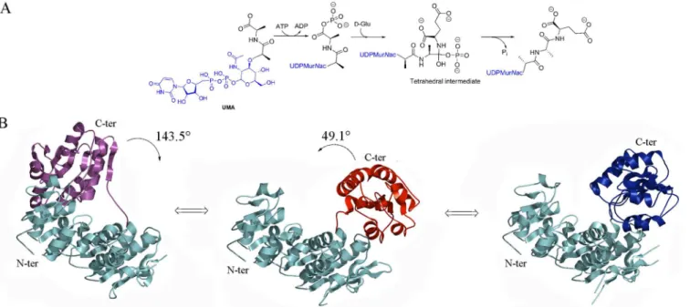
From a structural point of view, Mur ligases are three-domain enzymes that share a very well conserved ATP-binding site that harbors the Gly-X-X-Gly-Lys-Thr/Ser [12,14] motif and display notable structural similarities in terms of domain arrangement. [20–21] The N-termi-nal domain is involved in the binding of the UDP-precursor, the central domain in the binding of ATP, and the C-terminal domain in the binding of the amino acid or dipeptide. [9,19–21] Crystal structures of Mur ligases reveal that MurC, MurD and MurF display at least two dis-tinct conformations, namely‘open’and‘closed’, with the position of C-terminal domain dictat-ing the nature of the conformation. [20–21] This has led to the suggestion that formation of an active catalytic site requires closing of the structure upon substrate binding. [7]
MurD catalyzes the addition of D-glutamic acid to UDP-MurNAc-L-Ala, generating the dipeptide moiety L-Ala-D-Glu in almost all bacterial species. [22] As mentioned above, struc-tural information onE.coliMurD reveals two open conformations: an unbound one and an UDP-N-acetylmuramoyl-L-alanine (UMA)-complexed one. [10] In addition, several structures of substrate- or product-bound closed conformations have been published. [8–9,16]E.coli MurD has been the object of targeted molecular dynamics simulation studies that have shed light on the conformational changes that occur upon ligand binding as presented inFig 1B. These studies have revealed that upon binding of ATP and UMA, there is a‘closing’rotation of the C-terminal domain, which does not occur before ATP is bound. [23]E.coliMurD has been the object of extensive studies aiming at the development of new inhibitors, [24–43] but only a limited number of ligands could be crystallized within the MurD active site, [16,30,34,37,40, 42–43] or were shown to interact with MurD through NMR experiments. [38,44–45] In all solved X-ray structures of MurD with inhibitors, the ligase is in closed conformation. Notably, the exploitation of this tractable, potential antibiotic development target requires a precise knowledge of the structural modifications engendered by ligand recognition. In order to struc-turally characterize the different conformational changes of MurD in atomic detail, we solved two novel crystal structures ofE.coliMurD either in the presence or in the absence of ligands. This work led us to identify novel conformational properties of MurD involving an intermediate conformation in the presence of ADP and UMA, as well as an intermediate conformation in the absence of ligands. This work thus reveals that substrate binding is not the strict causative agent
Charles Nodier. The funders had no role in study design, data collection and analysis, decision to publish, or preparation of the manuscript. This work used the platforms of the Grenoble Instruct centre (ISBG; UMS 3518 CNRS-CEA-UJF-EMBL) with support from FRISBI (ANR-10-INSB-05-02) and GRAL (ANR-10-LABX-49-01) within the Grenoble Partnership for Structural Biology (PSB)[Lek Pharmaceuticals d.d.http://www.lek.si]. The funder provided support in the form of salaries for MK, but did not have any additional role in the study design, data collection and analysis, decision to publish, or preparation of the manuscript.
of domain closure, and that Mur enzymes display a variable amount of flexibility, both in the presence and absence of substrates, which could be essential for their activity in the cell. In addi-tion, our structural analyses indicate that the kinetic mechanism of MurD, which had previously been suggested as being ordered by similarity with MurC and MurF [7,13] may in fact be dis-tinct, since protein function could be potentially affected by domain flexibility.
Materials and Methods
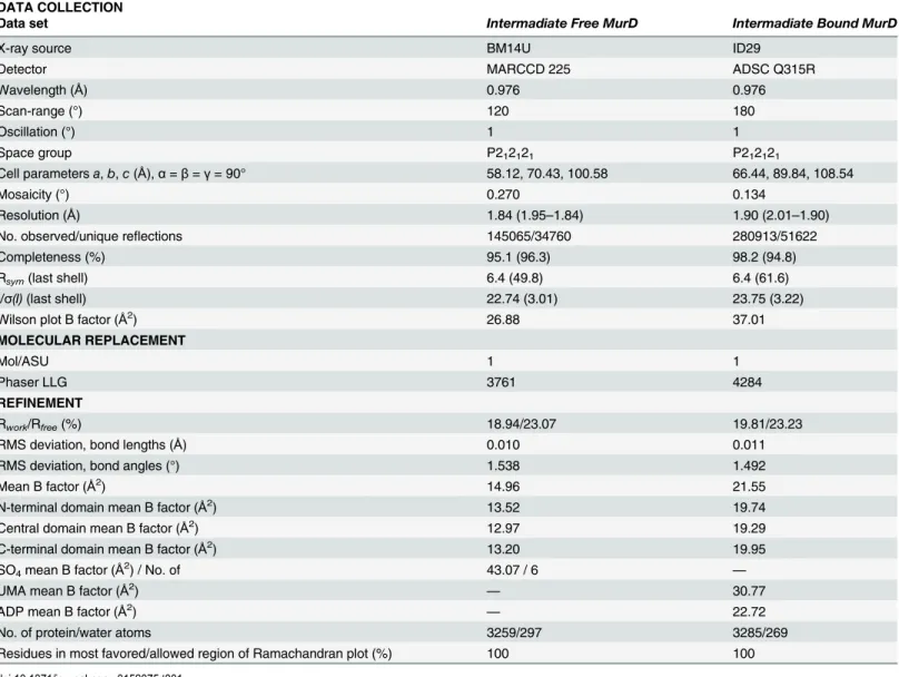
Crystallization and data collection
MurD was expressed and purified using the 6×His-tag system described. DH5αcells harbour-ing the pABD16/MurD vector were grown in 2-YT medium containharbour-ing ampicillin (100μg/
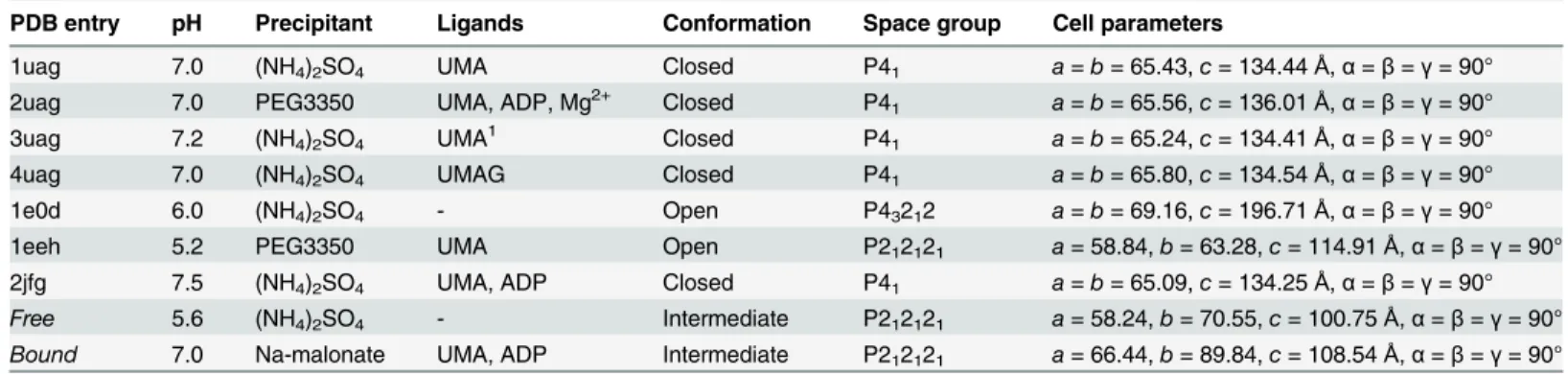
mL) at 37°C in a rotary shaker to reach A600nm of 3.5. Expression was induced by adding iso-propylβ-D-thiogalactopyranoside at a final concentration of 1 mM and growth was continued overnight at 20°C. The cells were lysed by sonication and the 6×His-tagged protein was puri-fied by affinity binding to a Ni2+-nitrilotriacetate-agarose column and elution with a discontin-uous gradient of imidazole. The enzyme was recovered in the 100 mM fraction. It was dialysed against 20 mM Hepes (pH 7.4), 200 mM NaCl, 5 mM DTT, 0.05% (w/v) NaN3. The amount of protein obtained was determined by the Bradford method, quantitative amino acid analysis, and by measuring the absorbance at 280 nm. The purity of the protein was checked by SDS-PAGE and MALDI-TOF mass spectrometry. [16] Both conformations crystallized in an orthorhombic space group with different cell parameters and contained one molecule per asymmetric unit (Table 1).
Crystallization of the intermediate conformation of ligand free MurD (Intermediate Free MurD):
We scanned various crystallization conditions in order to obtain different forms of MurD crystals, which could then be soaked with different inhibitor solutions. Different combinations of buffers, precipitants, co-precipitants, pH values and crystallization temperatures were tried
Fig 1. Reaction mechanism and known conformations of MurD.(A) Mechanism of reaction catalyzed by MurD. (B) Rotation of the C-terminal domain from Open UMA-bound (Magenta) (PDB entry: 1eeh [7]) to Open (Red) (PDB entry: 1e0d [7]) to Closed (Blue) (PDB entry: 3uag [6]) with identified angles of rotation. For the sake of clarity N-terminal and central domains are colored in cyan in all conformations.
in the presence or absence of MurD substrates. Several different forms of crystals grew over the time and after solution of all of the structures, new Intermediate Free MurD and Intermediate Bound MurD structures were identified. Concurrently, as a positive control, the crystals of open [10] and closed [8,9] forms of MurD were also produced using the crystallization condi-tions described previously.
Intermediate Free MurDwas crystallized by mixing 2μL of protein sample (3 mg/mL, in 20
mM HEPES, pH 5.6, 1 mM DTT, and 1 mM NaN3) and 2μL of reservoir solution (1.8 M
(NH4)2SO4, 7% (v/v) (±)-2-methyl-2,4-pentanediol and 0.1 M MES, pH 5.6) at 15°C by vapor diffusion using the hanging-drop method. Crystals grew in 48 hours. X-ray diffraction data were collected at the European Synchrotron Radiation Facility (ESRF, Grenoble, France) (Table 1).
Crystallization of intermediate conformations of MurD with ligands (Intermediate Bound MurD):
Crystals ofIntermediate Bound MurDwere obtained at 15°C by vapor diffusion using the hanging-drop method. Crystals were grown by mixing 2μL of protein (4 mg/mL, 1 mM UMA,
Table 1. X-ray diffraction data and structure refinement.
DATA COLLECTION
Data set Intermadiate Free MurD Intermadiate Bound MurD
X-ray source BM14U ID29
Detector MARCCD 225 ADSC Q315R
Wavelength (Å) 0.976 0.976
Scan-range (°) 120 180
Oscillation (°) 1 1
Space group P212121 P212121
Cell parametersa,b,c(Å),α=β=γ= 90° 58.12, 70.43, 100.58 66.44, 89.84, 108.54
Mosaicity (°) 0.270 0.134
Resolution (Å) 1.84 (1.95–1.84) 1.90 (2.01–1.90)
No. observed/unique reflections 145065/34760 280913/51622
Completeness (%) 95.1 (96.3) 98.2 (94.8)
Rsym(last shell) 6.4 (49.8) 6.4 (61.6)
I/σ(I)(last shell) 22.74 (3.01) 23.75 (3.22)
Wilson plot B factor (Å2
) 26.88 37.01
MOLECULAR REPLACEMENT
Mol/ASU 1 1
Phaser LLG 3761 4284
REFINEMENT
Rwork/Rfree(%) 18.94/23.07 19.81/23.23
RMS deviation, bond lengths (Å) 0.010 0.011
RMS deviation, bond angles (°) 1.538 1.492
Mean B factor (Å2
) 14.96 21.55
N-terminal domain mean B factor (Å2) 13.52 19.74
Central domain mean B factor (Å2) 12.97 19.29
C-terminal domain mean B factor (Å2) 13.20 19.95
SO4mean B factor (Å2) / No. of 43.07 / 6 —
UMA mean B factor (Å2) — 30.77
ADP mean B factor (Å2) — 22.72
No. of protein/water atoms 3259/297 3285/269
Residues in most favored/allowed region of Ramachandran plot (%) 100 100
5 mM AMP-PNP, 1 mM NaN3, 1 mM DTT, and 20 mM HEPES, pH 7.4) with 2μL of reservoir
solution (1.8 M Na-malonate, pH 7.0). Crystals appeared in 6 months and data was collected at the ESRF, as above (Table 1).
Structure determination and refinement
X-ray diffraction data sets were indexed and scaled with XDS. [46] The structure was solved by employing the structure of MurD ofE.coli(PDB entry: 3uag [9]) split into two search models (model 1: N-terminal and central domains (residues 1–299); model 2: C-terminal domain (resi-dues 300–437)) in a molecular replacement approach using PHASER. [47] The solution model was rebuiltde-novoin order to reduce bias from the model using ARP/wARP. [48] COOT [49– 51] was used for manual corrections of the model. Cycles of refinement employing three domains by TLS definition [52] (residues 1–92, 94–295 and 304–439) were performed by REFMAC 5.5 [53] as implemented in the CCP4-6.3.0 suite of programs. [53] In addition, water molecules were added to the residual electron density map using ARP/ wARP. [48] After sev-eral cycles of manual model building and refinement, Rworkand Rfreeconverged. The stereo-chemical quality of the refined model was verified with MOLPROBITY [54] and PROCHECK. [55] The secondary structure assignment was performed by DSSP [46] and STRIDE. [56] RMSD values for the best fit for the whole protein, separate domains, rotation angle and trans-lation of C-terminal domain and bending region analysis were calculated using DynDom server. [57] For identification of protein-ligand interactions and interactions between amino acid residues, LIGPLOT [58] and LigPlot+ [59] were used. X-ray diffraction data, structure solution and refinement statistics are found inTable 1. Figures containing protein structures were generated with PyMol. [60]
Results
Structures ofE.coliMurD solved by Bertrand and coworkers either in unbound form or in the presence of substrates or products suggest that ligand binding engenders a conformational movement of the C-terminal domain towards the center of the structure, thus leading to the closure of the enzyme. [9–10] Since the detailed knowledge of all of the forms of MurD is essential for optimal exploitation of this enzyme for potential antibiotic development, we initi-ated a study on the different conformations ofE.coliMurD, initially by attempting to trap MurD in states that were distinct from those which had been previously described. We achieved this by solving two intermediate conformations of MurD which we named Intermedi-ate Free MurD(i.e., intermediate conformation of ligand-free MurD) andIntermediate Bound MurD(i.e., intermediate conformation of MurD with ligands).
Results regarding these structures are presented in three separate sections in whichi) Inter-mediate Free MurDandii) Intermediate Bound MurDare both compared to the previously solved structures of MurD, andiii)similarities and differences between the two new conforma-tions of MurD are highlighted.
Intermediate Free MurD
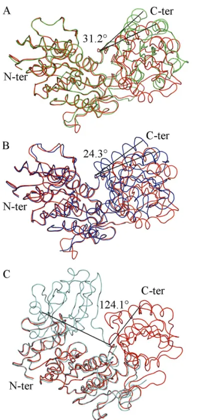
In the solving ofIntermediate Free MurD, 421 residues were refined. There is one minor region missing between residues Thr180─Tyr187 since it could not be traced in the electron density
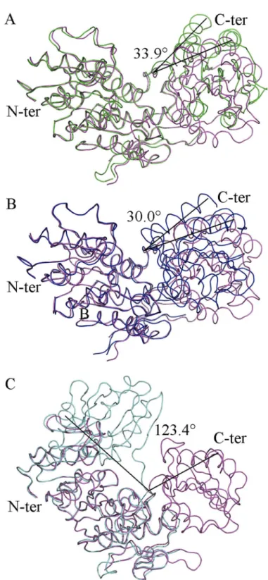
Fig 2. Superposition ofIntermediate Free MurD(red) to known MurD structures with identified angles
of rotation of the C-terminal domain.(A) open MurD (green) (PDB entry: 1e0d [7]). (B) closed MurD (blue) (PDB entry: 3uag [6]). (C) open MurD (cyan) (PDB entry: 1eeh [7]).
UMA-bound MurD [9] (Fig 2B). It appeared that it was not in a closed conformation. After superposition ofIntermediate Free MurDwith open UMA-bound MurD [10] (Fig 2C), we con-firmed that this new conformation of MurD is different from the previously described ones. These analyses revealed that, in spite of the absence of ligands, our new structure was not completely open, and rather displayed an intermediate conformation. This fact was quantified by the determination of the RMSD values of all of the superimposed structures (Table 2), both for the whole protein and for distinct domains.
We analyzed the conformations of key residues that contribute to the binding of UMA [9– 10] and discovered that they are the same as in closed UMA-bound MurD structures [9] We also compared the orientations of residues that contribute to ADP binding and found that they share basically the same conformation as previously reported. [9,16] Only Arg302 was found to be differently oriented so we focused on this residue because of its position in the hinge loop containing residues from Thr294 to Glu304. There is a difference in the orientation of Arg302 as well as its interactions, when compared to the previously solved closed conformations of MurD (Fig 3A–3D) and additionally orientation of Arg302 inIntermediate Free MurDis dif-ferent from all of the orientations found in the aforementioned structures of MurD (Fig 3E), which contributes towards the intermediate conformation adopted by the protein.
Intermediate Bound MurD
Intermediate Bound MurDwas co-crystallized with UMA and AMP-PNP in order to further explore the dynamics of MurD upon ligand binding. During the process of structure solution, 423 residues were refined. Structural inspection ofIntermediate Bound MurDrevealed that Lys198 is not carbamoylated, contrary to all previously published structures of MurD com-plexed to UMA [8–9,16] Previously, complex formation between MurD and UMA, [8] UDP-N-acetylmuramoyl-L-alanyl-D-glutamic acid (UMAG), [9] UMA-ADP, [13] UMA-ADP-Mg 2-+[9] and UMA-ADP-Mn2+[9] locked MurD in a closed structure, which gave weight to the
proposal that ligand binding was important for the movement of the C-terminal domain lead-ing to closure. [8–9] We investigated ourIntermediate Bound MurDstructure by superimpos-ing it to the previously solved structures (Fig 4). Superposition to the open unbound MurD structure [10] (Fig 4A) revealed that the new structure was clearly in a distinct conformation (Table 2). Further investigation consisted in superimposing it to the closed structure of
UMA-Table 2. Analysis of the superposition of theIntermediate Free MurDandIntermediate Bound MurD.
Aligned structures Overalla N-ter-centralb C-terc d(°) e(Å) Residue
3uag/Free 3.12 [417/421] 0.98 [276/293] 0.41 [141/144] 24.3 -1.8 298–301
1e0d/Free 3.70 [421/421] 1.21 [282/294] 0.43 [141/143] 31.2 1.5 298–301
1eeh/Free 15.33 [419/421] 1.06 [278/294] 1.32 [141/143] 124.1 -4.6 297–304
3uag/Bound 3.69 [420/423] 0.84 [279/294] 0.43 [141/144] 30.0 -0.7 298–300
1e0d/Bound 3.78 [419/423] 0.99 [287/294] 0.56 [141/143] 33.9 1.0 297–301
1eeh/Bound 15.21 [419/423] 0.64 [278/294] 1.40 [141/143] 123.4 -3.0 296–304
Free/Bound 1.30 [416/421] 1.01 [277/294] 0.65 [139/143] 7.5 0.2 118–122, 234–262, 291–292
aRMSD (Å) [No. Cα] of the two superimposed structures best fit bRMSD (Å) [No. Cα] of the
fixed domains of the two superimposed structures bestfit. Here N-terminal and central domains are seen as one non-rotating domain
cRMSD (Å) [No. Cα] of the rotating C-terminal domain of the two superimposed structures best fit
dRotation of the C-terminal domain around the inter-domain screw axis relative to central and N-terminal domain
eTranslation along the inter-domain screw axis. The minus sign represents translation in opposite direction along of the inter-domain screw axis
Fig 3. Orientation and LIGPLOT representations of Arg302 in MurD structures.(A) Open MurD (PDB entry: 1e0d [7]). (B) Closed ADP- and UMA-bound MurD (PDB entry: 3uag [6]). (C) Closed UMA-bound MurD (PDB entry 1uag [5]) (D) Open UMA-bound MurD (PDB entry 1eeh [7]). (E)Intermediate Free MurD. (F)
Intermediate Bound MurD.
Fig 4. Superposition ofIntermediate Bound MurD(magenta) to known MurD structures structures
with identified angles of rotation of the C-terminal domain.(A) open MurD (green) (PDB entry: 1e0d [7]). (B) closed MurD (blue) (PDG entry: 3uag [6]). (C) open MurD (cyan) (PDB entry: 1eeh [7]).
bound MurD [9] (Fig 4B). It appeared that it was not in a closed conformation. After superpo-sition ofIntermediate Bound MurDwith open UMA-bound MurD [10] (Fig 4C), we confirmed that this new conformation of MurD is different from the previously described ones, which was quantified by the determination of the RMSD values (Table 2) of all of the superimposed structures, both for the whole protein and for distinct domains.
In theIntermediate Bound MurDstructure, the H-bond pattern of UMA binding is almost the same as in the closed structures of MurD [8–9,16] and as in open UMA-bound MurD. [10] ADP-binding residues also occupy the same orientations as in previously solved struc-tures. [9,16] When focusing on the interactions and position of Arg302 inIntermediate Bound MurD(Fig 3F), we observed a novel H-bond pattern for Arg302.
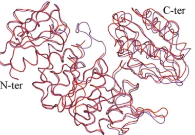
Intermediate Free MurD vs. Intermediate Bound MurD
Our newly solved intermediate structures of MurD were superimposed (Fig 5). Objectively we observed that our new structures share a similar conformational space with or without ligands, which was previously not considered. The Low RMSD value of best fit for the whole protein and low RMSD values for the best fits of individual non-rotating N-terminal and central domains and C-terminal domains (Table 2), quantitatively support our claim of conforma-tional similarities between theIntermediate Free MurDandIntermediate bound MurD.
Discussion and Conclusions
E.coliMurD has been studied in detail at the atomic level with several X-ray structures. [9–10, 16] Lately, a paradigm has been proposed that 3D structures of proteins should be represented as an ensemble of structures. [61] Therefore, obtaining structures of proteins in distinct confor-mations is very important for capturing a representative subset of true native conforconfor-mations. [62] Flexible regions described as“liquid-like”are the main reason for distinct conformations of proteins. Rigid regions in proteins described as“solid-like”do not contribute to distinct con-formations, which again suggests that a static structure of a protein is just one of the subset of native conformations. [63] A key event in MurD catalytic activity is the C-terminal domain rigid body rotation, thought to be triggered by the binding of ATP and UMA which together create the acyl phosphate intermediate. The tetrahedral intermediate is then generated upon D-Glu binding and is eventually disrupted by the release of Piand UMAG. [6,22] From the
Fig 5. Superposition ofIntermediate Free MurD(red) andIntermediate Bound MurD(magenta).
previously discovered MurD X-ray structures, two distinct conformations, open and closed, were inferred. [10] The kinetic mechanism proposed for MurD did not consider an ATP-bound non-closed conformation. [6,64] In the present paper we have described two new struc-tures ofE.coliMurD and compared them to all previously reported structures of unbound MurD and MurD complexed with ligands. Our analysis of different MurD conformations indi-cates that entire domain rotations are privileged as opposed to minor structural modifications. Thus, we were able to‘trap’an intermediate conformation of MurD that is not open despite the absence of ligands, nor closed in the presence of ligands. This suggests that it is not sub-strate recognition which causes MurD closure, as previously suggested, but that domain move-ment is an inherent feature of the enzyme that is independent of ligand binding.
Our discovery of new intermediate conformations ofIntermediate Free MurDand Interme-diate Bound MurDraises the question of what triggers C-terminal domain of MurD to adopt different conformations. Upon modification of crystallization conditions (Table 3), we man-aged to capture MurD in two new conformations from the ensemble of conformations that potentially exist within the bacterial cytoplasm. If we compare the conditions in which crystals were grown and the physiological cytoplasmic conditions ofE.coli, we conclude that since Mg2+, ATP, UMA and sulfate are continuously present in bacterial intracellular space, [65–67] it would not be reasonable to expect that any conformation of MurD would be advantageous with respect to the others; most probably, there is a dynamic equilibrium of different confor-mational states. This suggestion is supported by computational simulations that indicate that the energy difference between open and closed conformations of MurDin vacuumis around 10kcal/mol. [23]
However, the importance of carbamoylated Lys198 (KCX198) must not be neglected. KCX198 was only found in closed“active”conformations of MurD. [9–10] Its importance was studied by Dementinet al. [68], who established that KCX198 plays a role in the enzymatic reaction as it stabilizes Mg2+, an essential cation involved in the MurD activity, via two water molecules. Indeed, enzymatic activities of Lys198 MurD mutants were considerably less active than the native one, and the activity could be restored upon incubation with short-chain car-boxylic acids (chemical rescue). [68] Therefore, for the activation of MurD, not only binding of the substrates, but also carbamoylation of Lys198 is essential. Most probably,Intermediate Bound MurDrepresents the conformation which is adopted after substrates are bound, but it is not yet in the closed“active”conformation, since Lys198 is not carbamoylated. So far, there
Table 3. Review ofE.coliMurD crystallization conditions and its conformations.
PDB entry pH Precipitant Ligands Conformation Space group Cell parameters
1uag 7.0 (NH4)2SO4 UMA Closed P41 a=b= 65.43,c= 134.44Å,α=β=γ= 90° 2uag 7.0 PEG3350 UMA, ADP, Mg2+ Closed P41 a=b= 65.56,c= 136.01Å,α=β=γ= 90° 3uag 7.2 (NH4)2SO4 UMA1 Closed P41 a=b= 65.24,c= 134.41Å,α=β=γ= 90° 4uag 7.0 (NH4)2SO4 UMAG Closed P41 a=b= 65.80,c= 134.54Å,α=β=γ= 90° 1e0d 6.0 (NH4)2SO4 - Open P43212 a=b= 69.16,c= 196.71Å,α=β=γ= 90° 1eeh 5.2 PEG3350 UMA Open P212121 a= 58.84,b= 63.28,c= 114.91Å,α=β=γ= 90° 2jfg 7.5 (NH4)2SO4 UMA, ADP Closed P41 a=b= 65.09,c= 134.25Å,α=β=γ= 90° Free 5.6 (NH4)2SO4 - Intermediate P212121 a= 58.24,b= 70.55,c= 100.75Å,α=β=γ= 90°
Bound 7.0 Na-malonate UMA, ADP Intermediate P212121 a= 66.44,b= 89.84,c= 108.54Å,α=β=γ= 90°
1The structure in PDB also includes ADP and Mn2+, which were soaked into the protein after crystals were grown in conditions in the presence of (NH4)2SO4as precipitant and UMA.
was no evidence that carbamoylation of Lys198 is a transient event. Nevertheless, the crystal structures of open MurD (PDB ID: 1e0d and 1eeh, where Lys198 is not carbamoylated), and closed MurD (PDB ID: 1uag, 2uag, 3uag, 4uag, 2jfg, where Lys198 is carbamoylated), suggest that carbamoylation of Lys198 occurs during catalysis. Of course, the possibility of chemical degradation of KCX198 due to the long period in which the crystals grew cannot be totally ruled out.
Intermediate Bound MurDdisplays some features of the closed conformation of MurD. The hinge loop is similar to the one in open conformation of MurD, but with an additional H-bond to Pro300 and ADP, thus dictating the C-terminal domain to take a position closer to central domain than open conformation of MurD. [10] However, the C-terminal domain does not rotate to the closed conformation, because Arg302 is not H-bonded to Leu299 as it is the case in closed MurD. A common feature of the closed conformations with ligands is also the inter-action of Arg302 to sulfate (Fig 3C and 3E) orα-phosphate of ADP (Fig 3B). The higher num-ber of H-bonds between Arg302 and the residues in non-rotating central domain via ADP or sulfate result in conformations that are either closed or intermediate, while the lower number of H-bonds of Arg302 to the non-rotating domains restricts MurD into the open or intermedi-ate conformation. This claim is supported by computational simulations that suggest that the energy oscillations during the transition from open to closed statein vacuumwere on average between10and15kcal/mol. [23] The estimated energy is5–6kcal/mol for the bondin vacuum and0.5–1.5kcal/mol for the bond in aqueous solution, [69] which is approximately the esti-mated energy needed to induce C-terminal domain rotation, but probably not enough to fully lock it in the closed conformation. These estimations for the H-bond energy are calculated for water-free protein, but the energy of the H-bond in solvated proteins is 2- to 3-fold lower, [69] which gives even more weight to the assumption that H-bonding provides sufficient energy for conformation changes if the bond is formed or broken. [70] With total count of H-bonds and their energy in mind, Arg302 can be described as a molecular handle, which enables the C-ter-minal domain of MurD to rotate.
Another aspect of the biology of Mur ligases is their assembly inside the bacterial cytoplasm. It has been speculated that Mur ligases are assembledin vivoin the form of a multi-enzyme complex; [71] however, no evidence of the existence of such a complex between Mur ligases has been provided to date. Instead, interactions with other proteins involved in peptidoglycan synthesis (MurI, DdlA, MurG) or cell division (MreB) have been reported inCaulobacter cres-centus[72],Mycobacterium tuberculosis[73] andThermotoga maritima[15]. In the latter spe-cies, it was shown with surface plasmon resonance experiments that MurD, MurE and MurF interact with MurG and MreB. These experiments were performed in ligand-free environment, thus it was speculated that these two enzymes could act as scaffolds to maintain Mur ligases assembled to each other even in their open, inactive conformation. [15] Whether contact with these or other proteins affects the modifications of conformation we have observed is currently not known. Such a problem will be understood only by solving the structure of protein com-plexes in which Mur ligases are involved.
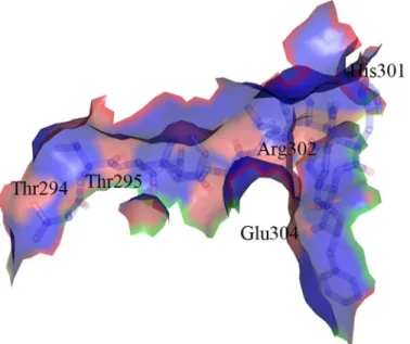
The crystal structures presented in this paper provide us with new tools in the urgent mis-sion to discover novel antibacterial agents. So far, inhibitors of MurD that were designed to bind in the substrate-binding sites have not expressed significant antibacterial activity. [6,22] Taking into account the detailed knowledge about the ability of MurD to take distinct confor-mations, new inhibitors can be designed not only as mimics of the natural MurD substrates, but also as smaller allosteric inhibitors. A region which could potentially be targeted by alloste-ric inhibitors is the hinge loop (Fig 6), which is involved in the C-terminal domain rotation. Thr294, Thr295, His301, Arg302 and Glu304 could be specifically targeted for potential polar interactions with inhibitors due to the presence of several lone electron pairs in their side chains. Structure-based designed allosteric inhibitors with affinity towards this loop could either prevent the binding of substrates or freeze the enzyme in the conformation where domain rotation and consequently catalytic activity are prevented.
Protein Data Bank accession numbers
Intermediate conformation of ligand free MurD (Intermediate Free MurD): 5a5e (S1 Checklist).
Intermediate conformation of MurD with ligands (Intermediate Bound MurD): 5a5f (S2 Checklist).
Supporting Information
S1 Checklist. Protein Data Bank Validation Reports 5a5e. (PDF)
S2 Checklist. Protein Data Bank Validation Reports 5a5f. (PDF)
Fig 6. Hinge loop containing residues Thr294−Glu304.Potential target for new allosteric MurD inhibitors.
Red regions represent the potential targets for H-bond interactions, blue regions represent hydrophobic surface of the hinge loop.
Acknowledgments
The authors thank the ESRF Grenoble for access to beamlines ID29 and BM14U and help with data collection.
Author Contributions
Conceived and designed the experiments: RS MK AD CCM. Performed the experiments: RS MK HB CCM. Analyzed the data: RS MK CCM. Contributed reagents/materials/analysis tools: AZ HB SG DB AD. Wrote the paper: RS MK CCM HB DB SG AD.
References
1. Contreras-Martel C, Dahout-Gonzalez C, Dos Santos Martins A, Kotnik M, Dessen A. PBP Active Site Flexibility as the Key Mechanism for beta-Lactam Resistance in Pneumococci. J Mol Biol. 2009; 38: 899–909.
2. Macheboeuf P, Fischer DS, Brown T, Zervosen A, Luxen A, Joris B, et al. Structural and mechanistic basis of penicillin-binding protein inhibition by lactivicins. Nat Chem Biol. 2007; 3: 565–569. PMID:
17676039
3. van Heijenoort J. Recent advances in the formation of the bacterial peptidoglycan monomer unit. Nat Prod Rep. 2001; 18: 503–519. PMID:11699883
4. Zapun A, Contreras-Martel C, Vernet T. Penicillin-binding proteins and beta-lactam resistance. FEMS Microbiol Rev. 2008; 32: 361–385. doi:10.1111/j.1574-6976.2007.00095.xPMID:18248419
5. Vollmer W, Blanot D, de Pedro MA. Peptidoglycan structure and architecture. FEMS Microbiol Rev. 2008; 32: 149–167. doi:10.1111/j.1574-6976.2007.00094.xPMID:18194336
6. Barreteau H, KovačA, Boniface A, Sova M, Gobec S, Blanot D. Cytoplasmic steps of peptidoglycan biosynthesis. FEMS Microbiol Rev. 2008; 32: 168–207. doi:10.1111/j.1574-6976.2008.00104.xPMID:
18266853
7. Anderson MS, Eveland SS, Onishi HR, Pompliano DL. Kinetic mechanism of theEscherichia coli UDP-MurNAc-tripeptide D-alanyl-D-alanine-adding enzyme: Use of a glutathione S-transferase fusion. Bio-chemistry 1996; 35: 16264–16269. PMID:8973200
8. Bertrand JA, Auger G, Fanchon E, Martin L, Blanot D, vanHeijenoort J, et al. Crystal structure of UDP-N-acetylmuramoyl-L-alanine: D-glutamate ligase fromEscherichia coli. EMBO J. 1997; 16: 3416– 3425. PMID:9218784
9. Bertrand JA, Auger G, Martin L, Fanchon E, Blanot D, La Beller D, et al. Determination of the MurD mechanism through crystallographic analysis of enzyme complexes. J Mol Biol. 1999; 289: 579–590. PMID:10356330
10. Bertrand JA, Fanchon E, Martin L, Chantalat L, Auger G, Blanot D, et al. "Open" structures of MurD: Domain movements and structural similarities with folylpolyglutamate synthetase. J Mol Biol. 2000; 301: 1257–1266. PMID:10966819
11. Bouhss A, Dementin S, van Heijenoort J, Parquet C, Blanot D. MurC and MurD synthetases of peptido-glycan biosynthesis: Borohydride trapping of acyl-phosphate intermediates. Methods Enzymol. 2002; 354: 189–196. PMID:12418226
12. Bouhss A, Mengin Lecreulx D, Blanot D, van Heijenoort J, Parquet C. Invariant amino acids in the mur peptide synthetases of bacterial peptidoglycan synthesis and their modification by site-directed muta-genesis in the UDP-MurNAc:L-alanine ligase fromEscherichia coli. Biochemistry 1997; 36: 11556– 11563. PMID:9305945
13. Emanuele JJ, Jin HY, Yanchunas J, Villafranca JJ. Evaluation of the kinetic mechanism ofEscherichia coliuridine diphosphate-N-acetylmuramate:L-alanine ligase. Biochemistry 1997; 36: 7264–7271. PMID:9188728
14. Eveland SS, Pompliano DL, Anderson MS. Conditionally lethalEscherichia colimurein mutants contain point defects that map to regions conserved among murein and folyl poly-gamma-glutamate ligases: Identification of a ligase superfamily. Biochemistry 1997; 36: 6223–6229. PMID:9166795
15. Favini-Stabile S, Contreras-Martel C, Thielens N, Dessen A. MreB and MurG as scaffolds for the cyto-plasmic steps of peptidoglycan biosynthesis. Environ Microbiol. 2013; 15: 3218–3228. doi:10.1111/ 1462-2920.12171PMID:23826965
17. Longenecker KL, Stamper GF, Hajduk PJ, Fry EH, Jakob CG, Harlan JE, et al. Structure of MurF from
Streptococcus pneumoniaeco-crystallized with a small molecule inhibitor exhibits interdomain closure.
Protein Sci. 2005; 14: 3039–3047. PMID:16322581
18. Mol CD, Brooun A, Dougan DR, Hilgers MT, Tari LW, Wijnands RA, et al. Crystal structures of active fully assembled substrate- and product-bound complexes of UDP-N-Acetylmuramic Acid: L-alanine ligase (MurC) fromHaemophilus influenzae. J Bacteriol. 2003; 185: 4152–4162. PMID:12837790
19. Sheng Y, Sun X, Shen Y, Bognar AL, Baker EN, Smith CA. Structural and functional similarities in the ADP-forming amide bond ligase superfamily: implications for a substrate-induced conformational change in folylpolyglutamate synthetase. J Mol Biol. 2000; 302: 427–440. PMID:10970743
20. Smith CA. Structure, function and dynamics in the Mur family of bacterial cell wall ligases. J Mol Biol. 2006; 362: 640–655. PMID:16934839
21. Kouidmi I, Levesque RC, Paradis-Bleau C. The biology of Mur ligases as an antibacterial target. Mol Microbiol. 2014; 94: 242–253. doi:10.1111/mmi.12758PMID:25130693
22. Šink R, Barreteau H, Patin D, Mengin-Lecreulx D, Gobec S, Blanot D. MurD: some recent
develop-ments. Biomol Concepts 2013; 4: 539–556. doi:10.1515/bmc-2013-0024
23. Perdih A,Šolmajer T. MurD ligase from Escherichia coli: C-terminal domain closing motion. Comput
Theor Chem. 2012; 979: 73–81.
24. Barreteau H, SosičI, Turk S, Humljan J, TomašićT, Zidar N, et al. MurD enzymes from different
bacte-ria: evaluation of inhibitors. Biochem Pharmacol. 2012: 84; 625–632. doi:10.1016/j.bcp.2012.06.006
PMID:22705647
25. BratkovičT, Lunder M, Urleb U,Štrukelj B. Peptide inhibitors of MurD and MurE, essential enzymes of
bacterial cell wall biosynthesis. J Basic Microbiol. 2008; 48: 202–206. doi:10.1002/jobm.200700133
PMID:18506905
26. Frlan R, KovačA, Blanot D, Gobec S, Pečar S, Obreza A. Design and synthesis of novel
N-benzylide-nesulfonohydrazide inhibitors of MurC and MurD as potential antibacterial agents. Molecules 2008; 13: 11–30. PMID:18259126
27. Frlan R, KovačA, Blanot D, Gobec S, Pečar S, Obreza A. Design, Synthesis and in vitro Biochemical
Activity of Novel Amino Acid Sulfonohydrazide Inhibitors of MurC. Acta Chim Slov. 2011; 58: 295–310. PMID:24062040
28. Frlan R, Perdih F, CirkvenčičN, Pečar S, Obreza A. Design and Synthesis of Novel UDP-Mur-NAc,
UDP-Mur-NAc-L-Ala and UDP-Mur-NAc-L-Ala-D-Glu mimetics. Acta Chim Slov. 2009; 56: 580–590. 29. Hrast M, SosičI,Šink R, Gobec S. Inhibitors of the peptidoglycan biosynthesis enzymes MurA-F.
Bioorg Chem. 2014; 55; 2–15. doi:10.1016/j.bioorg.2014.03.008PMID:24755374
30. Humljan J, Kotnik M, Contreras-Martel C, Blanot D, Urleb U, Dessen A, et al. Novel Naphthalene-N-sul-fonyl-D-glutamic Acid Derivatives as Inhibitors of MurD, a Key Peptidoglycan Biosynthesis Enzyme. J Med Chem. 2008; 51: 7486–7494. doi:10.1021/jm800762uPMID:19007109
31. Perdih A, KovačA, Wolber G, Blanot D, Gobec S,Šolmajer T. Discovery of novel benzene
1,3-dicar-boxylic acid inhibitors of bacterial MurD and MurE ligases by structure-based virtual screening approach. Bioorg Med. Chem Lett. 2009; 19: 2668–2673. doi:10.1016/j.bmcl.2009.03.141PMID:
19369074
32. Shanmugam A, Anbazhagan V, Natarajan J. Virtual screening of phenylsulfonamido-3-morpholinopro-pan-2-yl dihydrogen phosphate derivatives as novel inhibitors of MurC-MurF ligases from
Mycobacte-rium leprae. Med Chem Res. 2012; 21: 4341–4351.
33. Šink R, KovačA, TomašićT, Rupnik V, Boniface A, Bostock J, et al. Synthesis and biological evaluation of N-acylhydrazones as inhibitors of MurC and MurD ligases. ChemMedChem 2008; 3: 1362–1370. doi:10.1002/cmdc.200800087PMID:18651694
34. SosičI, Barreteau H, SimčičM,Šink R, Cesar J, Zega A, et al. Second-generation sulfonamide inhibi-tors of glutamic acid-adding enzyme: Activity optimisation with conformationally rigid analogues of D-glutamic acid. Eur J Med Chem. 2011; 46: 2880–2894. doi:10.1016/j.ejmech.2011.04.011PMID:
21524830
35. TomašićT, KovačA, Klebe G, Blanot D, Gobec S, Kikelj D, et al. Virtual screening for potential
inhibi-tors of bacterial MurC and MurD ligases. J Mol Model. 2012; 18: 1063–1072. doi: 10.1007/s00894-011-1139-8PMID:21667288
36. TomašićT, KovačA, SimčičM, Blanot D, Grdadolnik SG, Gobec S, et al. Novel
2-thioxothiazolidin-4-one inhibitors of bacterial MurD ligase targeting D-Glu- and diphosphate-binding sites. Eur J Med Chem. 2011; 46; 3964–3975. doi:10.1016/j.ejmech.2011.05.070PMID:21703731
37. TomašičT,Šink R, Zidar N, Fic A, Contreras-Martel C, Dessen A, et al. Dual Inhibitor of MurD and
38. TomašićT, Zidar N, KovačA, Turk S, SimčičM, Blanot D, et al. 5-Benzylidenethiazolidin-4-ones as
Multitarget Inhibitors of Bacterial Mur Ligases. ChemMedChem 2010; 5: 286–295. doi:10.1002/cmdc. 200900449PMID:20024979
39. TomašićT, Zidar N, Rupnik V, KovačA, Blanot D, Gobec S, et al. Synthesis and biological evaluation
of new glutamic acid-based inhibitors of MurD ligase. Bioorg Med Chem Lett. 2009; 19: 153–157. doi:
10.1016/j.bmcl.2008.10.129PMID:19014883
40. TomašićT, Zidar N,Šink R, KovačA, Blanot D, Contreras-Martel C, et al. Structure-Based Design of a
New Series of D-Glutamic Acid Based Inhibitors of Bacterial UDP-N-acetylmuramoyl-L-alanine:D-gluta-mate Ligase (MurD). J Med Chem. 2011; 54: 4600–4610. doi:10.1021/jm2002525PMID:21591605
41. Turk S, KovačA, Boniface A, Bostock JM, Chopra I, Blanot D, et al. Discovery of new inhibitors of the
bacterial peptidoglycan biosynthesis enzymes MurD and MurF by structure-based virtual screening. Bioorg Med Chem. 2009; 17: 1884–1889. doi:10.1016/j.bmc.2009.01.052PMID:19223185
42. Zidar N, TomašićT,Šink R, KovačA, Patin D, Blanot D, et al. New 5-benzylidenethiazolidin-4-one
inhibitors of bacterial MurD ligase: Design, synthesis, crystal structures, and biological evaluation. Eur J Med Chem. 2011; 46: 5512–5523. doi:10.1016/j.ejmech.2011.09.017PMID:21963114
43. Zidar N, TomašićT,Šink R, Rupnik V, KovačA, Turk S, et al. Discovery of Novel
5-Benzylidenerhoda-nine and 5-Benzylidenethiazolidine-2,4-dione Inhibitors of MurD Ligase. J Med Chem. 2010; 53: 6584– 6594. doi:10.1021/jm100285gPMID:20804196
44. SimčičM, Hodošček M, Humljan J, Kristan K, Urleb U, Kocjan D, et al. NMR and Molecular Dynamics
Study of the Binding Mode of Naphthalene-N-sulfonyl-D-glutamic Acid Derivatives: Novel MurD Ligase Inhibitors. J Med Chem. 2009; 52: 2899–2908. doi:10.1021/jm900117nPMID:19358612
45. SimčičM, SosičI, Hodošček M, Barreteau H, Blanot D, Gobec S, et al. The Binding Mode of
Second-Generation Sulfonamide Inhibitors of MurD: Clues for Rational Design of Potent MurD Inhibitors. PloS One 2012; 7: e52817. doi:10.1371/journal.pone.0052817PMID:23285193
46. Kabsch W. Automatic Processing of Rotation Diffraction Data from Crystals of Initially Unknown Sym-metry and Cell Constants. J Appl Crystallogr. 1993; 26: 795–800.
47. Storoni LC, McCoy AJ, Read RJ. Likelihood-enhanced fast rotation functions. Acta Crystallogr, Sect D: Biol Crystallogr. 2004; 60: 432–438.
48. Cohen SX, Ben Jelloul M, Long F, Vagin A, Knipscheer P, Lebbink J, et al. ARP/wARP and molecular replacement: the next generation. Acta Crystallogr, Sect D: Biol Crystallogr. 2008; 64: 49–60. 49. Buszek KR, Luo D, Kondrashov M, Brown N, VanderVelde D. Indole-derived arynes and their
diels-alder reactivity with Furans. Org Lett. 2007; 9: 4135–4137. PMID:17880092
50. Emsley P, Cowtan K. Coot: model-building tools for molecular graphics. Acta Crystallogr, Sect D: Biol Crystallogr. 2004; 60: 2126–2132.
51. Emsley P, Lohkamp B, Scott WG, Cowtan K. Features and development of Coot. Acta Crystallogr, Sect D: Biol Crystallogr. 2010; 66: 486–501.
52. Painter J, Merritt EA. TLSMD web server for the generation of multi-group TLS models. J Appl Crystal-logr. 2006; 39: 109–111.
53. Murshudov GN, Vagin AA, Dodson EJ. Refinement of macromolecular structures by the maximum-like-lihood method. Acta Crystallogr, Sect D: Biol Crystallogr. 1997; 53: 240–255.
54. Chen VB, Arendall WB, Headd JJ, Keedy DA, Immormino RM, Kapral GJ, et al. MolProbity: all-atom structure validation for macromolecular crystallography. Acta Crystallogr, Sect D: Biol Crystallogr. 2010; 66: 12–21.
55. Laskowski RA, Macarthur MW, Moss DS, Thornton JM. Procheck—a Program to Check the Stereo-chemical Quality of Protein Structures. J Appl Crystallogr. 1993; 26: 283–291.
56. Heinig M, Frishman D. STRIDE: a web server for secondary structure assignment from known atomic coordinates of proteins. Nucleic Acids Res. 2004; 32: W500–W502. PMID:15215436
57. Hayward S, Berendsen HJC. Systematic analysis of domain motions in proteins from conformational change: New results on citrate synthase and T4 lysozyme. Proteins: Struct, Funct, Genet. 1998; 30: 144–154.
58. Wallace AC, Laskowski RA, Thornton JM. LIGPLOT: a program to generate schematic diagrams of pro-tein-ligand interactions. Protein Eng. 1995; 8: 127–134. PMID:7630882
59. Laskowski RA, Swindells MB. LigPlot+: Multiple Ligand-Protein Interaction Diagrams for Drug Discov-ery. J Chem Inf Model. 2011; 51: 2778–2786. doi:10.1021/ci200227uPMID:21919503
60. The PyMOL Molecular Graphics System, Version 0.99rc6. Schrödinger, LLC.
61. Best RB, Lindorff-Larsen K, DePristo MA, Vendruscolo M. Relation between native ensembles and experimental structures of proteins. Proc Natl Acad Sci U. S. A. 2006; 103: 10901–10906. PMID:
62. Burra PV, Zhang Y, Godzik A, Stec B. Global distribution of conformational states derived from redun-dant models in the PDB points to non-uniqueness of the protein structure. Proc Natl Acad Sci U. S. A. 2009; 106: 10505–10510. doi:10.1073/pnas.0812152106PMID:19553204
63. Perdih A, Kotnik M, Hodošček M,Šolmajer T. Targeted molecular dynamics simulation studies of
bind-ing and conformational changes inE.coliMurD. Proteins: Struct, Funct, Bioinf. 2007; 68: 243–254. 64. Bally I, Rossi V, Lunardi T, Thielens NM, Gaboriaud C, Arlaud GJ. Identification of the C1q-binding
Sites of Human C1r and C1s A refined three-dimensional model of the C1 complex of complement. J Biol Chem. 2009; 284: 19340–19348. doi:10.1074/jbc.M109.004473PMID:19473974
65. Hurwitz C, Rosano CL. The intracellular concentration of bound and unbound magnesium ions in
Escherichia coli. J Biol Chem. 1967; 242: 3719–3722. PMID:5341484
66. Imhoff JF, Riedel T. Requirements for, and Cytoplasmic Concentrations of, Sulfate and Chloride, and Cytoplasmic Volume Spaces in the Halophilic BacteriumEctothiorhodospira Mobilis. J Gen Microbiol. 1989; 135: 237–244.
67. Dementin S, Bouhss A, Auger G, Parquet C, Mengin-Lecreulx D, Dideberg O, et al. Evidence of a func-tional requirement for a carbamoylated lysine residue in MurD, MurE and MurF synthetases as estab-lished by chemical rescue experiments. Eur J Biochem. 2001; 268: 5800–5807. PMID:11722566
68. Sheu SY, Yang DY, Selzle HL, Schlag EW. Energetics of hydrogen bonds in peptides. Proc Natl Acad Sci U. S. A. 2003; 100: 12683–12687. PMID:14559970
69. Hubbard RE, Kamran Haider M. Hydrogen Bonds in Proteins: Role and Strength. eLS; 2010. 70. Silver LL. Challenges in antibacterial discovery. Clin Microbiol Rev. 2001; 24: 71–109.
71. Chopra I. The 2012 Garrod lecture: discovery of antibacterial drugs in the 21st century. J Antimicrob Chemother. 2011; 68: 496–505.
72. White CL, Kitich A, Gober JW. Positioning cell wall synthetic complexes by the bacterial morphogenetic proteins MreB and MreD. Mol Microbiol. 2010; 76: 616–633. doi:10.1111/j.1365-2958.2010.07108.x
PMID:20233306
73. Munshi T, Gupta A, Evanghelopoulos D, Guzman JD, Gibbons S, Keep NH, et al. Characterisation of ATP-dependent Mur ligases involved in the biogenesis of cell wall peptidoglycan inMycobacterium
tuberculosis. PloS One 2013; 8: e60143. doi:10.1371/journal.pone.0060143PMID:23555903
74. Vaganay S, Tanner ME, van Heijenoort J, Blanot D. Study of the reaction mechanism of the D-glutamic acid-adding enzyme fromEscherichia coli. Microb Drug Resist. 1996; 2: 51–54. PMID:9158722
![Fig 3. Orientation and LIGPLOT representations of Arg302 in MurD structures. (A) Open MurD (PDB entry: 1e0d [7])](https://thumb-eu.123doks.com/thumbv2/123dok_br/17228344.244343/8.918.300.720.105.981/orientation-ligplot-representations-murd-structures-open-murd-entry.webp)