ESTHESIONEUROBLASTOMA
Case report
Jackson Gondim
1, Francisco Ramos Jr
2, Jorge Azevedo
3,
Fernando Porto Carrero Jr
3, Oswaldo Inácio Tella Jr
4ABSTRACT - Esthesioneuroblatoma (ENB) is a rare tumor arising from the olfactory epithelium of the nasal vault which frequently invades the cranial base, cranial vault and orbit. ENB has a bimodal age distribution between 11 and 20 years and between 51 and 60 years. ENB accounts for approximately 1 to 5% of intranasal cancers and no consensus has been reached regarding treatment of this tumor. We report on a 66 year old female patient with a Kadish stage C tumor with frontal lobe invasion submitted a total craniofacial resection w ith a combined head neck and neurosurgeon team. The purpose of this study is to analyze the natural history, treatment and prognosis of this tumor, based on the literature review.
KEY WORDS: esthesioneuroblastoma, craniofacial surgery, skull base surgery.
Estesioneuroblastoma: relato de caso
RESUMO - Estesioneuroblastoma é um tumor raro originado do epitélio olfactivo, frequentemente invadindo a base do crânio e a região orbitaria. É tumor que pode ser encontrado em qualquer idade mas apresentando dois picos de frequência entre 11 e 21 anos e 51 e 60 anos, raros em criança. A distribuição por sexo é praticamente igual mas com uma pequena predominância masculina. O diagnostico histopatológico é feito de forma definitiva por métodos imunohistoquímicos. A sintomatologia clínica corresponde ao de uma neoplasia intranasal ou frontobasal. Devido a raridade destes tumores não se chegou ainda a um consenso em relação ao tipo de tratamento. Relatamos o caso de uma paciente 66 anos de idade com um tumor classificado como Kadish tipo C, com invasão dos seios da face e lobo frontal, que foi submetida a ressecção crânio facial por uma equipe multidisciplinar. A historia natural o tratamento e o prognóstico dos pacientes portadores destes tumores serão analisados baseando-se numa revisão da literatura.
PALAVRAS-CHAVE: estesioneuroblastoma, cirurgia crânio facial, cirurgia da base do crânio.
1Neurocirurgião do Hospital Geral de Fortaleza, Fortaleza CE, Brazil (HGF) e M estrando em Neurocirurgia da Escola Paulista de M edicina, Universidade Federal de São Paulo, São Paulo SP, Brazil (UNIFESP-EPM ); 2Neurocirurgião do HGF; 3Cirurgião do Setor de Cabeça e Pescoço do HGF; 4Professor Livre-Docente da Disciplina de Neurocirurgia da UNIFESP-EPM .
Received 3 September 2001, received in final form 12 November 2001. Accepted 22 November 2001.
Dr. Jackson Gondim - Rua Dr. Pedro Sampaio 50 - 60181-560 Fortaleza CE - Brasil. E-mail: [email protected]
Esthesioneuroblastoma (olfactory neuroblasto-ma) (ENB) is a rare neuroepithelial tumor that arises from the olfactory epithelium in the cribriform place or nasal cavity1. First described in 1924 by Berger2 it has a histological pattern similar to that of sympathe-tic ganglia, retina, and adrenal medulla2 and only recently3 became recognized as a distinct pathologic entity probably as a result of immunohistochemis-try and by means of electron microscopy techniques. They have helped differentiate ENB from similar un-differentiated nasal cavity tumors3. ENB account for 1 to 5% of malignant neoplasm of the nasal cavity. Few er than 945 cases are reported in the w orld lit-erature4, and most of the reports were in small series. Unlike most other neuroectodermal tumors, w hich
manifest in childhood, ENB, has a bimodal age dis-tribution betw een 11-20 years and 51-60 years5. The symptoms are related to sites and invasion of the tumor. The staging system based on tumor exten-sion that w as presented by Kadish et al.6 in 1976 has been w idely accept ed. The treatment of choice is a multidisciplinary craniofacial surgical resection t hat has improved considerably t he prognosis.
The purpose of this study is to analyze the natu-ral history, treatment and prognosis of this tumor, based on the literature review .
CASE
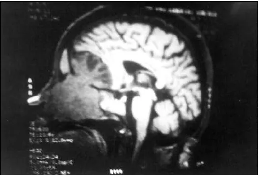
has present ing for about t w o mont hs visual blurring, lacrimatiom and ocular pain follow ed one w eek later by epistaxis. Physical examination revealed a bilateral nasal mass that w as endonasal endoscopy biopsy-proven to be an ENB. CT and M RI (Fig 1) demonstrated that the tumor filled the entire nasal cavity, ethmoid sinuses, sphenoid sinus, and left frontal sinus. The tumor invaded the cribri-form plate and the left frontal lobe. The patient under-w ent a craniofacial resection under-w ith a combined neurosur-geon and head neck surneurosur-geon team. A bicoronal incision
w ith frontal craniotomy, and in bloc resection of the frontal tumor and frontal base w ith preservation the pericranial flap, that w as placed along the floor of the anterior cra-nial fossa and sutured to the residual sphenoid bone as w ell as though the residual dura, combined w ith an ex-tended lateral rhinotomy, w ith totally resection of the in-fra cranial tumor. In the post operatively M RI (Fig 2), no tumor w as found. The patient receives 56 Gy of external beam radiotherapy over a 6-w eek period, and chemo-therapy. She show ed no recurrence after one year.
Fig 1. Preoperative sagittal MRI of the tumor, involving the ethmoid sinus, sphenoid sinus, nasal cavity and frontal lobe.
DISCUSSION
Esthesioneuroblastoma is a very rare malignancy of the neuroepithelium. It w as first described by Berger et al. in 1924, as “ l´ esthesioneuroepitheliome olfactif”2 and was only introduced into the American literature by Schall and Lineback7 in 1951. Nearly all of the 945 cases have been reported w ithin the last 40 years. Embryologically, the olfactory nerves de-velop from the olfactory placode present in the fetal olfactory mucosa8,9. Histologically, there are a num-ber of criterias that help in its diagnosis: neuroepi-thelial cells arranged in the classic pseudorosette pattern; fibrillar intracellular background; marked microvascularity; and round or fusiform cells appro-aching the size of lymphocytes w ith poorly defined, almost nonexistent cytoplasm1,10,11. Correct diagno-sis often requires confirmatory examination w ith electron microscopy for the detection of neurosecre-tory granules. M ore recently immunohistochemical methods for detection of neuronspecific enolase (NSE) and S-100 protein w ith negative epithelial, muscle, and lymphoid antigens allow ed further con-firmation of ENB1,12. This tumor must be differenti-ated from neoplasm of the nasal cavity and paranasal sinus, such as lymphoma, sarcoma, plasmacytoma, malignant melanoma, anaplastic carcinoma, rhab-domyosarcoma, and transitional cell carcinoma11,13. ENB has a bimodal age distribution w ith an early peak from 11 to 20 years and a later peak betw een 51 and 60 years of age10. Our patient w as a 66 years old w oman. There is a slight male predominance13. The staging system based on tumor extension that w as presented by Kadish et al.6 in 1976 has been w idely accepted. This staging system is predictive of disease-related mortality. The system classifies pa-tients with tumors limited to the nasal cavity as stage A. Patients w ith tumors involving the nasal cavity and extending into the paranasal sinuses are stage B, and stage C are tumors spreading beyond the nasal cavity and paranasal sinuses, as our patient. This sys-tem has been advocated by some, because of its sim-plicity and acceptable prognostic efficacy. Recently, Morita et al.14 justified a modified classification w ith stage D tumors, presenting metastases in cervical lymph nodes distant. Tw o other staging methods, the Biller method15 and the Dulguerov method16, have also been described and used.
ENB is a slow grow ing tumor and the patients may have a history of progressive symptomatology for months to years, our patient has a one-year evo-lution. The presenting symptoms are nonspecific and related to the sites and invasion of the tumor. The
most common finding on physical examination w as the presence of a nasal mass as our patient. Recur-rent epistaxis is sometimes present. Penetration into the cribriform plate can cause anosmia. Ophthalmo-logical symptoms as ptosis, diplopia, visual blurring, ocular pain, proptosis, and excessive lacrimatiom may be found. Ear pain and otitis can result from tumor obstructing the Eustachian tube. Frontal headache suggests involvement of the frontal sinus. Cranial nerves may also be affected inducing nerve paraly-sis14,17. Alteration of mental status may be present if frontal lobe is invaded.
The diagnosis and evaluation of staging of ENB can be done by CT, w hich provides the best infor-mation about the tumor invasion into bony struc-tures12. The tumor is presented as a homogeneous density mass, equal to or greater than the surround-ing soft tissues. There are no tumor cysts or calcifi-cations. Contrast enhancement w as usually moder-ate and homogeneous. Coronal images were of value in evaluating extension to the orbital and through the cribriform plate and the anterior cranial fossa12. M RI show s a tumor hypointense to gray matter on T1-w eighted images and iso or hyperintense on T2-w eighted images. Gadolinium enhancement T2-w as observed to some degree in all cases. Fat saturated T1-w eighted spin-echo images w ith and w ithout gadolinium enhancement of particular value in dif-ferentiating enhancing tumor from post-obstructive mucous debris and evaluating tumor extension to the non-enhancing orbital fat12. M RI is more accu-rate in depicting the exact margins of intracranial tumor extension because of its multiplanar display and superior tissue contrast12.
M etastasis occurs in about 10 to 30% of pati-ents18,19. The most common sites for metastasis spre-ad are the cervical lymph nodes, less frequent are lung and pleura, brain, bone, spinal column, breast, and abdominal viscera20,21. M etastasis to the central nervous system is infrequent and usually identified only at post-mortem examination5. In the spinal cord 80% of metastasis are in the cauda equine18.
w atertight dural seal. Fibrin glue is use to assist du-ral seal and to secure a w atertight closure. There are great variations in treatment for ENB. Some series advocate a protocol w ith surgery15,19-22, radiothera-py6,10,19,23 alone, combined surgery and pre opera-tive radiotherapy13,24, and combined surgery and pos-toperative radiotherapy22,25. The optimum manage-ment for ENB is probably surgery using the concept first described in 1971 by Doyle and Payton26: radi-cal surgery w ith a combined craniofacial approach12 taken by craniofacial team, including neurosurgery and head neck surgery. This technique has provided enhanced exposure and the possibility to achieve gross total resection. In patients w ithout extension of tumors to the superior nasal vault or the cribriform plate, an intracranial exploration and remove of the floor of the anterior cranial fossa must be performed. This approach has had a decrease incidence of local recurrence1. It is proved that the bone in this area may harbor tumor cells w ith a potential cause of recurrence. This w as done w ith our patient that had an involvement of cribriform plate and frontal lobe. We also used an adjuvant postoperative radiotherapy w ith 50-60 Gy resulted in effective local control as indicated by other authors10,27. Furthermore the ra-diotherapy is recommend for palliative treatment27. The role of systemic chemotherapy in the treatment of ENB has range from no response28-32, palliation20,33, partial14,33-37 and complete response36,38,39. In general chemotherapy is usually reserved for tumor spread-ing beyond the nasal cavity and paranasal sinuses12, or in the treatment of distant metastases40.
Treatment complications w ith ENB are high. Vi-sual impairment is the most common adverse ef-fect13, because the location of the tumor is difficult to deliver an adequate dose of radiation w ithout ex-ceeding the tolerance of critical structures such as the brain, optic chiasm and orbits.
The prognostic factors in the management of ENB are very controversy because of the small number of patients presented in each series. Morita1 and Foote41 advocated that the only reliable survival predictor is the tumor’s pathological grade (Hyams’ grading sys-tem42). Polin31 was unable to find significant differen-ce betw een survival of patients w ith low and high-grade tumors. Some authors16,43 affirm that the nega-tive prognostic include age, metastasis, recurrence, and extensions to the etmoidal, nasopharyngeal and orbital area. They also noted that the absence of me-tastasis does not necessarily confer a good progno-sis. Goldsw eig34 concluded that the degree of resec-tability of the tumor on primary surgery is the best
predictor of long-term survival. Irish determined a 100% 2-year survival rates in patients undergoing combined surgical and radiation treatment. Polin31 informs the 5 and 10 years survival rates of 87 and 54% respectively and a 97% one year survival. Pa-tients w ith stage C disease have 96% one-year sur-vival, 71% five years and 44% 10 years survival31. In the series of Jekunen40 the median survival time for 11 patients w as 27 months, and the median disease-free w as 27 months. In the literature the 5-year re-currence free survival is report to be betw een 52% and 90%16,29,40.
ENB recurs locally in up to 60% of patients w ho undergo surgery15, and its locally aggressive behav-ior is the most common cause of death44. M edian survival after recurrences w as only 12 months45,46. The majority of recurrences occur within the first few years after treatment.
In conclusion, ENB is a very uncommon malig-nant tumor arising from olfactory epithelium, that have a long natural history characterized by frequent local or regional recurrence. Radical craniofacial re-sections by a multidisciplinary surgical team com-bined w ith adjuvant radiotherapy w ith 50-60 Gy, is probably the most usual treatment. The role of sys-t emic chemosys-t herapy in sys-t he sys-t reasys-t mensys-t of dissys-t ansys-t metastasis should be further evaluated.
REFERENCES
1. Irish J, Dasgupa R, Freeman J, et al. Outcome and analysis of the surgi-cal management of esthesioneuroblastoa. J Otolaryng 1977;26,:1-7. 2. Berger L, Luc G, Richard D. L´esthesioneuroepitheliome olfactif. Bull
Assoc Franç Etude 1924;13:410-421.
3. Stewart F, Frierson H, Levin P, Spaulding C,. Esthesioneuroblastoma. In Williams JG, Krikorian MR, Green D (eds.) Textbook of uncommon cancer. Oxford: Wiley and Sons, 1988:631-652.
4. Broich G, Pagliari A, Ottaviani F. Esthesineuroblastoma: a general re-view of the cases published since the discovery of the tumour in 1924. Anticancer Res 1997;17:2383-2406.
5. Becker L, Hinton D. Primitive neuroectodermal tumors of the central nervous system. Hum Pathol 1983;14:538-550.
6. Kadish S, Goodman M, Wang C. Olfatory neuroblastoma: a clinical analysis of 17 cases. Cancer 1976;37:1571-1576.
7. Schall L, Lineback M. Primary intranasal neuroblastoma. Ann Otol Rhinol Laryngol 1951;60:221-229.
8. Moore K. The developing human. Philadelphia: Saunders, 1977:174-175. 9. O´Rahilly R, Muller F. The embryonic human brain. New York:
Wiley-Liss, 1994:91-337.
10. Elkon D, Hightower S, Meng L. Esthesioneuroblastoma. Cancer 1979; 44:1087-1094.
11. Harrison D. Surgical pathology of olfactory neuroblastoma. Head Neck Surg 1984;7:60-64.
12. Pickuth D, Heywang-Kobrunner H, Spielmann R. Computed tomog-raphy and magnetic resonance imaging features of olfactory neuro-blastoma: an analysis of 22 cases. Clin Otolaryngol 1999;24:457-461. 13. Simon J, Zhen W, McCulloch T, et al. Esthesioneuroblastoma: The
Uni-versity of Iowa experience 1978-1998. Laringoscope 2001;111:488-493. 14. Morita A, Ebersolod M, Olsen K, Foote R, Lewis J, Quasqst L. Esthe-sioneuroblastoma: prognosis and management. Neurosurgery 1993;32: 706-715.
16. Dulguerov P, Calcaterra T. Esthesioneuroblastoma: the UCLA experi-ence 1970-1990. Laryngoscope 1992;102:843-849.
17. Bastin K, Steeves R, Gilchrist K. Esthesioneuroblastoma: diagnosis, prognosis and treatment. Wis med J 1993;92:17-19.
18. Shaari C, Catalano P, Sen C, Post K. Central nervous system metastases from esthesioneuroblastoma. Otolaryngol Head Neck Surg 1996;114: 808-812. 19. Olsen K, De Santo L. Olfactory neurobllastoma: biologic and clinical
behavior. Arch Otolaryngol 1983;109:797-802
20. Beitler J, Fass D, Brenner H. Esthesioneuroblastoma: is there a role for elective neck treatment. Head Neck 1991;13:321-326.
21. Djalilian M, Zujko R, Weiland D, Devine K. Olfactory neuroblastoma. Surg Clin N Am 1977;57:751-762.
22. Skolnik E, Massari F, Tenta L. Olfactory neuroepithelioma: a review of the world literature and presentation of two cases. Arch Otolaryngol 1966;84:84-93.
23. Parsons J, Mendenhall W, Mancuso A. Malignant tumors of the nasal cavity and ethmoid sinuses. J Radiat Oncol Boil Phys 1988;14:11-22. 24. Cantrell R, Chorayeb B, Fitz-Hugh G. Esthesioneuroblastoma:
diagno-sis and treatment. Ann Otol Rhinol Laryngol 1977;86:760-765. 25. Oberman H, Rice D. Olfactory neuroblastoma: a clinico-pathologic
study. Cancer 1976;38:2494-2502.
26. Doyle P, Payton H. Combined surgical approach to esthesioneuroe-pithelioma. Trans Pa Acad Ophthalmol Otolaryngol 1971;75:526-531. 27. Eich H, Star S, Mickle O, Eich P, Stutzer H, Muller R. Radiotherapy of
esthesioneuroblastoma. Int J Radiation Boil Phys 2001;49:155-160. 28. Sheeran JM, Sheeram JP, Jane J, Polin R. Chemotherapy for
esthesioneu-roblastomas. Neurosurg Clin N Am 2000;11:693-701.
29. Levine P, Frierson H, Stewart F. Sinonasal undifferentiated carcinoma: a distinctive and highly aggressive neoplasm. Laryngoscope 1987;97: 905-908.
30. Mendeloff J. The olfactory neuroepithelial tumours. Cancer 1957;10:944-956. 31. Polin R, Sheeran J, Chenelle A. The role of pre-operative adjuvant treat-ment in the managetreat-ment of esthesioneuroblastoma: the University of Virginia experience. Neurosurgery 1998;42:1029-1037.
32. Polonowshi, Brasnu D, Roux F. Esthesioneuroblastoma: a complete tumor response after induction chemotherapy. Ear Nose Throat J 1990; 69:743-746.
33. Tingwald F. Olfactory placode tumors. Laryngoscope 1966;76:196-211. 34. Goldweig H, Sundarsean N. Chemotherapy of recurrent esthesioneuro-blastoma: case report review of the literature. Am J Clin Oncol 1990;13: 139-143.
35. Rodas R, Erkman-Balis B, Cahill D. Late intracranial metastasis from esthesioneuroblastoma: a case report and review of the literature. Neu-rosurgery 1986;9:622-627.
36. Wade P, Smith R, Johns M. Response of esthesioneuroblastoma to chemotherapy. Report of five cases and review of the literature. Can-cer 1984;53:1036-1041.
37. Weiden P, Yarington C, Richardson R. Olfactoy neuroblastoma: che-motherapy and radiation for extensive disease. Arch Otolaryngol Head Neck Surg 1984;11:759-760.
38. Grahne B. Olfactory neuroblastoma. Acta Otolaryngol (Stockh) 1965; 59:55-64.
39. Watne K, Hager B. Treatment of recurrence esthesioneuroblastoma with combined intracranial chemotherapy: a case report. J Neurooncol 1987; 5:47-50.
40. Jekunen A, Kalevi J, Kairemo J, Lehtonen H, Kajanti M. Treatment of olfactory neuroblastoma. Am J Clin Oncol 1966;19:375-378. 41. Foote R, Morita A, Ebersold M, et al. Esthesioneuroblastoma: the role
of adjuvant radiation therapy. Int J Radiat Oncol Biol 1993;27:835-842. 42. Hyams V, Batsakis J, Michaels L. Olfactory neuroblastoma. In Hyams V, Batsakis J, Michaels L (eds). Tumors of the upper respiratory tract and ear. Washington: Armed Forces Institute of Pathology, 1988:240-248. 43. Homzie M, Elkon D. Olfactory esthesioneuroblastoma: variables
pre-dictive of tumors control and recurrence. Cancer 1980; 46:2509-2513. 44. Ahern V, Pousen M. Olfactory neuroblastoma management of a rare
tumor at the Queensland Radium Institute and literature review. Australas Radiolol 1991;35:366-369.
45. Eriksen J, Bastholt L, Krogdahi A, Hansen O, Joergesen K. Esthesioneu-roblastoma What is the optimal treatment? Acta Oncologica 2000;2:231-235.