399
Holanda MMA et al. Basal encephalocele associated with clef palate
Radiol Bras. 2011 Nov/Dez;44(6):399–400
Basal sphenoethmoidal encephalocele in association
with midline cleft lip and palate: case report
*
Encefalocele basal esfenoetmoidal associada a fissura labiopalatina mediana: relato de caso
Maurus Marques de Almeida Holanda1, Artur Bastos Rocha2, Rayan Haquim Pinheiro Santos2,
Paulo Germano Cavalcanti Furtado3
Association of basal sphenoethmoidal encephalocele with midline cleft lip and palate is extremely rare. The authors report the case of a nine-year-old girl presenting a midline facial cleft with meningocele that was noticeable through the palatine defect as a medial intranasal pulsatile mass. An analysis of clinical and radiological findings of the present case of cranial dysraphism is carried out.
Keywords: Encephalocele; Cleft palate; Congenital malformation.
Associação de encefalocele basal esfenoetmoidal com fissura labiopalatina é extremamente rara. Relatamos um caso de uma criança de nove anos de idade apresentando uma fissura facial mediana com meningocele, que era evidente através da falha do palato como uma massa mediana intranasal pulsátil. Uma análise dos aspectos clínicos e radio-lógicos deste caso de disrafia craniana foi realizada.
Unitermos: Encefalocele; Fissura palatina; Malformação congênita. Abstract
Resumo
* Study developed at Universidade Federal da Paraíba (UFPB), João Pessoa, PB, Brazil.
1. PhD, Neurosurgeon, Associate Professor of Neurology at Universidade Federal da Paraíba (UFPB), João Pessoa, PB, Bra-zil.
2. Graduate Students of Medicine at Universidade Federal da Paraíba (UFPB), João Pessoa, PB, Brazil.
3. Associate Professor of Pediatric Surgery, Department of Pediatrics/Genetics, Universidade Federal da Paraíba (UFPB), João Pessoa, PB, Brazil.
Mailing Address: Dr. Maurus Marques de Almeida Holanda. Rua Borja Peregrino, 191, Torre. João Pessoa, PB, Brazil, 58040-050. E-mail: [email protected]
Received April 18, 2011. Accepted after revision May 31, 2011.
Holanda MMA, Rocha AB, Santos RHP, Furtado PGC. Basal sphenoethmoidal encephalocele in association with midline cleft lip and palate: case report. Radiol Bras. 2011 Nov/Dez;44(6):399–400.
0100-3984 © Colégio Brasileiro de Radiologia e Diagnóstico por Imagem
CASE REPORT
Free and informed consent was obtained from the child’s mother, and the Research Ethics Committee has approved the present study publication.
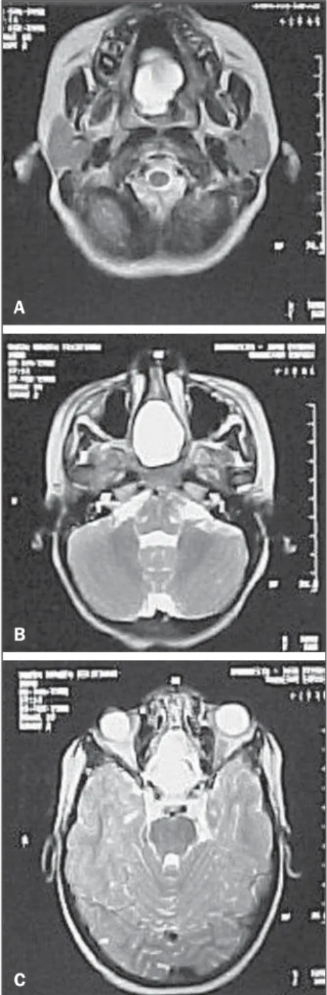
Computed tomography (CT) demon-strated metopic suture diastasis character-izing hypertelorism, and an empty sella turcica occupied by a cystic image. Mag-through the foramen cecum over the
crib-riform plate of the ethmoid. The second group includes basal encephaloceles with protrusion through the cribriform plate it-self or through the sphenoid body(2).
General encephaloceles prevalence is estimated to occur in one of every 35,000 to 40,000 live neonates. The basal presen-tations, that are even more rarely found, represent only 2% to 10% of such encepha-loceles(3,4). Considering the rarity of this malformations association, whose treat-ment leads to high morbidity and mortal-ity, and with few reports in the literature, the authors analyze the clinical and radio-logical findings of the present case of have cranial dysraphism.
CASE REPORT
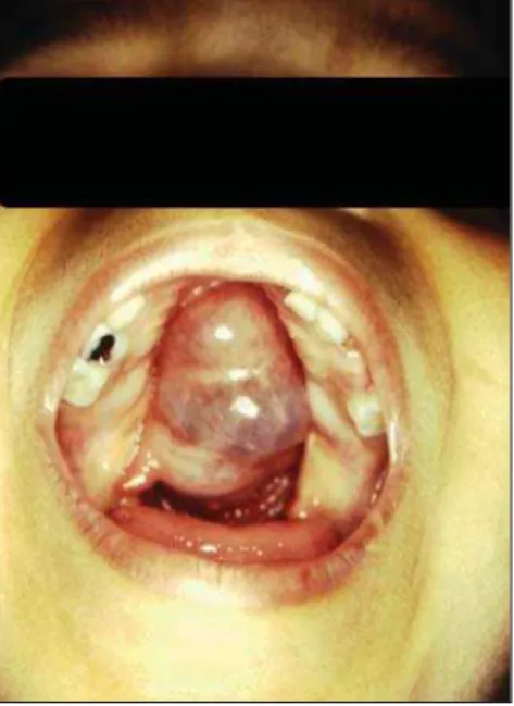
A female, nine-year-old child present-ing midline cleft lip and palate, hyperte-lorism, low height/weight development and normal intellectual development. Examina-tion of the oral cavity, demonstrated a wide cleft palate that allowed direct visualization of a round, pulsatile cystic mass, with a brownish color, regular and smooth sur-face, measuring about 4 cm in diameter, lo-cated on the nasal cavity midline (Figure 1).
INTRODUCTION
Sphenoethmoidal meningocele is a con-genital defect of the anterior cranial fossa with herniation of meninges and, eventu-ally, of parts of the brain (encephalome-ningocele), determining a protrusion to-ward the nasal cavity through a defect in the ethmoid and sphenoid sinus. Such a condition corresponds to a subtype of basal meningoencephalocele(1).
Different classification have been pro-posed for the anterior cranial fossa ence-phaloceles that, in a simplified way, are divided into two major groups, according to the herniation topography. The first group includes sincipital (frontal) encepha-loceles where the cranial contents herniate
400
Holanda MMA et al. Basal encephalocele associated with clef palate
Radiol Bras. 2011 Nov/Dez;44(6):399–400
Figure 2. MRI, sagittal section demonstrating im-age with fluid density in the anterior region of the skull base (T2).
netic resonance imaging (MRI) demon-strated fluid density in the anterior region of the skull base, showing ethmoid and sphenoid defect on the midline with nasal extension, besides a hard palate defect (Fig-ures 2 and 3).
The patient’s chronological age was nine years, while her osseous age was five years. Basal human growth hormone (HGH) levels after clonidine treatment was 0.2 ng/ml (reference: 0 to 10 ng/ml).
DISCUSSION
Data reporting such defects association are scarce in both the Brazilian and foreign literatures.
Geographic and racial factors influence both the frequency of this condition and the encephalic area to be involved, showing endemic levels in the Southern Asia. At two health centers in Bangkok, Thailand, in the period from 1992 to 1996, 120 cases of frontoethmoidal encephalomeningocele (sincipital). However, the basal presenta-tion is rarely observed in all of the racial groups(5).
Clinically, basal encephaloceles may manifest as a mass on the nasal cavity mid-line causing, or not, obstruction, feeding difficulties, cerebrospinal fluid leakage and meningitis. Such a condition may be asso-ciated with cleft lip and palate, microphthal-mos, bifid nose and optic nerve coloboma. Considering the presence of a nasal mass, glioma, dermoid cyst and polyp should be
taken into consideration in the differential diagnosis(1).
Advanced imaging studies are extremely necessary both to confirm de diagnosis and to analyze the hernial sac contents. Non-contrast-enhanced skull CT with 3D recon-struction is important to assess the skull base defect, and RMI plays a critical role in the analysis of the hernial sac elements(4).
In the present case, preoperative MRI played a key role, providing valuable infor-mation such as the location of vital struc-tures within the hernial sac. Only cere-brospinal fluid was observed within the hernial sac, without the presence of cere-bral structures, rigorously characterizing a basal meningoceles. The empty sella tur-cica explains the hypothalamic-hypophy-seal failure that may be present in 60% of cases(6).
Additionally, MRI has been largely uti-lized in the intrauterine diagnosis of cen-tral nervous system anomalies for its higher sensitivity as compared with ultrasonogra-phy(7,8).
The prognosis, in such cases, is re-served. Only a little more than half of treated cases present a favorable evolution. In a study including 114 patients, 59% have presented normal development, 18% have presented some degree of physical or men-tal deficiency, and 23% have a severe de-ficiency(2).
Additionally to the rarity of the present case, it is important to highlight the high índex of morbidity, mortality and risk for extremely severe complications in cases where the condition is equivocally diag-nosed as a dermoid cyst or nasal polyp and the surgeon inadvertently attempts to per-form its resection.
REFERENCES
1. Hoving EW. Nasal encephaloceles. Childs Nerv Syst. 2000;16:702–6.
2. Macfarlane R, Rutka JT, Armstrong D, et al. Encephaloceles of the anterior cranial fossa. Pediatr Neurosurg. 1995;23:148–58.
3. Monteiro M, Albuquerque AC, Nobre MC, et al. Meningoencefalocele transesfenoidal transpalatina. Arq Neuropsiquiatr. 2006;64:624–7.
4. Rathore YS, Sinha S, Mahapatra AK. Transsellar transsphenoidal encephalocele: a series of four cases. Neurol India. 2011;59:289–92.
5. Rocha LCM, Genes M. Disrafismo craniano – encefaloceles. Prat Hosp. 1989;4:6–16.
6. Lees MM, Hodgkins P, Reardon W, et al. Fronto-nasal dysplasia with optic disc anomalies and other midline craniofacial defects: a report of six cases. Clin Dysmorphol. 1998;7:157–62.
7. Peruzzi P, Corbitt RJ, Raffel C. Magnetic resonance imaging versus ultrasonography for the in utero evaluation of central nervous system anomalies. J Neurosurg Pediatr. 2010;6:340–5.
8. Rosen H, Chiou GJ, Stoler JM, et al. Magnetic resonance imaging for detection of brain abnor-malities in fetuses with cleft lip and/or cleft pal-ate. Cleft Palate Craniofac J. 2011;48:619–22. Figure 3. MRI, axial section demonstrating
show-ing ethmoid and sphenoid defect on the midline with nasal extension, besides a hard palate defect (T2).
A