Cop
yright
© ABE&M t
odos os dir
eit
os r
eser
vados
.
558
case report
Arq Bras Endocrinol Metab. 2013;57/7
Giant macroprolactinoma
and pregnancy
Macroprolactinoma gigante e gravidez
Joana Saraiva1, Leonor Gomes1, Sandra Paiva1, Luisa Ruas1, Manuela Carvalheiro1
SUMMARY
Prolactinomas are a common cause of gonadal dysfunction and infertility. We present the case of a 38-year-old woman with history of amenorrhea and infertility. At seven weeks of pregnancy she presented neuro-ophthalmologic complaints of headaches, diplopia, and right ptosis. The work-up study revealed an invasive pituitary macroadenoma with a maximum diameter of 9 cm and serum prolactin of 25,800 ng/mL (3-20). At 12 weeks, she was referred to the Endocrinology Department of the Coimbra University Hospital and started therapy with bromocriptine, initially 5 mg/day and then at crescent doses. Hyperprolactinemia was rapidly and drastically reduced to 254 ng/mL three weeks after taking bromocriptine 15 mg/day. Tumoral volume was reduced and there was improvement of III pair paresis. At 38 weeks, a male healthy baby was born. This is a relevant clinical case that illustrates the eficacy and safety of bromocriptine therapy during pregnancy, even in severe cases like this one. Arq Bras Endocrinol Metab. 2013;57(7):558-61
SUMÁRIO
Os prolactinomas são uma causa comum de anovulação e infertilidade. Apresenta-se o caso de uma mulher de 38 anos com antecedentes de amenorreia e infertilidade. Por volta das sete semanas de gestação, iniciaram-se clínica neuro-oftalmológica de cefaleias hemicranianas, ptose palpebral di-reita e diplopia. O estudo complementar revela a presença de macroadenoma hipoisário com cerca de 9 cm de maior diâmetro, de crescimento invasivo e destrutivo associado à hiperprolactinemia de 25.800 ng/mL (3-20). Às 12 semanas, foi referenciada à consulta de Endocrinologia e iniciou terapêu-tica com bromocriptina 5 mg/dia em doses crescentes. A prolactina diminuiu drasterapêu-ticamente para 254 ng/mL três semanas após, sob 15 mg/dia de bromocriptina. O volume tumoral também foi signiica-tivamente reduzido nas suas diferentes extensões. Clinicamente, houve regressão da ptose palpebral e da restante sintomatologia neuro-oftalmológica. O parto ocorreu por via vaginal às 38 semanas, com recém-nascido saudável. Este caso clínico é relevante, ilustrando bem a eicácia e a inocuidade da terapêutica com bromocriptina durante toda a gravidez, mesmo em casos graves como este. Arq Bras Endocrinol Metab. 2013;57(7):558-61
1 Department of Endocrinology,
Diabetes and Metabolism, Coimbra University Hospital, CHUC, EPE, Coimbra, Portugal
Correspondence to:
Joana Saraiva
Hospitais da Universidade de Coimbra, Praceta Mota Pinto 3000-075 – Coimbra, Portugal [email protected] [email protected]
Received on Aug/11/2012 Accepted on Nov/12/2012
INTRODUCTION
P
rolactinomas are the most common of hormone--secreting pituitary tumors, representing 30% to 40% of all pituitary tumors, and are a common cause of anovulation and female infertility (1,2). Hyperpro-lactinemia leads to gonadal dysfunction by means of several mechanisms: it reduces the amplitude and fre-quency of the GnRH secretory pulses and consequently the pulsatile secretion of LH; it also has direct action on the ovaries, inhibiting the production of progester-one and estrogen [3,4). The correction of hyperpro-lactinemia with dopaminergic agonists, cabergoline and bromocriptine, restores ovulation in about 90% of cases (3). Cabergoline is currently the treatment of choice for symptomatic microadenomas or macroadenomasbecause it has higher eficacy in normalizing PRL levels and in decreasing tumor size (5).
bromocrip-Cop
yright
© ABE&M t
odos os dir
eit
os r
eser
vados
.
559 Arq Bras Endocrinol Metab. 2013;57/7
tine (5,6). The use of bromocriptine throughout preg-nancy is only described in literature in about 100 cases (3). We describe the case of a giant macroprolactinoma diagnosed during early pregnancy that was successfully treated with bromocriptine throughout pregnancy.
CASE REPORT
In February 2001, a 38-year-old woman with macropro-lactinoma, 12-week pregnant, was referred to the Endo-crinology Department of the Coimbra University Hos-pital. In October 2000, she had been seen in gynecology appointment of another institution for primary infertil-ity and amenorrhea with about two years of evolution. She was given bromocriptine 5 mg id and clomiphene citrate 50 mg id before work-up was started, maintain-ing the amenorrhea, and she got pregnant. When preg-nancy was conirmed, bromocriptine was discontinued. In January 2001 (at around 7 weeks of gestation) she
started to complain about headaches, associated with progressively worsening right ptosis, diplopia, and otal-gia. Magnetic resonance imaging (MRI) showed a 9 x 5 x 5 cm lesion occupying the base of the skull, with inva-sive and destructive centrifugal multidirectional growth (sphenoidal sinus, rhinopharynx and nasal cavities, sellar loor and dorsum sellae, posterior fossa, cavernous si-nus, carotids, right temporal lobe and apex of the right orbit) (Figure 1). Transnasal biopsy was performed and revealed pituitary adenoma (immunohistochemistry positive synaptophysin and NSE, Ki67 < 1%). Prolactin (PRL) concentration was 25,800 (non-pregnant normal range 3.0-20.0 ng/mL). Other hormonal assays were unremarkable. At that time, an endocrinology appoint-ment was requested for therapeutic guidance.
She was a previously healthy woman without drug use, smoking, or alcohol intake habits. Family history was unremarkable. Ophthalmic examination revealed right ptosis (oculomotor nerve paresis) and strabismus.
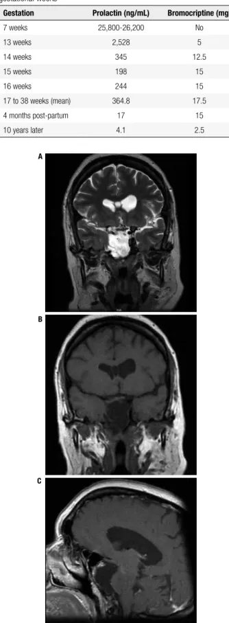
Figure 1. T1 (A) and T2 (B, C) weighted MRI pictures at 12 weeks, showing a large intra-sellar, right para-sellar and suprasellar tumor that ills the sphenoidal sinus, invading the nasal cavity and rinopharynx. Posteriorly, it invades the clivus. Laterally, it invades the right cavernous sinus and shapes the right temporal lobe and right hippocampus, measuring approximately 9 x 5 x 5 cm. After therapy, there was a marked decrease in the dimensions of the pituitary macroadenoma, with T1-weighted MRI (D, E) showing residual tumor intra-sellar paramedian right, extending into the sphenoid sinus and posterior portion of the nasal septum. The pituitary stalk and remaining gland are dislocated to the left.
A B C
D E
Cop
yright
© ABE&M t
odos os dir
eit
os r
eser
vados
.
560 Arq Bras Endocrinol Metab. 2013;57/7
The visual ields showed loss of ixation to the right. The obstetric assessment with ultrasound was normal with evidence of a fetus of 13 weeks of gestational age and no evidence of anomalies.
Given the tumor characteristics, gestation time and the mother’s desire to keep the baby, medical treatment with bromocriptine was chosen, starting with 5 mg at bedtime, together with close monitoring, initially as an inpatient. The dosage of bromocriptine was gradually increased with marked and fast improvement in average levels of PRL: 2,549 ng/mL with 5 mg/day, 346 ng/ mL with 12.5 mg/day to 254.5 ng/mL with 15 mg/ day. Bromocriptine was well-tolerated and improve-ment in headache and ptosis was observed. MRI three weeks after starting therapy showed moderate decrease in the macroadenoma size, in its various extensions, more evident in the nasal cavity extent.
The patient was discharged after 20 days, treated with bromocriptine 15 mg/day and folic acid 10 mg id, being followed up as outpatient in the Endocrinology-Obstetrics Practice. During follow-up, average PRL was 364.8 ng/mL and average dose of bromocriptine 17.5 mg/day (Table 1). She was also followed up in the Ophthalmology Practice with almost complete regres-sion of right ptosis.
At 38 weeks, she gave birth by vaginal delivery to a healthy male baby weighing 4,300 g, 51 cm, Apgar 9/10. The post-partum period was uneventful and she did not breastfeed. Treatment with bromocriptine 15 mg/day was maintained until assessment four months after delivery. She did not report any relevant symp-toms, and referred to regular cycles. Ophthalmological examination showed no ptosis and intermittent diplo-pia. Average PRL was 17 ng/mL. MRI showed marked reduction in tumor volume (3 x 2.5 x 1.5 cm) (Figure 1). During the follow-up at the Endocrinology Practice, prolactin assays always remained within the normal range, and the volume tumor decreased progressively and the dose of bromocriptine was gradually reduced.
In the last evaluation, in 2011, the patient showed an average PRL 4.1 ng/mL with 2.5 mg bromocriptine once day. The last MRI at 2011 (Figure 2) showed a cystic area, extending anterior to the sphenoid sinus on the right, and lack of visualization of the right caver-nous sinus and mass effect on the left cavercaver-nous sinus temporal lobe. Her 10 year-old son showed appropriate psychomotor development for his age, without comor-bidities.
Table 1. Prolactin levels in relation to the doses of bromocriptine and gestational weeks
Gestation Prolactin (ng/mL) Bromocriptine (mg)
7 weeks 25,800-26,200 No
13 weeks 2,528 5
14 weeks 345 12.5
15 weeks 198 15
16 weeks 244 15
17 to 38 weeks (mean) 364.8 17.5
4 months post-partum 17 15
10 years later 4.1 2.5
Figure 2. T2 (A) and T1 (B, C) weighted MRI, performed in 2011, showing a cystic area, extending anterior to the sphenoid sinus on the right, lack of visualization of the right cavernous sinus and mass effect on the left cavernous sinus and temporal lobe, measuring approximately 3.7 x 2.8 x 2.5 cm.
A
B
C
Cop
yright
© ABE&M t
odos os dir
eit
os r
eser
vados
.
561 Arq Bras Endocrinol Metab. 2013;57/7
DISCUSSION
We present a patient with an unusual clinical picture, in which the diagnosis of giant macroprolactinoma was only made after conception associated with the particu-larity of its large dimensions and degree of invasiveness. Prolactinoma diagnosis relies on inding and elevated PRL level after excluding hook effect (5). Biopsy is rarely needed, but in this case due to its imaging char-acteristics, accessibility trough nasal cavity and without a deinitive diagnosis, it was decided to perform such procedure.
Hyperprolactinemia is the cause of infertility in 15% to 20% of cases (2). The treatment with dopaminer-gic agonists is very effective in decreasing serum levels of PRL, and restoration of fertility may be immediate, even before the irst normal menstruation (4,6,7). In this particular case, the institution of therapy prior to the work-up of the cause of infertility is likely to have led to a decrease in PRL levels that, in combination with stimulation with clomiphene citrate, enabled the patient to get pregnant.
Soon after the diagnosis of pregnancy, the discon-tinuation of dopaminergic agonist decreased the time of fetal exposure to the drug but, in the presence of a macroadenoma, poses an increased risk of tumor growth. Therefore, during pregnancy and in the pres-ence of clinical evidpres-ence of tumor growth there are sev-eral challenges for the physician and the patient herself, and the decision should always be individualized. The latest guidelines recommend reinstitution of therapy with bromocriptine (5). Despite the limited experience of use in these situations, clinical evidence does not seem to show deleterious effects on the fetus. Trans-sphenoidal surgery may be an alternative, if there is no response to bromocriptine, but any surgery during pregnancy is associated with an increased risk of miscar-riage and other complications (2). If the fetus is near term, labor induction may be a valid option. In this patient, pregnancy occurred in an early and fortuitous way, precluding a previous diagnosis of macroprolacti-noma and its treatment.
The choice of bromocriptine was very effective in decreasing the levels of PRL and, namely, in the de-creasing the tumor size. It is not usual to use such high doses of bromocriptine, but due to such a large tumor it seemed a reasonable option, given the need of rapidly and eficiently decrease PRL levels to allow the correct progress of that pregnancy. Despite high doses of
bro-mocriptine used, there were no fetal adverse effects in the short-term, as well as in the subsequent develop-ment. Measurement of PRL during pregnancy is not usually recommended, because PRL levels may some-times not increase in case of tumor enlargement (4-6). In this particular case, given the initial treatment phase and magnitude of the increase in PRL (taking into ac-count the typical values described in the literature) (8), PRL measurement enabled dose adjustment of bro-mocriptine therapy.
Pregnancy and treatment with bromocriptine con-ferred a high risk of pituitary apoplexy in this particular case. The inpatient treatment for almost three weeks allowed us to closely monitor this possibility. The ex-tended follow-up of this patient, about 10 years, has shown clinical, analytical and radiologic stability, with a low dose of the dopaminergic agonist. Similar situ-ations that show the beneicial effect of pregnancy on the natural history of prolactinomas are also described in the literature (2).
In conclusion, it seems that this case, despite its se-verity (in relation with the extension, invasion, surgi-cal accessibility dificulties, and neuro-ophthalmologic complications), illustrates the effectiveness and safety of therapy with bromocriptine. However, it is important to emphasize that the diagnosis should, whenever pos-sible, be prior to pregnancy so that the tumor can be controlled with the consequent reduction of maternal and fetal risks.
Disclosure: no potential conlict of interest relevant to this article was reported.
REFERENCES
1. Colao A. The prolactinoma. Best Pract Res Clin Endocrinol Metab. 2009;23:575-96.
2. Shibli-Rahhal A, Schechte J. Hyperprolactinemia and infertility. Endoc Metab Clin N Am. 2011;40:837-46.
3. Molitch M. Prolactinoma in pregnancy. Best Pract Res Clin Endocrinol Metab. 2011;25:885-96.
4. Mancini T, Casanueva F, Giustina A. Hyperprolactinemia and prolactinomas. Endocrinol Metab Clin N Am. 2008;37:67-99. 5. Melmed S, Casanueva F, Hoffman A, Kleinberg DL, Montori VM,
Schlechte JA, et al. Diagnosis and treatment of hyperprolactinemia: an Endocrine Society clinical practice guideline. J Clin Endocrinol Metab. 2011;96:273-88.
6. Casanueva F, Molitch ME, Schlechte JA, et al. Guidelines of the Pituitary Society for the diagnosis and management of prolactinomas. Clin Endocrinol. 2006;65:265-73.
7. Thorner M, Harrison D, Bronstein M. Hyperprolactinemia. 2011;6: 1-8. Disponível em: www.endotext.org.
8. Bronstein M. Prolactinomas and pregnancy. Pituitary. 2005;8:31-8.