Aging in Sensory and Motor Neurons Results
in Learning Failure in
Aplysia californica
Andrew T. Kempsell, Lynne A. Fieber*
University of Miami, Rosenstiel School of Marine and Atmospheric Science, Department of Marine Biology and Ecology, Miami, Florida, United States of America
Abstract
The physiological and molecular mechanisms of age-related memory loss are complicated by the complexity of vertebrate nervous systems. This study takes advantage of a simple neural model to investigate nervous system aging, focusing on changes in learning and memory in the form of behavioral sensitization in vivo and synaptic facilitation in vitro. The effect of aging on the tail withdrawal reflex (TWR) was studied inAplysia californicaat matu-rity and late in the annual lifecycle. We found that short-term sensitization in TWR was ab-sent in agedAplysia. This implied that the neuronal machinery governing nonassociative learning was compromised during aging. Synaptic plasticity in the form of short-term facilita-tion between tail sensory and motor neurons decreased during aging whether the sensitiz-ing stimulus was tail shock or the heterosynaptic modulator serotonin (5-HT). Together, these results suggest that the cellular mechanisms governing behavioral sensitization are compromised during aging, thereby nearly eliminating sensitization in agedAplysia.
Introduction
Brain aging is associated with the progressive decline of neurophysiological processes and an increased prevalence of memory impairments. Some loss of neurons occurs with age, but more relevant is a reduction in synaptic contact and the efficiency of synaptic transmission between these contacts with age. In mammals, the number of neuronal synapses and density of dendrit-ic spines decrease during aging [1–4] resulting in weaker synapses that are less capable of short-term plasticity [5]. In the rodent hippocampus, aging is associated with a decrease in syn-apses in the dentate gyrus and area CA1, a decrease in NMDA-receptor-evoked responses at perforant path synapses onto dentate gyrus granule cells, and changes in Ca2+regulation in area CA1 [6]. As these changes in synaptic structure and function accumulate during aging, cellular analogs of learning and memory, including term potentiation (LTP) and long-term depression (LTD), are disrupted. Hippocampal aging is associated with deficits in the in-duction and maintenance of LTP and lower thresholds for LTD [7]. Altered synaptic plasticity in aged hippocampal networks results in age-related memory loss including reduced perfor-mance in spatial memory tasks [8,9].
OPEN ACCESS
Citation:Kempsell AT, Fieber LA (2015) Aging in Sensory and Motor Neurons Results in Learning Failure inAplysia californica. PLoS ONE 10(5): e0127056. doi:10.1371/journal.pone.0127056
Academic Editor:Riccardo Mozzachiodi, Texas A&M University - Corpus Christi, UNITED STATES
Received:December 19, 2014
Accepted:April 10, 2015
Published:May 13, 2015
Copyright:© 2015 Kempsell, Fieber. This is an open access article distributed under the terms of the
Creative Commons Attribution License, which permits unrestricted use, distribution, and reproduction in any medium, provided the original author and source are credited.
Data Availability Statement:All data files are available from Figshare. doi:10.6084/m9.figshare. 1383211.
Funding:This work was funded by National Institutes of Health Grant P40 OD010952, and Maytag and Korein Foundation fellowships to A.T. Kempsell. The funders had no role in study design, data collection and analysis, decision to publish, or preparation of the manuscript.
Neural networks in mammalian models are vastly complex, making it challenging to con-nect behavioral aging with physiological aging. The marine snailAplysia californica(Aplysia) provides a unique animal model to study the neurobiology of aging with the advantages of a simple nervous system and short, one-year lifespan.Aplysiais an opistobranch mollusk and a member of the large superclade of bilaterian animals, the Lophotrochozoa. While Ecdysozoans includingDrosophilaandC.eleganshave undergone severe gene loss and sequence divergence [10], Lophotrochozoans do not have such derived genomes [11]. A lower amino acid replace-ment rate and thus a slower evolving genome when compared to the Ecdysozoans imply that
Aplysiamay have more genes in common with higher order species including humans. One of the best mapped neuronal circuits in theAplysianervous system serves the tail with-drawal reflex (TWR). Direct stimulation to the tail initiates TWR, a monosynaptic reflex in-volving identified primary mechanosensory neurons (SNs) in the pleural ganglia and motoneurons (MNs) in the pedal ganglia [12,13]. Sensitization in TWR is a simple form of nonassociative learning in which tail withdrawal becomes enhanced after harmful stimuli such as tail shock [14–16]. Three temporal phases of memory for sensitization can be induced de-pending on the amount and pattern of tail shocks [17]. A single tail shock induces short-term memory lasting30 min, whereas repeated tail shocks induce intermediate-term memory last-ing90 min and long-term memory lasting24 h. The neuromodulator serotonin (5-HT) is released by facilitatory interneurons onto the sensorimotor synapse during behavioral sensiti-zation, resulting in increased tail SN excitability and heterosynaptic facilitation of tail SN-MN transmission [14,18–23].
Three temporal forms of synaptic facilitation can be induced and the underlying mecha-nisms governing this proxy for synaptic plasticity are well-studied [24,25]. In short-term facili-tation (30 min), a brief pulse of 5-HT activates adenylyl cyclase in presynaptic neurons, causing an increase in cyclic adenosine monophosphate (cAMP) and activation of protein ki-nase A (PKA). PKA mediates short-term facilitation by covalent modifications of channel acti-vation, resulting in altered channel activity including closure of K+ channels. Reduced K + current increases action potential (AP) duration and thus raises the amount of presynaptic cytosolic Ca2+. Ca2+influx activates Ca2+/calmodulin-dependent protein kinase II, which then phosphorylates synapsin I. Phosphoylated synapsin I loses its affinity for synaptic vesicles, re-leasing them from the cytoskeleton and leading to increased synaptic release and temporary synaptic strengthening. Longer exposures to 5-HT cause the prolonged activation of PKA as well as activation of protein kinase C (PKC), inducing intermediate-term facilitation (90 min) and long-term facilitation (24 h). Intermediate-term facilitation requires translation while long-term facilitation requires both transcription and translation. Prolonged PKA activi-ty results in the phosphorylation of transcription factors such as CREB-1, stimulating RNA and protein synthesis.
This study investigated whether behavioral sensitization in TWR undergoes age-related de-clines that involve changes in synaptic facilitation between SNs and MNs known to be involved in the reflex circuit. We measured short-term memory for sensitization in TWR in identified animals as they aged. We also measured short-term changes in tail SN excitability and synaptic facilitation of tail SN-MN transmission. The results indicate that aging of the neural circuit for TWR resulted in learning failure, including the ability to sensitize TWR in intact animals, and related impairment of synaptic facilitation of the SN-MN circuit underlying TWR.
Materials and Methods
an ad-libitum diet consisting ofGracilaria feroxandAgardhiella subulataand reared as de-scribed previously [26]. Four hatchery-reared animals of different weights were assigned to each cage to allow for identification of individual animals by monthly weight measurements. Sexual maturity for a cohort of animals was designated as the day the first egg mass was laid. Behavioral and electrophysiological experiments were done at two points in the adult life of this annual animal: mature and aged II, as described in Kempsell and Fieber (2014) [27]. Ma-ture animals were age 7–8 mos and had reached sexually maturity<1 month earlier. Aged II animals were age 12–13 mos, designated as advanced age animals. They had significantly re-duced performance in the righting reflex, TWR, and biting response compared to mature and aged I siblings, as described in the previous paper [27].
Sensitization of TWR in freely behaving Aplysia
Animals were placed individually in 48 x 27 x 20 cm translucent plastic cages filled with 20–
21°C seawater to a depth of 15 cm and allowed to acclimate for 5 min prior to measurements. A trained experimenter who was unaware of the age of the animals conducted behavioral mea-surements with the help of an assistant. The experimenter measured TWR amplitude and du-ration while the assistant recorded each measurement. We opted to score responses from live observations. Measuring TWR amplitude and duration from video recordings was not done due to difficulties in establishing a camera angle that properly recorded all animals.
The same 18 animals were measured for sensitization in TWR at mature and aged II time points following previously defined protocols [16,27]. An animal was placed on its foot in the center of the cage and allowed to acclimate for 5 min. The animal’s resting body length was mea-sured with a ruler. A 500 ms tap to the tip of the tail at a stimulus pressure of 75 grams/mm2 caused tail withdrawal towards the center of the body and signified the start of the reflex. The re-tracted body length was then measured. The time to relax the tail to ~30% of original tail length was recorded, and signified the end of the reflex and the reflex duration. TWR amplitude was calculated as the fraction of starting body length withdrawn following tail touch. TWR ampli-tude and TWR duration were measured at 15, 10 and 5 min before sensitizing electrical shocks to the tail (-15, -10, -5 min) and the average of these 3 measurements was designated as baseline. Five minutes after the last baseline tail tap, sensitizing tail shocks were then delivered, consisting of five 1.5 sec, 100 mA electrical shocks, with an interstimulus interval of 1 sec, delivered to the tail immediately posterior to the parapodial convergence at a different site than that used to mea-sure baseline TWR (Fig 1A). This site was ~2–3 cm anterior to the tip of the tail. Next, TWR was again elicited by tail tap 5, 15, 30, and 60 min following sensitizing shocks, and amplitude and duration were measured.
General Intracellular recording methods
Glass capillary microelectrodes of 5–15 MOresistance were used for intracellular recordings in tail SNs and MNs. All recordings were made at room temperature of 21–23°C using pClamp10 software with BRAMP-01R and ELC-01MX amplifiers (ALA Scientific Instruments, Farming-dale, NY) connected to a PC and Digidata 1440A A/D converter.
Behavioral experiments were conducted at 20–21°C while electrophysiological experiments were conducted at room temperature (21–23°C).
Intracellular recording of TWR in semi-intact tail preparation
differences in tail SN and MN responses compared to the more reduced preparation that isolat-ed SN and MN innervation to the pleural and pisolat-edal ganglion. Animals were anesthetizisolat-ed by in-jection of isotonic MgCl2(~50% animal weight by volume) into the body cavity. Pleural-pedal
ganglia, nerve p9, and attached tail were removed and pinned tightly to a Sylgarded dish. The other ganglia were removed from the remaining tissue to euthanize the animal, and unneeded ganglia and tissue were discarded. The pins were positioned in the reduced tail preparation to minimize tail contraction following mechanical and electrical stimulation. The protective sheath surrounding the ganglia was removed mechanically. Ganglia were maintained for the duration of the experiment in artificial seawater (ASW) via a gravity-fed perfusion pipette lo-cated ~5 mm from the ganglia. Tail SNs and MNs were identified according to previously de-fined methods [12–14]. Tail SNs of the ventral caudal region of the pleural ganglia (PVC) were not spontaneously active and had resting potentials of -40–-55 mV, but when receptive fields on the tail were stimulated, produced AP of 60–100 mV amplitude [12]. Tail pedal motoneu-rons P5-7 were spontaneously active with resting potentials of -40–-70 mV and exhibited in-creased AP firing in response to mechanical stimulation of the tail [14].
Electrophysiological experiments involved tail SN and MN responses to injected current or mechanical tail stimulation before and after sensitizing stimuli. Sensitizing stimuli consisted of tail shocks (described above), or application of 5-HT. In all electrophysiological experiments, three pre-treatment (-shock or -5-HT) tests were recorded with a 5 min interval between tests, and the baseline response was calculated from their average. Post-treatment test responses were then evoked at 5, 15, and 30 min after one of these treatments: tail shocks, or 5-HT appli-cation for 10 min from the perfusion pipette. Tail shocks were delivered as in freely behaving
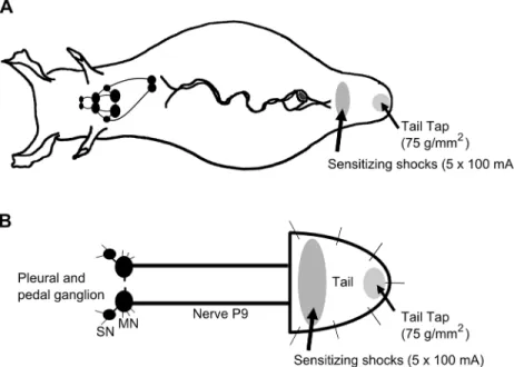
Fig 1. Schematic diagram ofAplysiaillustrating regions of the tail subject to mechanical test stimulation and sensitizing electrical shocks.A) Behavioral experiments were performed in the intact animal. TWR amplitude and TWR duration were monitored in response to tail tap (75 g/mm2). After 3 baseline pretest taps, each animal received sensitization training consisting of five 1.5 sec, 100 mA shocks delivered to a site immediately posterior to the parapodial convergence. TWR amplitude and TWR duration in response to test tail tap were recorded 15, 30, 45, and 60 min after training and compared to baseline. B)
Electrophysiological experiments consisted of a reduced preparation that involved the pleural-pedal ganglia, nerve P9, and attached tail. Tail SN and MN responses to mechanical tail tap were assessed before and after sensitization training as described in A.
animals to a site on the tail immediately posterior to the parapodia, ipsilateral to the ganglion under investigation, at a position that did not include the mechanoreceptive field of the tail SN. In electrophysiological experiments investigating heterosynaptic modulation by 5-HT, 5-HT (20μM) was perfused onto the pleural-pedal ganglion for 10 min. This concentration was
pre-viously found to induce forms of modulation including synaptic facilitation, increased AP du-ration and increased excitability in tail SNs [29,30]. Tail motoneurons were held at -90 mV to arrest AP generation.
To measure excitability in tail PVC SNs, a protocol was modified from previous experi-ments [31,32]. Depolarizing current was applied to PVC SNs in increasing increments of 0.1 nA for 500 ms until the neuron fired a single AP, then the single AP evoked by this depolar-ization was confirmed 3 times (baseline response; 5 min interval between tests). After treat-ments of either sensitizing tail shocks or 5-HT, the same depolarizing current as in baseline was applied, and the number of AP fired by the SN was counted.
To assess monosynaptic transmission between tail SNs and MNs, synaptic facilitation was evaluated by measuring the amplitude of a single SN-evoked excitatory postsynaptic potential (EPSP) in the MN and comparing EPSP amplitude to baseline. A single AP was evoked in the tail SN by 50 ms depolarizing current injection and the amplitude of the resulting EPSP was measured at time points corresponding to 15, 10 and 5 min before and 5, 15, and 30 min after the end of sensitizing tail shocks or 5-HT treatment and compared. EPSP latency was defined as the time from tail SN AP initiation to tail MN EPSP initiation. No homosynaptic depression was observed with the interstimulus intervals used in these experiments.
To assess synaptic transmission between tail SNs and MNs during mechanical tail tap, pro-tocols were modified from previous experiments [14,28]. The responses to a single 500 ms tail tap at a stimulus pressure of 75 grams/mm2at -15, -10, and -5 (before), and 5, 15, and 30 min following sensitizing tail shocks were compared in mature and aged II tail SNs and MNs. Tail tap evoked AP in tail SNs and complex EPSP in tail MNs. The number of AP fired in response to tail tap was compared in mature and old tail SNs. For analysis of complex EPSP in tail MNs, the amplitude of the first evoked EPSP was averaged in mature and in aged II MNs and compared.
In electrophysiological experiments investigating modulation of tail SN biophysical proper-ties by 5-HT, resting membrane potential was monitored while 5-HT (20μM) in ASW was
per-fused onto the pleural-pedal ganglion. After 3 min 5-HT perfusion, a single AP was evoked in tail SNs by 3 ms depolarizing intracellular current injection. Repolarization amplitude, afterhy-perpolarization (AHP) amplitude, and AP duration were compared before and after 5-HT treatment in mature and aged II tail SNs.
Solutions
Extracellular solution consisted of ASW containing (mM) 417 NaCl, 10 KCl, 10 CaCl2, 55
MgCl2, and 15 HEPES-NaOH, pH 7.6. The pipette solution for intracellular recordings in
in-tact ganglia consisted of 3 M KCl. Solutions containing 20μM 5-HT in ASW were prepared
daily from 0.5 M stocks as previously reported [33]. All reagents were from Sigma-Aldrich (St. Louis, MO, USA).
Data Acquisition and Analysis
time point. For electrophysiological experiments, significant differences to determine facilita-tion of averaged neuron responses were assessed via one-way within subjects ANOVA with Tukey’s posthoc test. Baseline responses were compared to responses after treatments of sensi-tizing tail shocks or 5-HT. To compare mature and aged II responses, 2-sample t-tests were used. All analyses were performed using the open source R statistical program (R Foundation for Statistical Computing, Vienna, Austria). P-values are all 2-tailed. Differences at p0.05 were accepted as significant.
Results
Sexual maturity occurred by age 7 mos and median lifespans were 12.9 and 12.7 mos for the 2 cohorts studied. The percentage of animals that died before the aged II time point was 8.3% and 6.3% respectively. Morphological and aging characteristics of these cohorts were reported in a previous study [27]. Briefly, growth was steady and peaked shortly after first sexual maturi-ty and maximal egg production. A significant reduction in body mass was observed from age 11–13 mos in these cohorts (p0.05, 2-sample t-tests), however, no difference in body mass was observed between stages mature and aged II.
Sensitizing shocks in intact animals increased TWR in mature but not
aged II
Aplysia
Baseline TWR amplitude in 18 sibling animals was significantly weaker (Fig 2A, p0.01, 2-sample t-test) and TWR duration was significantly slower (Fig 2B, p0.05, 2-sample t-test) when the animals reached aged II compared to their performance when mature, as noted previ-ously [27]. Prior work inAplysiahas demonstrated that sensitizing tail stimuli increased TWR amplitude and duration [14–16]. To investigate if short-term memory for sensitization in TWR was disrupted during aging, the amplitude and duration of tail withdrawal in response to tail tap were measured before and after 5 sensitizing shocks (1.5 s each, 100 mA, AC, 1 s inter-stimulus interval). No change in response amplitude or duration was found at a 5 min interval between baseline tail taps (Fig 2C and 2D). In mature animals, five sensitizing shocks delivered to the anterior tail resulted in a significant increase in mean TWR amplitude at 15, 30, 45, and 60 min post-shock (Fig 2C; p0.05 at 15 and 60 min time points compared to baseline, p0.01 at 30 and 45 min time points; Tukey’s posthoc analysis). TWR duration also increased signifi-cantly in mature animals after sensitizing shocks (Fig 2D; p0.05 at each time point compared to baseline; Tukey’s posthoc analysis). Shocks did not significantly elevate TWR amplitude (Fig 2C) or TWR duration (Fig 2D) in aged IIAplysiacompared to baseline. Thus, short forms (60 min) of memory for sensitization in TWR were absent in aged IIAplysia.
Between group differences in TWR amplitude and duration also were significant following sensitizing tail shocks. TWR amplitude and duration measured at each of 15–60 min following sensitizing shocks were significantly greater in mature compared to aged II animals (p0.05; Tukey’s posthoc analysis).
Sensitizing shocks in reduced ganglia-tail preparations increased
excitability in mature but not aged II tail SNs
aged II; input resistance: 10.5±2.1 MOfor mature, 9.9±2.7 MOfor aged II). These values were consistent with those previously reported at ages mature and aged II [27].
Sensitizing shock has been shown to increase excitability in tail SNs [14,34–36]. To deter-mine whether aging affects this phenomenon, excitability of individual SNs was evaluated in mature and aged II reduced tail preparations before and after 5 rapidly applied sensitizing shocks (Fig 3; 1 sec interstimulus interval). In preparations from mature animals, the same de-polarizing test pulse that elicited a single AP in each control tail SN elicited multiple AP in the
Fig 2. Sensitization in TWR of intact animals declined during aging.A) TWR amplitude in baseline pretests decreased, while (B) TWR duration increased significantly during aging.*and**denote significant difference compared to mature at p0.05 and p0.01, respectively, via 2-sample t-tests. C) TWR amplitude and (D) TWR duration increased significantly in mature animals following five sensitizing shocks (1.5 s each, 100 mA, AC, 1 s interstimulus interval) delivered to the anterior tail, while in the same animals at stage aged II no shock-induced change in amplitude or duration was observed.*and**denote increase compared to baseline at p0.05 and p0.01, respectively, via Tukey’s posthoc tests following repeated measures ANOVA (n = 18).
SN after sensitizing shocks (Fig 3A and 3B; p0.05 at each time point compared to baseline; Tukey’s posthoc analysis). In contrast, no change in tail SN excitability was found following sensitizing shocks in preparations from aged II animals.
Between group differences in tail SN excitability following sensitizing shocks also were sig-nificant following sensitizing tail shocks (p0.05; Tukey’s posthoc analysis).
Fig 3. Synaptic facilitation between tail SNs and MNs following sensitizing tail shocks declined during aging.A) Representative responses during depolarizing current injection in tail SNs before and 5 min after sensitizing tail shocks in mature and aged II animals. B) In mature tail SNs, the same depolarizing current injection that yielded a single AP in baseline conditions evoked significantly more AP 5, 15, and 30 min after sensitizing tail shocks.*
denotes significant increase compared to baseline at p0.05, Tukey’s posthoc tests (n = 16). No change in AP number was observed following sensitizing tail shocks in aged II tail SNs (n = 20). C) Monosynaptic facilitation between tail SNs and MNs following sensitizing tail shocks decreased during aging. A single AP evoked in tail SNs by 50 ms injected current resulted in a monosynaptic EPSP in tail MNs in mature (n = 14) and aged II (n = 13) preparations. Tail MNs were hyperpolarized to -90 mV. D) Baseline EPSP amplitude was significantly decreased in MNs of aged II preparations.*denotes significant decrease compared to mature at p0.05, 2-sample t-test. E) Baseline EPSP latency did not change during aging. F) Following sensitizing tail shocks, EPSP amplitude increased significantly in mature but not in aged II tail MNs.*denotes significant increase compared to baseline at p0.05, Tukey’s posthoc tests.
Sensitizing shocks facilitated mature but not aged II tail SN-MN
synapses
Previous studies showed that monosynaptic EPSP amplitude increased following sensitizing stimulation in tail MNs [14,37]. Monosynaptic EPSP were evoked in tail MNs by applying de-polarizing current sufficient to initiate a single AP in tail SNs (Fig 3C). Baseline EPSP ampli-tude was significantly smaller in each MN studied from aged II preparations compared to mature (Fig 3D; p0.05; 2-sample t-test). The latency between AP initiation in tail SNs and evoked EPSP in tail MNs did not change during aging (Fig 3E). EPSP amplitude increased sig-nificantly in mature preparations at all time points after sensitizing shocks compared to pre-shock values (Fig 3F, p0.05 at each time point compared to baseline; Tukey’s posthoc analy-sis), whereas in aged II preparations no change was noted in EPSP amplitude after sensitizing shocks. EPSP amplitude also was significantly increased in mature animals following sensitiz-ing shocks compared to aged II animals (p0.05; Tukey’s posthoc analysis). Thus, short-term facilitation (30 min) of tail SN-MN transmission induced by shocks was absent in aged II
Aplysia.
Sensitizing shocks increased the response to tail tap in mature but not
aged II SNs and MNs
The excitatory response of tail SNs and MNs to weak tail mechanical stimulation has been shown to increase after sensitizing shock [14,28]. To further investigate changes in synaptic fa-cilitation during aging, tail SNs and MNs were monitored during mechanical tail tap before and after sensitizing shocks in mature and aged II preparations. The sites for shocks and tail taps were separated (Fig 1B) to reduce activation of subpopulations of tail SNs with overlap-ping receptive fields [14,17]. Tail tap evoked AP in individual tail SNs and complex EPSP in individual tail MNs (Fig 4A). In response to baseline tail tap, the number of AP fired in tail SNs and complex EPSP amplitude in tail MNs were significantly different between mature and aged II preparations, with aged II SNs exhibiting reduced AP firing (Fig 4B1) and aged II MNs exhibiting reduced complex EPSP amplitude (Fig 4B2; p0.05 in each case; 2-sample t-tests). Aging affected the response to sensitizing shocks as well. Whereas sensitizing shocks increased the responses of mature SNs and MNs to tail tap (Fig 4C and 4D; p0.01 at each time point compared to baseline; Tukey’s posthoc analysis), SNs and MNs from aged II preparations were unaffected. Furthermore, SN and MN responses were significantly greater in mature animals following sensitizing shocks compared to aged II animals (p0.01 at each mature time point compared to aged II time point; Tukey’s posthoc analysis). Thus, while tail SN and MN sponses to mechanical tail tap were facilitated after sensitizing shocks in mature animals, re-sponses from aged II animals were not.
5-HT treatment increased excitability in tail SNs of mature but not aged II
Aplysia
5-HT treatment. Tail SN excitability was significantly greater in mature animals when excit-ability was assessed 5 and 15 min after 5-HT treatment compared to aged II animals (p0.01 at 5 and 15 min; Tukey’s posthoc analysis).
5-HT facilitated mature but not aged II tail SN-MN synapses
Short-term synaptic facilitation induced by 5-HT applied to tail SN-MN synapses has been widely used as a cellular model of short-term memory for sensitization. Monosynaptic EPSP were evoked in tail MNs by applying depolarizing current sufficient to initiate a single AP in each tail SN studied (Fig 5C). After 10 min 5-HT perfusion of the pleural-pedal ganglion, MN EPSP amplitude increased significantly in mature (Fig 5D; p0.05 at 5 and 15 min time points compared to baseline; Tukey’s posthoc analysis) but not in aged II preparations. EPSP ampli-tude was significantly greater in mature animals after 5-HT treatment compared to aged II ani-mals (p0.05 at 5 and 15 min in mature compared to aged II; Tukey’s posthoc analysis). Thus, short-term facilitation (30 min) of tail SN-MN transmission after 5-HT treatment was absent in aged IIAplysia.
Fig 4. Tail tap-evoked responses of tail SNs and MNs decreased during aging.A) A single 500 ms tap to the tail at a stimulus pressure of 75 g/mm2 evoked AP generation in tail SNs and complex EPSPs in tail MNs from mature and aged II reduced tail preparations. B) The number of AP in response to baseline tail tap in tail SNs (B1) as well as the complex EPSP amplitude elicited in MNs (B2) decreased significantly during aging.*denotes significant decrease compared to mature at p0.05, 2-sample t-test. C) After sensitizing tail shocks, the number of AP in response to tail tap in tail SNs as well as (D) the complex EPSP amplitude in MNs increased significantly in mature but not in aged II preparations.**denotes significant increase compared to baseline at p0.01, Tukey’s posthoc tests (n = 14 for mature, n = 18 for aged II).
5-HT altered biophysical properties of mature but not aged II tail SNs
5-HT is an important neuromodulator implicated in forms of learning and memory including behavioral sensitization in TWR. Previous studies showed that 5-HT treatment induces changes in biophysical properties of tail SNs that enhance excitability, including resting mem-brane potential and duration of evoked AP [34,38]. In mature tail SNs, 5-HT treatment re-sulted in a slow and maintained depolarization of resting membrane potential, significantly different at 3 min in 5-HT from the initial measurement (Fig 6A and 6B; p0.05; paired t-test). In aged II tail SNs, the effects of 5-HT treatment were much more variable. No significant changes in resting membrane potential were observed in aged II tail SNs after 5-HT (Fig 6A and 6B).
Another hallmark of 5-HT induced facilitation of excitability in tail SNs was increased AP duration [38]. A single AP was evoked by 3 ms depolarizing intracellular current injection in tail SNs before and after 3 min 5-HT treatment (Fig 6C). Depolarization amplitude,
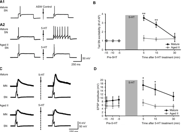
Fig 5. Synaptic facilitation between tail SNs and MNs following 5-HT treatment declined during aging.A) Representative responses during 500 ms depolarizing current injection in tail SNs before and 5 min after conclusion of 10 min perfusion of vehicle (ASW; A1) or 5-HT (20μM; A2) onto the
pleural-pedal ganglion. B) In mature tail SNs, the same depolarizing pulse that produced a single AP during baseline conditions evoked significantly more AP when assessed 5 and 15 min after 5-HT treatment.**denotes significant increase compared to baseline at p0.01, Tukey’s posthoc tests (n = 16). No change in AP number was observed in aged II tail SNs following 5-HT treatment (n = 17). C) Monosynaptic facilitation between tail SNs and MNs following 5-HT treatment decreased during aging. A single AP evoked in tail SNs by 50 ms injected current resulted in a monosynaptic EPSP in tail MNs. D) Following 5-HT treatment, tail MN EPSP amplitude increased significantly in mature but not in aged II preparations.*denotes significant increase compared to baseline at p0.05, Tukey’s posthoc tests (n = 20 for mature, n = 17 for aged II).
afterhyperpolarization (AHP) amplitude, and AP duration were measured. In ASW before 5-HT treatment, AP duration (mature: 3.8±0.6 ms, aged II: 4.0±0.9 ms) and depolarization am-plitude (mature: 88±11 mV, aged II: 82±13 mV) were not different between mature and aged II (Fig 6C and 6D). AHP amplitude (mature: 4.2±0.7 mV, aged II: 4.7±0.9 mV) also was not dif-ferent, consistent with studies in other aging models that found no change in fast components of AHP amplitude during aging [39].
In mature tail SNs, 5-HT caused a significant increase in AP duration of 2.0±0.9 ms com-pared to ASW controls (Fig 6D; p0.05, paired t-test), but not in aged II animals. Thus changes in biophysical properties of tail SNs induced by 5-HT that facilitate excitability were absent in aged IIAplysia.
Discussion
The experiments described here took advantage of a simple neural model to investigate nervous system aging, focusing on learning-induced changes that accompany behavioral sensitization in vivo and synaptic facilitation in vitro. These experiments were performed in cohorts of half sibling animals. The consistency of the results demonstrates the promise for aging studies of carefully rearedAplysia. Short-term memory for sensitization in TWR and short-term facilita-tion of the tail SN-MN synapse were examined during aging. The findings suggest that aging of the neural circuit for TWR resulted in learning failure, including the ability to sensitize TWR in intact animals, and related impairment of synaptic facilitation of the SN-MN circuit under-lying TWR.
Fig 6. 5-HT caused increased excitability in SNs of mature but not aged IIAplysia.A) Membrane potential in SNs in response to 5-HT (20μM) perfusion
onto the pleural-pedal ganglion. B) 3 min of 5-HT significantly depolarized mature but not aged II tail SNs.*denotes significant difference from ASW control at p0.05 via paired t-test. C) Injection of 3 ms depolarizing current evoked a single AP in tail SNs that was compared before (ASW) and after 5-HT treatment. D) AP duration increased significantly in mature but not aged II tail SNs after 3 min 5-HT.*denotes significant difference from ASW control at p0.05, paired t-test (n = 25 for mature, n = 23 for aged II).
Baseline TWR declined in amplitude and slowed in duration during aging, and appeared to have related neural correlates. Tail SN excitability and tail MN EPSP amplitude decreased dur-ing agdur-ing, suggestdur-ing that agdur-ing negatively affects tail SN-MN synaptic transmission. Previous-ly we noted that current injection to aged MNs did not change either the number of AP elicited, nor the threshold of aged MNs to fire AP [27]. The earlier results, combined with those here, suggest that aged MNs could be experimentally driven to perform similar to mature MNs, but that some aspects of their intrinsic excitability, such as EPSP amplitude, declined in aging. These results add to the growing relevance ofAplysiaas a model with characteristic and measureable declines in defined neural circuits with age [27,40–44].
We also found that short-term synaptic facilitation between tail SN-MN declined during aging whether the facilitating agent was tail shock or 5-HT, suggesting deficits in the underly-ing circuit for sensitization in TWR. Diagnostically, sensitizunderly-ing shocks failed to increase excit-ability in aged II tail SNs or to increase the efficacy between tail SN-MN synapses, as occurred in mature preparations. Furthermore, treatment with the neuromodulator 5-HT did not change tail SN excitability or SN-MN efficacy in aged II preparations. In mature preparations, 5-HT depolarized tail SNs, increased SN AP duration, and increased excitability during intra-cellular current injection. 5-HT also increased monosynaptic EPSP amplitude in mature tail MNs. The absence of synaptic facilitation in aged animals may be the result of reduced sensory neuron performance including decreased baseline excitability. Such a sensory deficit would cause the sensitizing training stimulus to be more weakly transmitted in aged animals, thereby interfering with the induction of short-term facilitation and short-term sensitization. Stronger, longer, or more frequent sensitizing stimuli, however, did not induce memory for sensitization or synaptic facilitation in aged animals. When aged SNs were driven to fire the same number of AP as mature in baseline responses, sensitization training failed in aged SNs. These results suggest that impaired sensory input represents only part of the decline in synaptic facilitation in aged animals.
The result that 5-HT was ineffective at inducing synaptic facilitation in aged neurons sug-gests that the 5-HT receptor-initiated signaling cascade is compromised during aging. During synaptic facilitation, 5-HT released at presynaptic terminals activates molecular pathways that include PKA and PKC signaling, resulting in changes in synaptic plasticity and learning. In short-term facilitation, 5-HT activates adenylyl cyclase through activation of a G protein, Gs,
causing an increase in intracellular cAMP and activation of PKA [24]. Age-related deficits in any part of this signaling cascade would alter induction of synaptic facilitation. 5-HT (20μM)
perfused directly onto ganglia was ineffective in inducing short-term facilitation, suggesting that low 5-HT was not responsible for aging declines in SN excitability or SN-MN efficacy, but that compromised 5-HT receptor physiology or G-protein coupled signaling may have been. Responsiveness to neuromodulators that act through G-protein coupled signaling, including to 5-HT, has been previously shown to decrease with advanced age in several animal models [45–
51]. Studies have noted a progressive age-related decline in the postsynaptic response to 5-HT in the rodent hippocampus [46]. Our findings that aged tail SNs did not depolarize or display increased AP duration after 5-HT suggest that components of this signaling pathway deterio-rated in aging and that 5-HT receptor performance or immediate downstream components were compromised in aged tail SNs.
Aging-related defects in synaptic facilitation could involve altered Gs-5-HT-receptor
cou-pling, or failure to activate sufficient adenylyl cyclase, leading to insufficient cAMP or poorly activated PKA. Biochemical studies in rodent hippocampus have described defective
observed in agedAplysiamay reflect modifications in the Gs-adenylyl
cyclase-cAMP-PKA-de-pendent second messenger cascade. Modifications in other molecular pathways, including PKC signaling, also likely contribute to the age-related reduction in synaptic facilitation.
An additional source of aging declines pertinent to SN-MN physiology is glutamate recep-tors. Recent studies in vertebrates have reported the loss of subtypes of the AMPA and NMDA types of glutamate receptors in the brain during aging, as well as a decline in glutamate-mediat-ed excitatory transmission [56–64]. AMPA and NMDA receptors contribute to the induction and expression, respectively, of long-term synaptic plasticity, and age-related loss of functional receptors results in impaired synaptic plasticity and learning deficits [60,65–66]. InAplysia, glutamatergic responses declined during aging in several SN types including tail SNs [27,41]. It is likely that changes in glutamate receptor composition contribute to the age-related changes in learning and synaptic facilitation observed in this study.
TheAplysiamodel has direct parallels to vertebrate models of aging in the receptors and second messenger cascades involved in age-related memory loss. The 5-HT receptor that medi-ates synaptic facilitation inAplysia, 5-HTapAC1, is linked to Gs, whose activation initiates the
adenylyl cyclase-cAMP-PKA signaling cascade [67]. 5-HTapAC1is most similar to 5-HT4and
5-HT7receptors in vertebrates [67]. Activation of 5-HT4and 5-HT7receptors and the resultant
activation of the Gscascade had therapeutic effects on age-related learning and memory
defi-cits, enhancing the formation of memories [68,69]. The parallels betweenAplysiaand verte-brate neurophysiologies support the prospect thatAplysiaaging studies may identify molecular targets with therapeutic potential in age-related memory failure. The connection between be-havior and the cellular communication accessible by studies of membrane excitability and re-ceptor physiology is most direct in animal models with simple brains. Our results of failure of synaptic enhancement in the same aged animals that showed behavioral declines demonstrate theAplysiamodel's use in connecting individual neurons and synapses more directly to behav-ioral aging than is possible in vertebrate models.
In conclusion, the result that aging disrupted the ability to induce behavioral sensitization in TWR suggests that this simple form of nonassociative learning and short-term forms of memory were compromised in agedAplysia, as in other organisms [66,70]. Short-term synap-tic plassynap-ticity in the form of facilitation between tail SNs and MNs decreased during aging, whether the sensitizing stimulus was tail shock or the heterosynaptic modulator 5-HT. These results suggest that the cellular mechanisms involved in the induction and maintenance of syn-aptic facilitation were compromised during aging. Their failure eliminated a simple form of memory in agedAplysia.
Acknowledgments
We gratefully acknowledge assistance from the staff of the University of MiamiAplysia
Resource.
Author Contributions
Conceived and designed the experiments: ATK LAF. Performed the experiments: ATK. Ana-lyzed the data: ATK LAF. Contributed reagents/materials/analysis tools: ATK LAF. Wrote the paper: ATK LAF.
References
2. Anderson B, Rutledge V (1996) Age and hemisphere effects on dendritic structure. Brain 119 (Pt 6): 1983–1990.
3. Leuba G (1983) Aging of dendrites in the cerebral cortex of the mouse. Neuropathol Appl Neurobiol 9: 467–475. PMID:6656999
4. Page TL, Einstein M, Duan H, He Y, Flores T, Rolshud D, et al. (2002) Morphological alterations in neu-rons forming corticocortical projections in the neocortex of aged Patas monkeys. Neurosci Lett 317: 37–41. PMID:11750991
5. Mostany R, Anstey JE, Crump KL, Maco B, Knott G, Portera-Cailliau C (2013) Altered synaptic dynam-ics during normal brain aging. J Neurosci 33: 4094–4104. doi:10.1523/JNEUROSCI.4825-12.2013 PMID:23447617
6. Rosenzweig ES, Barnes CA (2003) Impact of aging on hippocampal function: plasticity, network dy-namics, and cognition. Prog Neurobiol 69: 143–179. PMID:12758108
7. Kumar A, Foster TC (2007) Neurophysiology of Old Neurons and Synapses. In: Riddle DR, editor. Brain Aging: Models, Methods, and Mechanisms. Boca Raton (FL).
8. Almaguer W, Estupinan B, Uwe Frey J, Bergado JA (2002) Aging impairs amygdala-hippocampus in-teractions involved in hippocampal LTP. Neurobiol Aging 23: 319–324. PMID:11804717
9. Barnes CA, Rao G, Houston FP (2000) LTP induction threshold change in old rats at the perforant path—granule cell synapse. Neurobiol Aging 21: 613–620. PMID:11016529
10. Technau U, Rudd S, Maxwell P, Gordon PM, Saina M, Grasso LC, et al. (2005) Maintenance of ances-tral complexity and non-metazoan genes in two basal cnidarians. Trends Genet 21: 633–639. PMID: 16226338
11. Takahashi T, McDougall C, Troscianko J, Chen WC, Jayaraman-Nagarajan A, Shimeld SM, et al. (2009) An EST screen from the annelid Pomatoceros lamarckii reveals patterns of gene loss and gain in animals. BMC Evol Biol 9: 240. doi:10.1186/1471-2148-9-240PMID:19781084
12. Walters ET, Bodnarova M, Billy AJ, Dulin MF, Diaz-Rios M, Miller MW, et al. (2004) Somatotopic organi-zation and functional properties of mechanosensory neurons expressing sensorin-A mRNA inAplysia californica. J Comp Neurol 471: 219–240. PMID:14986314
13. Walters ET, Byrne JH, Carew TJ, Kandel ER (1983) Mechanoafferent neurons innervating tail of Aply-sia. I. Response properties and synaptic connections. J Neurophysiol 50: 1522–1542. PMID:6663341
14. Walters ET, Byrne JH, Carew TJ, Kandel ER (1983) Mechanoafferent neurons innervating tail of Aply-sia. II. Modulation by sensitizing stimulation. J Neurophysiol 50: 1543–1559. PMID:6663342
15. Philips GT, Sherff CM, Menges SA, Carew TJ (2011) The tail-elicited tail withdrawal reflex ofAplysiais mediated centrally at tail sensory-motor synapses and exhibits sensitization across multiple temporal domains. Learn Mem 18: 272–282. doi:10.1101/lm.2125311PMID:21450911
16. Watkins AJ, Goldstein DA, Lee LC, Pepino CJ, Tillett SL, Ross FE, et al. (2010) Lobster attack induces sensitization in the sea hare,Aplysia californica. J Neurosci 30: 11028–11031. doi:10.1523/
JNEUROSCI.1317-10.2010PMID:20720109
17. Sutton MA, Ide J, Masters SE, Carew TJ (2002) Interaction between amount and pattern of training in the induction of intermediate- and long-term memory for sensitization inAplysia. Learn Mem 9: 29–40. PMID:11917004
18. Brunelli M, Castellucci V, Kandel ER (1976) Synaptic facilitation and behavioral sensitization inAplysia: possible role of serotonin and cyclic AMP. Science 194: 1178–1181. PMID:186870
19. Glanzman DL, Mackey SL, Hawkins RD, Dyke AM, Lloyd PE, Kandel ER (1989) Depletion of serotonin in the nervous system ofAplysiareduces the behavioral enhancement of gill withdrawal as well as the heterosynaptic facilitation produced by tail shock. J Neurosci 9: 4200–4213. PMID:2592997
20. Levenson J, Byrne JH, Eskin A (1999) Levels of serotonin in the hemolymph ofAplysiaare modulated by light/dark cycles and sensitization training. J Neurosci 19: 8094–8103. PMID:10479709
21. Mackey SL, Kandel ER, Hawkins RD (1989) Identified serotonergic neurons LCB1 and RCB1 in the ce-rebral ganglia ofAplysiaproduce presynaptic facilitation of siphon sensory neurons. J Neurosci 9: 4227–4235. PMID:2592999
22. Marinesco S, Carew TJ (2002) Serotonin release evoked by tail nerve stimulation in the CNS ofAplysia: characterization and relationship to heterosynaptic plasticity. J Neurosci 22: 2299–2312. PMID: 11896169
23. Mercer AR, Emptage NJ, Carew TJ (1991) Pharmacological dissociation of modulatory effects of sero-tonin inAplysiasensory neurons. Science 254: 1811–1813. PMID:1662413
25. Glanzman DL (2008) New tricks for an old slug: the critical role of postsynaptic mechanisms in learning and memory inAplysia. Prog Brain Res 169: 277–292. doi:10.1016/S0079-6123(07)00017-9PMID: 18394481
26. Gerdes R, Fieber LA (2006) Life history and aging of captive-reared California sea hares (Aplysia cali-fornica). J Am Assoc Lab Anim Sci 45: 40–47. PMID:17089990
27. Kempsell AT, Fieber LA (2014) Behavioral aging is associated with reduced sensory neuron excitability inAplysia californica. Front Aging Neurosci 6: 84. doi:10.3389/fnagi.2014.00084PMID:24847260
28. Clatworthy AL, Walters ET (1993) Rapid amplification and facilitation of mechanosensory discharge in
Aplysiaby noxious stimulation. J Neurophysiol 70: 1181–1194. PMID:8229167
29. Schacher S, Castellucci VF, Kandel ER (1988) cAMP evokes long-term facilitation inAplysiasensory neurons that requires new protein synthesis. Science 240: 1667–1669. PMID:2454509
30. Goldsmith BA, Abrams TW (1992) cAMP modulates multiple K+ currents, increasing spike duration and excitability inAplysiasensory neurons. Proc Natl Acad Sci U S A 89: 11481–11485. PMID: 1333612
31. Cleary LJ, Lee WL, Byrne JH (1998) Cellular correlates of long-term sensitization inAplysia. J Neurosci 18: 5988–5998. PMID:9671684
32. Chin J, Angers A, Cleary LJ, Eskin A, Byrne JH (1999) TGF-beta1 inAplysia: role in long-term changes in the excitability of sensory neurons and distribution of TbetaR-II-like immunoreactivity. Learn Mem 6: 317–330. PMID:10492013
33. Carlson SL, Kempsell AT, Fieber LA (2012) Pharmacological evidence that D-aspartate activates a cur-rent distinct from ionotropic glutamate receptor curcur-rents inAplysia californica. Brain Behav 2: 391–401. doi:10.1002/brb3.60PMID:22950043
34. Baxter DA, Byrne JH (1989) Serotonergic modulation of two potassium currents in the pleural sensory neurons ofAplysia. J Neurophysiol 62: 665–679. PMID:2549212
35. Klein M, Hochner B, Kandel ER (1986) Facilitatory transmitters and cAMP can modulate accommoda-tion as well as transmitter release inAplysiasensory neurons: Evidence for parallel processing in a sin-gle cell. Proc Natl Acad Sci U S A 83: 7994–7998. PMID:16593772
36. Scholz KP, Byrne JH (1987) Long-term sensitization inAplysia: biophysical correlates in tail sensory neurons. Science 235: 685–687. PMID:2433766
37. Walters ET (1987) Multiple sensory neuronal correlates of site-specific sensitization inAplysia. J Neu-rosci 7: 408–417. PMID:3819818
38. Baxter DA, Byrne JH (1990) Differential effects of cAMP and serotonin on membrane current, action-potential duration, and excitability in somata of pleural sensory neurons ofAplysia. J Neurophysiol 64: 978–990. PMID:2172477
39. Power JM, Oh MM, Disterhoft JF (2001) Metrifonate decreases sI(AHP) in CA1 pyramidal neurons in vitro. J Neurophysiol 85: 319–322. PMID:11152731
40. Akhmedov K, Rizzo V, Kadakkuzha BM, Carter CJ, Magoski NS, Capo TR, et al. (2013) Decreased re-sponse to acetylcholine during aging ofAplysianeuron R15. PLoS One 8: e84793. doi:10.1371/ journal.pone.0084793PMID:24386417
41. Fieber LA, Carlson SL, Capo TR, Schmale MC (2010) Changes in D-aspartate ion currents in the Aply-sianervous system with aging. Brain Res 1343: 28–36. doi:10.1016/j.brainres.2010.05.001PMID: 20452331
42. Kadakkuzha BM, Akhmedov K, Capo TR, Carvalloza AC, Fallahi M, Puthanveettil SV (2013) Age-asso-ciated bidirectional modulation of gene expression in single identified R15 neuron ofAplysia. BMC Ge-nomics 14: 880. doi:10.1186/1471-2164-14-880PMID:24330282
43. Moroz LL, Kohn AB (2010) Do different neurons age differently? Direct genome-wide analysis of aging in single identified cholinergic neurons. Front Aging Neurosci 2.
44. Rattan KS, Peretz B (1981) Age-dependent behavioral changes and physiological changes in identified neurons inAplysia californica. J Neurobiol 12: 469–478. PMID:7276930
45. Ayyagari PV, Gerber M, Joseph JA, Crews FT (1998) Uncoupling of muscarinic cholinergic phosphoi-nositide signals in senescent cerebral cortical and hippocampal membranes. Neurochem Int 32: 107– 115. PMID:9460709
46. Bickford-Wimer PC, Miller JA, Freedman R, Rose GM (1988) Age-related reduction in responses of rat hippocampal neurons to locally applied monoamines. Neurobiol Aging 9: 173–179. PMID:3374734
48. Foster TC (1999) Involvement of hippocampal synaptic plasticity in age-related memory decline. Brain Res Brain Res Rev 30: 236–249. PMID:10567726
49. Nicolle MM, Colombo PJ, Gallagher M, McKinney M (1999) Metabotropic glutamate receptor-mediated hippocampal phosphoinositide turnover is blunted in spatial learning-impaired aged rats. J Neurosci 19: 9604–9610. PMID:10531462
50. Shen J, Barnes CA (1996) Age-related decrease in cholinergic synaptic transmission in three hippo-campal subfields. Neurobiol Aging 17: 439–451. PMID:8725906
51. Stern WC, Pugh WW, Morgane PJ (1985) Single unit activity in frontal cortex and caudate nucleus of young and old rats. Neurobiol Aging 6: 245–248. PMID:4058653
52. Asanuma M, Nishibayashi S, Iwata E, Kondo Y, Nakanishi T, Vargas MG, et al. (1996) Alterations of cAMP response element-binding activity in the aged rat brain in response to administration of rolipram, a cAMP-specific phosphodiesterase inhibitor. Brain Res Mol Brain Res 41: 210–215. PMID:8883954
53. Godefroy F, Bassant MH, Weil-Fugazza J, Lamour Y (1989) Age-related changes in dopaminergic and serotonergic indices in the rat forebrain. Neurobiol Aging 10: 187–190. PMID:2471092
54. Luine V, Bowling D, Hearns M (1990) Spatial memory deficits in aged rats: contributions of monoamin-ergic systems. Brain Res 537: 271–278. PMID:2085779
55. Bach ME, Barad M, Son H, Zhuo M, Lu YF, Shih R, et al. (1999) Age-related defects in spatial memory are correlated with defects in the late phase of hippocampal long-term potentiation in vitro and are at-tenuated by drugs that enhance the cAMP signaling pathway. Proc Natl Acad Sci U S A 96: 5280– 5285. PMID:10220457
56. Barnes CA, Rao G, Foster TC, McNaughton BL (1992) Region-specific age effects on AMPA sensitivi-ty: electrophysiological evidence for loss of synaptic contacts in hippocampal field CA1. Hippocampus 2: 457–468. PMID:1284976
57. Barnes CA, Rao G, Shen J (1997) Age-related decrease in the N-methyl-D-aspartateR-mediated excit-atory postsynaptic potential in hippocampal region CA1. Neurobiol Aging 18: 445–452. PMID: 9330977
58. Barria A, Malinow R (2002) Subunit-specific NMDA receptor trafficking to synapses. Neuron 35: 345– 353. PMID:12160751
59. Clayton DA, Browning MD (2001) Deficits in the expression of the NR2B subunit in the hippocampus of aged Fisher 344 rats. Neurobiol Aging 22: 165–168. PMID:11164294
60. Clayton DA, Mesches MH, Alvarez E, Bickford PC, Browning MD (2002) A hippocampal NR2B deficit can mimic age-related changes in long-term potentiation and spatial learning in the Fischer 344 rat. J Neurosci 22: 3628–3637. PMID:11978838
61. Jouvenceau A, Dutar P, Billard JM (1998) Alteration of NMDA receptor-mediated synaptic responses in CA1 area of the aged rat hippocampus: contribution of GABAergic and cholinergic deficits. Hippocam-pus 8: 627–637. PMID:9882020
62. Magnusson KR, Nelson SE, Young AB (2002) Age-related changes in the protein expression of sub-units of the NMDA receptor. Brain Res Mol Brain Res 99: 40–45. PMID:11869807
63. Newcomer JW, Krystal JH (2001) NMDA receptor regulation of memory and behavior in humans. Hip-pocampus 11: 529–542. PMID:11732706
64. Potier B, Poindessous-Jazat F, Dutar P, Billard JM (2000) NMDA receptor activation in the aged rat hip-pocampus. Exp Gerontol 35: 1185–1199. PMID:11113601
65. Adams MM, Smith TD, Moga D, Gallagher M, Wang Y, Wolfe BB, et al. (2001) Hippocampal dependent learning ability correlates with N-methyl-D-aspartate (NMDA) receptor levels in CA3 neurons of young and aged rats. J Comp Neurol 432: 230–243. PMID:11241388
66. Glisky EL (2007) Changes in Cognitive Function in Human Aging. In: Riddle DR, editor. Brain Aging: Models, Methods, and Mechanisms. Boca Raton (FL).
67. Lee YS, Choi SL, Lee SH, Kim H, Park H, Lee N, et al. (2009) Identification of a serotonin receptor cou-pled to adenylyl cyclase involved in learning-related heterosynaptic facilitation inAplysia. Proc Natl Acad Sci U S A 106: 14634–14639. doi:10.1073/pnas.0907502106PMID:19706550
68. Moser PC, Bergis OE, Jegham S, Lochead A, Duconseille E, Terranova JP, et al. (2002) SL65.0155, a novel 5-hydroxytryptamine(4) receptor partial agonist with potent cognition-enhancing properties. J Pharmacol Exp Ther 302: 731–741. PMID:12130738
69. Perez-Garcia G, Meneses A (2008) Ex vivo study of 5-HT(1A) and 5-HT(7) receptor agonists and an-tagonists on cAMP accumulation during memory formation and amnesia. Behav Brain Res 195: 139– 146. doi:10.1016/j.bbr.2008.07.033PMID:18723050