Research Article
Effect of Inhaling
Cymbopogon martinii
Essential
Oil and Geraniol on Serum Biochemistry Parameters and
Oxidative Stress in Rats
Bruna Fernanda Murbach Teles Andrade,
1Camila Pereira Braga,
2Klinsmann Carolo dos Santos,
2Lidiane Nunes Barbosa,
1Vera Lúcia Mores Rall,
1José Maurício Sforcin,
1Ana Angélica Henrique Fernandes,
2and Ary Fernandes Júnior
11Department of Microbiology and Immunology, Institute of Biosciences, UNESP, 18618-970 Botucatu, SP, Brazil 2Department of Chemistry and Biochemistry, Institute of Biosciences, UNESP, 18618-970 Botucatu, SP, Brazil
Correspondence should be addressed to Ary Fernandes J´unior; [email protected]
Received 13 October 2014; Accepted 23 November 2014; Published 9 December 2014
Academic Editor: Tzi Bun Ng
Copyright © 2014 Bruna Fernanda Murbach Teles Andrade et al. his is an open access article distributed under the Creative Commons Attribution License, which permits unrestricted use, distribution, and reproduction in any medium, provided the original work is properly cited.
he efects of the inhalation ofCymbopogon martiniiessential oil (EO) and geraniol on Wistar rats were evaluated for biochemical parameters and hepatic oxidative stress. Wistar rats were divided into three groups(� = 8): G1 was control group, treated with saline solution; G2 received geraniol; and G3 receivedC. martiniiEO by inhalation during 30 days. No signiicant diferences were observed in glycemia and triacylglycerol levels; G2 and G3 decreased(� < 0.05)total cholesterol level. here were no diferences in serum protein, urea, aspartate aminotransferase activity, and total hepatic protein. Creatinine levels increased in G2 but decreased in G3. Alanine aminotransferase activity and lipid hydroperoxide were higher in G2 than in G3. Catalase and superoxide dismutase activities were higher in G3.C. martiniiEO and geraniol increased glutathione peroxidase. Oxidative stress caused by geraniol may have triggered some degree of hepatic toxicity, as veriied by the increase in serum creatinine and alanine aminotransferase. herefore, the beneicial efects of EO on oxidative stress can prevent the toxicity in the liver. his proves possible interactions between geraniol and numerous chemical compounds present inC. martiniiEO.
1. Introduction
Plants synthesize around 200,000 secondary metabolites or specialized phytochemicals, of which essential oils (EOs) constitute an important group [1]. hese compounds can be extracted from plant tissues (e.g., stem, leaves, lowers, and roots) by several procedures (e.g., hydrodistillation and steam distillation) [2]. hese compounds are mostly terpenes, which are commonly used in pharmaceutical industries and have therapeutic beneits and promote welfare, especially when used in aromatherapy procedures [3].
Cymbopogon martinii(Roxb.), Watson, popularly known as palmarosa, exhibits beneicial efects on several central nervous system pathologies, mainly neuralgia, epileptic, and anorexia [4]. here are a few reports on its efects; still C.
martiniihas attracted many researchers’ attention due to its antimicrobial, antigenotoxic, and antioxidant activities [5– 8]. Countries such as India, Brazil and Madagascar have the practice to produce EOs from this plant.
Geraniol, the major constituent ofC. martiniiEO, is an acyclic monoterpenoid that is abundant in many plants [9]. It may represent a new class of therapeutic agents against pancreatic [10] and colon cancers [11] and has several biologi-cal properties, including antimicrobial, antioxidant and anti-inlammatory activities [12]. Geraniol is also an important constituent of ginger, lemon, lime, lavender, nutmeg, orange and rose EOs [13]. Also, it is used as a lavoring agent and was determined to be safe at the current levels of intake by the Joint Expert Committee on Food Additives
of Food and Agriculture Organization—FAO/World Health Organization—WHO [14].
Aromatherapy is a traditional treatment that uses EOs. Its efects begin when the aromatic molecule passes through the nasal cavity and adheres to the olfactory epithelium, causing nerve stimulation directly to the hippocampus and limbic amygdaloidal body. his consequently triggers stimuli that control the autonomic nervous system and internal secretory control by changing a number of vital reactions [15]. he inhalation of aromatic compounds present in EOs is the reason for the name “aromatherapy” and this therapy may have sedating or stimulating efects on the individual [16].
Reports in the literature describe the beneits of using EOs in aromatherapy on the wellbeing of individuals, including improvements in mood, stress, anxiety, depression, and chronic pain, and promote so therapeutic, psychological, and physiological efects [17]. he inhalation of EOs elevated blood pressure and renal sympathetic activity, which enforces the idea that these components act in the central nervous system and pass through the blood-brain barrier [18].
Volatile organic compounds are highly lipophilic and may easily cross the blood-brain barrier and easily exert their neuropharmacological and toxicological efects. While studies on the toxic efects of these compounds are relatively easy to perform, the central efects induced by the perception of odor (e.g., in aromatherapy) are inherently complex. his is why the toxicological studies performed using volatile compounds are much more advanced [19].
Many studies have been conducted in vitro with the purpose of verifying the biological properties of EOs [2,17, 20]; usually they are performed using in vitro assays. On the other hand, when the tests are performed in vivo, the products are usually administered in their liquid forms (e.g., by gavage or intraperitoneal), with few studies in the volatile state (i.e., by inhalation) [21]. Furthermore, EOs are used as lavoring agents in food products [14] and are also used in dermatology and in the fragrance and cosmetics industries. Speciically, geraniol is extensively used in the manufacture of both household and cosmetics products [12].
Since people are oten use EOs, it is important to evaluate the possible hepatotoxic efects of these oils. Liver is the main detoxiication organ; the catabolism of both endogenous and exogenous compounds takes place in the liver. As a result it is exposure to toxic agents which can cause drug-induced hepatic dysfunction. herefore, studies on serum activity of alanine aminotransferase (ALT) and aspartate aminotrans-ferase (AST), which are biomarkers of liver damage, are important. he serum activity of ALT and AST is frequently used in clinical settings for diagnostic hepatic toxicity [22].
Numerous chronic degenerative diseases are associated with oxidative stress, which occurs when there is excess for-mation of reactive oxygen species (e.g., superoxide, hydroxyl, and hydrogen peroxide) and insuicient defense by the antioxidant system (enzymatic and nonenzymatic). his imbalance between pro- and antioxidants may cause cell injury and death, which consequently lead to tissue dysfunc-tion [23,24]. It is well established that oxidative stress plays a fundamental role in the pathogenesis of hepatic disease,
especially nonalcoholic steatohepatitis [25]. In addition, dur-ing hepatic catabolism of xenobiotics, excessive production of reactive oxygen species (ROS) occurs [26].
Our aim was to investigate the efect of inhalation of theC. martiniiEO and geraniol on serum biochemical parameters, biomarkers of hepatotoxicity, and oxidative stress in hepatic tissue.
2. Materials and Methods
2.1. Cymbopogon martinii EO and Geraniol. C. martiniiEO was supplied by the companyBy Samia Aromaterapia (S˜ao Paulo, SP) and showed the following chemical composition: geraniol (57.5%), geranyl acetate (13.6%), linalool (1.7%),
�-caryophyllene (1.1%), and ocimene (0.3%) found by gas chromatography-mass spectrometer (GC-MS). he geraniol with 98% of purity was purchased from Sigma Aldrich (St. Louis, MO, USA).
2.2. Animals and Experimental Procedure. he experimental procedure was approved by the Ethical Committee from Institute of Biological Science, S˜ao Paulo State University, Botucatu, Brazil, and the animals experiments were carried out in accordance with the principles and guidelines of the Canadian Council on Animal Care as outlined in the Guide to the Care and Use of Experimental Animals.
Male Wistar rats (290–310 g) were reared in polypropy-lene cages maintained in a controlled environment (temper-ature22 ± 3∘C; 50–55% humidity; and a 12-hour light : dark cycle), with free access to water and food (Purina Ltd., Campinas, SP, Brazil).
he rats were randomly distributed into three groups (� =
8). he rats in the control group (G1) received saline solution by inhalation (saline = 0.9% g/v). he G2 group received geraniol by inhalation and the G3 group receivedC. martinii
EO by inhalation.
he rats from all groups were placed individually into chambers (180 mm×300 mm×290 mm) adapted from de Almeida et al. [27] and submitted to inhalation of geraniol (8.36 mg geraniol/L of air, which corresponds to 136.2�L of geraniol/perspex box 14.5 L of air) and C. martinii EO (13.73 mg of C. martinii EO/L of air, which corresponds to 227�L of C. martinii EO/perspex box 14.5 L of air) for 10 minutes every 48 hours for 30 days. he geraniol concentration was calculated from the amount of geraniol found in theC. martiniiEO.
Food and water consumption were measured daily at the same time and body weights were determined once a week.
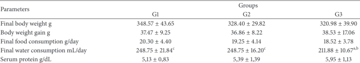
Table 1: General characteristics and serum protein levels ater 30 days for all experimental groups.
Parameters Groups
G1 G2 G3
Final body weight g 348.57±43.65 328.40±29.82 320.98±39.90
Body weight gain g 37.47±9.25 36.86±8.22 38.53±17.06
Final food consumption g/day 20.30±4.40 19.25±4.14 18.52±3.78
Final water consumption mL/day 248.75±21.84c 248.75±16.20c 211.88±10.67a,b
Serum protein g/dL 5,13±0,83 5,39±1,39 5,95±1,13
Values are given as the mean±SD for each group of eight animals.aSigniicantly diferent from G1;� ≤ 0.05;bsigniicantly diferent from G2;� ≤ 0.05; and
csigniicantly diferent from G3;� ≤ 0.05. G1: untreated control; G2: treated with geraniol; and G3: treated with essential oil.
was estimated using the biuret reagent and the total choles-terol concentration was determined using the cholescholes-terol esterase/oxidase enzymatic procedure. Triacylglycerols levels were measured by enzymatic hydrolysis and the inal forma-tion of quinoneimine, which is proporforma-tional to the concentra-tion of triacylglycerols present in the sample. Serum urea was determined by addition of urease and phenol-hypochloride, which leads to the formation of an indophenol-blue complex. he serum creatinine levels were estimated using a reaction with picric acid in alkaline bufer to form a yellow-orange complex, whose color intensity is proportional to the crea-tinine concentration in the sample. ALT and AST activities were determined by using pyruvate and oxaloacetate as sub-strates, wherein NADH is converted into NAD+ proportional to the activities of these enzymes. Hepatic samples (200 mg) were removed and homogenized in 0.1 M phosphate bufer, pH 7.4, using a Telon-glass Potter-Elvehjem homogenizer. he homogenate was centrifuged (10,000 g for 15 minutes) and the supernatant was used to determine the concentration of hepatic lipid hydroperoxide (LH) and activities of antiox-idant enzymes. Lipid hydroperoxide activity was determined by the oxidation of Fe+2 in the presence of a reactive mixture containing methanol, xylenol orange, sulfuric acid, and butylated hydroxytoluene. Catalase activity was assayed using phosphate bufer containing hydrogen peroxide. he activity of glutathione peroxidase (GSH-Px) was determined in the presence of phosphate bufer, NADPH2, reduced glu-tathione, and glutathione reductase. Superoxide dismutase (SOD) activity was assayed according to the method by measuring the rate of reduction of nitroblue-tetrazole (NBT) in the presence of free radicals generated by hydroxylamine.
2.4. Statistical Analysis. Results are expressed as the mean
± SD. he statistical signiicance between the groups was assessed using one-way analysis of variance (ANOVA) with Tukey’s test to compare the means of the experimental group. he probability with� ≤ 0.05was considered signiicant.
3. Results
Inhalation of geraniol (G2) and ofC. martiniiEO (G3) had no efects on inal body weight, body weight gain, and food intake of the rats (Table 1). No alteration in total hepatic protein was observed. While no signiicant diferences were observed in the glycemia and triacylglycerol levels, geraniol
0 20 40 60 80 100 120 140 160 180
(m
g/dL)
b, c
a a
Glucose Total cholesterol Triglycerides
G1 G2
G3
Figure 1: Serum glucose, total cholesterol, and triglycerides levels ater 30 days for all experimental groups. Values are given as the mean±SD for each group of eight animals.aSigniicantly diferent
from G1;� ≤ 0.05;bsigniicantly diferent from G2;� ≤ 0.05; and
csigniicantly diferent from G3;� ≤ 0.05. G1: untreated control; G2:
treated with geraniol; and G3: treated with essential oil.
(G2) andC. martinii EO (G3) decreased (� ≤ 0.05) total cholesterol levels when compared with the control group G1 (Figure 1). here were no signiicant diferences in serum urea levels between the groups. Creatinine levels increased in the presence of geraniol but decreased in the presence ofC. martiniiEO (G3;Figure 2).
ALT activity was higher in the group exposed to geraniol when compared to the other groups, which did not difer from each other. No change was found in the AST activity between the groups (Figure 3). LH was higher in the G2 group than in the G3 group (Figure 4). Catalase and SOD activities were higher in the G3 group when compared to the other groups. Both geraniol andC. martiniiEO increased GSH-Px when compared to the control rats (Figure 5).
4. Discussion
0 2 4 6 8 10 12
Urea Creatinine
c b
(m
g/dL)
G1 G2
G3
Figure 2: Serum urea and creatinine levels ater 30 days for all experimental groups. Values are given as the mean±SD for groups of eight animals each.aSigniicantly diferent from G1;� ≤ 0.05; bsigniicantly diferent from G2;� ≤ 0.05;csigniicantly diferent
from G3;� ≤ 0.05. G1: untreated control; G2: treated with geraniol; G3: treated with essential oil.
0 20 40 60 80 100 120 140
(U/L)
ALT AST
b b
a, c
G1 G2
G3
Figure 3: Serum activity of ALT and AST ater 30 days for all experimental groups. Values are given as the mean±SD for each group of eight animals.aSigniicantly diferent from G1;� ≤ 0.05;
bsigniicantly diferent from G2; � ≤ 0.05; and csigniicantly
diferent from G3;� ≤ 0.05. G1: untreated control; G2: treated with geraniol; and G3: treated with essential oil.
EOs on an organism when administered by inhalation. Since EOs have been extensively used in aromatherapy due to their therapeutic properties and also used in food products, in dermatology, and in the fragrance and cosmetic industries [14], there is interest in investigating their hepatic toxicity, as well as dyslipidemia.
C. martiniiEO reduced the inal water intake of the rats, but without altering other parameters that we studied. No signiicant changes were observed in inal body weight, body weight gain, inal food intake, or total serum protein level, indicating no dehydration and no deiciencies of nutritionally
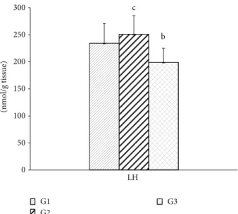
0 50 100 150 200 250 300
(nmo
l/g tissue)
LH c
b
G1 G2
G3
Figure 4: Hepatic lipid hydroperoxide levels ater 30 days for all experimental groups. Values are given as the mean±SD for each group of eight animals.aSigniicantly diferent from G1;� ≤
0.05;bsigniicantly diferent from G2;� ≤ 0.05; andcsigniicantly diferent from G3;� ≤ 0.05. G1: untreated control; G2: treated with geraniol; and G3: treated with essential oil.
0 10 20 30 40 50 60 70
SOD
(nmol/mg tissue) (nmol/mg tissue)
GSH-Px b
a, c
b b
a, c
b
b, c a
a
Catalase (�g/tissue)
G1 G2
G3
Figure 5: Hepatic activities of catalase, SOD, and GSH-Px ater 30 days for all experimental groups. Values are given as the mean±SD for each group of eight animals.aSigniicantly diferent from G1;� ≤
0.05;bsigniicantly diferent from G2;� ≤ 0.05; andcsigniicantly diferent from G3;� ≤ 0.05. G1: untreated control; G2: treated with geraniol; and G3: treated with essential oil.
demonstrated that C. martinii extracts exhibit antihyper-glycemic activities through inhibition of�-glucosidase under diabetic conditions [30]. Assays with C. citratus aqueous extract (500 mg/kg/day; via oral) showed that the mecha-nism by which the extract induced hypoglycemia could be attributed to increased insulin synthesis and secretion or increased peripheral glucose utilization [31].
he inhalation of both geraniol (G2) andC. martiniiEO (G3) reduced total cholesterol, but no changes in the serum triacylglycerol concentration were observed. Our results are in agreement with the result of Adeneye and Agbaje [31] and Burke et al. [10], who observed hypocholesterolemic efects using an aqueous extract of Cymbopogon citratus by oral administration. his reduction caused by EO can possibly be attributed to inhibition of 3-hydroxy-3-methylglutaryl CoA reductase, a key enzyme that regulates hepatic cholesterol synthesis [32,33], or by reduction in the expression of these enzymes [34]. Yu et al. [35] demonstrated that geraniol inhib-ited the formation of mevalonate, a metabolic intermediate in the biosynthesis of cholesterol, in hepatomas. On the other hand, the administration of the highest EO dose (100 mg/kg) from Cymbopogon resulted in no change in total serum cholesterol [36].
Our results are also in agreement with Costa et al. [36] about total serum cholesterol; they reported that the biochemical parameters did not change ater treatment with
Cymbopogonbut showed that there was a signiicant reduc-tion in it (F(4,27) = 3.061;� ≤ 0.05) ater the administration of the highest EO dose (100 mg/kg) by gavage over a period of 21 days.
Since urea is formed in the liver and excreted by kidneys, estimation of this nitrogenous compound in the bloodstream is important to estimate both hepatic and renal functions. Animals treated with geraniol and EO did not show altered serum urea levels, suggesting a normal degree of protein catabolism, which was conirmed by a normal concentration of hepatic protein. Although there was no alteration in the levels of serum urea in the G2 group, we cannot exclude the involvement of possible changes in the glomerular iltration rate in these animals. In clinical practice, serum creatinine, a biomarker for renal failure, is used as an indicator of renal function [37]. Curiously, serum creatinine levels were higher in the G2 group and lower in the G3 group, suggesting a decrease in renal excretion and some degree of renal insuiciency or early stages of kidney dysfunction when the major compound ofC. martiniiEO was administrated alone. he animals treated with both products had a tendency to have decreased levels of serum urea in our study. Serum creatinine was higher in G2, which may indicate lower renal excretion since the creatinine production was relatively constant [38].
Plasma membrane damage from some cells types, such as hepatic cells, is accompanied by release of cytosolic enzymes into bloodstream, a phenomenon that always occurs under several pathophysiological conditions [39]. he aminotrans-ferases ALT and AST are used for diagnosis of hepatic injury ater toxic agents exposure [40]. here was a signiicant increase in serum ALT activity in animals that inhaled geraniol (Figure 3). Since serum enzymatic activity of ALT is
oten used as a biomarker of hepatic toxicity, we can assume that there was some injury in hepatic tissue induced by geraniol ater 30 days.
ROS, such as superoxide anion (O2−), hydroxyl rad-icals (OH−), and hydrogen peroxide (H2O2), are formed through mitochondrial respiration during normal cellular metabolism. However, the cells have an enzymatic antioxi-dant defense system against ROS, but, under certain patho-logical conditions the excess formation of ROS results in suppression of antioxidant enzymes, the increase of ROS can occur in this way leading to oxidative stress [41].
Lipid peroxidation is an important toxic event because involves the removal of hydrogen from fatty acid chains mediated by ROS [42,43] this way can lead to cell death and tissue damage.
he endogenous antioxidant enzyme includes superox-ide dismutase that catalyzes the dismutation of superoxsuperox-ide radicals [23]. Glutathione peroxidase catalyzes the reduction of hydrogen peroxidase to water through the oxidation of reduced glutathione. Catalase also participates in this conversion [44].
Signiicantly high LH was observed in rats exposed to geraniol (G2), while the beneicial efect ofC. martiniiEO was evidenced by the reduced LH in these animals. he reductions observed in G3 can be attributed to a synergis-tic mechanism: a concomitant antioxidant action between other compounds, for example, linalool and�-caryophyllene, present in theC. martiniiEO that showed antioxidant activity in other researches [45, 46]. Since free radical scavenger ability depends on the number of hydroxyl radicals in the molecule [43], the inhalation of the total C. martinii EO contributed to the reduction in the formation of ROS.
Rats exposed to geraniol (G2) had higher catalase, SOD, and GSH-Px activities, indicating that antioxidant enzyme activities were not suicient to inhibit the ROS action and, consequently, the lipoperoxide generation in liver of these animals.
According to Koek et al. [47] the activity of antioxidants enzymes is increased early in nonalcoholic steatohepatitis but tends to decrease with progression of pathogenesis. he activity of the SOD and catalase did not change in the G3 group, while GSH-Px increased in these animals, which showed lower values for LH. Buch et al. [4] observed increases in both SOD and catalase in the brains of rats treated with
C. martiniEO. Terpenoids, which are important components of EOs, lowered malondialdehyde levels and improved SOD activity in gastric mucosa [48]. Experimental data have shown that terpenoids, which are main components of EOs, are responsible for their antioxidant action [49,50].
Since lipid hydroperoxide has been widely studied as marker of lipoperoxidation [51], a process that involves removal of hydrogen from fatty acids side chains by ROS, the result is referring to the mixture of compounds present in C. martinii EO (G3) that was efective in controlling oxidative stress and, therefore, lipoperoxidation by reducing the concentration of LH through a mechanism independent of the endogenous antioxidant enzymatic system.
antioxidant potential in inlammatory lung diseases, in which oxidative stress plays key role in these pathogenesis [14]. Moreover, the possible synergism between the compounds present in EOs can inluence biological responses [52]. his can explain the results obtained for the G3 group. hus, the efects we observed could be attributed to a constituent in a smaller proportion or synergism between compounds that are present in the oil [53]. For biological purposes, it is more informative to study the whole oil than some of its components because the concept of synergism appears to be more signiicant in the research on natural products [2].
5. Conclusion
In conclusion, C. martinii EO and geraniol maintained the glycemia, triacylglycerol protein, and urea levels but decreased cholesterol levels in Wistar rats. he oxidative stress caused by geraniol alone appears to trigger, to some degree, hepatic toxicity, as can be veriied by the increase of serum creatinine and ALT. hese results suggest that beneicial actions ofC. martiniiEO on oxidative stress can prevent the toxicity in liver. his proves the possible interac-tions between geraniol and numerous chemical compounds present inC. martiniiEO.
Abbreviations
C. martinii: Cymbopogon martinii
EO: Essential oil EOs: Essential oils
ALT: Alanine aminotransferase AST: Aspartate aminotransferase ROS: Reactive oxygen species LH: Lipid hydroperoxide GSH-Px: Glutathione peroxidase SOD: Superoxide dismutase NBT: Nitroblue-tetrazole.
Conflict of Interests
he authors declare that there is no conlict of interests regarding the publication of this paper.
References
[1] P. Sharma, N. Sangwan, S. Bose, and R. Sangwan, “Biochemical characteristics of a novel vegetative tissue geraniol acetyltrans-ferase from a monoterpene oil grass (Palmarosa,Cymbopogon martiniivar. Motia) leaf,”Plant Science, vol. 203, pp. 63–73, 2013. [2] F. Bakkali, S. Averbeck, D. Averbeck, and M. Idaomar, “Bio-logical efects of essential oils—a review,”Food and Chemical Toxicology, vol. 46, no. 2, pp. 446–475, 2008.
[3] M. Arruda, H. Viana, N. Rainha et al., “Anti-acetylcholin-esterase and antioxidant activity of essential oils from Hedy-chium gardnerianumsheppard ex ker-gawl,”Molecules, vol. 17, no. 3, pp. 3082–3092, 2012.
[4] P. Buch, V. Patel, V. Ranpariya, N. Sheth, and S. Parmar, “Neu-roprotective activity ofCymbopogon martiniiagainst cerebral
ischemia/reperfusion-induced oxidative stress in rats,”Journal of Ethnopharmacology, vol. 142, no. 1, pp. 35–40, 2012. [5] S. Sinha, D. Biswas, and A. Mukherjee, “Antigenotoxic and
antioxidant activities of palmarosa and citronella essential oils,” Journal of Ethnopharmacology, vol. 137, no. 3, pp. 1521–1527, 2011. [6] M. H. Lodhia, K. R. Bhatt, and V. S. haker, “Antibacterial activ-ity of essential oils from palmarosa, evening primrose, lavender and tuberose,”Indian Journal of Pharmaceutical Sciences, vol. 71, no. 2, pp. 134–136, 2009.
[7] A. Prashara, P. Hili, R. G. Veness, and C. S. Evans, “Antimi-crobial action of palmarosa oil (Cymbopogon martinii) on Saccharomyces cerevisiae,”Phytochemistry, vol. 63, no. 5, pp. 569–575, 2003.
[8] M. C. Duarte, E. E. Leme, C. Delarmelina, A. A. Soares, G. M. Figueira, and A. Sartoratto, “Activity of essential oils from Brazilian medicinal plants onEscherichia coli,”Journal of Ethnopharmacology, vol. 111, no. 2, pp. 197–201, 2007.
[9] J. M. Mat´es, J. A. Segura, F. J. Alonso, and J. M´arquez, “Natural antioxidants: therapeutic prospects for cancer and neurological diseases,”Mini-Reviews in Medicinal Chemistry, vol. 9, no. 10, pp. 1202–1214, 2009.
[10] Y. Burke, M. Stark, S. L. Roach, S. E. Sen, and P. L. Crowell, “Inhi-bition of pancreatic cancer growth by the dietary isoprenoids farnesol and geraniol,”Lipids, vol. 32, no. 2, pp. 151–156, 1997. [11] S. Carnesecchi, Y. Schneider, J. Ceraline et al., “Geraniol, a
com-ponent of plant essential oils, inhibits growth and polyamine biosynthesis in human colon cancer cells,” he Journal of Pharmacology and Experimental herapeutics, vol. 298, no. 1, pp. 197–200, 2001.
[12] W. Chen and A. M. Viljoen, “Geraniol—a review of a commer-cially important fragrance material,”South African Journal of Botany, vol. 76, no. 4, pp. 643–651, 2010.
[13] F. Sol´orzano-Santos and M. G. Miranda-Novales, “Essential oils from aromatic herbs as antimicrobial agents,”Current Opinion in Biotechnology, vol. 23, no. 2, pp. 136–141, 2012.
[14] M. Tiwari and P. Kakkar, “Plant derived antioxidants—geraniol and camphene protect rat alveolar macrophages against t-BHP induced oxidative stress,”Toxicology in Vitro, vol. 23, no. 2, pp. 295–301, 2009.
[15] D. Jimbo, Y. Kimura, M. Taniguchi, M. Inoue, and K. Urakami, “Efect of aromatherapy on patients with Alzheimer’s disease,” Psychogeriatrics, vol. 9, no. 4, pp. 173–179, 2009.
[16] G. Buchbauer, L. Jirovetz, W. J¨ager, H. Dietrich, and C. Plank, “Aromatherapy: evidence for sedative efects of the essential oil of lavender ater inhalation,”Zeitschrit f¨ur Naturforschung C, vol. 46, no. 11-12, pp. 1067–1072, 1991.
[17] G. Bagetta, L. A. Morrone, L. Rombol`a et al., “Neuropharma-cology of the essential oil of bergamot,”Fitoterapia, vol. 81, no. 6, pp. 453–461, 2010.
[18] M. Tanida, A. Niijima, J. Shen, T. Nakamura, and K. Nagai, “Olfactory stimulation with scent of essential oil of grape-fruit afects autonomic neurotransmission and blood pressure,” Brain Research, vol. 1058, no. 1-2, pp. 44–55, 2005.
[19] M. E. Mafei, J. Gertsch, and G. Appendino, “Plant volatiles: pro-duction, function and pharmacology,”Natural Product Reports, vol. 28, no. 8, pp. 1359–1380, 2011.
[21] S. Inouye, T. Takizawa, and H. Yamaguchi, “Antibacterial activity of essential oils and their major constituents against respiratory tract pathogens by gaseous contact,” Journal of Antimicrobial Chemotherapy, vol. 47, no. 5, pp. 565–573, 2001. [22] C. A. Burtis, E. R. Ashwood, and D. E. Bruns,Tietz:
Fundamen-tos de Qu´ımica Cl´ınica, Elsevier Editora Ltda, 6th edition, 2008. [23] J. S. Johansen, A. K. Harris, D. J. Rychly, and A. Ergul, “Oxidative stress and the use of antioxidants in diabetes: linking basic science to clinical practice,”Cardiovascular Diabetology, vol. 4, article 5, 2005.
[24] J. M. Mat´es, J. A. Segura, F. J. Alonso, and J. M´arquez, “Intracellular redox status and oxidative stress: implications for cell proliferation, apoptosis, and carcinogenesis,”Archives of Toxicology, vol. 82, no. 5, pp. 273–299, 2008.
[25] A. Wieckowska, A. J. McCullough, and A. E. Feldstein, “Nonin-vasive diagnosis and monitoring of nonalcoholic steatohepati-tis: present and future,”Hepatology, vol. 46, no. 2, pp. 582–589, 2007.
[26] P. Muriel, “Role of free radicals in liver diseases,”Hepatology International, vol. 3, no. 4, pp. 526–536, 2009.
[27] R. N. de Almeida, S. C. Motta, C. D. B. Faturi, B. Catallani, and J. R. Leite, “Anxiolytic-like efects of rose oil inhalation on the elevated plus-maze test in rats,”Pharmacology Biochemistry and Behavior, vol. 77, no. 2, pp. 361–364, 2004.
[28] A. Brenes and E. Roura, “Essential oils in poultry nutrition: main efects and modes of action,”Animal Feed Science and Technology, vol. 158, no. 1-2, pp. 1–14, 2010.
[29] S. Asnaashari, A. Delazar, B. Habibi et al., “Essential oil from Citrus aurantifoliaprevents ketotifen-induced weight-gain in mice,”Phytotherapy Research, vol. 24, no. 12, pp. 1893–1897, 2010. [30] V. Ghadyale, S. Takalikar, V. Haldavnekar, and A. Arvindekar, “Efective control of postprandial glucose level through inhi-bition of intestinal alpha glucosidase by Cymbopogon mar-tinii(Roxb.),”Evidence-Based Complementary and Alternative Medicine, vol. 2012, Article ID 372909, 6 pages, 2012.
[31] A. A. Adeneye and E. O. Agbaje, “Hypoglycemic and hypolipi-demic efects of fresh leaf aqueous extract of Cymbopogon citratusStapf. in rats,”Journal of Ethnopharmacology, vol. 112, no. 3, pp. 440–444, 2007.
[32] P. L. Crowell, “Prevention and therapy of cancer by dietary monoterpenes,”Journal of Nutrition, vol. 129, no. 3, pp. 775S– 778S, 1999.
[33] P. Lu, M. L. Schrag, D. E. Slaughter, C. E. Raab, M. Shou, and A. D. Rodrigues, “Mechanism-based inhibition of human liver microsomal cytochrome P450 1A2 by zileuton, A 5-lipoxygenase inhibitor,”Drug Metabolism and Disposition, vol. 31, no. 11, pp. 1352–1360, 2003.
[34] S.-Y. Cho, H.-J. Jun, J. H. Lee, Y. Jia, K. H. Kim, and S.-J. Lee, “Linalool reduces the expression of 3-hydroxy-3-methylglutaryl CoA reductase via sterol regulatory element binding protein-2-and ubiquitin-dependent mechanisms,”FEBS Letters, vol. 585, no. 20, pp. 3289–3296, 2011.
[35] S. G. Yu, L. A. Hildebrandt, and C. E. Elson, “Geraniol, an inhibitor of mevalonate biosynthesis, suppresses the growth of hepatomas and melanomas transplanted to rats and mice,” Journal of Nutrition, vol. 125, no. 11, pp. 2763–2767, 1995. [36] C. Costa, L. T. Bidinotto, R. K. Takahira, D. M. F. Salvadori,
L. F. Barbisan, and M. Costa, “Cholesterol reduction and lack of genotoxic or toxic efects in mice ater repeated 21-day oral intake of lemongrass (Cymbopogon citratus) essential oil,”Food and Chemical Toxicology, vol. 49, no. 9, pp. 2268–2272, 2011.
[37] R. D. Perrone, N. E. Madias, and A. S. Levey, “Serum creatinine as an index of renal function: new insights into old concepts,” Clinical Chemistry, vol. 38, no. 10, pp. 1933–1953, 1992. [38] S. B. Heymsield, C. Arteaga, C. M. McManus, J. Smith, and S.
Moitt, “Measurement of muscle mass in humans: validity of the 24-hour urinary creatinine method,”he American Journal of Clinical Nutrition, vol. 37, no. 3, pp. 478–494, 1983.
[39] R.-Z. Yang, S. Park, W. J. Reagan et al., “Alanine aminotrans-ferase isoenzymes: molecular cloning and quantitative analysis of tissue expression in rats and serum elevation in liver toxicity,” Hepatology, vol. 49, no. 2, pp. 598–607, 2009.
[40] J. Ozer, M. Ratner, M. Shaw, W. Bailey, and S. Schomaker, “he current state of serum biomarkers of hepatotoxicity,”Toxicology, vol. 245, no. 3, pp. 194–205, 2008.
[41] B. Halliwell, “Antioxidants in human health and disease,” Annual Review of Nutrition, vol. 16, pp. 33–50, 1996.
[42] P. M. Abuja and R. Albertini, “Methods for monitoring oxidative stress, lipid peroxidation and oxidation resistance of lipoproteins,”Clinica Chimica Acta, vol. 306, no. 1-2, pp. 1–17, 2001.
[43] L. A. Faine, H. G. Rodrigues, C. M. Galhardi et al., “Efects of olive oil and its minor constituents on serum lipids, oxidative stress, and energy metabolism in cardiac muscle,” Canadian Journal of Physiology and Pharmacology, vol. 84, no. 2, pp. 239– 245, 2006.
[44] J.-C. Preiser, “Oxidative stress,”Journal of Parenteral and Enteral Nutrition, vol. 36, no. 2, pp. 147–154, 2012.
[45] S. Jana, K. Patra, S. Sarkar et al., “Antitumorigenic potential of linalool is accompanied by modulation of oxidative stress: an in vivo study in sarcoma-180 solid tumor model,”Nutrition and Cancer, vol. 66, pp. 835–848, 2014.
[46] M. A. Calleja, J. M. Vieites, T. Montero-Melendez et al., “he antioxidant efect of beta-caryophyllene protects rat liver from carbon tetrachloride-induced ibrosis by inhibiting hepatic stellate cell activation,”British Journal of Nutrition, vol. 109, pp. 394–401, 2013.
[47] G. H. Koek, P. R. Liedorp, and A. Bast, “he role of oxidative stress in non-alcoholic steatohepatitis,”Clinica Chimica Acta, vol. 412, no. 15-16, pp. 1297–1305, 2011.
[48] N. Rocha, G. de Oliveira, F. Y. de Ara´ujo et al., “(-)-� -Bisabolol-induced gastroprotection is associated with reduction in lipid peroxidation, superoxide dismutase activity and neutrophil migration,”European Journal of Pharmaceutical Sciences, vol. 44, no. 4, pp. 455–461, 2011.
[49] H. Fadel, F. Marx, A. El-Sawy, and A. El-Ghorab, “Efect of extraction techniques on the chemical composition and antiox-idant activity of Eucalyptus camaldulensis var. brevirostris leaf oils,”Zeitschrit f¨ur Lebensmitteluntersuchung und -Forschung A, vol. 208, no. 3, pp. 212–216, 1999.
[50] J. Grassmann and G. Litwack, “Terpenoids as plant antioxi-dants,”Plant Hormones, vol. 72, pp. 505–535, 2005.
[51] K. Nageswari, R. Banerjee, and V. P. Menon, “Efect of saturated,
�-3 and�-6 polyunsaturated fatty acids on myocardial infarc-tion,”he Journal of Nutritional Biochemistry, vol. 10, no. 6, pp. 338–344, 1999.
[52] J. Gershenzon and N. Dudareva, “he function of terpene natural products in the natural world,”Nature Chemical Biology, vol. 3, no. 7, pp. 408–414, 2007.
Submit your manuscripts at
http://www.hindawi.com
Hindawi Publishing Corporation
http://www.hindawi.com Volume 2014
Anatomy
Research International
Peptides
Hindawi Publishing Corporation
http://www.hindawi.com Volume 2014
Hindawi Publishing Corporation http://www.hindawi.com
International Journal of
Volume 2014
Zoology
Hindawi Publishing Corporation
http://www.hindawi.com Volume 2014
Molecular Biology International
Genomics
International Journal of
Hindawi Publishing Corporation
http://www.hindawi.com Volume 2014
The Scientiic
World Journal
Hindawi Publishing Corporationhttp://www.hindawi.com Volume 2014
Hindawi Publishing Corporation
http://www.hindawi.com Volume 2014
Bioinformatics
Advances inMarine Biology
Journal ofHindawi Publishing Corporation
http://www.hindawi.com Volume 2014
Hindawi Publishing Corporation
http://www.hindawi.com Volume 2014
Signal Transduction
Journal ofHindawi Publishing Corporation
http://www.hindawi.com Volume 2014
BioMed
Research International
Evolutionary Biology International Journal of
Hindawi Publishing Corporation
http://www.hindawi.com Volume 2014
Hindawi Publishing Corporation
http://www.hindawi.com Volume 2014 Biochemistry Research International
Archaea
Hindawi Publishing Corporationhttp://www.hindawi.com Volume 2014
Hindawi Publishing Corporation
http://www.hindawi.com Volume 2014 Genetics
Research International
Hindawi Publishing Corporation
http://www.hindawi.com Volume 2014
Advances in
Virology
Hindawi Publishing Corporation http://www.hindawi.com
Nucleic Acids
Journal ofVolume 2014
Stem Cells
International
Hindawi Publishing Corporation
http://www.hindawi.com Volume 2014
Hindawi Publishing Corporation
http://www.hindawi.com Volume 2014
Enzyme
Research
Hindawi Publishing Corporation
http://www.hindawi.com Volume 2014