Original Article
INFLUENCE OF AEROBIC EXERCISE TRAINING ON SERUM MARKERS OF OXIDATIVE STRESS IN DIABETIC RATS
INFLUÊNCIA DO TREINAMENTO FÍSICO AERÓBIO NOS MARCADORES SÉRICOS DE ESTRESSE OXIDATIVO EM RATOS DIABÉTICOS
Aron da Silva Pereira1, Alexandre Roveratti Spagnol1, Eliete Luciano1 e José Alexandre Curiacos de Almeida Leme1
1
Universidade Estadual Paulista, Rio Claro-SP, Brasil.
RESUMO
Para verificar o efeito do exercício físico sobre biomarcadores de estresse oxidativo no sangue de ratos diabéticos foram utilizados 40 ratos Wistar distribuídos nos grupos controle, treinado, diabéticos e diabéticos treinado. O treinamento físico consistiu de natação 1 hora/dia, 5 dias/semana por 8 semanas. O diabetes foi induzido utilizando aloxana (32mg/kg). Foram analisados os parâmetros séricos: glicose, colesterol, triglicerídeos, proteína c reativa, catalase, superóxidodismutase (SOD) e malondialdeído (MDA). O diabetes aumentou as concentrações séricas de glicose e TBARs, porém diminuiu as atividades da SOD e catalase. O treinamento físico reduziu os níveis séricos de glicemia, colesterol e aumentou e catalase nos animais diabéticos. A atividade da SOD nos animais diabéticos foi menor que nos controles, sendo este parâmetro recuperado pelo treinamento físico. Não houve diferenças significativas nos demais parámetros estudados. Pode ser concluído que o treinamento físico foi eficaz em amenizar a hiperglicemia e o estresse oxidativo emanimais diabéticos.
Palavras-chave: Exercício. Diabetes. Estresse oxidativo. Sangue. Ratos.
ABSTRACT
To verify the effects of exercise on blood biomarkers of oxidative stress in diabetic rats, 40 rats Wistar, were distributed into four groups: control, diabetes, trained control and trained diabetes. Training was constituted of swimming 1 hour/day, 5 days/week for 8 weeks with intensity equivalents to minimum lactate. Diabetes was induced with intravenous alloxan (32 mg/kg). It was evaluated the following blood parameters: catalase, superoxide dismutase (SOD), Malondialdehyde (MDA), glucose, cholesterol, triglycerides and C-reactive-protein. Diabetes induced hyperglycemia, reduction in body mass, catalase and SOD activities and increased TBARs concentrations. Physical training, on the other hand, reduced blood glucose and cholesterol, increased body mass and blood catalase. SOD activity was lower in diabetic rats than control rats and physical training was able to recovery this parameter. The other parameters showed no differences. In conclusion, physical exercise protocol contributes in controlling blood glucose, and ameliorates oxidative stress by increasing catalase levels and recovery SOD activity.
Keywords: Exercise. Diabetes. Oxidative stress. Blood. Rats.
Introduction
Diabetes mellitus (DM) is chronic degenerative disease with the highest prevalence, and it is estimate that around 415 million people suffering of diabetes in the world1. The estimation for the year 2040 is that the disease reaches 642 million people1.
The metabolic disorders caused by diabetes are diverse as dyslipidemias, subclinical inflammation, increased cardiac risk, in addition to loss of function that may evolve to failure of several organs, caused by the lack of production or the action of insulin and high concentration of glucose in the blood2. Among the consequences of hyperglycemia is oxidative stress, defined as an unbalance between antioxidants and pro-oxidant.
acid and glucose. Its action is selective and destructive on the pancreatic β cells leading to degeneration and definitive death6. After the diabetes induction, the animals begin to show classic signs of diabetes as polydipsia, polyphagia, weight loss and elevated glycaemia, which, if not treated, weakens the animal by high concentrations of glucose in the blood and its complications as the formation of stress oxidativo5,7.
The acute and chronic physical exercise may facilitate the uptake of glucose, the reduction of gluconeogenesis and body fat mass, which promote the production of reactive oxygen species (EROS) 8,10.
The physical exercise stimulates the production of reactive oxygen species (EROS), but in fact, these high levels stimulate the increase of expression and activity of antioxidant enzymes11.
Some of endogenous antioxidant enzymes are the glutathione peroxidase (GPX) (GPx; dependent or not of selenium), catalase (CAT-hemi-enzyme), superoxide dismutase (SOD) (CuZn- Cytosolic and extracellular SOD and Mn-SOD-mitochondrial). Their function is to decompose superoxide, hydrogen peroxide and hydroxides radicals and to control the activity of lipid peroxidation, through the levels of species reagents to thiobarbituric acid (TBARS) 12.
Evaluating the markers of oxidative stress in blood can be interesting, because according to Aguilar-da-Silva and collaborators13, red blood corpuscles are good indicators of oxidative stress, because they are in contact with numerous molecules and organic structures and they act as carriers of gases, being very susceptible to damage by oxygen reactive species and, as a consequence, their functionality will be impaired.
Despite the knowledge about the damage caused by oxidative stress in diabetic body and the beneficial effects of moderate physical training to alleviate this process, few studies have evaluated the effects of physical training on parameters related to oxidative stress in diabetic animals. In this way, the present study aimed to investigate the effects of physical training on markers of oxidative stress in alloxan diabetic rats.
Materials and methods
Animals
It was used 40 wistar rats with 45 days of age at the beginning of the experiment, selected from the Universidade Estadual Paulista (UNESP), Campus of Botucatu. The animals were kept in cages of polyethylene measuring 37 x 31 x 16 (five rats per cage), they were kept at ambient temperature of 21 C and photoperiod of 12 hours light/dark, they were fed with Purina standard balanced ration and water ad libitum at the animal facility of Biodynamic laboratory of Physical Education Department (Institute of Biosciences) - UNESP - Rio Claro. The experimental period occurred between the months of April and September 2013.
All experimental procedures are in accordance with the standards of the Brazilian College of Animal Experimentation (COBEA) and they were submitted and approved by the Committee of Ethics in Animal Experimentation (CEUA) of the Institute of Biosciences of UNESP - Campus of Rio Claro (N° 633/2012 protocol).
Experimental design
buffer 0.01M, pH 4.5. After the induction to DM by alloxan, the animals remained in the cages and receiving during the first day a solution of water and glucose (15%) and food. Seven days after the drug administration, blood glucose was tested (commercial kit- Laborlab® enzymatic colorimetric method of glucose oxidase-peroxidase) to verify the diabetic state of the animals.
It was considered diabetic only those animals which had blood glucose levels greater than or equal to 200 mg/dL. After these procedures, it was registered the animal body weight and the animals were randomly distributed into the following groups (n=10): sedentary control - SC; Trained Control - TC; sedentary Diabetic - SD; Trained diabetic - TD.
Training
The training protocol consisted of swimming for 1 hour/day, 5 days/week, with load corresponding to minimum lactate14,15 for the period of 8 weeks, preceded by aquatic environment and loads adaptation phase for a week.
Swimming sessions were held in tanks with 100 cm length, 70 cm width and 60 cm depth, containing water at a depth of 50 cm, to avoid the rats to suspend themselves in their tail on the bottom of the container. The water temperature was between 31° and 32° C, being considered thermally neutral in relation to the rat’s temperature 16.
Samples and analyzes
Forty-eight hours after the last training session, the rats were anesthetized with thiopental sodium (40 mg/kg of body weight via intraperitoneal injection) and blood samples were collected by cardiac puncture to determinate and assess the following biomarkers: Catalase - CAT17 and superoxide dismutase - SOD using laboratory kit (Cayman Chemical, Ann Arbor, MI, USA), as well as the reagent products for thiobarbituric acid18. For analyzes of serum glucose and cholesterol C reactive protein were used colorimetric enzymatic method using commercial kit (Laborlab®).
Statistical analysis
It was conducted the Shapiro-Wilk normality test, and one way ANOVA, followed by the complementary test of Tukey. The data are expressed as mean and standard deviation and the values were considered significant when P<0.05. For analyzes, we used the Statistica software 7.0® and the GraphPad Prism®.
Results
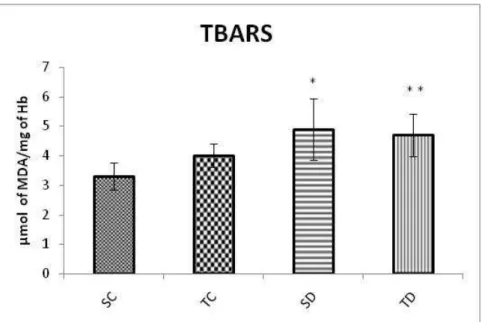
Figure 2 showed that diabetes promoted greater production of this substance and that the physical training did not change these values in these animals.
Table 1. Values of blood glucose (mg/dl), total cholesterol (mg/dl) and C-reactive protein (mg/dl) from animals of the groups sedentary control (SC), trained control (TC), sedentary diabetic (SD) and trained diabetic (TD) in the end of the experimental period.
Groups
Parameters
SC TC SD TD
Glycemia 166.7 ± 18.88 170.4 ± 14.92 446.2 ± 79.49 ab 279.4 ± 98.64abc
Total cholesterol
98.8 ± 9.7 82.0 ±: 13.09 95.5 ± 31.6 75.5 ±5.,21a
C- reactive protein
0.44 ± 0.18 0.43 ± 0.31 0.54 ± 0.44 0.39 ± 0.14
Values were expressed as mean ± standard deviation. a ≠ SC; b ≠ TC; c ≠ SD (ANOVA, Tukey post-hoc; p<0.05).
Figure 2. Concentrations of reagents to thiobarbituric acid (TBARS) in the blood of animals belonging to the groups: sedentary control (SC), trained control (TC), sedentary diabetic (SD) and trained diabetic (TD) to the end of the experimental period. Results were expressed as mean and standard deviation. Significant differences: (*) and (**) in relation to SC (P < 0.05, Post-hoc Tukey test).
Figure 3 shows serum values of catalase enzyme activity (SC: 3.3 ± 0.45; TC: 4.07 ± 0.4; SD: 4.9 ± 1.03; TD: 4.70 ± 0.73). These values were increased in diabetic animals (SD and TD), and there was no significant effect of physical training on this parameter.
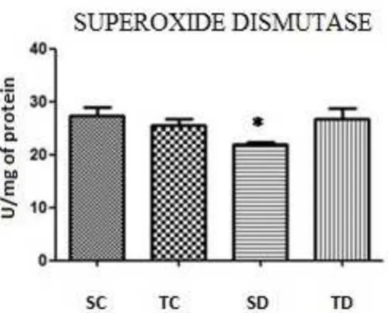
Figure 4. Activity of superoxide dismutase enzyme in animals belonging to the groups: sedentary control (SC), trained control (TC), sedentary diabetic (SD) and trained diabetic (TD) to the end of the experimental period. Results were expressed as mean and standard deviation. Significant differences: (*) in relation to the SC (P < 0.05, Post-hoc Tukey test).
Finally in the analysis of the activity of the enzyme superoxide dismutase (SC: 27.33 ± 4.19; TC: 21.95 ± 3.72; SD: 21.95 ± 0.90; TD: 26.76 ± 5.16). The animals of SD group had lower values than the SC group (figure 4). However, the physical training recovered these values in diabetic animals, not being observed a difference compared to SC.
Discussion
The present study explores the effects of physical training on oxidative stress of diabetic rats. Alloxan was used for induction of diabetes, taking as reference several studies that showed the mechanisms of injury in beta cells that culminate with the dual stage of hyperinsulinaemia in the first hours, follow by hypoinsulinaemia and hyperglycemia4-6. The diabetic hyperglycemia can causes an increase in the rate of cellular respiration, producing EROS; furthermore, sugars oxidized react with lipoprotein components and membrane receptors, stimulating the formation of superoxide anions and hydrogen peroxide due to permanent hyperglycemia that induces the self-glucose oxidation.19-21
Similarly, the aerobic physical training was based on previous results that determined the load used as predominantly aerobic for diabetic animals (4, 2% b.m.) and no diabetic (5.5% b.m.)14,15,22,23. The aerobic exercise facilitates the glucose uptake independently of insulin, by increasing the tyrosine-kinase activity of insulin receptors, increasing the translocation of the skeletal muscle glucose transporters (GLUT-4)8.
In the present study, there was an induction of hyperglycemia in diabetic animals, while the physical training was capable of reducing this hyperglycemia in the animals. The training contributes to potentiate other points of insulin signaling pathway, as increasing of IRS phosphorylation, PI3-kinase activity that culminate with increasing in the translocation of GLUT-4 adjacent to the membrane8,24,25.
Among the symptoms are included the dyslipidemias. According to Luciano and collaborators3 in an exercise protocol similar to groups used in this work (moderate to light), no differences in some lipid parameters were found between the groups, but when HDL was analyzed, it was found that the exercise is able to stimulate its increase.
The acute physical exercise can aggravate this mechanism, but the body adapts and produces antioxidant enzymes as catalase in response to the event, having a protective effect against the lipid peroxidation26.
The reasons for the choice of blood tissue are: it is be widely exposed to metabolites and gases, such as O2, and in this case to the high levels of glycaemia makes it more likely the EROS and because the red blood cells have small metabolism but physiologically significant, keeping the structure of the membrane and hemoglobin, in order to protect against oxidative damage13. In addition, the iron found in the free form or connected to a heme can convert the radical superoxide anion and hydrogen peroxide, by Fenton's reaction, in the hydroxyl radical, one of the most reactive species known12.
The cell membrane may suffer oxidative damage due peroxidation of residues of unsaturated fatty acids of phospholipids and a nonspecific test of products that react to thiobarbituric acid (TBARS) is widely used as an indicator of oxidative attack10,26,27.
In our study, serum concentration of TBARs was high in diabetic groups, which indicates that the diabetes increased peroxidation and the physical training was not efficient in mitigating this process. Corroborating with the present study, Kakkar and collaborators28, showed that the concentration of TBARs was higher in diabetic animals.
The effects of exercise are dependent on several factors such as duration, type and intensity of exercise. Prada and collaborators10, using an exercise protocol of swimming, observed an increase of lipid peroxidation, finding out the TBARS significantly high in the blood of trained animals. Coskun and collaborators29 showed that the exercise of swimming with moderate intensity was more effective at reducing levels of MDA in diabetic rats induced by streptozotocin, comparing to light and heavy exercise. Additionally the authors demonstrated that moderate intensity swimming was better in protecting the pancreatic β cells.
The serum concentration value of catalase enzyme showed that the trained groups had a higher enzyme expression. This data does not corroborate with the study of Prada and colleagues10 who claimed that there is a possibility of different responses due to the type, intensity and duration of exercise. However, Torres et al30 found catalase values is significantly higher in the diabetic group, agreeing with Kakkar and collaborators28 who reported an increase independently of the time of DM induction, after all according to the authors it would reduce during the course of the disease.
With regard to the activity of superoxide dismutase (SOD), the diabetic animals had a reduction in the values of this parameter. This data corroborates previous studies that present a possible glycation of this enzyme or action of S2O2 leading to inactivation of it in hyperglycemic animals31. In the present study, the moderate physical training has mitigated the activity reduction of this enzyme, demonstrating a beneficial adaptation of the physical training to diabetic organism.
Conclusion
The set of data collected in this study allow us to conclude that the aerobic physical exercise protocol contributes to glycemic control and partially alleviate the oxidative stress demonstrated through increased catalase levels in diabetic animals and recovery of SOD.
Acknowledgment: This work was supported by CNPq (131037/2013-7):
References
1. International Diabetes Federation. IDF Diabetes Atlas.Key Messages, 7.ed. Brussels, 2015. 2. Gross JL, Silveiro SP, Camargo Jl, Reichelt AJ, Azevedo MJ. Diabetes mellitus: diagnosis,
classification and evaluation of glycemic control. Arq Bras Endocrinol Metab 2010;46,(1):16- 26.
3. Luciano E. Atividade física e metabolismo lipídico em ratos Diabéticos experimentais. Rev Bras Ativ Fís Saúde 1996;1(4):19-26.
4. Lima MA, Lima LMB, Rita DPC, Navarro FC, Tatsukawa RS, Pereira GA, et al. Análise quantitativa das células das ilhotas pancreáticas em ratos sob efeito de aloxana. Med 2001;34(3/4):308-314.
5. Leme JACA, Castellar A, Remedio RN, Barbosa RA, Moura LP, Dalia RA et al., Efeitos em curto prazo da aplicação de aloxana para indução de diabetes em ratos Wistar. Biosci J 2010;26(3):451-456.
6. Lenzen S. The mechanisms of alloxan- and streptozotocin-induced Diabetes. Diabetologia 2008;51(2):216–226.
7. Halliwell B, Gutteridge JMC. Free Radicals in Biology and Medicine. 4. ed. Oxford: Oxford University Press; 2007.
8. Goodyear LJ, Kahn BB. Exercise, glucose transport, and insulin sensitivity. Annu Rev Med 1998;49(1):235-61.
9. Moura L, Bertolini N, Ghezzi AC, Bertucci D, Bonfim M, Serafim TH, et al. Glucose Homeostasis in Type 1 Diabetic Rats after Acute PhysicalActivity. JEP online
2011;14(6):8-19.
10. Prada FJA, Voltarelli FA, Oliveira CAM, Gobatto CA, Macedo DV, Mello MAR. Condicionamento aeróbio e estresse oxidativo em ratos treinados por natação em intensidade equivalente ao limiar anaeróbio. R Bras Cien e Mov 2004;12(2):29-34. 11. Voltarelli FA, Araujo MB, Yamada A, Carolina MSS, Mello MAR. Estresse Oxidativo e
ExercicioFisico: Efeitos Sobre o Musculo Esqueletico. RBNE 2011;4(20):05-12. 12. Halliwell B, Gutteridge JMC Free radicals in biology and medicine. 2. ed. Oxford:
Clarendon Press;1989.
13. Aguilar-Da-Silva, RH, Moraes, TP, Moraes, G. Implicações do estresse oxidativo sobre o metabolismo eritrocitário de pessoas com Síndrome de Down. Rev Bras Hematol
Hemoter 2003;25(4):231-237.
14. Gobatto CA,Sibuya CY, Azevedo JRM, Luciano E, KokubunE,MelloMAR.
Caracterização da intensidade de exercício e do efeito de treinamento físico no modelo de natação de ratos Wistar. Motri 2001;7(1):32- 36.
15. Araujo GG, Papoti M, Manchado FDE, Mello MAR, Gobatto CA. Protocols for
hyperlactatemia induction in the lactate minimum test adapted to swimming rats. Comp Biochem Physiol A Mol Integr Physiol 2007;148(4):888-892.
16. Azevedo JRM. Determinação de parâmetros bioquímicos em ratos sedentários e treinados após exercício agudo de natação. [Tese de Doutorado]. Campinas: Universidade Estadual de Campinas. Departamento de Fisiologia e Biofísica;1994.
18. Mataix J, Quiles JL, Huertas JR, Battino M, Manas M. Tissue specific interactions of exercise, dietary fatty acids, and vitamin E in lipid peroxidation. Free Rad Biol Med 1998;24(4):511-521.
19. Baynes J. Role of oxidative stress in development of complications of diabetes. Diabetes 1991;40(4):405-412.
20. Brownlee M. Biochemistry and molecular cell biology of diabetic complications. Nature 2001;414(6865):813-820.
21. Wajchenberg, BL. Disfunção endotelial no diabetes tipo 2. Arq Bras Endocrinol Metab 2002; 46(5):514-519.
22. Moura LP, Gomes RJ, Leme JA, Voltarelli FA, Ribeiro C, Moura RF, et al. Insulina pancreática de ratos diabéticos tipo 1 submetidos a um protocolo de treinamento físico individualizado. Motri 2012; 8(1):23-32.
23. Gomes RJ, Oliveira CA, Ribeiro C, Mota CS, Moura LP, Tognoni LM, et al. Effects of
exercise training on hippocampus concentrations of insulin and IGF-1 in diabetic rats. Hippocampus 2009;19(10):981-987.
24. Jessen N, Goodyear LJ. Contraction signaling to glucose transport in skeletal muscle. J Appl Physiol 2005;99(1):330-337.
25. Luciano E, Carneiro EM, Carvalho CR, Carvalheira JB, Peres SB, Reis MA, Saad MJ, Boschero AC, Velloso LA. Endurance training improves responsiveness to insulin and modulates insulins ignal transduction through the phosphatidylinositol 3-kinase/Akt-1 pathway. Eur J Endocrinol, v. 147, n. 1, p. 149-157, 2002.
26. Araújo MB. Estresse Oxidativo em ratos exercitados em diferentes intensidades. [Dissertação de Mestrado]. Rio Claro: Universidade Estadual Paulista. Instituto de Biociências; 2008.
27. Aguirre F, Martin I, Grinspon D, Ruiz M, Hager A, Paoli T, et al. Oxidative damage, plasma antioxidant capacity, and glucemic control in elderly. NIDDM patients. Free Radic Biol Med 1998;24(4):580-585.
28. Kakkar R, Mantha SV, Radhi J, Prasad K, Kalra J. Increased oxidative stress in rat liver and pancreas during progression of streptozotocin-induced diabetes. Clinical Science 1998;94(6):623-632.
29.Coskun O, Ocakci A, Bayraktaroglu T, Kanter M. Exercise Training Prevents and Protects Streptozotocin-Induced Oxidative Stress and β-Cell Damage in Rat Pancreas. Tohoku J Exp Med 2004;203(3):145-154.
30. Torres MD, Canal JR, Pérez C. Oxidative Stress in Normal and Diabetic Rats. Physiol Res 1999;48(1):203 -208.
31. Rajasekaran S, Sivagnanam K, Subramanian S. Antioxidant effect of Aloe vera gel extract in streptozotocin-induced diabetes in rats. Pharmacol Rep 2005;57(1):90-96.
Received on Jul, 11, 2015. Reviewed on Sep, 16, 2015. Accepted on Jan, 14, 2016.
Author address: Aron da Silva Pereira. Departamento de Educação Física, Instituto de Biociências, Universidade Estadual Paulista.