Granulocyte-Macrophage Colony Stimulatory
Factor Enhances the Pro-Inflammatory
Response of Interferon-
γ
-Treated
Macrophages to
Pseudomonas aeruginosa
Infection
Sonali Singh1, Helen Barr2, Yi-Chia Liu1, Adrian Robins1, Stephan Heeb1, Paul Williams1, Andrew Fogarty3, Miguel Cámara1
*, Luisa Martínez-Pomares1
*
1School of Life Sciences, University of Nottingham, Nottingham, NG7 2RD, United Kingdom,2School of Medicine, University of Nottingham, Nottingham, NG7 2RD, United Kingdom,3School of Community Health Sciences, University of Nottingham, Nottingham, NG7 2RD, United Kingdom
*luisa.martinez-pomares@nottingham.ac.uk(LMP);miguel.camara@nottingham.ac.uk(MC)
Abstract
Pseudomonas aeruginosais an opportunistic pathogen that can cause severe infections at compromised epithelial surfaces, such those found in burns, wounds, and in lungs dam-aged by mechanical ventilation or recurrent infections, particularly in cystic fibrosis (CF) pa-tients. CF patients have been proposed to have a Th2 and Th17-biased immune response suggesting that the lack of Th1 and/or over exuberant Th17 responses could contribute to the establishment of chronicP.aeruginosainfection and deterioration of lung function. Ac-cordingly, we have observed that interferon (IFN)-γproduction by peripheral blood mononu-clear cells from CF patients positively correlated with lung function, particularly in patients chronically infected withP.aeruginosa. In contrast, IL-17A levels tended to correlate nega-tively with lung function with this trend becoming significant in patients chronically infected withP.aeruginosa. These results are in agreement with IFN-γand IL-17A playing protective and detrimental roles, respectively, in CF. In order to explore the protective effect of IFN-γin CF, the effect of IFN-γalone or in combination with granulocyte-macrophage colony-stimu-lating factor (GM-CSF), on the ability of human macrophages to controlP.aeruginosa growth, resist the cytotoxicity induced by this bacterium or promote inflammation was inves-tigated. Treatment of macrophages with IFN-γ, in the presence and absence of GM-CSF, failed to alter bacterial growth or macrophage survival uponP.aeruginosainfection, but changed the inflammatory potential of macrophages. IFN-γcaused up-regulation of mono-cyte chemoattractant protein-1 (MCP-1) and TNF-αand down-regulation of IL-10 ex-pression by infected macrophages. GM-CSF in combination with IFN-γpromoted IL-6 production and further reduction of IL-10 synthesis. Comparison of TNF-αvs. IL-10 and IL-6 vs. IL-10 ratios revealed the following hierarchy in regard to the pro-inflammatory potential of human macrophages infected withP.aeruginosa: untreated<treated with GM-CSF< treated with IFN-γ<treated with GM-CSF and IFN-γ.
OPEN ACCESS
Citation:Singh S, Barr H, Liu Y-C, Robins A, Heeb S, Williams P, et al. (2015) Granulocyte-Macrophage Colony Stimulatory Factor Enhances the Pro-Inflammatory Response of Interferon-γ-Treated Macrophages toPseudomonas aeruginosaInfection. PLoS ONE 10(2): e0117447. doi:10.1371/journal. pone.0117447
Academic Editor:Yuanpu Peter Di, University of Pittsburgh, UNITED STATES
Received:March 12, 2014
Accepted:December 23, 2014
Published:February 23, 2015
Copyright:© 2015 Singh et al. This is an open access article distributed under the terms of the Creative Commons Attribution License, which permits unrestricted use, distribution, and reproduction in any medium, provided the original author and source are credited.
Data Availability Statement:All relevant data are within the paper and its Supporting Information files.
Introduction
Pseudomonas aeruginosais an opportunistic, Gram-negative bacterial pathogen that poses a se-rious healthcare challenge. It can colonise and adapt to many different environments due to its versatile metabolism and ability to form biofilms and produce a wide range of virulence factors [1,2]. Cystic fibrosis (CF) patients are especially susceptible to chronic, often fatal, respiratory infections caused byP.aeruginosa[3] with this bacterium being considered the primary cause of morbidity and mortality in the CF population [4].
In CF patients, high systemic and pulmonary expression of the T helper cell 2 (Th2) cyto-kine interleukin 4 (IL-4) correlates with chronicP.aeruginosainfection and poor pulmonary function, while high expression of the Th1 cytokine interferon-γ(IFN-γ) correlates with im-proved pulmonary function [5–7]. Infections using animal models also show that IFN-γor a Th1 bias is protective inP.aeruginosarespiratory infections [8,9]. It is widely accepted that macrophages present during Th1 and Th2 responses differ greatly: IFN-γinduces M1/classical-ly activated macrophages, which are highM1/classical-ly phagocytic, microbicidal against intracellular path-ogens, and pro-inflammatory, while the Th2 cytokines IL-4 and IL-13 induce M2/alternatively activated macrophages, which are much less microbicidal and inflammatory [10,11]. CF pa-tients infected withP.aeruginosahave a higher percentage of M2 macrophages in their lungs than those who are not and this inversely correlates with lung function [12]. In line with this, CF patients infected withP.aeruginosawho are on azithromycin, a drug shown to induce M2 macrophage activation, present a very high percentage of M2 macrophages and very poor lung function [12] [13].
Recently, CF patients have also been shown to display a Th17-biased immune response that could contribute to deterioration of lung function [14–17] with one report highlighting the co-existence of Th17 and Th2 cytokine profiles in CF [18]. IL-17A is important for protection against extracellular bacterial and fungal pathogens, particularly in mucosal tissue [19]. In par-ticular, IL-17A promotes the generation, recruitment, and activation of neutrophils by induc-ing the expression of: (i) the colony-stimulatinduc-ing factors: granulocyte colony-stimulatinduc-ing factor (G-CSF) and granulocyte-macrophage colony-stimulating factor (GM-CSF), and (ii) the che-mokines: IL-8, growth-related oncogeneα(GRO-α), and granulocyte chemotactic protein 2 (GCP-2) [20]. It has been suggested that overproduction of IL-17A may drive chronic pulmo-nary inflammation in CF [21].
Macrophages play important roles both as immunomodulatory cells that influence inflam-matory responses and as effector cells that phagocytose and kill pathogens [22]. Numerous studies have demonstrated that human and murine macrophages phagocytoseP.aeruginosa [23–25] and that murine macrophages produce cytokines and chemokines in response toP. aeruginosaor its products [26,27]. An important consideration is how macrophage activation affects responses toP.aeruginosa.
The study presented in this manuscript shows that IFN-γproduction by peripheral blood mononuclear cells (PBMCs) from CF patients positively correlates with lung function, whereas the opposite is the case for IL-17A production. We hypothesised that IFN-γcould be protective in CF by modifying the response of macrophages toP.aeruginosainfection which in turn could affect the inflammatory response in the infected lung. To test this, the effect of IFN-γon the interaction ofP.aeruginosawith primary human macrophages from healthy donors was investigated. GM-CSF was also used to treat macrophages because: (i) it has an activating effect Competing Interests:The authors have declared
on myeloid cell function [28,29]; (ii) it has therapeutic potential in CF [30]; (iii) high serum GM-CSF levels correlate with increased IFN-γexpression and better pulmonary function in CF patients [6], and (iv) GM-CSF complements IFN-γin macrophage resistance to mycobacterial infection [31], strongly suggesting that the two factors may co-operate in host defence.
Our results show that IFN-γin the presence and absence of GM-CSF does not alter the abili-ty of human macrophages to controlP.aeruginosagrowth or improve macrophage survival during infection. IFN-γpromotes the pro-inflammatory response of human macrophages toP. aeruginosainfection by increasing the TNF-αvs IL-10 and IL-6 vs IL-10 ratios and these effects are enhanced by GM-CSF. This study provides further information on what constitutes a bene-ficial immune response againstP.aeruginosain humans and how cytokine(s)-based immuno-therapy [32] could influence effector immune responses against this pathogen.
Materials and Methods
Ethics statement
Study using two groups of CF patients and control PBMCs was approved by the Nottingham University Hospital NHS Trust Research and Development (ID: 09RM001) and the University of Nottingham Medical School Research Ethics Committee (Ethics Reference No:
BT28092010), respectively. All donors provided informed written consent and samples were anonymised.
CF patients and controls
Study subjects were18 years old. In the present study, CF patients were divided in two groups to compare those with chronic P. aeruginosa infection (referred to in this study as CF/ Chronic PA) versus those with intermittent infection or free of infection (referred to in this study as CF/Intermittent-Free PA). P. aeruginosa infection status was defined according to the Leeds criteria [33]. CF patients were clinically stable and not on intravenous antibiotics at the time of the study. Healthy volunteers were age-, sex-, and ethnicity-matched to patients had no history of respiratory disease, were not suffering from infection or on medication at the time of sample collection. SeeTable 1for demographic details for CF patients, including classification criteria, and matched healthy controls utilised in the present study.
Assessment of lung function
Spirometry was performed using a micro-medical microlab (ML 3500) spirometer according to the joint ERS/ATS criteria [34].
Preparation of PAO1-L lysates for PBMC stimulation
The strain ofP.aeruginosaPAO1, subline Lausanne (PAO1-L), was used throughout this study. PAO1 is a serogroup O2/O5, type b flagellated strain [33,34] exhibiting moderate viru-lence [35]. The genome of PAO1-L has been fully sequenced and found closest to Holloway’s original isolate (data not shown) [36,37]. Mid-log phase cultures of PAO1-L (3 hour, h) in Luria-Bertani (LB) broth were washed with PBS without Ca2+or Mg2+(Sigma-Aldrich), resus-pended in RPMI-1640 without phenol red (Sigma-Aldrich) such that cultures were 20x con-centrated, and lysed using a French Press. Lysates were centrifuged to remove debris, filtered through a 0.2μm filter, aliquoted, snap frozen on dry ice, and stored at -80°C until used. Total protein content in lysates was quantified using the bicinchoninic acid assay (Thermo
PBMC stimulation and culture
Venous blood was collected in EDTA vacutainers (BD Diagnostics-Preanalytical Systems), the plasma was removed, and cells were processed within 24 h. 2 x 10⁵PBMCs in 100μl/well were cultured in RPMI-1640 containing 10% human AB serum, 2 mM L-glutamine, 100 U/ml peni-cillin, 100μg/ml streptomycin, and 10 mM HEPES in 96-well U-bottom tissue culture plates (Corning Life Sciences) and stimulated with 2% (v/v) phytohaemagglutinin (PHA, M-form, Invitrogen), 10 ng/ml staphylococcal enterotoxin B (SEB, Sigma-Aldrich) or two concentra-tions (6,250 ng/ml and 24 ng/ml total protein) of two independently prepared PAO1-L lysates at 37°C, 5% CO2for 6 days. On Day 6 PBMCs were re-stimulated with 15 ng/ml phorbol
12-myristate 13-acetate (PMA, Sigma-Aldrich) overnight, and supernatants harvested for cytokine analysis on Day 7.
Generation and activation of monocyte-derived macrophages
PBMCs were isolated from buffy coats (Blood Transfusion Service, Sheffield, UK) by Histopa-que-1077 (Sigma-Aldrich) density gradient centrifugation, and monocytes purified using human CD14 MicroBeads and LS MACS columns (Miltenyi Biotec). Monocytes were cultured in Teflon bottles (Nalgene, Fisher Scientific, 107monocytes/ bottle) in RPMI-1640 (Sigma-Aldrich) con-taining 15% human AB serum (Sigma-Aldrich or PAA Laboratories Ltd), 2 mM L-glutamine, 100 U/ml penicillin, 100μg/ml streptomycin (Sigma-Aldrich), 10 mM HEPES (Invitrogen), and 50 ng/ml human macrophage colony-stimulating factor (M-CSF, R&D Systems) at 37°C, 5% CO2for 7 days. Fresh medium containing 50 ng/ml M-CSF was added on Day 3. Macrophages
expressed the expected surface markers (S1A and B Fig.) and generated oxidative burst products in response to zymosan (S1C Fig.). On Day 7, macrophages were plated on 24-well tissue culture plates (Corning Life Sciences) at 2.5 x 10⁵macrophages per well in X-Vivo 15 (Lonza) with M-CSF (50 ng/ml) or GM-M-CSF (50 ng/ml, Miltenyi Biotec), in the presence and absence of IFN-γ (200 U/ml = 10 ng/ml, R&D Systems) and cultured at 37°C, 5% CO2for 48 h.
Table 1. Demographic details for CF patients and matched healthy controls utilised in the present study.
Healthy controls
CF/Chronic PA CF/Intermittent-Free PA
Number (n) 13 15 15
Intermittent n = 2 Free n = 11 Never n = 2 Classification
criteria [33]
N/A Chronic:>or = 50% of cultures positive forP.
aeruginosain past 12 months
Intermittent:<50% cultures positive forP.aeruginosain past 12 months
Free:P.aeruginosanot isolated in past 12 months but has been isolated prior to this study
Never:P.aeruginosanever isolated
Sex (F/M) 6/7 6/9 8/7
Mean age±SD (years)
29.6±4.8 27.7±8.1 27.5±11.4
Genotype N/A ΔF508 homozygous: 6 ΔF508 homozygous: 7
ΔF508 heterozygous: 4 ΔF508 heterozygous: 2
Other: 2 Other: 1
Unknown: 3 Unknown: 5
F, female; M, male; SD, standard deviation; N/A, not applicable.
Inocula preparation
To avoid potential interference from microbe-associated molecular patterns (MAMPs) and danger-associated molecular patterns (DAMPs) [35] contained in commonly used bacterial growth media such as LB broth,P.aeruginosainocula were grown and prepared in the same serum-free medium (X-Vivo 15) in which the infections were performed.P.aeruginosagrowth and expression of key quorum sensing regulatory genes and quorum sensing controlled genes encoding virulence factors in X-Vivo 15 were evaluated and found suitable for the study (S2
andS3Figs. andS1 Table).P.aeruginosastrain PAO1-L from glycerol stocks was streaked on to an LB agar plate and incubated at 37°C overnight. The following day a single colony was in-oculated into 5 ml of X-Vivo 15 and incubated overnight at 37°C, 200 rpm. The overnight cul-ture was adjusted to OD600nm= 1, diluted 1:100 in 25 ml of fresh X-Vivo 15 in a 250 ml flask,
and incubated at 37°C, 200 rpm for 3 h. This 3 h culture was washed twice with cold PBS with-out Ca2+or Mg2+, resuspended in X-Vivo 15, counted using a Thoma bacterial counting cham-ber (Hawksley) and a Nikon Eclipse TE200 microscope, and the density of the culture adjusted to achieve a multiplicity of infection (MOI) = 1.
Macrophage infection
Macrophages plated in 24-well plates were washed three times with cold PBS without Ca2+or Mg2+. PAO1-L mid-log phase inoculum containing the appropriate cytokine(s) was then added to the wells (MOI = 1, 350μl inoculum per well) and cultures were incubated at 37°C, 5% CO2for up to 6 h. SinceP.aeruginosais mainly an extracellular pathogen, non-internalised
bacteria were maintained throughout the course of the infection. Controls were uninfected macrophages treated with the same cytokines, and bacteria only cultures—wells that contained the PAO1-L inoculum with the appropriate cytokine(s) but no macrophages. To measure the macrophage-associated bacteria, macrophages were lysed by adding cold ultra-pure distilled water (Invitrogen). Appropriate dilutions of the macrophage supernatants and lysates and bac-teria only controls were prepared in PBS and plated on LB agar plates and incubated at 37°C overnight to determine viable cfu. Both extracellular and macrophage-associated bacteria were expressed as a percentage of the bacteria only controls for that time point.
Quantification of macrophage cytotoxicity
Lactate dehydrogenase (LDH) activity was measured using the Cytotoxicity Detection Kit (Roche Applied Science) as per the manufacturer’s instructions. A standard curve prepared from an LDH positive control (Promega) was used to calculate the LDH concentration in sam-ples. Lysates of macrophages prepared using 0.1% Triton X-100 were used as 100% lysis con-trols. Bacterial contribution to LDH readings was negligible (S4 Fig.)
Microscopic assessment of infected macrophages
Cytokine quantification
Cytokines and chemokines in supernatants from PBMC stimulation assays and macro-phages infection assays were quantified using the FlowCytomix bead-based assay (eBioscience). Supernatants had to be diluted 1:10 in order to measure TNF-αand IL-18 accurately as these cytokines were produced at quantities that exceeded the standard range of the respective cytokine kits. Results were analysed using the eBioscience FlowCytomix Pro 2.4 software.
Statistical analysis
Statistical analyses were performed in GraphPad Prism v 6.02. For CF and healthy control PBMC cytokine responses significance was calculated using Kruskal-Wallis test with Dunn’s post test. Correlations between CF pulmonary function and cytokine responses were calculated by Spearman rank test. For macrophage experiments significance was calculated by one-way ANOVA with Tukey’s post test (for unpaired data) or repeated measures one-way ANOVA with Tukey’s or Dunnett’s post test (for paired data). LDH release at different times post-infection was analysed using a paired Student’s t test. p0.05 was considered significant in all cases.
Results
IFN-γ
and IL-17A production by stimulated PBMC cultures from CF
patients and healthy controls
Correlation between IFN-γ
and IL-17A production by stimulated PBMC
cultures and lung function in CF patients
To ascertain the potential clinical relevance of the levels of IFN-γand IL-17A produced by stimulated CF PBMCs, their possible correlation with pulmonary function in these patients Fig 1. Production of IFN-γand IL-17A in CF and control PBMCs in response to several stimuli.PBMCs were stimulated with 2% (v/v)
phytohaemagglutinin (PHA), 10 ng/ml staphylococcal enterotoxin B (SEB), or two concentrations of PAO1-L lysates—PA Hi (6,250 ng/ml total protein) and PA Lo (24 ng/ml total protein)—for 6 days, re-stimulated with 15 ng/ml phorbol 12-myristate 13-acetate (PMA) overnight, and supernatants collected on day 7 for cytokine analysis.A. CF patient PBMCs produced less IFN-γthan healthy control PBMCs in response to PHA and SEB, but not PAO1-L lysates.B. CF patient PBMCs produced less IL-17A than healthy control PBMCs in response to PHA, SEB and PAO1-L lysates. Graph depicts 5–95 percentile with median; healthy controls: n = 13, CF/Chronic PA and CF/Intermittent-Free PA: n = 15 each. Significance calculated by Kruskal-Wallis test with Dunn’s post test;*= p0.05,**= p0.01,***= p0.001.
was studied. When the CF patients were considered as a single population, a highly significant positive correlation was observed between pulmonary function (measured as forced expiratory volume in one second, FEV1) and IFN-γproduction by PBMCs in response to PHA (Fig. 2A)
(IFN-γvs. FEV1(L): r = 0.5809, p = 0.0019; IFN-γvs. % predicted FEV1: r = 0.5138, p =
0.0061). CF/Intermittent-Free PA patients had significantly better lung function than Fig 2. Correlation between IFN-γand IL-17A production by PHA-stimulated PBMC cultures and lung function in CF patients. A. Highly significant
positive correlation was observed between IFN-γproduction in response to phytohaemagglutinin (PHA) and lung function in CF patients as measured by absolute and % predicted forced expiratory volume in one second (FEV1). A trend towards a negative correlation was observed between IL-17A production in
response to PHA and lung function in CF patients as measured by absolute and % predicted FEV1.B. Significant positive correlation between IFN-γ
expression by CF/Chronic PA PBMCs in response to PHA with lung function as measured by absolute and % predicted FEV1. Significant negative correlation
between IL-17A expression by CF/Chronic PA PBMCs in response to PHA with lung function as measured by % predicted FEV1. CF/Chronic PA: n = 14 (as
lung function data was not available for 1 of the 15 CF/Chronic PA patients), CF/Intermittent-Free PA: n = 12 (as lung function data was not available for 3 of the 15 CF/Intermittent-Free PA patients). Spearman test was used to determine correlation;*= p0.05,**= p0.01.
CF/Chronic PA patients (S6 Fig.), and when both groups of CF patients were analysed sepa-rately, the correlation between IFN-γproduction in response to PHA and lung function only remained significant in the case of CF/Chronic PA patients (Fig. 2B;S7 Fig.) (CF/Chronic PA patients, IFN-γvs. FEV1(L): r = 0.6484, p = 0.0144; IFN-γvs. % predicted FEV1: r = 0.6201,
p = 0.0157). A positive, significant correlation between levels of IFN-γproduced by CF/Chron-ic PA PBMCs in response to PA Hi and FEV1(absolute, r = 0.5611, p = 0.0394 and % predicted,
r = 0.5256, p = 0.0465) was also observed. Levels of IFN-γin response to PA Lo did not corre-late with lung function (data not shown).
In the case of IL-17A, a trend towards a negative correlation between IL-17A levels in re-sponse to PHA and lung function was observed when the CF patients were considered as a sin-gle population (Fig. 2A), and this trend became significant only when CF/Chronic PA patients were considered and FEV1was expressed as % predicted (Fig. 2B;S8 Fig.) (CF/Chronic PA
pa-tients, IFN-γvs. % predicted FEV1: r = -0.552, p = 0.0341). IFN-γand IL-17A production in
re-sponse SEB and IL-17A production in rere-sponse to PA lysates did not correlate with lung function (data not shown).
These results are consistent with an association between chronicP.aeruginosainfection and poor lung function in CF patients, and with a protective role for IFN-γand a detrimental role for IL-17A in CF, particularly when lung function declines.
Pro-inflammatory response of human macrophages to
P.
aeruginosa
infection
The protective role of IFN-γin CF led to investigate how this cytokine could assist in the main-tenance of lung function in the context ofP.aeruginosainfection. Given that macrophage acti-vation appears to correlate withP.aeruginosainfection status and lung function in CF patients [12], the impact of IFN-γtreatment on the interaction of healthy human macrophages withP. aeruginosawas investigated. Towards this aim, the response of untreated macrophages toP. aeruginosainfection was studied in the first instance. Human macrophages were infected at MOI = 1 as described in materials and methods and the following parameters were analysed at 2, 4, and 6 hours post-infection (hpi): cytokine production by macrophages, bacterial cfu both in the supernatants and cell-associated fraction, and macrophage death.
Several cytokines and chemokines were quantified in supernatants to obtain a general over-view of the type of inflammatory response elicited by liveP.aeruginosain human macro-phages. These include chemoattractants for neutrophils (IL-8, also termed CXCL8) and monocytes (MCP-1 and MIP-1α, also termed CCL2 and CCL3, respectively) and pro-inflam-matory (TNF-α, IL-6, IL1-βand IL-18) and anti-inflammatory (IL-10) cytokines (Fig. 3). IL-8 and MCP-1 were found to be constitutively expressed by uninfected macrophages and their levels increased uponP.aeruginosainfection, at which time MIP-1αproduction was also de-tected (Fig. 3A). PAO1-L infection stimulated a rapid and vigorous cytokine response in mac-rophages (Fig. 3B). In addition to TNF-αand IL-6,P.aeruginosainfection also induced IL-10 expression but it was generally low except in one donor (Fig. 3B). Inflammasome activation was indicated by the production of both IL-1βand IL-18 (Fig. 3C). Expression of IL-8, MCP-1, MIP-1αTNF-α, IL-10 and IL-18, peaked at 4 hpi, while that of IL-6 and IL-1βexpression con-tinued to increase up to 6 hpi, which was the last time-point measured in this study. The exper-iments were not continued beyond 6 hpi as widespread damage of the macrophage monolayer was observed microscopically (see below).
donors, the total bacteria in the presence of macrophages equalled or exceeded the bacteria in control wells. Most bacteria were planktonic with only a small proportion detected in the cell-associated fraction (Fig. 4A). Phagocytosis by macrophages was minimal probably due to the absence of opsonins and low MOI used. Progressive macrophage death occurred, evidenced by increasing LDH levels in supernatants and microscopic analysis of cells (Fig. 4B and C;S8 Fig.).
Thus,P.aeruginosainfection induced a robust pro-inflammatory cytokine response in un-treated human macrophages. Macrophages temporarily restrictedP.aeruginosagrowth but widespread macrophage death occurred in spite of the low MOI used.
GM-CSF in combination with IFN-γ
enhances the pro-inflammatory
response of human macrophages to P. aeruginosa
To further investigate the contribution of human macrophages to the positive correlation be-tween IFN-γand lung function in CF in the context ofP.aeruginosainfection, the ability of Fig 3. Production of chemokines and cytokines by human macrophages upon infection withP.aeruginosa.Supernatants from uninfected and infected macrophages (MOI = 1) were collected at 2, 4, and 6 hpi and tested for the presence of IL-8, MCP-1, and MIP-1α(A)TNF-α, IL-6 and IL-10(B)and IL-1βand IL-18(C). IL-8 and MCP-1 were detected in supernatants from uninfected macrophages. All cytokines and chemokines tested were upregulated upon PAO1-L infection, with most peaking at 4 hpi. Data presented are mean±SD for n = 4 for all cytokines except IL-8 and IL-18 where n = 2. Significance calculated by repeated measures one-way ANOVA with Tukey’s post test;*= p0.05,**= p0.01,****= p0.0001.
IFN-γto enhance the response of macrophages toP.aeruginosainfection was studied. The pro-inflammatory cytokine GM-CSF was used in addition to IFN-γto treat macrophages. Thus, four types of differentially activated macrophages were investigated in the present study: untreated macrophages, macrophages treated with IFN-γ, macrophages treated with GM-CSF, and macrophages treated with GM-CSF and IFN-γ. The effect of cytokine treatment on the in-teraction ofP.aeruginosaand human macrophages was studied at 4 hpi based on results ob-tained with untreated macrophages (Fig. 3). The four types of differentially activated macrophage populations differed in their ability to produce chemokines in the absence of in-fection (Fig. 5). IFN-γsignificantly increased constitutive MCP-1 expression (IFN-γ: p = 0.0106; GM-CSF and IFN-γ: p = 0.0098), while GM-CSF significantly increased constitutive Fig 4. Human macrophages limit PAO1-L growth transiently but undergo significant cell death. A. Bacterial growth in the presence and absence of macrophages was quantified at 2, 4, and 6 hpi. In the presence of macrophages, a significant reduction in total (significance reported as black asterisks) and extracellular (significance reported as grey asterisks) PAO1-L was observed at 2 hpi (extracellular bacteria: p = 0.0005; total bacteria: p = 0.0022) and 4 hpi (extracellular and total bacteria: p<0.0001), with maximum reduction at 4 hpi. However, this inhibition was transient and by 6 hpi there were no significant
differences between extracellular or total PAO1-L growth in the presence or absence of macrophages (extracellular bacteria: p = 0.5618; total bacteria: p = 0.8323). At all three time points bacteria in infected macrophage wells were predominantly planktonic rather than cell-associated. Data presented are mean± SD for n = 4. Significance was calculated by repeated measures one-way ANOVA with Dunnett’s post test;**= p0.01,***= p0.001,****= p0.0001. B. LDH release at 4 and 6 hpi by infected macrophages was significantly higher than by uninfected macrophages (4 hpi: p = 0.0072; 6 hpi: p = 0.0067). Data presented are mean±SD for n = 4. Significance calculated by a paired Student’s t test;**= p0.01.C. Analysis of infected macrophage cultures by light microscopy showed a monolayer of healthy, phenotypically heterogeneous macrophages with very few bacteria visible at 1 hpi. By 6 hpi, however,P.
aeruginosadominated the culture and macrophages showed a reduction in the size of nuclei and disappearance of cytoplasm. Magnification = 400x.
IL-8 expression (GM-CSF: p = 0.0020; GM-CSF and IFN-γ: p<0.0001) (Fig. 5A). GM-CSF
ap-peared to have a minor effect on constitutive MCP-1 expression, with GM-CSF-treated macro-phages producing approximately 2-fold more MCP-1 than untreated macromacro-phages, but this difference was not significant (p = 0.6700) (Fig. 5A). Hence, untreated macrophages produced low MCP-1/low IL-8, macrophages treated with IFN-γproduced high MCP-1/low IL-8, mac-rophages treated with GM-CSF produced moderate MCP-1/high IL-8, and macmac-rophages treat-ed with IFN-γand GM-CSF produced high MCP-1/high IL-8 (Fig. 5A).
AfterP.aeruginosainfection IFN-γsignificantly boosted MCP-1 production, regardless of the presence of GM-CSF (IFN-γ: p = 0.0020; GM-CSF and IFN-γ: p = 0.0105) (Fig. 5A). In Fig 5. Differential production of MCP-1 and IL-8 by macrophages under steady state and in response toP.aeruginosainfection upon activation with IFN-γin the presence and absence of GM-CSF.Macrophages were plated in X-Vivo 15, treated with IFN-γin the presence and absence of GM-CSF for 48 h and infected with PAO1-L for 4 h.A. IFN-γactivation significantly increased expression of MCP-1 in uninfected and PAO1-L-infected macrophages. In contrast, constitutive IL-8 production was up-regulated by GM-CSF but was not significantly altered by macrophage activation after PAO1-L infection. Dashed lines indicate significant differences between responses of uninfected macrophages; solid lines indicate significant differences between responses of PAO1-L-infected macrophages.B. Fold increase in MCP-1 and IL-8 expression in infected macrophages compared with uninfected macrophages. Upon infection the median IL-8 expression by untreated and IFN-γ-treated macrophages was up-regulated by 14-fold and 18-fold above baseline levels, respectively, while that by macrophages treated with GM-CSF in the presence and absence of IFN-γonly increased by 3 to 4-fold above baseline levels. No significant differences were observed in MCP-1 up-regulation among the four macrophage populations. Please note the differences in scale between the lower (0–50) and higher values (50–350) in the graph representing IL-8 fold increase. Graphs depict 5–95 percentile with median for n = 4 to 8. Significance calculated by one-way ANOVA with Tukey’s post test.*= p0.05,**= p0.01,***= p0.001,****= p0.0001. Unt: untreated macrophages; (-): uninfected; (+): infected.
contrast, macrophages produced equivalent amounts of IL-8 after infection irrespective of cy-tokine treatment (Fig. 5A). Upon infection the median IL-8 expression by untreated macro-phages and macromacro-phages treated with IFN-γwas increased by 14-fold and 18-fold above baseline levels, respectively, while that by macrophages treated with GM-CSF in the absence and presence of IFN-γonly increased by 3 to 4-fold above baseline levels (Fig. 5B). These dif-ferences in fold increases are consistent with the levelling of IL-8 production among the differ-ent macrophage populations. In contrast, the fold increase in MCP-1 production uponP. aeruginosainfection did not differ among the differentially activated macrophages (Fig. 5B). IL-1β, IL-18, and MIP-1αexpression in response toP.aeruginosawas not affected by macro-phage activation (S9 Fig.).
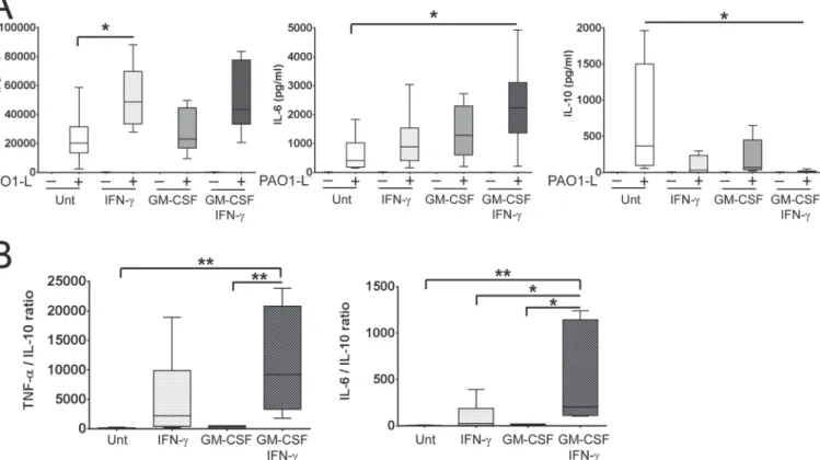
Macrophage activation up-regulated TNF-αand IL-6 and down-regulated IL-10 production upon PAO1-L infection and differences between the effects of IFN-γand GM-CSF were ob-served (Fig. 6A). TNF-αproduction was up-regulated approximately 2-fold by IFN-γ specifi-cally; this was significant for macrophages treated with IFN-γalone (p = 0.0378) (Fig. 6A). IL-6 production was weakly enhanced by IFN-γor GM-CSF alone, but was significantly increased when used in combination (p = 0.0287) (Fig. 6A). Finally, while treatment with IFN-γor CSF alone did not reduce IL-10 production significantly (IFN-γ: ~11-fold, p = 0.0556; GM-CSF: ~5-fold, p = 0.2084), both cytokines in combination caused a significant 72-fold reduction
Fig 6. Activation with IFN-γin the presence and absence of GM-CSF enhances the pro-inflammatory cytokine profile of macrophages in response
to liveP.aeruginosa.Macrophages were plated in X-Vivo 15, treated with IFN-γin the presence and absence of GM-CSF for 48 h and infected with PAO1-L for 4 h.A. Macrophage activation upregulated the production of TNF-αand IL-6 and downregulated IL-10 production in response to PAO1-L infection. Negligible to no production of these cytokines was seen in uninfected macrophages under all activation conditions.B. Ratios of TNF-αvs. IL-10 and IL-6 vs IL-10 revealed that the combination of IFN-γand GM-CSF produced the most inflammatory macrophage population. In donors where IFN-γactivation completely abrogated IL-10 expression, the minimum detectable limit of IL-10 for the assay (1.9 pg/ml) was used in order to calculate TNF-α/IL-10 and IL-6/ IL-10 ratios. Graphs depict 5–95 percentile with median for n = 6 to 8. Significance calculated by one-way ANOVA with Tukey’s post test;*= p0.05,**= p0.01. Unt: untreated macrophages; (-): uninfected; (+): infected.
(p = 0.0420) (Fig. 6A). In some donors, IL-10 was undetectable in infected cultures of macro-phage treated with GM-CSF and IFN-γ. The effect of IFN-γand GM-CSF on IL-6 expression appeared additive, while on IL-10 expression it appeared synergistic. TNF-αvs. IL-10 and IL-6 vs. IL-10 ratios revealed the following hierarchy regarding the inflammatory potential of acti-vated macrophages in response toP.aeruginosainfection: untreated<treated with GM-CSF <treated with IFN-γ<treated with GM-CSF and IFN-γ(Fig. 6B). GM-CSF and IFN-γdid not affectP.aeruginosagrowth in X-Vivo 15 based on results from independent growth curves and the quantification of cfu in bacteria-only control cultures done in parallel to macrophage infections (S10 Fig.).
These results illustrate the complementary capabilities of IFN-γand GM-CSF for modulat-ing the inflammatory potential of human macrophages under steady state conditions and in re-sponse toP.aeruginosainfection and support a role for macrophages in boosting
inflammation in response toP.aeruginosaduring an IFN-γ-dominated response, particularly in the presence of GM-CSF.
IFN-γ
in the presence and absence of GM-CSF does not alter
P.
aeruginosa
growth or macrophage survival
Treatment with IFN-γin the presence and absence of GM-CSF did not affect the ability of human macrophages to restrictP.aeruginosagrowth at 4 hpi. A consistent reduction in extra-cellular bacteria was observed in the presence of macrophages, irrespective of donor or macro-phage treatment (Fig. 7A). As shown inFig. 4A, in most instances the cell-associated bacterial fraction was minimal (Fig. 7B). However, in certain donors, extra growth of the cell-associated fraction was observed and compensated for the reduction in the extracellular fraction, such that in these cases the presence of macrophages did not affect the total amount of bacteria (Fig. 7C). Finally, comparison of LDH concentrations in supernatants from infected macro-phages at 4 hpi revealed that activation of macromacro-phages with IFN-γin the presence and absence of GM-CSF did not significantly affect macrophage survival at this time point (IFN-γ: p = 0.9475; GM-CSF: p = 0.9438; GM-CSF and IFN-γ: p = 0.9001) (Fig. 7D). These results suggest that the altered cytokine / chemokines responses obtained upon activation with IFN-γin the presence and absence of GM-CSF (Figs.5and6) were not due to differences in cell survival or bacterial load and that cellular activation does not protect macrophages fromP.aeruginosa -in-duced cytotoxicity nor affectsP.aeruginosagrowth in the presence of macrophages.
Discussion
Cytokine responses in CF PBMCs
in the bronchoalveolar lavage of CF patients chronically infected withP.aeruginosacompared toP.aeruginosa-free CF patients [7]. These discrepancies between studies may be due the use of different patient cohorts and classification criteria forP.aeruginosainfection status. For in-stance Moser et al. and Hartl et al. recruited patients who were free ofP.aeruginosainfection rather than those intermittently infected or free ofP.aeruginosaas in the study presented here. Robust Th1 responses to the mitogen PHA, correlated with better pulmonary status in CF pa-tients, particularly in CF patients chronically infected withP.aeruginosa(Fig. 2). Moser et al. reported a similar significant correlation in their cohort of CF patients chronically infected withP.aeruginosa[5]. While reduced IFN-γin CF samples is suggestive of a Th2 bias in CF PBMCs, we did not observe increased production of the Th2-associated cytokine IL-13 (S5 Fig.), nor did we detect any correlation between IFN-γand IL-13 levels in response to PHA, SEB and PA Lo (data not shown). Our culture conditions did not support the detection of IL-4 but further work looking at IL-5 levels [18] could help to address this issue.
The reduced levels of IL-17A detected in CF cultures in response to all stimuli used (Fig. 1B) were surprising based on previous reports in support of IL-17-driven inflammation in CF Fig 7. IFN-γactivation in the presence and absence of GM-CSF did not affect macrophage survival orP.aeruginosagrowth.Macrophages were plated in X-Vivo 15, treated with IFN-γin the presence and absence of GM-CSF for 48 h and infected with PAO1-L for 4 h. No significant differences were observed in the percentage of extracellular(A), cell-associated(B), and total(C)bacterial growth among cultures of differentially activated infected macrophages. The cell-associated fraction was found to be increased in certain donors. n = 6 to 10 for all bacterial growth data.D. Levels of LDH in infected macrophage supernatants were not significantly affected by macrophage activation, n = 6 to 10. Significance was calculated by one-way ANOVA with Tukey’s post test. Unt: untreated macrophages.
[14–18]. It could be argued that IL-17A production at peripheral sites might not correlate with IL-17A produced by circulating immune cells upon stimulation; particularly because IL-17A-producing cells might be recruited to the lung or peripheral lymph nodes [14]. Bayes et al. de-tectedPseudomonas-specific Th1, Th17 and Th22 cells in both healthy and CF individuals and observed reduced percentages of Th17 memory CD4+ T cells specific forP.aeruginosain the peripheral blood of CF patients compared to healthy controls [37]. Additionally IL-17A pro-duction in the lung could be mediated by a variety of cells. For instance, Tams et al. identified IL-17+ neutrophils,γδT cells and natural killer cells in addition to Th17 lymphocytes in bron-chial biopsies of children with CF [17]. However our results are in clear disagreement with studies that identified increased numbers of circulating IL-17A-producing T cells in CF pa-tients [16,18] and the intrinsic predisposition of CF T cells to differentiate into Th17 cells re-cently described [16].
The usefulness of IFN-γand IL-17A production by PBMC as indicators of lung health in CF may be limited by factors such as the stimulus used. PHA-induced IFN-γand IL-17A produc-tion in CF samples appears to be biologically relevant because of the positive and negative cor-relations between lung function and PHA-induced IFN-γand IL17A production, respectively, observed for CF/Chronic PA samples (Fig. 2). In contrast IFN-γand IL17A production in re-sponse to SEB or PA Lo did not show any correlation with lung function (data not shown), in-dicating a stimulus-dependent effect. While PHA induces polyclonal T cell activation in the presence of soluble factors, such as IL-6, produced by accessory cells [38] this would not be the case for SEB andP.aeruginosalysate. The superantigen SEB is a toxin produced by Staphylo-coccus aureus[39] that cross-links the T cell receptor (TCR) onαβT cells bearing particular Vβ chains with non-cognate MHC molecules, thus allowing promiscuous T cell activation [39]. SEB can activate up to 20% of allαβT cells (predominantly CD4+ T cells) therefore only a sub-population of T cells was being probed in our assays. SEB is also capable of engaging iNKT cells through MHC class II, but not CD1d [40]. SEB requires expression of HLA-DR at the cell surface of monocytes andP.aeruginosalysates require antigen processing and presentation by antigen presenting cells in the PBMC cultures. It is possible that CF monocytes could not effec-tively support SEB and/orP.aeruginosalysates-mediated lymphocyte activation as they have been shown to express reduced levels of MHCII and might be impaired in antigen presentation [41].
flagellated vs. non-flagellated) as infection with flagellatedP.aeruginosawas associated with MDSC induction in CF patients and flagellum induced the generation of MDSC [42].
The potential contribution of human macrophages to maintenance of
lung function during IFN-γ-dominated immune responses in the context
of
P.
aeruginosa
infection
In the study presented here it was next questioned how IFN-γcould positively impact on lung function in CF patients (Fig. 2). Preservation of organ function during infection results from a delicate balance between pro-inflammatory processes that promote clearance of infectious agents but have the potential to cause substantial tissue damage, and anti-inflammatory pro-cesses, aimed at minimising tissue damage and promote resolution that could compromise anti-microbial immunity. Thus IFN-γcould be beneficial in CF by promoting an inflammatory response capable of controlling infection while minimising lung damage. Macrophages play a central role in the regulation of inflammation and it was hypothesised that investigation of the effect of IFN-γon the behaviour of macrophages duringP.aeruginosainfection could provide information regarding the potential benefits of an IFN-γ-driven inflammatory response against P.aeruginosa. GM-CSF was included in the study because of its established activating effect on myeloid cell function [28,29] and the reported correlation between high serum GM-CSF with increased IFN-γexpression and better pulmonary function in CF patients [6]. Also, recent pub-lications indicate that GM-CSF production by Th17 cells is responsible for their pathological role during chronic inflammation [47,48]. Although there are clear limitations when trying to translatein vitrofindings toin vivoconditions it is possible to speculate regarding the biologi-cal consequences of the work presented here and make predictions that can be tested using more complex systems or clinical samples.
This manuscript shows that the response of macrophages toP.aeruginosainfection can be modulated by IFN-γand GM-CSF and that both cytokines synergise to increase the inflamma-tory potential of macrophages in response toP.aeruginosa. In particular macrophages treated with IFN-γproduce more TNF-αand less IL-10 in response toP.aeruginosainfection than un-treated macrophages and macrophages un-treated with GM-CSF. GM-CSF synergises with IFN-γ in reducing IL-10 synthesis by macrophages in response to infection. Hence it is possible that exposure of macrophages to IFN-γand GM-CSF in the lung could trigger an aggressive inflam-matory response characterised by high TNF-α/IL-10 and IL-6/IL-10 ratios (Fig. 6B) that pro-motes tissue damage. In agreement with this idea GM-CSF can be expressed in association with IFN-γby human T cells during chronic inflammation [49]. The drastic reduction in IL-10 production induced by IFN-γand GM-CSF could also be linked to the detrimental role of IL-17A in CF. GM-CSF can be produced by Th17 cells and if we consider IL-IL-17A as a surrogate marker for the presence of GM-CSF, it could be speculated that the negative correlation be-tween IL-17A levels and lung function in CF (Fig. 2) could be potentially caused by increased levels of GM-CSF in these patients. Further work would benefit from the identification of T cell subsets in the lungs of CF patients with and withoutP.aeruginosainfection with particular focus on T cells that co-express IFN-γand GM-CSF and their relation to Th17 cells [49].
macrophages exposed to IFN-γor GM-CSF could recruit monocytes and/or neutrophils, even before encountering a pathogen. In infected macrophages treatment with IFN-γin the presence or absence of GM-CSF did not affect the production of IL-8 indicating that neutrophil recruit-ment might not be affected by these cytokines during infection. On the other hand, IFN-γ treatment could promote recruitment of monocytes by infected macrophages regardless of the presence of GM-CSF which might limit tissue destruction caused by neutrophils. The results from the present manuscript are in agreement with a previous report on the differential regula-tion of IL-8 and MCP-1 expression in human macrophages, with MCP-1 synthesis being in-duced in response to IFN-γand IL-8 in response to LPS [50,51].
Activation with IFN-γin the presence and absence of GM-CSF did not alter the production of the inflammasome-associated cytokines IL-1βor IL-18 (S9 Fig.). This is relevant in the con-text ofP.aeruginosarespiratory infections as it has been demonstrated in murine acuteP. aeru-ginosapneumonia models that IL-1βand IL-18 released upon alveolar macrophage
inflammasome activation are deleterious for the host [52–54]. Cellular responses to IFN-γand GM-CSF are dominated by STAT-1 [55] and STAT-5 [56], respectively, with both cytokines conferring a‘primed state’to macrophages that enhances their ability to respond to stimula-tion. It would be of interest to characterise the interplay between the signalling pathways trig-gered by IFN-γand GM-CSF in order to determine the molecular basis for their additive and synergistic effects with particular emphasis on how they cooperate to regulate
IL-10 expression.
Macrophages could potentially control the growth ofP.aeruginosathrough phagocytosis, production of reactive oxygen species, release of hydrolases and iron sequestration [57,58]. Macrophage activation with IFN-γin the presence and absence of GM-CSF did not affect bac-terial growth or protect macrophages fromP.aeruginosa-induced cytotoxicity (Figs.4B-Cand
7D). Hence, an IFN-γ-dominated (Th1) immune response is unlikely to safeguard lung func-tion by promoting the ability of macrophages to controlP.aeruginosainfection or by increas-ing resistance of macrophages to the cytotoxic effect ofP.aeruginosa. This is in agreement with a previous study describing a negative effect of IFN-γon the phagocytosis and killing ofP. aer-uginosaby human macrophages [59]. The inability of macrophages treated with GM-CSF to restrictP.aeruginosagrowth contrasts with the role played by this cytokine in protection against lethalP.aeruginosapneumonia in vaccinated neutropenic mice [60]. It is possible that the protective role of GM-CSF in the murine pneumonia model could be facilitated by the pres-ence of opsonins such as antibodies specific forP.aeruginosaor lung collectins, such as SP-A, which could promote phagocytic uptake and cellular activation, or the accumulation of mono-cytes/macrophages in the infected lungs of vaccinated animals [60].
and might account for the increased cell-associated growth ofP.aeruginosain several experi-ments (Fig. 7B). Preliminary evidence suggests this may be donor-dependent. We propose that these changes in bacterial behaviour could be caused by the inflammatory conditions prevalent in these particular infections and be related to the levels of reactive oxygen species or anti-mi-crobial peptides present [61].
In conclusion, we have observed that IFN-γproduction by CF PBMCs positively correlated with lung function, particularly in patients chronically infected withP.aeruginosa, while IL-17A levels tended to correlate negatively with lung function, with this trend becoming signifi-cant in patients chronically infected withP.aeruginosa. These results are in agreement with IFN-γand IL-17A playing protective and detrimental roles, respectively, in CF. In addition, our study provides information regarding parameters that need to be taken into consideration when looking at systemic immune responses in clinical samples. For instance, our results illus-trate the importance of the stimuli used for activation of immune cells. Stimuli that require support from antigen presenting cells for T cell activation in the form of cytokines and/or high surface MHCII expression would provide a more comprehensive evaluation of the immunolog-ical status of patients as T cell responses are highly dependent on the activation state of antigen presenting cells. This would be lost when employing anti-CD3 and CD28 antibodies to stimu-late T cells which would induce polyclonal activation of all T cells but will only allow the analy-sis of the potential levels of T cell-derived cytokines that can be produced in patients.
Results using human macrophages infected withP.aeruginosaindicate that IFN-γmight promote an inflammatory response characterised by a balanced production of pro- and anti-inflammatory cytokines and recruitment of mononuclear cells. This is agreement with early work showing the ability of IFN-γto decrease the inflammatory response in chronicP. aerugi-nosapneumonia in rats [8]. In contrast IFN-γin combination with GM-CSF might promote tissue damage because of a reduced anti-inflammatory response, unable to safeguard tissues from the deleterious effects of inflammation.
Future work in this area will benefit from the use of isolated tissue macrophages from healthy donors and CF patients and clinical isolates ofP.aeruginosa. It would also be pertinent to investigate the effect of macrophage-derived cytokines and other inflammatory mediators on other immune cells such as neutrophils. Particular emphasis should be placed in establish-ing activation regimes consistestablish-ing of specific combinations of effector cytokines that promote the microbicidal capabilities of immune cells but minimise the potential for tissue damage. Co-cultures of phagocytes (macrophages and neutrophils) exposed to different combination of cy-tokines would provide a highly valuable tool to perform preclinical studies towards this aim.
Supporting Information
S1 Fig. Characterisation of human monocyte-derived macrophages differentiated in sus-pension (Teflon bottles) for 7 days.A. CD68 and mannose receptor (MR) expression in mac-rophages demonstrated by immunofluorescence. B. Flow cytometric analysis showed that CD14, CD16, HLA-DR, and MR surface expression was significantly upregulated upon mono-cyte (open circles) differentiation into macrophages (open squares). MFI = mean fluorescence intensity. Significance was calculated by unpaired Student’s t test for CD14, HLA-DR, and mannose receptor, and Mann-Whitney test for CD16. C. Reactive oxygen species production by zymosan-stimulated macrophages differentiated in M-CSF or GM-CSF.
(TIF)
S2 Fig. PAO1-L growth in LB broth and X-Vivo 15 is similar under oxygenated conditions.
measured at the times indicated. (TIF)
S3 Fig. PAO1-N expresses quorum sensing molecules and virulence factors in X-Vivo 15 but the las and rhl systems are lower than in LB.PAO1-N (Nottingham subline) reporter strains carrying lux promoter fusions (seeS1 Table) were used to determine the expression of quorum sensing and virulence factor genes in LB broth and X-Vivo 15. Luminescence and
OD600nmwere measured for each culture every 30 min. To negate any differences in
lumines-cence due to differences in growth of the reporter strains, gene expression was reported as lu-minescence (relative light units, RLU) divided by OD600nmfor that culture. Only every second
data point is plotted for ease of viewing. Data presented are mean of three independent experi-ments. Bacterial strains are described inS1 Table.
(TIF)
S4 Fig. LDH released by PAO1-L upon freezing and thawing and sonication to lyse cells.
Three independently prepared mid-log phase cultures of PAO1-L in X-Vivo 15 at a density of 5 x 10⁸cfu/ml (which was much higher than the total bacterial load ever achieved in any of the in vitro infection assays used in this study) released on average only 12.2 ± 3.6 ng/ml LDH (mean ± SD, n = 3) upon freezing and thawing followed by sonication.
(TIF)
S5 Fig. IL-13 (A) and IL-10 (B) production by healthy and CF PBMCs stimulated with PHA, SEB orP.aeruginosalysates.Graphs depict 5–95 percentile with median. For IL-10 (all stimuli), Healthy controls: n = 13, CF / Intermittent-Free PA and CF / Chronic PA: n = 15. For IL-13 (SEB, PA Hi, PA Lo), Healthy controls: n = 13, CF / Intermittent-Free PA: n = 15, CF / Chronic PA: n = 11, while for IL-13 (PHA): Healthy controls: n = 7, CF / Intermittent-Free PA: n = 13, CF / Chronic PA: n = 9 as IL-13 production by the remaining donors under this condi-tion was above the standard range of the assay. Significance calculated by Kruskal-Wallis test with Dunn’s post test.= p0.05,= p0.01.
(TIF)
S6 Fig. Lung function in CF patients.CF / Intermittent-Free PA patients demonstrated signif-icantly better lung function than CF / Chronic PA patients. This was particularly evident when pulmonary function was described as a percentage of predicted FEV1. Data presented are mean ± SD. Significance calculated by an unpaired Student’s t test.
(TIF)
S7 Fig. Correlation between lung function and IFN-γand IL-17A production in response to PHA in CF / Intermittent-Free PA patients.Correlation between IFN-γand IL-17A pro-duction in response to PHA and lung function was non-significant in the subset of intermit-tent-free CF patients. Correlation calculated by Spearman rank test.
(TIF)
S8 Fig. Analysis of PAO1-L-infected macrophage cultures by light microscopy.PAO1-L clusters can be observed at 2 hpi (black arrows) and increase in number and size at later times post-infection. Magnification = 400x.
(TIF)
for IFN-γ, n = 4 for GM-CSF +/- IFN-γ. MIP-1α: p = 0.3786, n = 6 for +/- IFN-γand n = 4 for GM-CSF +/- IFN-γ. Unt: untreated controls.
(TIF)
S10 Fig. PAO1-L growth in X-Vivo 15 under stationary conditions is not affected by the ad-dition of macrophage-activating cytokines.A. Bacteria were cultured in a 96-well microtitre plate at 37°C for 24 h in a Tecan Infinite M1000 PRO plate reader which measured the absor-bance (OD600nm) of the cultures every 30 min. Only every second data point is plotted for ease
of viewing. Data presented are mean ± SD for two experiments. B. Number of cfu in bacteria only wells at 4 hpi during macrophage infection assays. No significant difference was observed in the presence of different cytokines (p = 0.4890, one-way ANOVA with Tukey’s post test). (TIF)
S1 Table.P.aeruginosastrains used in this study.
(TIF)
Acknowledgments
The authors are grateful to Darryl Jackson for technical assistance
Author Contributions
Conceived and designed the experiments: SS AF MC LM-P. Performed the experiments: SS HB Y-CL. Analyzed the data: SS HB PW AF MC LM-P. Contributed reagents/materials/analysis tools: HB AR SH AF. Wrote the paper: SS HB PW AF MC LM-P.
References
1. Lyczak JB, Cannon CL, Pier GB (2000) Establishment of Pseudomonas aeruginosa infection: lessons from a versatile opportunist. Microbes Infect 2: 1051–1060. PMID:10967285
2. Sadikot RT, Blackwell TS, Christman JW, Prince AS (2005) Pathogen-host interactions in Pseudomo-nas aeruginosa pneumonia. Am J Respir Crit Care Med 171: 1209–1223. PMID:15695491
3. Hauser AR, Jain M, Bar-Meir M, McColley SA (2011) Clinical significance of microbial infection and ad-aptation in cystic fibrosis. Clin Microbiol Rev 24: 29–70. doi:10.1128/CMR.00036-10PMID:21233507 4. Lyczak JB, Cannon CL, Pier GB (2002) Lung infections associated with cystic fibrosis. Clin Microbiol
Rev 15: 194–222. PMID:11932230
5. Moser C, Kjaergaard S, Pressler T, Kharazmi A, Koch C, et al. (2000) The immune response to chronic Pseudomonas aeruginosa lung infection in cystic fibrosis patients is predominantly of the Th2 type. APMIS 108: 329–335. PMID:10937769
6. Moser C, Jensen PO, Pressler T, Frederiksen B, Lanng S, et al. (2005) Serum concentrations of GM-CSF and G-GM-CSF correlate with the Th1/Th2 cytokine response in cystic fibrosis patients with chronic Pseudomonas aeruginosa lung infection. APMIS 113: 400–409. PMID:15996157
7. Hartl D, Griese M, Kappler M, Zissel G, Reinhardt D, et al. (2006) Pulmonary T(H)2 response in Pseu-domonas aeruginosa-infected patients with cystic fibrosis. J Allergy Clin Immunol 117: 204–211. PMID:16387607
8. Johansen HK, Hougen HP, Rygaard J, Hoiby N (1996) Interferon-gamma (IFN-gamma) treatment de-creases the inflammatory response in chronic Pseudomonas aeruginosa pneumonia in rats. Clin Exp Immunol 103: 212–218. PMID:8565302
9. Moser C, Johansen HK, Song Z, Hougen HP, Rygaard J, et al. (1997) Chronic Pseudomonas aerugi-nosa lung infection is more severe in Th2 responding BALB/c mice compared to Th1 responding C3H/ HeN mice. APMIS 105: 838–842. PMID:9393554
10. Gordon S (2003) Alternative activation of macrophages. Nat Rev Immunol 3: 23–35. PMID:12511873 11. Martinez FO, Helming L, Gordon S (2009) Alternative activation of macrophages: an immunologic
12. Murphy BS, Bush HM, Sundareshan V, Davis C, Hagadone J, et al. (2010) Characterization of macro-phage activation states in patients with cystic fibrosis. J Cyst Fibros 9: 314–322. doi:10.1016/j.jcf. 2010.04.006PMID:20570573
13. Murphy BS, Sundareshan V, Cory TJ, Hayes D Jr, Anstead MI, et al. (2008) Azithromycin alters macro-phage phenotype. J Antimicrob Chemother 61: 554–560. doi:10.1093/jac/dkn007PMID:18230686 14. Chan YR, Chen K, Duncan SR, Lathrop KL, Latoche JD, et al. (2013) Patients with cystic fibrosis have
inducible IL-17+IL-22+ memory cells in lung draining lymph nodes. J Allergy Clin Immunol 131: 1117– 1129, 1129 e1111–1115. doi:10.1016/j.jaci.2012.05.036PMID:22795370
15. Decraene A, Willems-Widyastuti A, Kasran A, De Boeck K, Bullens DM, et al. (2010) Elevated expres-sion of both mRNA and protein levels of IL-17A in sputum of stable Cystic Fibrosis patients. Respir Res 11: 177. doi:10.1186/1465-9921-11-177PMID:21143945
16. Kushwah R, Gagnon S, Sweezey NB (2013) Intrinsic predisposition of naive cystic fibrosis T cells to dif-ferentiate towards a Th17 phenotype. Respir Res 14: 138. doi:10.1186/1465-9921-14-138PMID: 24344776
17. Tan HL, Regamey N, Brown S, Bush A, Lloyd CM, et al. (2011) The Th17 pathway in cystic fibrosis lung disease. Am J Respir Crit Care Med 184: 252–258. doi:10.1164/rccm.201102-0236OCPMID: 21474644
18. Tiringer K, Treis A, Fucik P, Gona M, Gruber S, et al. (2013) A Th17- and Th2-skewed cytokine profile in cystic fibrosis lungs represents a potential risk factor for Pseudomonas aeruginosa infection. Am J Respir Crit Care Med 187: 621–629. doi:10.1164/rccm.201206-1150OCPMID:23306544
19. Iwakura Y, Ishigame H, Saijo S, Nakae S (2011) Functional specialization of interleukin-17 family mem-bers. Immunity 34: 149–162. doi:10.1016/j.immuni.2011.02.012PMID:21349428
20. Linden A, Laan M, Anderson GP (2005) Neutrophils, interleukin-17A and lung disease. Eur Respir J 25: 159–172. PMID:15640338
21. Dubin PJ, McAllister F, Kolls JK (2007) Is cystic fibrosis a TH17 disease? Inflamm Res 56: 221–227. PMID:17607545
22. Galli SJ, Borregaard N, Wynn TA (2011) Phenotypic and functional plasticity of cells of innate immunity: macrophages, mast cells and neutrophils. Nat Immunol 12: 1035–1044. doi:10.1038/ni.2109PMID: 22012443
23. Speert DP, Wright SD, Silverstein SC, Mah B (1988) Functional characterization of macrophage recep-tors for in vitro phagocytosis of unopsonized Pseudomonas aeruginosa. J Clin Invest 82: 872–879. PMID:3138287
24. Speert DP, Gordon S (1992) Phagocytosis of unopsonized Pseudomonas aeruginosa by murine mac-rophages is a two-step process requiring glucose. J Clin Invest 90: 1085–1092. PMID:1522217 25. Kannan S, Audet A, Huang H, Chen LJ, Wu M (2008) Cholesterol-rich membrane rafts and Lyn are
in-volved in phagocytosis during Pseudomonas aeruginosa infection. J Immunol 180: 2396–2408. PMID: 18250449
26. Power MR, Li B, Yamamoto M, Akira S, Lin TJ (2007) A role of Toll-IL-1 receptor domain-containing adaptor-inducing IFN-beta in the host response to Pseudomonas aeruginosa lung infection in mice. J Immunol 178: 3170–3176. PMID:17312165
27. Raoust E, Balloy V, Garcia-Verdugo I, Touqui L, Ramphal R, et al. (2009) Pseudomonas aeruginosa LPS or flagellin are sufficient to activate TLR-dependent signaling in murine alveolar macrophages and airway epithelial cells. PLoS One 4: e7259. doi:10.1371/journal.pone.0007259PMID:19806220 28. Hamilton JA (2008) Colony-stimulating factors in inflammation and autoimmunity. Nat Rev Immunol 8:
533–544. doi:10.1038/nri2356PMID:18551128
29. Hamilton JA (2002) GM-CSF in inflammation and autoimmunity. Trends Immunol 23: 403–408. PMID: 12133803
30. Heslet L, Bay C, Nepper-Christensen S (2012) The immunomodulatory effect of inhaled granulocyte-macrophage colony-stimulating factor in cystic fibrosis. A new treatment paradigm. J Inflamm Res 5: 19–27. doi:10.2147/JIR.S22986PMID:22334793
31. Vogt G, Nathan C (2011) In vitro differentiation of human macrophages with enhanced antimycobacter-ial activity. J Clin Invest 121: 3889–3901. doi:10.1172/JCI57235PMID:21911939
32. Leentjens J, Kox M, Koch RM, Preijers F, Joosten LA, et al. (2012) Reversal of immunoparalysis in hu-mans in vivo: a double-blind, placebo-controlled, randomized pilot study. Am J Respir Crit Care Med 186: 838–845. doi:10.1164/rccm.201204-0645OCPMID:22822024
34. Miller MR, Crapo R, Hankinson J, Brusasco V, Burgos F, et al. (2005) General considerations for lung function testing. Eur Respir J 26: 153–161. PMID:15994402
35. Medzhitov R, Janeway CA Jr (2002) Decoding the patterns of self and nonself by the innate immune system. Science 296: 298–300. PMID:11951031
36. Brazova J, Sediva A, Pospisilova D, Vavrova V, Pohunek P, et al. (2005) Differential cytokine profile in children with cystic fibrosis. Clin Immunol 115: 210–215. PMID:15885645
37. Bayes HK, Bicknell S, MacGregor G, Evans TJ (2014) T helper cell subsets specific for Pseudomonas aeruginosa in healthy individuals and patients with cystic fibrosis. PLoS One 9: e90263. doi:10.1371/ journal.pone.0090263PMID:24587305
38. Ceuppens JL, Baroja ML, Lorre K, Van Damme J, Billiau A (1988) Human T cell activation with phytohe-magglutinin. The function of IL-6 as an accessory signal. J Immunol 141: 3868–3874. PMID:3263438 39. Herman A, Kappler JW, Marrack P, Pullen AM (1991) Superantigens: mechanism of T-cell stimulation
and role in immune responses. Annu Rev Immunol 9: 745–772. PMID:1832875
40. Hayworth JL, Mazzuca DM, Maleki Vareki S, Welch I, McCormick JK, et al. (2012) CD1d-independent activation of mouse and human iNKT cells by bacterial superantigens. Immunol Cell Biol 90: 699–709. doi:10.1038/icb.2011.90PMID:22041925
41. del Fresno C, Gomez-Pina V, Lores V, Soares-Schanoski A, Fernandez-Ruiz I, et al. (2008) Monocytes from cystic fibrosis patients are locked in an LPS tolerance state: down-regulation of TREM-1 as puta-tive underlying mechanism. PLoS One 3: e2667. doi:10.1371/journal.pone.0002667PMID:18628981 42. Rieber N, Brand A, Hector A, Graepler-Mainka U, Ost M, et al. (2013) Flagellin induces myeloid-derived suppressor cells: implications for Pseudomonas aeruginosa infection in cystic fibrosis lung disease. J Immunol 190: 1276–1284. doi:10.4049/jimmunol.1202144PMID:23277486
43. Gabrilovich DI, Nagaraj S (2009) Myeloid-derived suppressor cells as regulators of the immune system. Nat Rev Immunol 9: 162–174. doi:10.1038/nri2506PMID:19197294
44. Hirahara K, Poholek A, Vahedi G, Laurence A, Kanno Y, et al. (2013) Mechanisms underlying helper T-cell plasticity: implications for immune-mediated disease. J Allergy Clin Immunol 131: 1276–1287. doi: 10.1016/j.jaci.2013.03.015PMID:23622118
45. Lund FE, Randall TD (2010) Effector and regulatory B cells: modulators of CD4+ T cell immunity. Nat Rev Immunol 10: 236–247. doi:10.1038/nri2729PMID:20224569
46. Verschoor CP, Johnstone J, Millar J, Dorrington MG, Habibagahi M, et al. (2013) Blood CD33(+)HLA-DR(-) myeloid-derived suppressor cells are increased with age and a history of cancer. J Leukoc Biol 93: 633–637. doi:10.1189/jlb.0912461PMID:23341539
47. Codarri L, Gyulveszi G, Tosevski V, Hesske L, Fontana A, et al. (2011) RORgammat drives production of the cytokine GM-CSF in helper T cells, which is essential for the effector phase of autoimmune neu-roinflammation. Nat Immunol 12: 560–567. doi:10.1038/ni.2027PMID:21516112
48. El-Behi M, Ciric B, Dai H, Yan Y, Cullimore M, et al. (2011) The encephalitogenicity of T(H)17 cells is dependent on IL-1- and IL-23-induced production of the cytokine GM-CSF. Nat Immunol 12: 568–575. doi:10.1038/ni.2031PMID:21516111
49. Piper C, Pesenacker AM, Bending D, Thirugnanabalan B, Varsani H, et al. (2014) T cell expression of granulocyte-macrophage colony-stimulating factor in juvenile arthritis is contingent upon Th17 plastici-ty. Arthritis Rheumatol 66: 1955–1960. doi:10.1002/art.38647PMID:24692225
50. Bauermeister K, Burger M, Almanasreh N, Knopf HP, Schumann RR, et al. (1998) Distinct regulation of IL-8 and MCP-1 by LPS and interferon-gamma-treated human peritoneal macrophages. Nephrol Dial Transplant 13: 1412–1419. PMID:9641170
51. Han YL, Li YL, Jia LX, Cheng JZ, Qi YF, et al. (2012) Reciprocal interaction between macrophages and T cells stimulates IFN-gamma and MCP-1 production in Ang II-induced cardiac inflammation and fibro-sis. PLoS One 7: e35506. doi:10.1371/journal.pone.0035506PMID:22567105
52. Cohen TS, Prince AS (2013) Activation of inflammasome signaling mediates pathology of acute P. aer-uginosa pneumonia. J Clin Invest 123: 1630–1637. doi:10.1172/JCI66142PMID:23478406
53. Schultz MJ, Rijneveld AW, Florquin S, Edwards CK, Dinarello CA, et al. (2002) Role of interleukin-1 in the pulmonary immune response during Pseudomonas aeruginosa pneumonia. Am J Physiol Lung Cell Mol Physiol 282: L285–290. PMID:11792633
54. Schultz MJ, Knapp S, Florquin S, Pater J, Takeda K, et al. (2003) Interleukin-18 impairs the pulmonary host response to Pseudomonas aeruginosa. Infect Immun 71: 1630–1634. PMID:12654774
56. Lehtonen A, Matikainen S, Miettinen M, Julkunen I (2002) Granulocyte-macrophage colony-stimulating factor (GM-CSF)-induced STAT5 activation and target-gene expression during human monocyte/mac-rophage differentiation. J Leukoc Biol 71: 511–519. PMID:11867689
57. Britigan BE, Rasmussen GT, Olakanmi O, Cox CD (2000) Iron acquisition from Pseudomonas aerugi-nosa siderophores by human phagocytes: an additional mechanism of host defense through iron se-questration? Infect Immun 68: 1271–1275. PMID:10678937
58. Recalcati S, Locati M, Cairo G (2012) Systemic and cellular consequences of macrophage control of iron metabolism. Semin Immunol 24: 393–398. doi:10.1016/j.smim.2013.01.001PMID:23375134 59. Speert DP, Thorson L (1991) Suppression by human recombinant gamma interferon of in vitro
macro-phage nonopsonic and opsonic phagocytosis and killing. Infect Immun 59: 1893–1898. PMID: 1645327
60. Kamei A, Wu W, Traficante DC, Koh AY, Van Rooijen N, et al. (2013) Collaboration between macro-phages and vaccine-induced CD4+ T cells confers protection against lethal Pseudomonas aeruginosa pneumonia during neutropenia. J Infect Dis 207: 39–49. doi:10.1093/infdis/jis657PMID:23100569 61. Limoli DH, Rockel AB, Host KM, Jha A, Kopp BT, et al. (2014) Cationic antimicrobial peptides promote
microbial mutagenesis and pathoadaptation in chronic infections. PLoS Pathog 10: e1004083. doi:10. 1371/journal.ppat.1004083PMID:24763694
62. Franchi L, Stoolman J, Kanneganti TD, Verma A, Ramphal R, et al. (2007) Critical role for Ipaf in Pseu-domonas aeruginosa-induced caspase-1 activation. Eur J Immunol 37: 3030–3039. PMID:17935074 63. Fink SL, Cookson BT (2005) Apoptosis, pyroptosis, and necrosis: mechanistic description of dead and