BRIEF
COMMUNICATION
Interferon therapy shifts natural killer subsets among
Egyptian patients with chronic hepatitis C
Authors
Amal Fathy1 Mohamed Mohy Eldin2 Lobna Metwally3 Mohamed Eida4 Marwa Abdel-Rehim2 Gamal Esmat4
1Department of Clinical Pathology, Suez Canal University, Egypt. 2Department of Internal Medicine, Suez Canal University, Egypt. 3Department of Microbiology, Suez Canal University, Egypt. 4Department of Tropical Medicine, Suez Canal University, Egypt.
Submitted on: 07/28/2009 Approved on: 11/17/2009
Correspondence to:
Amal Fathy Clinical Pathology Department, Suez Canal University, P.O Box 351, Ismailia, 41511 – Egypt Phone: +20-64-3917138 Fax: +20-64-3378524 E-mail:
We declare no confl ict of interest.
ABSTRACT
Natural killer cells can be divided into fi ve subpopulations based on the relative expression of CD16 and CD56 markers. The majority of natural killer cells are CD56dim, which are considered to be the main cytotoxic effectors. A minority of the natural killer cells are CD56bright, and function as an important source of immune-regulatory cytokines. Shifts of these subsets have been reported in patients with chronic hepatitis C virus infection. We sought to investigate the shift of natural killer subsets among Egyptian patients with chronic HCV and to analyze the infl uence of interferon therapy on this shift. We applied a fl ow cytometric analysis of peripheral blood natural killer sub-sets for 12 interferon-untreated and 12 interferon-treated patients with chronic HCV, in compari-son to 10 control subjects. Among interferon-untreated patients, there was a signifi cant reduction of CD56-16+ (immature natural killer) cells. Among interferon-treated patients, the absolute count of natural killer cells was reduced, with expansion of the CD56bright subset and reduction of the CD56dim16+ subset. Natural killer subset counts were not signifi cantly correlated to HCV viral load and were not signifi cantly different among interferon responders and non-responders. In conclusion, HCV infection in Egyptian patients has been observed to be statistically and signifi cantly associated with reduction of the CD56-16+NK subset, while a statistically signifi cant expansion of CD56bright and reduction of CD56dim16+ subsets were observed after interferon therapy. Further studies are required to delineate the molecular basis of interferon-induced shift of natural killer subsets among patients with HCV.
Keywords: natural killer cells, natural killer subsets, chronic hepatitis C, innate immunity, interferon.
[Braz J Infect Dis 2010;14(4):398-405]©Elsevier Editora Ltda.
INTRODUCTION
Natural killer (NK) cells form 10-15% of the peripheral blood lymphocyte population. They are found in several lymphoid and non-lym-phoid compartments of organs, such as spleen, liver, and lungs.1 The identifi cation of NK cells
can be based on their morphology, function, or phenotype. Morphologically, NK cells are typed as large granular lymphocytes having a ki dney-shaped nucleus, a high cytoplasm to nucleus volume ratio, and containing large intracyto-plasmic azurophilic granules.2 Functionally, NK
cells are thought to have two important roles: target cell killing and cytokine production. With regards to target cell killing, NK cells dif-fer from T cells in that their cytolytic response does not require prior sensitization, and they are capable of destroying MHC I-mismatched haematopoietic and lymphoid cells.3 Regarding
cytokine production, NK cells secrete mainly interferon-γ (IFN-γ), tumor necrosis factor-α (TNF-α) and granulocyte monocyte colony-stimulating factor (GM-CSF), which enable them to regulate the host immune mechanism locally.4 Phenotypically, they are distinguished
from T and B lymphocytes by the absence of the T-cell receptor/CD3 complex and immu-noglobulin receptors on their cell surface.5
Be-sides the lack of certain markers on their sur-face, the presence of CD56 is used as a marker for NK cells in humans. CD56 has been shown to be an isoform of the neural cellular adhe-sion molecule.6 The expression of CD16, which
is involved in the antibody dependent cellular cytotoxicity (ADCC) mediated by NK cells, is also used as a phenotypic marker.7
There-fore, NK cells are phenotypically defi ned as CD56+CD16+CD3- cells. In human peripheral
iden-tified on the basis of the relative expression of CD16 and CD56 markers; CD56bright CD16-, CD56bright CD16+, CD56-dim CD16+, CD56dim CD16-, and CD56- CD16+. CD56dim
cells are the main mediators of NK cytotoxicity, as they contain high levels of perforin, whilst CD56bright act as an
important source of immunoregulatory cytokines.8,9
Hepatitis C virus (HCV) infects over 170 million peo-ple worldwide.10 A minority of infected persons may
re-solve the acute stage of infection and clear the virus, but most patients develop a life-long infection, making HCV a leading cause of chronic liver disease, cirrhosis and hepatocellular carcinoma.11 The host immune response
to HCV antigens is thought to determine whether viral clearance or chronic infection occurs. Hepatotropic vi-ruses induce production of type I IFN by hepatocytes and other cells in the liver, which, in turn, promotes infiltra-tion of NK cells in virus infected livers.12 The production
of type I IFN and other cytokines (including IL-12, IL-15, and IL-18) by hepatocytes activates NK cells and induces IFN-γ production by them. The IFN-γ produced by NK cells recruits activated T cells to the liver. It is noteworthy that IFN-γ produced by NK cells plays a major role in liv-er infiltration of CD4 and CD8 T cells. It has been shown in animal models that deliberate depletion of NK cells be-fore a hepatotropic viral infection leads to inhibition of a virus-specific T cell response, as well as inhibition of liver injury.13 Human NK cells co-cultured with HCV replicon
inhibit the replicon expression at protein and RNA levels by secreting antiviral factors, including IFN-γ.14 Thus, NK
cells could also potentially contribute towards control of HCV replication. It is possible that an adequate NK cell response may control hepatotropic viruses such as HCV, even in the absence of virus-specific immune responses. This notion is supported by the observation that, like hu-mans, a certain percentage of HCV-infected chimpanzees can clear the virus spontaneously, and this clearance does not correlate with the appearance of acquired immuni-ty.15 However, the potential importance of NK cells in
the control of HCV was disputed in a study that demon-strated that depletion of CD8 T cells by mAb aggravates HCV infections in animal models.16 It is noteworthy that
about one third of human and chimpanzee NK cells ex-press CD8, and depletion of CD8 T cells by anti-CD8 an-tibodies would also deplete CD8 NK cells. Therefore, the results from such studies should be interpreted with care. Meier et al.17 identified significantly reduced numbers of
total NK cells and a striking shift in NK subsets, with a marked decrease in the CD56dim cell fraction compared
to CD56bright cells in HCV infections. This shift influenced
the phenotype and functional capacity (IFN-γ produc-tion and killing) of the total NK pool. The aim of this work was to assess the influence of interferon therapy and HCV infection on NK subsets among Egyptian patients.
MATERIAL AND METHODS
Study design
This is a cross-sectional analytical study, as we are measur-ing exposure (± interferon) and outcome (NK subsets) at the same time.
Study subjects
Twenty four patients, attending either Suez Canal Univer-sity Hospital or Kasr El-Aini Hospital, were divided into two groups. Patients with chronic HCV who had never received interferon at any time during their course of illness comprised an interferon-untreated group (n = 12). Patients who suffered from chronic HCV infection and had been receiving standard interferon therapy for 24 weeks comprised an interferon-treat-ed group (n = 12). The standard treatment protocol includinterferon-treat-ed: interferon (interferon-alpha 2a), three million units, three times per week for 48 weeks. Patients were eligible for inclusion in this study after 24 weeks of therapy. Patients co-infected with HIV or HBV were excluded from this study. A normal control group was comprised by healthy HCV negative blood donors (n = 10). Written informed consent was obtained from all subjects.
Clinical history and examination
A detailed history and clinical examination were carried out for all groups. They were diagnosed as chronic hepatitis if the following criteria were present after a six month follow-up: a) signifi cant and persistent symptoms, b) fl uctuating or persistently elevated ALT and AST (> 1.5 fold of normal levels), normal serum albumin and prothrombin time, c) ul-trasonography revealed an enlarged bright texture ± portal tract thickening and ± normal spleen. Liver biopsy was done for the untreated group. Hepatic injury was assessed using the histological activity index (HAI) as modifi ed by Ishak.18
This consisted of a necro-infl ammatory grading score (range 0 to 18; 0 = no activity, 18 = severe activity) and a fi brosis staging score (range 0 to 6; 0 = no activity, 6 = cirrhosis).
Laboratory investigations
Venous blood samples (10 mL in EDTA and plain tubes) were drawn from each subject. Serum samples were obtained after clotting of the blood. For PCR testing, serum samples were stored at -80 °C until the time of assay. All samples were subjected to the following laboratory investigations:
1 – Complete blood count using an automated cell coun-ter (Cell Dyne, Abbott Diagnostics).
2 – Liver function tests:
a) Alanine aminotransferase (ALT), aspartate ami notransferase (AST), total bilirubin, direct bilirubin, se-rum albumin, and alkaline phosphatase (Cobas Integra Auto Analyzer, Roche).
3 – Viral hepatitis markers: HCV antibodies (HCV-EIA, Abbott Laboratories and HBsAg (Elecsys1010, Roche Diag-nostic GmbH) were assessed.
4 – Flow cytometry analysis: NK cell subsets were assessed by fl ow cytometry using FACS Calibur (BD Bio-sciences, San José, CA, USA). Cells were labeled with mAb against CD3 APC (UCHT1), CD16 FITC (B-E16), and CD56 PE (MOC-1). Monoclonal antibodies against CD3, CD16 and CD56 were purchased from IQProduct (Groningen, Neth-erlands). A sample of 20 µL of each monoclonal reagent was added to 100 µL of whole EDTA blood in 12×75 test tubes and incubated at room temperature for 15 min in the dark. Red blood cells were lysed using FACS Lyse reagent (BD, USA) and centrifuged at 2000 rpm for 5 min. The superna-tants were discarded and the cells were washed twice with phosphate-buffered saline (PBS), re-suspended in 300 µL of PBS. Data were analyzed using FACS Calibur and Flowjo software (TreeStar, Ashland, OR, USA). The typical forward and side scatters were used for lymphocytes gating. Gated lymphocytes were further gated by selecting CD3- popula-tion. CD3- cells were divided according to the expression of CD56 and/or CD16. Accordingly, NK cells were subgrouped into CD56bright or CD56dim and further defi ned by CD16
ex-pression.
5 – PCR: Quantitative HCV real-time PCR was per-formed for untreated patients before the start of interferon treatment, using a Light Cycler-RNA Master Hybridization Probe system in accordance with the manufacturers’ instruc-tion (Roche diagnostic, Mannheim, Germany). The lower detection limit is < 1000 IU/mL. Qualitative PCR was per-formed for treated patients at 24 weeks of treatment.
HCV-RNA was isolated by the QIAmp HCV-RNA viral kit (Qiagen, Hildene, Germany). HCV-RNA was reverse transcribed into cDNA, amplifi ed by PCR (Qiagen), and detected by electro-phoresis in 2% agarose gel stained with ethidium bromide. The PCR product (270 bp) was detected using an ultraviolet trans-illuminator.
Statistical analysis
Statistical analysis was done using SPSS program version 11. Data were presented as mean ± standard deviation. The Kruskal-Wallis test for analysis of non parametric data was used to compare between groups followed by Dunnett’s post-hoc test. Pearson correlation analysis was used when appropriate. Statistical signifi cance was considered at the 5% level of probability (p < 0.05).
RESULTS
The interferon-untreated patients (n = 12) had ages rang-ing from 29 to 53 (42.7 ± 8) years. Seven were female and five were male, and their duration of hepatic illness ranged from 12 to 50 (28 ± 16) months. The interferon-treated patients (n = 12) had ages ranging from 35 to 58 (45.5 ± 7) years. Three were female and nine were male, and the duration of their hepatic illness ranged from 18 to 53 (31 ± 15) months. Control subjects (n = 10) had ages rang-ing from 38 to 50 (44.7 ± 5) years. Seven were male and three were female. The study subjects were matched for age and sex.
Clinical, histological and virological features of INF-un-treated and INF-un-treated patients are listed in Table 1, and the routine laboratory data of the studied groups are listed in Table 2.
Table 1. Clinical, histological and virological features of IFN-untreated and IFN-treated patients
IFN-untreated IFN-treated
n = 12 n = 12
Risk factor
Anti-Bilhazial injection 2 3
Blood transfusion 2 0
Tooth extraction 5 6
Tattooing 2 2
Surgical intervention 5 2
Histology
Grading 6.9 ± 1.8 ND
Staging 2.9 ± 1.3 ND
Viral load
Quantitative HCV - RNA(IU/mL×103) 296 ± 349 ND
Qualitative PCR ND 7 cases negative HCV-RNA
5 cases positive HCV-RNA
Table 2. The laboratory data of IFN-untreated, IFN-treated and controls
Laboratory variables IFN-untreated IFN-treated Controls
n = 12 n = 12 n = 10
Albumin (mg/L) 4.4 ± 0.3 4.4 ± 0.36 4.3 ± 0.3
Alkaline phosphatase (u/L) 129 ± 66† 118 ± 70* 71 ± 13
AST (u/L) 44.5 ± 17.4§ 28.9 ± 8.9† 18.3 ± 6.1
ALT (u/L) 63.5 ± 29§ 30.9 ± 10* 22.1 ± 5.6
T.Bilirubin (mg/dL) 1.39 ± 2.0* 2.1 ± 4.3* 0.53 ± 0.20
D.Bilirubin (mg/dL) 0.45 ± 0.34* 1.5 ± 3.9† 0.17 ± 0.18
AFP (ng/mL) 9.3 ± 8.3 17.9 ± 25.1 5.2 ± 2.1
Hemoglobin (g/dL) 14.0 ± 0.72* 11.2 ± 1.73§ 14.7 ± 0.60
Leukocytes count (cells/uL) 6483 ± 1625 3625 ± 2092† 6440 ± 1649
Platelets count ×103 168 ± 55† 136 ± 73† 250 ± 38
Values are expressed as mean ± standard deviation. * Significant p < 0.05 compared with controls. † Significant p < 0.01 compared with controls.
§ Significant p < 0.001 compared with controls.
Table 3. Lymphocytes, NK and NK subsets in IFN-untreated, IFN-treated and controls
IFN-untreated IFN-treated Controls
n = 12 n = 12 n = 10
Percentage of lymphocytes 32.1 ± 8.0 31.7 ± 7.8 40.7 ± 13.6
CD3- 35.6 ± 11.1 39.2 ± 11.7 42.8 ± 10.7
NK 43.7 ± 15.2 53.6 ± 14.4 44.3 ± 14
CD56bright 5.4 ± 5.2 20.2 ± 9.9§ 3.3 ± 1.72
CD56dimCD16+ 84.4 ± 10.5 68.3 ± 10.7† 80.9 ± 5.3
CD56dimCD16- 5.9 ± 4.8 7.0 ± 3.1 7.0 ± 4.3
CD56-CD16+ 3.9 ± 3.2† 4.2 ± 2.3† 8.4 ± 2.7
Absolute counts of lymphocytes 2632 ± 413 1085 ± 559† 2526 ± 784
CD3- 920 ± 269 623 ± 780† 1096 ± 457
NK 392 ± 155 204 ± 76† 471 ± 207
CD56bright 19 ± 19 44 ± 29† 13 ± 8
CD56dimCD16+ 335 ± 150 127 ± 64† 388 ± 187
CD56dimCD16- 21 ± 18 20 ± 22 27 ± 14
CD56-CD16+ 15 ± 12† 7 ± 3§ 40 ± 23
CD3- is expressed as % of gated lymphocytes, total NK is expressed as % of CD3- and NK subsets are expressed as % of NK. The calculated absolute numbers are expressed as cells/µL. Values are expressed as M ± SD.
* Significant p < 0.05 compared with controls. † Significant p < 0.01 compared with controls.
§ Significant p < 0.001 compared with controls.
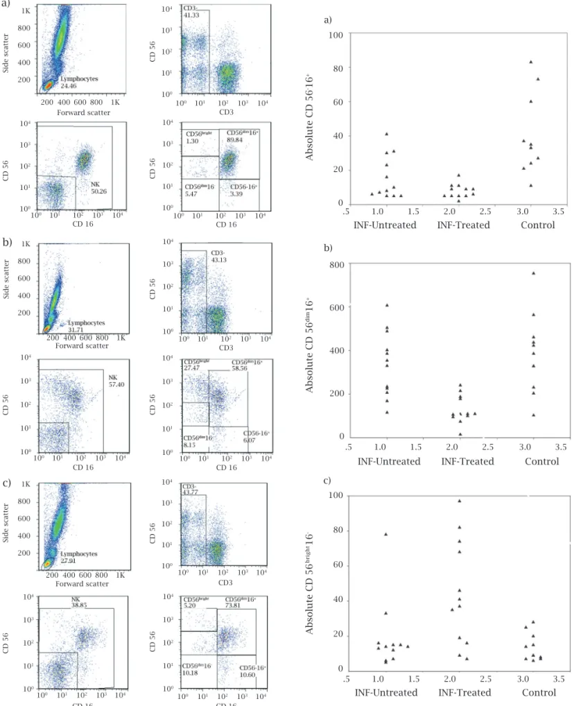
The proportion of total NK and NK subsets, as well as the absolute numbers of various NK cells in interferon-untreated, interferon treated chronic HCV patients, and healthy controls are presented in Table 3. Figure 1 (a,b,c)
shows the flow cytometry analysis for a subject from each group. The relative and absolute counts of CD56-CD16+
Figure 1: Flow cytometry analysis of NK cells and NK subsets. a) Representative dot plots of NK and NK subsets in untreated chronic hepatitis C subject.
b) Representative dot plots of NK and NK subsets in interferon-treated subject.
c) Representative dot plots of NK and NK subsets in normal healthy subject.
Figure 2: A scatter diagram compares the absolute counts of NK subsets in the studied groups
a) Representative of absolute CD56-16+. b) Representative of absolute CD56dim 16+. c) Representative of absolute CD56bright.
a) 1K 800 600 400 200 104 103 102 101 100 104 103 102 101 100 104 103 102 101 100 Side scatter CD 56 CD 56 CD 56 Forward scatter CD 16 CD3 CD 16 200 400 600 800 1K
100 101 102 103 104
100 101 102 103 104
100 101 102 103 104
b) c) 1K 800 600 400 200 1K 800 600 400 200 Side scatter Side scatter Forward scatter Forward scatter 200 400 600 800 1K
200 400 600 800 1K 104 103 102 101 100 CD 56 CD 16 100 101 102 103 104
104 103 102 101 100 104 103 102 101 100 104 103 102 101 100 104 103 102 101 100 104 103 102 101 100 CD 56 CD 56 CD 56 CD 56 CD 56 CD3 CD3 CD 16 CD 16 CD 16 100 101 102 103 104
100 101 102 103 104
100 101 102 103 104 100 101 102 103 104
100 101 102 103 104
a)
b)
INF-Untreated INF-Treated Control
INF-Untreated INF-Treated Control
Absolute CD 56
-16
+
Absolute CD 56
dim 16 + 100 80 60 40 20 0 100 80 60 40 20 0 800 600 400 200 0
.5 1.0 1.5 2.0 2.5 3.0 3.5
.5 1.0 1.5 2.0 2.5 3.0 3.5
c)
INF-Untreated INF-Treated Control
Absolute CD 56
bright
16
whereas no significant difference was detected in these subsets between interferon-untreated and treated pa-tients (data not shown). Similarly, the proportion of total NK and NK subsets, as well as the absolute numbers of various NK cells in interferon responder patients versus non responders, did not show any significant difference (data not shown). The absolute numbers of total NK cells and both the relative and absolute numbers of CD56
dim-CD16+ cell subsets were decreased in interferon-treated
patients when compared to interferon-untreated patients and controls (Figure 2b). Conversely, the relative and ab-solute counts of CD56bright cell subsets were increased in
interferon-treated patients when compared to interferon untreated patients and controls (Figure 2c).
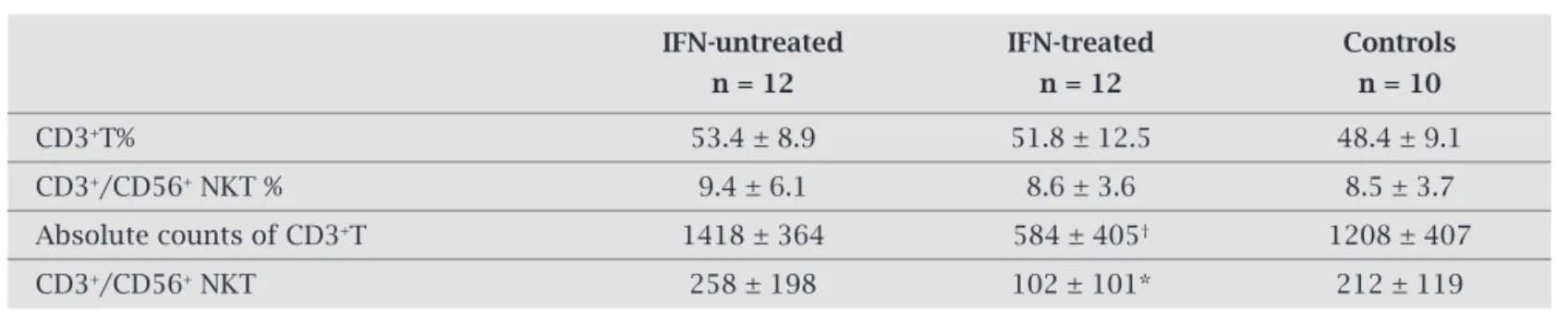
The relative and absolute numbers of CD3+ T cells and
CD3+/CD56+ NKT in untreated,
interferon-treated chronic HCV patients and healthy controls are presented in Table 4. There was a statistically significant decrease in the absolute counts of CD3+ T cells and CD3+/
CD56+NKT of interferon-treated patients compared with
interferon-untreated patients and controls.
No significant correlations between NK subsets and HCV viremia were seen. However, a significant positive correlation between CD56bright counts and hepatic in-jury, based on HAI, was detected (r = 0.63, p = 0.025). A significant negative correlation between CD56dimCD16+
counts and hepatic fibrosis was also detected (r = -0.61, p = 0.03).
DISCUSSION
NK cells constitute the fi rst line of host defense against in-vading pathogens. They eliminate virus-infected cells by direct and indirect killing methods. Direct killing of target cells is achieved through the release of cytotoxic molecules, such as perforin and granzymes. Alternatively, apoptosis may be induced by over expression of Fas/FasL, TNF-α, and TNF-related apoptosis-inducing ligand (TRAIL)/death re-ceptor4 (DR-4) and DR-5 interactions.19-21 Indirect killing
of target cells may also be achieved by secretion of soluble factors such as IFN-γ and TNF-α. In addition, NK cells play a role in inhibiting virus replication by the induction of an antiviral state in host cells.22 Furthermore, NK cells play
im-portant immunoregulatory roles through their interaction with T cells, B cells and antigen-presenting cells.
In this study, we found that CD3-CD56-CD16+ cell
counts were decreased in HCV infected patients. In con-trast, Gonzalez et al.23 and Zarife et al.24 reported increased
frequency of CD3-CD56-CD16+ NK cells in patients with
chronic HCV/HIV, or in blood donors with HCV among European and Brazilian populations, respectively. This dif-ference may be attributed to either host or viral heterogene-ity, as our study was done on Egyptian populations, in which HCV genotype 4 is the most prevalent genotype.25
A signifi cant reduction of circulating NK cells and ex-pansion of CD56bright, and reduction of CD56dimCD16+
sub-sets were seen in patients with chronic HCV after interferon treatment. These results agreed with the data reported by Gonzalez et al.,23 who indicated that the CD56bright
immu-noregulatory NK cells subset temporarily expanded in re-sponse to interferon treatment. This also agreed with the data reported by Saraste et al.,26 who observed an
expan-sion of CD56bright NK cells with a concomitant decrease of
CD56dim cells in multiple sclerosis patients after 12 months
of treatment with IFN-b. In contrast to our fi ndings, Meier et al.17 and Golden-Mason et al.27 reported a signifi cant
re-duction of total NK cells count and a striking shift in NK subsets, with a marked decrease in the CD56dim cell fraction
compared to CD56bright cells in HCV infection. This can be
explained by the observation that Meier et al.17 did not
ex-clude patients with history of interferon therapy and includ-ed patients who were not on current or recent (within the last six months) IFN-α treatment.
CD56bright NK cells are very likely precursor cells of the
CD56dim subset. Indeed, CD56dim NK cells display shorter
telomeres than CD56bright NK cells from peripheral blood,
which implies that the latter are less mature than the former.
Table 4. CD3+T cells and CD3+/CD56+ NKT in IFN-untreated, IFN-treated and controls
IFN-untreated IFN-treated Controls
n = 12 n = 12 n = 10
CD3+T% 53.4 ± 8.9 51.8 ± 12.5 48.4 ± 9.1
CD3+/CD56+ NKT % 9.4 ± 6.1 8.6 ± 3.6 8.5 ± 3.7
Absolute counts of CD3+T 1418 ± 364 584 ± 405† 1208 ± 407
CD3+/CD56+ NKT 258 ± 198 102 ± 101* 212 ± 119
Expansion of CD56bright subsets was reported in patients
who are treated daily with a low dose of IL-2.27 Since the
mechanism of interferon-induced NK cells shift is not fully understood, we suggest that interferon may infl uence the differentiation of CD56bright to CD56dim. On the other hand,
the CD56dim16- subset was not altered either by HCV
infec-tion or interferon therapy; the main funcinfec-tion of this subset is largely unknown.
In this study, no signifi cant difference in total NK cells and NK subsets among interferon responders were seen when compared to non-responders. Different results were reported by Panasiuk et al.,28 who found a slight increase in
NK cell counts in patients who did not clear HCV. Also, Kha-koo et al.,29 showed that genes encoding the NK cell
recep-tor directly infl uence resolution of HCV infection. However, in our study, we did not assess the function of NK cells, as the immunodefi cient NK cells may not be able to kill virus-infected cells. Further studies in this area, to correlate NK function with interferon response, are required.
In this study, a signifi cant positive correlation between the relative counts of CD56bright subsets with HAI was found.
Lin et al.,30 reported that increasing activated bright NK
cells and increasing activated apoptotic bright NK cells were both signifi cantly associated with increasing hepatic necro-infl ammatory grade. This fi nding may be explained by the fact that CD56bright subsets have immunoregulatory
func-tions and produce potent pro-infl ammatory cytokines, like INF-γ and TNF-α.31 Absence of correlation between NK or
its subsets with HCV viral load agrees with similar data re-ported by Pernollet et al.,32 who found that NK populations
did not correlate with any biochemical or viral parameters. In conclusion, this study has shown that, in Egyptian pa-tients, HCV infection is associated with reduction of CD56
-16+ NK subsets, and interferon-alpha therapy is associated
with expansion of CD56bright and reduction of CD56dim16+.
Further studies are required to delineate the molecular basis of interferon-induced shift of NK subsets among patients with HCV. As this study was conducted on two distinct groups of chronic HCV patients before and after interferon treatment, a further longitudinal study is recommended to address the limitations which are inherent in cross sectional studies of this type.
ACKNOWLEDGEMENTS
We thank Dr Omar Fathy for technical assistance.
REFERENCES
1. Yokoyama WM. Natural killer cell receptors. Current Opinion Immunology 1998; 10:298-305.
2. Timonen T. Natural killer cells: Endothelial interactions, migration, and target cell recognition. J Leukocyte Biology 1997; 62:693-701.
3. Rolstad B, Seaman WE. Natural killer cells and recognition of MHC class I molecules: New perspectives and challenges in immunology. Scand J Immunology 1998; 47:412-25.
4. Robertson MJ, Cameron C, Lazo S et al. Costimulation of human natural killer cell proliferation: Role of accessory cy-tokines and cell contact-dependent signals. Natural Immu-nity 1996; 15:213-26.
5. Phillips JH, Lanier LL. Dissection of the lymphokine-activat-ed killer phenomenon. Relative contribution of peripheral blood natural killer cells and T lymphocytes to cytolysis. J Exp Medicine 1986; 164:814-25.
6. OShea J, Ortaldo JR. The biology of natural killer cells: in-sights into the molecular basis of function. In: Lewis CE, Mc-gee JOD. The Natural Immune System. The Natural Killer Cell. Oxford: Oxford University Press, 1992.
7. Trinchieri G. Biology of natural killer cells. Adv Immunol 1998; 47:187-376.
8. Cooper MA, Fehniger TA, Caligiuri MA. The biology of human natural killer-cell subsets. Trends Immunol 2001; 22:633-40.
9. Caligiuri M. Human natural killer cells. Blood 2008; 112:461-9.
10. Cohen J. The scientifi c challenge of hepatitis C. Science 1999; 285:26-30.
11. Tong MJ, El-Farra NS, Reikes AR, Co RL. Clinical outcomes after transfusion-associated hepatitis C. N Engl J Med 1995; 332:1463-66.
12. Salazar-Mather TP, Orange JS, Biron CA. Early murine cy-tomegalovirus (MCMV) infection induces liver natural killer (NK) cell infl ammation and protection through macrophage infl ammatory protein 1α (MIP-1α)-dependent pathways. J Exp Med 1998; 187:1-14.
13. Liu ZX, Govindarajan S, Okamoto S, Dennert G. NK cells cause liver injury and facilitate the induction of T cell-me-diated immunity to a viral liver infection. J Immunol 2000; 164:6480-6.
14. Li Y, Zhang T, Ho C et al. Natural killer cells inhibit hepatitis C virus replicon expression mediated by interferon. 8th An-nual Meeting of the Society for Natural Immunity, Noordwi-jkerhout, the Netherlands; Abstract No. C035, Abstract Book, 54, 2004.
15. Thomson M, Nascimbeni M, Havert MB et al. The clearance of hepatitis C virus infection in chimpanzees may not neces-sarily correlate with the appearance of acquired immunity. J Virol 2003; 77:862-70.
16. Shoukry NH, Grakoui A, Houghton M et al. Memory CD8 T cells are required for protection from persistent hepatitis C virus infection. J Exp Med 2003; 197:1645-55.
17. Meier UC, Owen RE, Taylor E et al. Shared Alterations in NK Cell Frequency, Phenotype, and Function in Chronic Human Immunodefi ciency Virus and Hepatitis C Virus Infections. J Virol 2005; 19:12365-74.
18. Ishak KG. Chronic hepatitis: Morphology and nomenclature. Mod Pathol 1994; 7:690-713.
19. Arase H, Arase N, SaitoT. Fas-mediated cytotoxicity by fresh-ly isolated natural killer cells. J Exp Med 1995; 181:1235-8. 20. Zamai L, Ahmad M, Bennett IM et al. Natural killer (NK)
cell-mediated cytotoxicity: differential use of TRAIL and Fas ligand by immature and mature primary human NK cells. J Exp Med 1998; 188:2375-80.
22. Guidotti LG, Chisari FV. Noncytolytic control of viral infec-tions by the innate and adaptive immune response. Annu Rev Immunol 2001; 19:65-91.
23. Gonzalez VD, Falconer K, Michaëlsson J et al. Expansion of CD56- NK cells in chronic HCV/HIV-1 coinfection: reversion by antiviral treatment with pegylated IFN-alpha and ribavirin. Clin Immunol 2008; 1:46-56.
24. Zarife MA, Reis EA, Carmo TM et al. Increased frequency of CD56 Bright NK-cells,CD3-CD16+CD56-NK-cells and acti-vated CD4+T-cells or B-cells in parallel with CD4+CD25High T-cells control potentially viremia in blood donors with HCV. J Med Virol 2009; 81:49-59.
25. el-Zayadi A, Simmonds P, Dabbous H et al. Response to in-terferon-alpha of Egyptian patients infected with hepatitis C virus genotype 4. J Viral Hepat 1996; 3:261-4.
26. Saraste M, Irjala H, Airas L. Expansion of CD56bright natural killer cells in the peripheral blood of multiple sclerosis patients treated with interferon-beta. Neurol Sci 2007; 28:121-6.
27. Carson W, Caligiuri M. Natural killer cell subsets and develop-ment. Methods 1996; 9:327-43.
28. Panasiuk A, Prokopowicz D, Zak J. Immunological response in chronic hepatitis C virus infection during interferon alpha therapy. Hepatogastroenterology 2004; 58:1088-92.
29. Khakoo SI, Thio CL, Martin MP et al. HLA and NK cell in-hibitory receptor genes in resolving hepatitis C virus infection. Science 2004; 305:872-4.
30. Lin AW, Gonzalez SA, Cunningham-Rundles S et al. CD56+dim and CD56+bright cell activation and apoptosis in hepatitis C vi-rus infection. Clin Exp Immunol 2004; 137:408-16.
31. Jacobs R, Hintzen G, Kemper A et al. CD56bright cells differ in their KIR repertoire and cytotoxic features from CD56dim NK cells. Eur J Immunol 2001; 31:3121-7.