without Affecting Pathogen Clearance
Benjamin Weber1, Steffen Schuster2, Daniel Zysset1, Silvia Rihs1, Nina Dickgreber1, Christian Schu¨rch1,3, Carsten Riether3, Mark Siegrist3, Christoph Schneider4, Helga Pawelski4, Ursina Gurzeler5,
Pascal Ziltener6, Vera Genitsch1, Fabienne Tacchini-Cottier2, Adrian Ochsenbein3,7, Willy Hofstetter3, Manfred Kopf4, Thomas Kaufmann5, Annette Oxenius6, Walter Reith8, Leslie Saurer1*,
Christoph Mueller1*
1Division of Experimental Pathology, Institute of Pathology, University of Bern, Bern, Switzerland,2Department of Biochemistry, University of Lausanne, Epalinges, Switzerland,3Department of Clinical Research, University of Bern, Bern, Switzerland,4Institute of Molecular Health Sciences, ETH Zurich, Zurich, Switzerland,5Institute of Pharmacology, University of Bern, Bern, Switzerland,6Institute of Microbiology, ETH Zurich, Zurich, Switzerland,7Department of Medical Oncology, University of Bern, Bern, Switzerland,8Department of Pathology and Immunology, Centre Medical Universitaire, Geneva, Switzerland
Abstract
Triggering receptor expressed on myeloid cells-1 (TREM-1) is a potent amplifier of pro-inflammatory innate immune reactions. While TREM-1-amplified responses likely aid an improved detection and elimination of pathogens, excessive production of cytokines and oxygen radicals can also severely harm the host. Studies addressing the pathogenic role of TREM-1 during endotoxin-induced shock or microbial sepsis have so far mostly relied on the administration of TREM-1 fusion proteins or peptides representing part of the extracellular domain of TREM-1. However, binding of these agents to the yet unidentified TREM-1 ligand could also impact signaling through alternative receptors. More importantly, controversial results have been obtained regarding the requirement of TREM-1 for microbial control. To unambiguously investigate the role of TREM-1 in homeostasis and disease, we have generated mice deficient inTrem1.Trem12/2mice are
viable, fertile and show no altered hematopoietic compartment. In CD4+T cell- and dextran sodium sulfate-induced models of colitis, Trem12/2 mice displayed significantly attenuated disease that was associated with reduced inflammatory
infiltrates and diminished expression of pro-inflammatory cytokines.Trem12/2
mice also exhibited reduced neutrophilic infiltration and decreased lesion size upon infection withLeishmania major. Furthermore, reduced morbidity was observed for influenza virus-infectedTrem12/2mice. Importantly, while immune-associated pathologies were significantly reduced,
Trem12/2mice were equally capable of controlling infections withL. major, influenza virus, but alsoLegionella pneumophila
asTrem1+/+controls. Our results not only demonstrate an unanticipated pathogenic impact of TREM-1 during a viral and parasitic infection, but also indicate that therapeutic blocking of TREM-1 in distinct inflammatory disorders holds considerable promise by blunting excessive inflammation while preserving the capacity for microbial control.
Citation:Weber B, Schuster S, Zysset D, Rihs S, Dickgreber N, et al. (2014) TREM-1 Deficiency Can Attenuate Disease Severity without Affecting Pathogen Clearance. PLoS Pathog 10(1): e1003900. doi:10.1371/journal.ppat.1003900
Editor:Robert Modlin, University of California, Los Angeles, United States of America
ReceivedJune 26, 2013;AcceptedDecember 10, 2013;PublishedJanuary 16, 2014
Copyright:ß2014 Weber et al. This is an open-access article distributed under the terms of the Creative Commons Attribution License, which permits unrestricted use, distribution, and reproduction in any medium, provided the original author and source are credited.
Funding:This work was supported by grants of the Swiss National Foundation. The funders had no role in study design, data collection and analysis, decision to publish, or preparation of the manuscript.
Competing Interests:The authors have declared that no competing interests exist. * E-mail: [email protected] (LS); [email protected] (CM)
Introduction
Innate immune cells express several cell surface receptors and intracellular sensing molecules that allow for autonomous recog-nition of pathogen- and danger-associated molecular patterns (PAMPs and DAMPs) and initiation of pro-inflammatory anti-microbial reponses. Toll-like receptors (TLR) and nucleotide-binding oligomerization domain (NOD)-like receptors, which recognize a diverse group of highly conserved microbial structures, represent only two examples of large innate immune receptor families with activating functions. Over the last decade, an addi-tional family of evolutionary conserved innate immune receptors has been identified and characterized, the so-called triggering receptors expressed on myeloid cells (TREMs). TREMs belong to the immunoglobulin (Ig) superfamily of receptors and contain both inhibitory and activating receptors [1,2,3]. In contrast to the fairly
cellular activation, TREM-1 potently synergizes with distinct TLR ligands for a substantial amplification of oxidative burst and pro-duction of pro-inflammatory mediators such as TNF, IL-1b, IL-6, IL-8, MCP-1 and Mip-1a[4,9,10].
In vivo, the role of TREM-1 has been mostly characterized in experimental models of endotoxin-induced shock or microbial sepsis where blockade of TREM-1 signaling conferred significant protection [9,11,12]. The detection of TREM-1 in inflammatory lesions caused by bacterial or fungal agents, but not in psoriasis or immune-mediated vasculitis [9], has further led to the general concept that TREM-1 is primarily involved in microbial diseases, particularly, since elevated levels of the serum soluble form of the shed TREM-1 surface receptor (sTREM-1) also appear to associate with bacterial infections in patients with pneumonia or suspected sepsis [13,14].
However, increasing evidence is now emerging that TREM-1 may additionally play a role in non-infectious inflammatory con-ditions. Thus, expression of TREM-1 can also be induced by the non-microbial agent monosodium urate monohydrate crystals (MSU) or by hypoxic cell culture conditions in vitro [15,16]. Augmented sTREM-1 levels have been reported for patients with rheumatoid arthritis, acute pancreatitis, chronic obstructive pul-monary disease and cardiac arrest [17,18,19,20]. Furthermore, we have previously described an involvement of TREM-1 in human inflammatory bowel diseases (IBD) and in models of experimental colitis [21,22,23].
Investigations on the precise function of TREM-1 in distinct diseases have so far been complicated by the still unidentified ligand(s) for TREM-1. Putative ligands for TREM-1 have been described on the surface of human platelets and on murine granulocytes during experimental peritonitis and endotoxaemia [12,24,25]. In addition, necrotic cell lysates also appear to stimu-late pro-inflammatory responses in a TREM-1-dependent manner, which may relate to association of TREM-1 with the High Mobility Group Box 1 (HMGB1) protein [26,27]. Hence, it can be speculated
that not only PAMPs but also DAMPs induce signalingvia TREM-1 and that several ligands for TREM-TREM-1 may exist.
In the absence of clearly defined ligands for TREM-1, studies addressing the impact of TREM-1 in disease have so far mostly relied on the use of TREM-1/Ig fusion proteins or synthetic peptides mimicking part of the extracellular domain of TREM-1. Although by the use of these agents substantial protection from endotoxin-induced shock, microbial sepsis or experimental colitis could be conferred [9,11,12,22], several aspects regarding the true biological role of TREM-1 remain unclear. First, considering the redundancy of innate immune receptor-ligand interactions, the possibility exists that in these previous studies not only signaling through TREM-1 but through additional, potentially more rele-vant receptors was prevented. Second, controversial findings have been obtained with respect to the impact of impaired TREM-1 signaling on microbial control [9,28,29,30].
In order to investigate the role of TREM-1 in homeostasis and disease, we have generated a TREM-1-deficient (Trem12/2) mouse by targeted deletion of exon 2. Here we show, employing distinct inflammation and infection models ranging from experimental colitis to infections with Leishmania major, influenza virus and
Legionella pneumophila, that complete absence of TREM-1 signifi-cantly attenuates morbidity and immune-mediated pathologies while microbial control remains unimpaired. These findings not only demonstrate an unanticipated clear role for TREM-1 in chronic inflammatory disorders, parasitic and viral infections, but also illustrate the potential for a novel therapeutic intervention in various disease settings.
Results
Deletion ofTrem1has no apparent impact under homeostatic conditions
To account for potential embryonically lethal effects of a total deletion of theTrem1 gene, a targeting vector was designed for conditional deletion of exon 2 (Fig. S1). Exon 2 encodes the extracellular domain of TREM-1 and also contains the putative ligand binding site [31]. Breeding ofTrem1+/flox
chimeric offspring mice with deleter mice that expressed Cre ubiquitously yielded viable Trem1+/2 x Cre+/2 offspring. Moreover, interbreeding of Trem1+/2 mice gave rise to Trem12/2 mice at the expected
Mendelian frequencies, andTrem12/2 mice were equal in size, weight and fertility to littermateTrem1+/+
controls. We thus con-tinued to characterizeTrem12/2mice with a ubiquitously deleted
Trem1 gene by elementary flow cytometry analyses. Deletion of
Trem1resulted in a gene-dose-dependent loss of TREM-1 surface expression by peripheral blood neutrophils and Ly6Clomonocytes (Fig. 1). Accordingly, TREM-1 was still expressed at ,2-fold reduced levels inTrem1+/2mice, while surface TREM-1
expres-sion was absent on myeloid cells in Trem12/2 mice (Fig. 1). Absence of Trem1 did not appear to affect the composition of various immune compartments, since numbers of distinct myeloid and lymphoid cell subsets isolated from the peripheral blood, bone marrow (BM) and spleen of Trem12/2 and Trem1+/+
mice were identical (Fig. S2). However, to formally exclude a potential effect of TREM-1 on hematopoiesis, the BM ofTrem12/2andTrem1+/+
mice was analysed in more depth with respect to hematopoietic stem cell and myeloid progenitor numbers following lineage depletion and depletion of lymphoid progenitors (Fig. 2). Stem cell-enriched cells were identified by their lineage2(lin2) Sca-1+
c-kithi phenotype (LSK cells) while common myeloid progenitors
(CMP), granulocyte/macrophage progenitors (GMP) and mega-karyocyte/erythrocyte precursors (MEP) were discriminated with-in the Sca-12 c-kithi population according to their differential
Author Summary
Triggering receptor expressed on myeloid cells-1 (TREM-1) is an immune receptor expressed by myeloid cells that has the capacity to augment pro-inflammatory responses in the context of a microbial infection. While a TREM-1-amplified response likely serves the efficient clearance of pathogens, it also bears the potential to cause substantial tissue damage or even death. Hence, TREM-1 appears a possible therapeutic target for tempering deleterious host-pathogen interactions. However, in models of bacterial sepsis controversial findings have been obtained regarding the requirement of TREM-1 for bacterial control - depend-ing on the overall degree of the TREM-1 blockade that was achieved. In order to conclusively investigate harmful versus essential functions of TREM-1 in vivo, we have generated mice deficient in Trem1. Trem12/2 mice were subjected to experimentally-induced intestinal inflamma-tion (as a model of a non-infectious, yet microbial-driven disease) and also analysed following infections with
Leishmania major, influenza virusand Legionella pneumo-phila. Across all models analysed,Trem12/2mice showed
substantially reduced immune-associated disease. We thus describe a previously unanticipated pathogenic role for TREM-1 also during a parasitic and viral infection. Importantly, our data suggest that in certain diseases microbial control can be achieved in the context of blunted inflammation in the absence of TREM-1.
expression of FccR and CD34, respectively (Fig. 2a). Compared to
Trem1+/+
mice,Trem12/2mice exhibited equal numbers of LSK cells, CMP, GMP and MEP (Fig. 2B). Moreover, similar numbers
of colony forming units could be observed in lineage-depleted (lin2) BM cells isolated fromTrem12/2mice (Fig. 2B). Although these analyses indicated again that TREM-1 is unlikely to play a substantial role in hematopoietic processes, we were intrigued by the selective expression of surface TREM-1 by GMP, but not by CMP (Fig. 2A). As a final measure, we therefore established mixed bone marrow chimeras with either Trem12/2 (x GFP2/2) and
Trem1+/+
x GFP+/+
BM cells or, as a control,Trem1+/+
(x GFP2/2) andTrem1+/+x GFP+/+
BM cells. Analysis of chimeric mice at 10 and 31 weeks post reconstitution and calculation of the respective ratios of GFP2to GFP+
peripheral blood neutrophil, Ly6Chior Ly6Clo monocyte numbers demonstrated an equal capacity of
Trem12/2BM to give rise to distinct myeloid subsets asTrem1+/+
BM. Thus, while the potential role of TREM-1 expression by GMP still remains to be explored, deficiency inTrem1does not appear to affect hematopoietic processes under homeostatic conditions.
We next addressed whether absence ofTrem1could affect other receptors that use DAP12 for signaling, either by the potential presence of increased levels of intracellularly available DAP12 or by the lack of counterregulatory signals conferred by TREM-1. Indeed, the hyperresponsive phenotype of DAP12-deficient macrophages is largely ascribed to a lack of inhibitory signals by TREM-2 which also employs DAP12 [32]. Due to the important role of TREM-2 in osteoclast formation and function [33,34], we reasoned that lack of TREM-1 expression inTrem12/2mice could possibly manifest in altered osteoclastogenesis. However, as determined by Xray and MicroCT analyses, no differences in bone density could be detected betweenTrem12/2mice and their age- and sex-matchedTrem1+/+
controls (data not shown). Taken together, these analyses revealed no apparent phenotype ofTrem12/2mice under homeostatic conditions.
Trem12/2x Rag22/2 mice are largely protected from a CD4 T cell-induced colitis
We have previously demonstrated a substantial accumulation of TREM-1 expressing macrophages in the inflamed, but not healthy intestinal mucosa of patients with IBD and of mice with experimental colitis [22,23]. Hence, one of our major interests in the characterization of theTrem12/2 mouse was to unambi-gously investigate the role ofTrem1in the pathogenesis of IBD. To this end, CD4+ CD252 CD45RBhi
T cells were adoptively transferred into Helicobacter-positive Trem1+/+
x Rag22/2 and
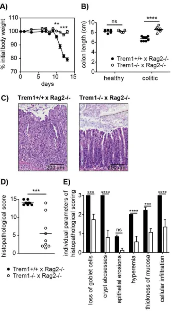
Trem12/2x Rag22/2recipient mice and animals were monitored regularly for clinical signs of colitis. Importantly, whereasTrem1+/+ x Rag22/2mice lost,20% of their initial body weight at the end of the observation period, weight loss inTrem12/2x Rag22/2mice was minimal and only transient (Fig. 3A). Furthermore, shortening of the colon was substantially attenuated inTrem12/2x Rag22/2
mice compared to controls (Fig. 3B). While some of theTrem12/2 x Rag22/2mice still exhibited moderate histopathological signs of
intestinal inflammation, individual parameters of the histopatho-logical scoring as well as the overall histopathohistopatho-logical score were significantly reduced (Fig. 3C–E).
In order to gain insight in the potential underlying mechanism of the highly attenuated colitis in Trem12/2 x Rag22/2 mice,
colonic lamina propria cells that were isolated from both groups of mice in the absence of an adoptive CD4+
T cell transfer (healthy colon) or 12–13 days post colitis induction were analysed by FACS. As depicted in Fig. 4A, the colonic lamina propria of healthyTrem1+/+
x Rag2+/+
mice andTrem12/2 x Rag22/2 mice contained a similar proportion of CD11b+
MHCIIhi cells and Gr1+cells were virtually absent. In contrast, Gr1+cells,
repre-senting infiltrating Ly6Chi Gr1int monocytes and Ly6Cint Gr1hi neutrophils, were readily detected inTrem1+/+
x Rag2+/+
mice and
Figure 1. TREM-1 surface expression by peripheral blood myeloid cell subsets from wildtype versusTrem1-deficient mice.
Peripheral blood cells obtained from wildtype (Trem1+/+) andTrem1 -deficient (Trem1+/2 andTrem12/2) mice (n = 2 mice for each group) were stained for surface expression of TREM-1 and analysed by FACS. (A) Representative gating strategy to identify neutrophils and LyC6lo and Ly6Chimonocytes. (B) Representative histograms showing TREM-1
Trem12/2 x Rag22/2 mice at 12–13 days post colitis induction (Fig. 4A). Notably, the relative frequency of Gr1+ cells among
CD45+CD11b+colonic LP cells was
,5-fold lower inTrem12/2x
Rag22/2 mice (Fig. 4A). In further contrast to the colonic LP of healthy mice, among LP MHCII+Gr12cells of colitic mice two
populations of MHCIIhiLy6Cloand MHCIIintLy6Chicells could be discriminated, likely representing intestinal macrophages and monocytes in the process of differentiation (Fig. 4A) [35,36]. Also within this gate of MHCII+
Gr12 cells, substantial differences could be detected between the two groups of mice. Accordingly, the relative frequency of MHCIIintLy6Chicells was increased in colitic Trem1+/+
x Rag22/2 mice whereas Trem12/2 x Rag22/2
mice exhibited a larger proportion of MHCIIhi Ly6Clo macro-phages (Fig. 4A).
When colonic LP cells of n = 9 mice of both groups were systematically analysed at 12–13 days post colitis induction, substantially reduced numbers of various cell subsets could be seen inTrem12/2x Rag22/2mice (Fig. 4B). Thus,Trem12/2x Rag22/2
mice not only exhibited reduced infiltrating CD4+T cells but also
significantly decreased numbers of neutrophils, Ly6Chimonocytes and MHCIIintLy6Chicells (Fig. 4B). These differences were not apparent for the colonic LP of healthyTrem1+/+
x Rag22/2 and
Trem12/2 x Rag22/2 mice which mainly contained MHCIIhi Ly6Clocells anyway (Fig. 4A and 4C).
Figure 2. Unimpaired hematopoiesis inTrem12/2mice.(A) Representative dot plots show the FACS-based identification of lineage-depleted (lin2) Sca1+c-kithi(LSK) cells and lin2Sca12c-Kithimyeloid progenitors inTrem1+/+(top panels) andTrem12/2(bottom panels) bone marrow (BM) following lineage depletion and depletion of lymphoid progenitors by MACS. Common myeloid progenitors (CMP), granulocyte-macrophage progenitors (GMP) and megakaryocyte/erythrocyte progenitors (MEP) were further discriminated according to their expression of FccR and CD34. Filled histograms show TREM-1 surface expression by LSK cells, CMP, GMP and MEP progenitors fromTrem12/2mice in comparison toTrem1+/+mice (lines). (B) Absolute cell numbers of total BM cells, lin2BM cells, lin2Sca12c-kithimyeloid progenitors, LSK cells, CMP, GMP and MEP and colony
forming units (CFU) of hematopoietic precursors isolated from the BM ofTrem1+/+andTrem12/2mice were determined as described in the Materials and Methods section. Mean values of n = 2 mice analysed are shown with error bars indicating the range. (C) Mixed BM chimeras were generated by i.v. transfer of 1:1 mixedTrem1+/+x GFP+/+andTrem12/2x GFP2/2BM cells (white circles, dotted lines) into irradiated recipient mice. As control, and to account for potential interfering effects of the GFP expression, mixed BM fromTrem1+/+x GFP+/+
andTrem1+/+x GFP2/2mice (black circles and lines) was transferred into additional recipient mice. BM chimeras were analyzed after 10 and 31 weeks of chimerism. Neutrophils, Ly6Chiand Ly6Clo
monocytes were identified in the peripheral blood according to the depicted gating strategy and the GFP2: GFP+
cell ratio in the respective cell subsets was determined. Mean values of n = 4–5 mice analyzed per group are shown with error bars indicating the SEM. ns, no statistically significant difference. Data depicted in Figure 2 are representative of two independent experiments.
doi:10.1371/journal.ppat.1003900.g002
To gain more insight which myeloid TREM-1-expressing cell subset could potentially be involved in driving intestinal inflam-mation inTrem1+/+
x Rag22/2mice, TREM-1 surface expression was analysed on colonic LP neutrophils, Ly6Chimonocytes as well as CD11b+
Ly6C+
Gr12 and CD11b+
Ly6C2 Gr12 cells. As reported previously [23,37], in the healthy colonic LP TREM-1
expression was hardly detectable owing to the absence of infil-trating neutrophils and Ly6C+
cells (Fig. 4A and 4D). In colitic
Trem1+/+
x Rag22/2mice, TREM-1 expression was observed on neutrophils, Ly6Chi monocytes and CD11b+
Ly6C+
Gr12 cells (Fig. 4D). Moreover, CD11b+Ly6C2Gr12cells, likely
represent-ing intestinal macrophages, that were isolated from coliticTrem1+/+ x Rag22/2 mice exhibited a ,3-fold upregulated expression of surface TREM-1 (Fig. 4D).
In line with the reduced infiltrating cell numbers, mRNA expres-sion for various innate and adaptive pro-inflammatory chemokines and cytokines was significantly decreased in the lamina propria of
Trem12/2 x Rag22/2 compared to Trem12/2 x Rag22/2 mice (Fig. 4E).
Dextran sodium sulfate (DSS)-induced colitis is attenuated inTrem12/2mice
While protection from colitis inTrem12/2x Rag22/2mice was associated with reduced expression of several pro-inflammatory mediators, previous data generated in our laboratory have demonstrated that TNF produced by nonlymphoid cells plays a non-redundant pathogenic role in the CD4+
T cell transfer model of colitis sinceTnf2/2 x Rag22/2 mice are completely protected from colitis induction [38]. However, in acute models of intestinal inflammation such as the DSS-induced colitis, Tnf2/2 mice exhibit aggrevated disease [39,40], presumably, because early anti-microbial and repair responses following DSS-induced breaching of the epithelial barrier are fundamentally impaired. Due to the central function of TREM-1 in amplifying pro-inflammatory cytokine production and oxidative burst, we hypothesized that during acute intestinal inflammation complete absence ofTrem1
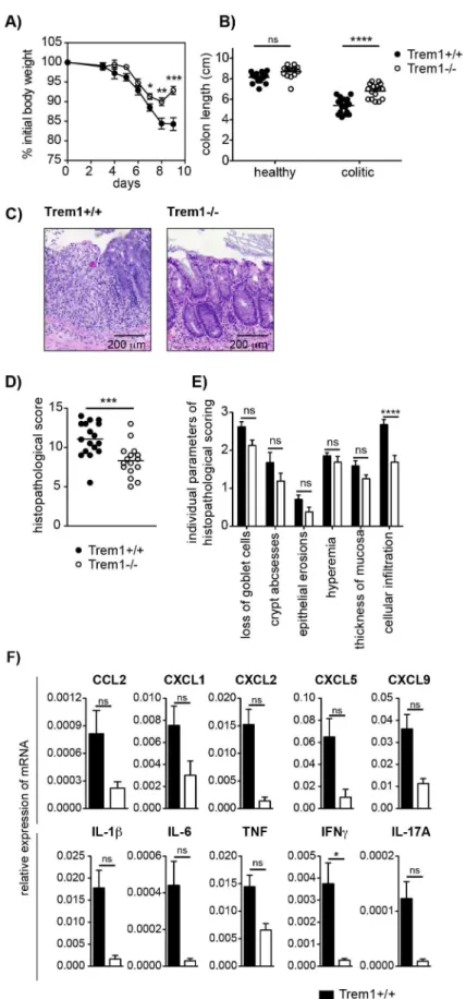
could also prove detrimental. Intriguingly, although upon admin-istration of 3% DSS Trem12/2 mice initially lost weight to a similar extent as Trem1+/+
mice, weight loss was considerably attenuated at 7 days post colitis induction and at 9 daysTrem12/2
mice had already improved again (Fig. 5A). InTrem12/2 mice, shortening of the colon was markedly attenuated and total histopathological colitis scores were significantly decreased (Fig. 5B and 5C–E). Furthermore, analogous to the CD4 T cell-induced colitis (Fig. 4E), TREM-1 deficiency resulted in reduced colonic mRNA expression of several pro-inflammatory mediators (Fig. 5F). Hence, in contrast toTnf2/2 mice [39,40],Trem12/2
mice still showed an adequate host response to DSS-induced epithelial injury while exhibiting reduced immune-mediated pathologies.
Infection withLeishmania majorleads to smaller
inflammatory lesions with decreased neutrophilic cellular infiltrates inTrem12/2mice
The observations made in the acute DSS model of colitis raised our interest whetherTrem12/2mice would also be able to control
bona fidemicrobial infections, in particular, since maximal silencing of TREM-1 by a siRNA approach had proven deleterious in a fecal peritonitis model [29]. Since the rapid kinetics of this model hardly allowed to simultaneously look at beneficial effects of the
Trem1deficiency on immune-mediated tissue damage or to assess potential adverse consequences for the priming of adaptive immune responses, we chose theLeishmania majorinfection model. Following infection with L. major, C57BL/6 mice develop local cutaneous lesions that spontaneously resolve within 4–8 weeks. Central to the resolution is the TNF-mediated control of the early inflammatory response or the clearance of neutrophils and the later IFNc-mediated and Th1-driven elimination of the parasite by infected macrophages [41,42].
Figure 3.Trem12/2x Rag22/2mice are protected from a CD4+T
cell-induced colitis.Colitis was induced inTrem1+/+x Rag22/2(filled circles) andTrem12/2x Rag22/2mice (white circles) by i.p. injection of 26105CD4+CD45RBhiT cells. (A) Weight loss relative to the initial body
weight. Mean values of n = 9 mice analysed per group are shown with error bars indicating the SEM. (B) Colon lengths were determined in individual mice (symbols). Lines show mean values for each group of mice. (C) Representative H&E-stained colonic tissue sections of a Trem1+/+x Rag22/2(histopathological score: 14) andTrem12/2x Rag22/2 mouse (histopathological score: 2). (D) Total histopathological scores. Symbols show total scores for individual mice and lines indicate the mean value for each group of mice. Histopathological scores were determined for individual mice by a pathologist according to parameters defined in the Materials and Methods section. (E) Individual parameters of histopathological scoring. Columns show mean values for n = 9 mice analysed per group and error bars indicate the SEM. ****, p,0.0001; ***, p,0.001; **, p,0.01. One representative experiment out of three independent experiments is shown.
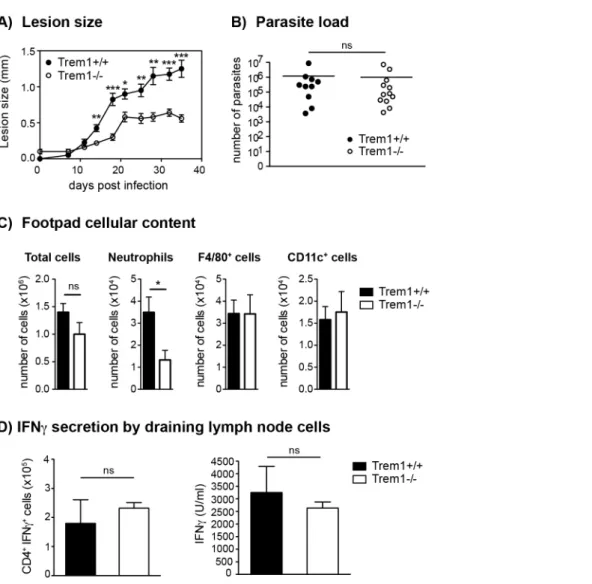
Upon injection of 36106 L. major promastigotes s.c. in the footpad ofTrem1+/+
andTrem12/2mice, an attenuation in lesion
development was apparent inTrem12/2 mice already at 14 days post infection. From thereof, Trem12/2 mice showed a signifi-cantly decreased lesion size (Fig. 6A). Notably, however, parasite counts did not differ between Trem12/2 and Trem1+/+
mice (Fig. 6B). We further analysed the cellular composition of infected footpads from Trem12/2 and Trem1+/+ mice at 21 days post
infection. While the overall cell counts were comparable, the cellular infiltrate inTrem12/2mice exhibited,3-fold reduced neu-trophil numbers (Fig. 6C). To look at the potential impact of the
Trem1-deficiency on the priming of Th1 responses, expression of IFNcwas analysed in cells isolated from the draining lymph node. The frequency of IFNc-secreting CD4+T cells was similar in Trem12/2andTrem1+/+mice (Fig. 6D). In addition, comparable
levels of IFNcwere detected in cells of both groups of mice upon re-stimulation in vitrowith the parasitic antigen (Fig. 6D). These data are in line with comparable parasite killing observed in
Trem12/2 and Trem1+/+ mice. Thus, in the L. major infection
model, absence of Trem1 does not appear to have adverse consequences on the priming of adaptive immune responses and on parasite control while neutrophil-mediated inflammatory lesion development is substantially reduced.
StimulationviaTREM-1 induces TNF secretion and resistance to apoptosis in SCF-condHoxb8 progenitor-derived neutrophils
The reduced neutrophil numbers atL. major-infected sites and the decreased lesion size inTrem12/2mice agree with the notion that neutrophils play a central role in inflammatory lesion development. Indeed, the presence of non-healing lesions in L. major susceptible BALB/c strains is associated with elevated numbers of neutrophils [43]. Since neutrophils constitutively express high levels of TREM-1 (Fig. 1B, 4D), we aimed to investigate the consequences of TREM-1-mediated stimulation on their functional responses in more detail. Analysis of mouse neutrophils has so far been complicated by the limited numbers of cells that can be retrieved from the peripheral blood, their short life span or the distinct differentiation stages of BM-derived neutrophils. Hence, we made use of a recently described system by which neutrophils can be differentiated ex vivoin large numbers using conditional Hoxb8 [44]. Using a slightly modified protocol, BM-derived progenitors were lentivirally transduced with condi-tional Hoxb8 in the presence of SCF, resulting in immortalized myeloid progenitor lines, termed SCF-condHoxb8. Upon shutdown of Hoxb8 expression by withdrawal of 4-OHT, cells differentiate into mature neutrophils in the presence of SCF. As shown in Figure 7A and 7B, withdrawal of 4-OHT in fact induced the appearance of cells bearing the characteristic phenotype of mouse neutrophils after in vitro differentiation for 5 days. Moreover,
Trem1+/+
SCF-condHoxb8-derived mature neutrophils also expressed
distinct levels of surface TREM-1 (Fig. 7B). Stimulation ofTrem1+/+
, but not ofTrem12/2, SCF-condHoxb8-derived mature neutrophils with a plate-bound agonistic anti-TREM-1 antibody resulted in pronounced mRNA expression of iNOS and TNF (Fig. 7C) and induced rapid secretion of TNF at levels comparable to those triggered by LPS (Fig. 7D).
Since neutrophil survival versus apoptosis could represent a deciding factor in the control of inflammation not only in theL. majorinfection model but also in the pathogenesis of experimental colitis [45], we compared the susceptibility of Trem1+/+
and
Trem12/2 neutrophils to spontaneous apoptosis. Following pro-longed culture of fully differentiated SCF-condHoxb8-derived neutrophils in vitro, an increasing and comparable frequency of AnnexinV+DAPI+cells was detected for Trem1+/+andTrem12/2
neutrophils (Fig. 7E). However, in the presence of TREM-1-mediated stimulation a reduced apoptosis rate based on dimin-ished Caspase 3/7 activity could be observed forTrem1+/+
neu-trophils (Fig. 7F). Furthermore, agonistic anti-TREM-1 stimula-tion ofTrem1+/+
but notTrem12/2neutrophils resulted in,2-fold upregulated mRNA expression of Myeloid Cell Leukemia-1 (Mcl-1) (Fig. 7G), analogously to what has recently been described for human TREM-1-stimulated monocytes [46].
Thus, TREM-1-mediated activation of mouse neutrophil appears to contribute to their survival.
Influenza virus-infectedTrem12/2 mice exhibit reduced morbidity but an equal capacity for viral clearance
Intrigued by the substantially diminished inflammatory lesions, yet intact parasite clearance inL. major-infectedTrem12/2 mice, we aimed to substantiate these findings in an altogether different infection model. Since high expression of TREM-1 by alveolar macrophages and previously published data [28,30,47,48] suggest a potential role for TREM-1 in lung inflammatory responses, we infected Trem1+/+ and Trem12/2 mice intratracheally with
50 PFU influenza A virus PR8. Hypothermia and body weight loss, which are characteristically associated with experimental influenza virus infection, were observed inTrem1+/+
mice at 7 days post infection (Fig. 8A and 8B). While the body temperature also dropped inTrem12/2mice, a quicker recovery from hypothermia was seen in theTrem12/2group (Fig. 8A). Moreover, weight loss inTrem12/2mice was significantly attenuated andTrem12/2mice further exhibited reduced levels of IL-6 in bronchoalveolar lavage fluid (BALF) at 10 days post infection (Fig. 8B and 8C). Notably, in spite of the reduced morbidity observed, Trem12/2 mice were equally competent in clearing the influenza virus infection as
Trem1+/+
controls (Fig. 8D).
Trem12/2mice are equally capable of clearingL. pneumophilaasTrem1+/+controls
After having established that deficiency in TREM-1 attenuates disease but does not impair pathogen control during a parasitic
Figure 4. Upon colitis induction,Trem12/2x Rag22/2mice exhibit substantially reduced inflammatory infiltrates and diminished expression of pro-inflammatory mediators.(A–C) Lamina propria cells were isolated from the colon ofTrem1+/+x Rag22/2andTrem12/2x Rag22/2mice 12–13 days post adoptive transfer of colitogenic CD4 T cells or from untransferred mice (healthy colons) and analysed by FACS. (A) After exclusion of doublets and dead cells, CD11b+
cells were discriminated from CD4+
T cells and further subgated into MHCIIloGr1+
(gate 1) and MHCIIhiGr12(gate 2) cells. In gate 1, monocytes and neutrophils were identified according to their Ly6ChiGr1intand Ly6CintGr1hiphenotype,
respectively. In gate 2, MHCII+
cells were further subdivided into two populations of MHCIIintLy6Chiand MHCIIhiLy6Clocells. (B, C) Absolute numbers
of total cells recovered from individual mice (symbols; lines indicate mean values per group) and mean values6SEM for CD45+cells, CD4+T cells, CD11b+
cells and subsets defined within the CD11b+
gate as illustrated in (A). Per group, n = 9 mice adoptively transferred with CD4 T cells (B) and n = 4 untransferred (C) mice were analysed. (D) TREM-1 surface expression by neutrophils (Ly6CintGr1hi), monocytes (Ly6ChiGr1int) and CD11b+Gr12 Ly6C+
versus Ly6C2subsets identified in the lamina propria (according to the gating strategy depicted in D) of colitic (n = 9) versus healthy (n = 4) Trem1+/+x Rag22/2mice. (E) Colonic tissues were assessed for the expression of pro-inflammatory mediators by qRT-PCR. Bars show mean values
6
and viral infection, we last sought to address the significance of TREM-1 in a bacterial infection model. Indeed, controversial results have been obtained with respect to the importance of TREM-1 in microbial control following infection of experimental animals withPseudomonas aeruginosa[28,30]. Here, we employed a
Legionella pneumophilainfection model which also causes severe upper airway inflammation in permissive mice and critically depends on neutrophil-mediated control [49,50,51]. As shown in Figure 9, at 3 days post infection with 56106CFU ofL. pneumophila, CFU and neutrophil numbers in the BALF did not significantly differ between
Trem1+/+andTrem12/2mice. Furthermore, at day 5 post infection
CFU were reduced to,500/BALF in both groups of mice with no significant differences observed betweenTrem1+/+
and Trem12/2
mice (data not shown).
Discussion
The significance of TREM-1 as a central amplifier of acute pro-inflammatory responses during endotoxin-induced shock and microbial sepsis is well established. However, increasing evidence, stained colonic tissue sections of a Trem1+/+
(histopathological score: 13) and Trem12/2 mouse (histopathological score: 5.5). (D) Total histopathological scores. Symbols show total scores for individual mice and lines indicate the mean value for each group of mice. Histopathological scores were determined for individual mice by a pathologist according to parameters defined in the Materials and Methods section. (E) Individual parameters of histopathological scoring. Columns show mean values and error bars indicate the SEM. (F) Colonic tissues were assessed for the expression of pro-inflammatory mediators by qRT-PCR. Bars show mean values6SEM for n = 7 mice per group from one independent experiment. (A, B, D) Pooled data from three independent experiments are shown. ****, p,0.001; ***, p,0.001; *, p,0.05.
doi:10.1371/journal.ppat.1003900.g005
Figure 6.Trem12/2mice develop smaller inflammatory lesions and show decreased cellular infiltrates atL. majorinfection sites.(A) Trem1+/+andTrem12/2mice were inoculated with 3
6106L. majorpromastigotes s.c. in the footpad and lesion development was measured over time. Each data point represents the mean lesion size6SEM of n = 5 mice analysed per group. (B) Parasite load was assessed at 35 days post infection (p.i.) by limiting dilution analysis. (C) Infected footpads fromTrem1+/+andTrem12/2mice (n = 4–5 mice per group) were isolated 21 days p.i., digested and the cellular content was analysed by flow-cytometry. Data show mean values6SEM and are representative of two independent experiments. (D) Draining lymph node cells fromTrem1+/+
andTrem12/2mice (n = 4 mice per group) were isolated 35 days p.i.; the frequency of CD4+ IFNc+
T cells was analysed by intracellular FACS staining or cells were re-stimulated with UV-treatedL. majorparasites and IFNclevels in the supernatants were assessed by ELISA. Data show mean values6SEM of triplicate measurements. Representative data from one out of three independent experiments are shown. ***, p,0.001; **, p,0.01; *, p,0.05.
Figure 7. TREM-1 mediates TNF secretion and resistance to apoptosis in SCF-condHoxb8 progenitor-derived neutrophils.
SCF-condHoxb8Trem1+/+
andTrem12/2progenitor lines were generated by lentiviral transduction of Hoxb8 into BM-derived hematopoietic cells obtained from the respective mice and cultured in the presence of SCF and 4-hydroxytamoxifen (4-OHT). Forin vitroneutrophil differentiation, SCF-condHoxb8 cells were cultured for additional 5–6 days in the absence of 4-OHT. (A) H&E stained cytospins ofTrem1+/+
SCF-condHoxb8 progenitor cells (left) and differentiated neutrophils (right). (B) FACS-based characterization of Trem1+/+ (top panels) and Trem12/2 (bottom panels) SCF-condHoxb8 progenitor cells (left) and differentiated neutrophils (right). (C) TNF and iNOS mRNA expression by Trem1+/+
and Trem12/2 SCF-condHoxb8 differentiated neutrophils following 2 h stimulation with plate-bound agonistic anti-TREM-1 mAb or an isotype control antibody was
determined by qRT-PCR. (D) TNF secretion byTrem1+/+andTrem12/2SCF-condHoxb8 differentiated neutrophils in response to stimulation with an
agonistic anti-TREM-1 mAb or an irrelevant isotype control mAb in the presence or absence of LPS (100 ng/ml) was assessed by ELISA. (E) Spontaneous apoptosis ofTrem1+/+andTrem12/2SCF-condHoxb8 neutrophilsin vitrowas analysed at 5 days post differentiation with SCF (0 h) and
including the recently reported association of TREM-1 with the DAMP protein HMGB1 [26,27], also suggests a potential role for TREM-1 during non-infectious and chronic inflammatory condi-tions. In line with this notion, we have previously described a crucial involvement of TREM-1 in IBD as based on the significant amelioration of experimental colitis upon blockade of TREM-1 with the antagonistic LP17 peptide [22]. Blocking TREM-1 signaling by daily administration of TREM-1-Ig fusion proteins or synthetic analogues in chronic disease models is not only costly and straining but also fails to cover for the possibility that the yet unidentified TREM-1 ligand may signal through alternative receptors.
Here, we have generated aTrem12/2mouse to unambiguously investigate the impact of a complete TREM-1-deficiency on the pathogenesis of experimental colitis but also of two other distinct sub-acute disease settings where the role of TREM-1 has so far not been addressedin vivo, i.e. inflammation induced by a parasitic and viral infection. Our findings demonstrate thatTrem12/2mice not only show a highly attenuated CD4+T cell- and DSS-induced
colitis but also display significantly reduced lesion size and diminished morbidity during infections withL. majorand influenza virus, respectively.
The substantial attenuation of illness and immune-mediated pathologies inTrem12/2mice across these distinct models suggests a common mechanism by which TREM-1 signaling promotes
inflammation irrespective of the original trigger. Several non-exclusive scenarios can be considered that may account for the attenuated disease inTrem12/2mice:A priorireduced chemotactic recruitment ofTrem12/2neutrophils and monocytes, diminished pro-inflammatory activities, and/or reduced life-span of myeloid cell subsets. Although we observed significantly decreased num-bers of distinct myeloid cell subsets in the LP of coliticTrem12/2x Rag22/2mice and atL. major-infected sites inTrem12/2mice, we consider it unlikely that deficiency in TREM-1 causes an intrinsic primary defect in chemotaxis. When we analysedL. majorinfected sites at an early time-point, i.e. 3 days post infection, no differences in cellularity were detected betweenTrem1+/+
andTrem12/2mice (data not shown). Moreover, a recent study clearly demonstrated that TREM-1/3 proteins are not required for transendothelial migration of neutrophils [30]. Nonetheless, the markedly de-creased expression of mRNA for monocyte- (CCL2), granulocyte-(CXCL1, CXCL2, CXCL5) but also T cell (CXCL9) chemoat-tractants in the LP of Trem12/2 x Rag22/2 mice will in a secondary manner certainly have contributed to the decreased accumulation of inflammatory cells. Besides the diminished expression of chemotactic mediators, the colonic LP of colitic
Trem12/2 x Rag22/2 mice also exhibited substantially reduced mRNA levels of several innate cytokines, including IL-1b, IL-6 and TNF. The reduced expression of pro-inflammatory mediators in the entire colonic LP inTrem12/2x Rag22/2mice at the late in vitroreplicates from one representative experiment out of three independent experiments. (F) Caspase 3/7 activity was assessed upon 24 h stimulation of differentiatedTrem1+/+andTrem12/2SCF-condHoxb8 neutrophils with plate-bound agonistic anti-TREM-1 mAb, an isotype control
antibody, or LPS (100 ng/ml) as a positive survival control. Bars show mean values6SEM for n = 3in vitroreplicates from one representative experiment out of three independent experiments. n.d., not detected. (G) Expression of Mcl-1 mRNA was assessed by qRT-PCR 2 h post stimulation with plate-bound anti-TREM-1 or an isotype control mAb in the presence or absence of LPS. Bars show mean values6SEM of three independent experiments for anti-TREM-1 vs. isotype control mAb stimulated cells. n.d., not detected. ****, p,0.01; **, p,0.01; *, p,0.05.
doi:10.1371/journal.ppat.1003900.g007
stage of colitis induction certainly also mirrors the decreased cellular infiltration. However, considering the capacity of TREM-1 to augment the production of several chemotactic mediators either directly in infiltrating myeloid cells or likely also indirectly, in a paracrine manner (e.g. through secretion of IL-1b) [4,22,52], we believe that TREM-1-amplified production of pro-inflamma-tory cytokines represents a key early pathogenic event that will ultimately determine the later disease course and may largely account for the attenuated disease in Trem12/2 mice. In this respect, it is noteworthy that the colonic LP of colitic Trem12/2
mice contained markedly fewer MHCIIint Ly6Chi cells or inflammatory macrophages with the capacity for expression of pro-inflammatory mediators [35,36].
As we have employed a CD4+
T cell-dependent colitis model and indeed observed considerably reduced CD4+
T cell numbers and correspondingly also mRNA levels for IFNcand IL-17 in the colonic LP of transferredTrem12/2x Rag22/2mice, the question arises whether deficiency in TREM-1 may directly impact the priming of adaptive immune responses. Whereas in the colitis model we have not analysed CD4+
T cell responses in more detail, CD4+
T cells isolated from L. major-infected and CD8+
T cells retrieved from influenza virus-infected Trem12/2 mice,
respec-tively, exhibited an unimpaired capacity for IFNc production compared to T cells fromTrem1+/+mice.
The deciding role of neutrophils in theL. majorinfection model [42] and the substantially decreased lesion size inTrem12/2mice have prompted us to investigate the impact of TREM-1 on neutrophil-mediated functions in more detail. In particular, we were interested in the potential modulatory effect of TREM-1 ligation on neutrophil survival as delayed neutrophil apoptosis could also represent a critical pathogenic factor in intestinal inflam-mation [45]. In agreement with a previous report [30], deficiency in TREM-1 caused no intrinsic predisposition for increased sponta-neous neutrophil apoptosis. However, agonistic TREM-1 stimulation significantly promoted survival of SCF-condHoxb8 progenitor-derived neutrophils in vitro. We have also sought to assess the frequency of apoptotic neutrophils in Trem1+/+
versus Trem12/2
micein situby staining colonic tissue sections from colitic mice for cleaved caspase 3 (data not shown). However, due to the likely very rapid clearance of apoptotic neutrophils by intestinal phagocytes, cleaved caspase 3 positive cells were rare in Trem1+/+
mice and even more scarce inTrem12/2 mice. Moreover, the substantially decreased cellular infiltrate inTrem12/2mice further precluded an objective comparison and quantification of apoptotic cellsin situ. While we cannot present definitive evidence that TREM-1 prolongs neutrophil survivalin vivo, we still believe that the reduced presence of neutrophils inTrem1-deficient mice in the CD4 T cell induced colitis and L. major infection model relates to both: A reduced secondary recruitment based on the diminished expression of neutrophil chemotactic mediators and to a reduced life-span in the absence of TREM-1 ligation, with the former mechanism numerically perhaps being more relevant.
Considering the various effector cell types and mechanisms by which TREM-1-mediated stimulation could contribute to disease, it may appear intriguing that deficiency in TREM-1 did not completely protect from disease. Accordingly, the degree of protection from colitis was not much higher inTrem12/2 mice
compared to mice that were treated with the antagonistic LP17 peptide in our previous study [22]. We hypothesize that the absence of complete protection inTrem12/2mice may primarily relate to the role of TREM-1 as an amplifier but not inducer of pro-inflammatory reactions [3,5]. While we cannot rule out a potential participation of TREM-3, we believe that the protective effects seen in Trem12/2 mice are too substantial for a major involvement of TREM-3 in the inflammatory models analysed.
One of the most striking findings of the present study was the observation that microbial control in the models analysed was apparently not impaired inTrem12/2mice in spite of the blunted inflammatory responses. Hence, while Tnf2/2 or anti-TNF-treated mice exhibit an aggrevated acute DSS-induced colitis [39,40] and also show enhanced parasite and bacterial burdens upon infection with L. major and L. pneumophila, respectively [41,53],Trem12/2mice appeared equally capable of controlling a parasitic, viral and bacterial infection asTrem1+/+controls. This
observation is in line with the main function of TREM-1 as an inflammatory fine-tuner which still allows for pro-inflammatory TLR or NOD-like receptor-induced reactions in its absence. Moreover, TREM-1 does not appear to be involved in phagocytic or direct antimicrobial activity of myeloid cells [30,54,55]. Still, conflicting data on the effect of a TREM-1 blockade on microbial control have been reported from various bacterial challenge models. Injection of a TREM-1/IgG fusion protein allowed for sufficient control of an E. coli-induced peritoneal infection and conferred protection [9] whereas maximal but not half-dose siRNA silencing of TREM-1 increased mortality in a fecal peritonitis model [29]. Similarly, administration of the antagonistic LP17 peptide protected rats from a P. aeruginosa-induced pneu-monia [28], whereas complete deficiency in TREM-1/3 led to markedly increased mortality inPseudomonas aeruginosa-challenged mice due to defective transepithelial migration of neutrophils [30]. It has been proposed that the degree of TREM-1 blockade was a likely critical parameter to account for these disparant findings [29,30]. However, our findings demonstrate that microbial control must not necessarily be impaired in mice with a complete deficiency in TREM-1, even when employing a L. pneumophila
infection model where early neutrophil accumulation is also crucial for bacterial clearance [49,50,51]. Thus, we speculate that analogous to the divergent results obtained for endotoxin-challenged DAP12-deficient mice [56,57], possibly the infection dose, the nature of the microbial agent and/or the kinetics of the infection may be critical parameters regarding the requirement for
Figure 9. Trem12/2 mice are equally capable of clearing L. pneumophila as Trem1+/+
controls. Trem1+/+ and Trem12/2 mice were infected intranasally with 56106CFUL. pneumophila. 3 days post
infection CFU (A) and neutrophils (B) were quantified in the BALF. ns, no statistically significant difference. Pooled data from two independent experiments are shown.
doi:10.1371/journal.ppat.1003900.g009
TREM-1. Accordingly, in the presence of low abundance and/or low affinity TLR ligands or fast replicating agents, the inflamma-tory response may more stringently depend on TREM-1-amplified signaling. Alternatively, the expression of TREM-1 and its association with DAP12 may only preferentially be induced in situations of high abundance and/or high affinity TLR ligands whereas low level signaling would favour the engagement of TREM-2.
In summary, while the impact of TREM-1 on microbial control still needs further investigations across different experimental models, our extensive characterisation ofTrem12/2mice shows an
unanticipated prominent role for TREM-1 in parasitic and viral infections. Our findings thus suggest that therapeutic targeting of TREM-1 holds considerable promise for distinct non-infectious and infectious inflammatory disorders and may bypass the increased risk for impaired microbial control which is associated with the general targeting of TNF.
Materials and Methods
Mice
Breedings and cohort maintenance were performed under SPF conditions in isolated ventilated cages in the central animal facility of the Medical School, University of Bern. All studies were conducted with age- and sex-matched animals.Trem1+/+
mice that were used as wildtype controls were originally derived from the interbreeding of Trem1+/2 x Cre+/2 mice, thus carrying the
identical C57BL/6 genetic background and representing former littermates ofTrem12/2mice. To nonetheless adjust for potential differences in the microflora composition inTrem12/2vs.Trem1+/
+
mice, the bedding ofTrem1+/+
andTrem12/2mice was regularly exchanged between cages three weeks prior to the start of the experiments.
Ethics statement
All animal experiments were approved by the Veterinary Offices of the Cantons of Bern, Lausanne and Zurich and per-formed in compliance with Swiss laws for animal protection.
Generation ofTrem1-deficient mice
The generation of Trem1-deficient mice was designed and carried out in collaboration with the TaconicArtemis GmbH (Ko¨ln, Germany). To account for potential lethal effects of a total deletion of theTrem1gene and to allow for a possible cell-specific ablation ofTrem1expression, a targeting vector was designed for conditional deletion of exon 2, which encodes the extracellular part of Trem1and also contains the putative ligand binding site [31]. As illustrated in the supplemental material Fig. S1, the targeting vector was constructed on the basis of the cloning vector KS loxP ftr Neo BS to flank exon 2 with loxP sites, to comprise additional restriction sites (AseI and AvaI) and to contain PuroR (flanked by F3 sites) and Neomycin (flanked by FRT sites) positive selection marker cassettes to control for homologous recombina-tion upstream and downstream of exon 2, respectively. For counterselection, a Tk cassette was included downstream of exon 4. As a template for the PCR reactions a BAC-based plasmid containing the entire genomic mouse Trem1 locus (RP23-32N8) was obtained from BACPAC Resources Center BPRC (Oakland, USA). The targeting vector was electroporated into a C57BL/ 6N.tac embryonic stem cell line (TaconicArtemis). On day 2, cells were selected with Puromycin and G418 and on day 8 counter-selection with Gancyclovir was initiated. Isolated and expanded ES clones were screened for complete integration of the targeted allele by standard Southern blotting analyses with probes located
upstream of exon 1 (5e2) or exon 3 (ila1) (Fig. S1). Primer sequences for generation of the 5e2 Southern probes were: CGGATTTGACCAGGAATGACAG(sense) and CTTCCAG-TTCATTCATGGACAGC (antisense) and for the ila1 Southern probe: AGCTCCTCTTGTCTGCCATTCAAGGC (sense) and GGCTACAACCTTGTTCTGCAG (antisense). Eight positive clones could be identified and the ES clone A-A5 was subsequently injected into Balb/c derived blastocytes which were then trans-ferred to pseudopregnant NMRI females. Chimeric offspring were bred to C57BL/6 female mice (C57BL/6-Tg(ACTB-Flpe)2Arte, TaconicArtemis) transgenic for Flp recombinase to achieve deletion of the FRT and F3 flanked selection cassettes PuroR and Neomycin, respectively. Germline transmission of the targeted
Trem1allele was identified by coat color contribution and by PCR using oligo 1_sense (GTGCTCAGAGAATGTCTTTGTATCC) and oligo 4_antisense (CCCTGGTCAGACCATTTACC) which either yield a 1.3 kb fragment for the wildtype (WT) allele or a 1.6 kb fragment for the conditionalTrem1floxallele. (Fig. S1). Cycling conditions were: 59at 95uC followed by 35 cycles consisting of 300at 95uC, 300 at 60uC, 19 at 72uC, followed by 109 at 72uC. Thus identified Trem1+/flox
mice (C57BL/6-TREM-1tm1821_33.1Arte) were mated with male mice carrying the Cre recombinase under the control of the ROSA26 locus (C57BL/6-Gt(ROSA)26Sortm16(Cre)arte, TaconicArtemis) to obtain systemic deletion of one Trem1 allele (Trem1+/2). Trem1+/2 x Cre+/2 mice were interbred to achieve
deletion of Cre and to obtain wildtype controls (Trem1+/+
) and heterozygous (Trem1+/2) and homozygous (Trem12/2) Trem1
-deficient mice. Deletion of exon 2 inTrem1+/2andTrem12/2mice
was assessed by the genotyping PCR strategy described above and depicted in Fig. S1.Trem1+/+
andTrem12/2mice were subsequently expanded for experiments. For the CD4 adoptive transfer model of colitis, Trem12/2 x Rag22/2 mice were generated by crossing
Trem12/2mice with Helicobacter+Rag22/2mice and
interbreed-ing of the F1 offsprinterbreed-ing. The Helicobacter+status of theTrem-12/2x Rag22/2 offspring was confirmed by PCR testing (Microbios GmbH, Reinach, Switzerland).
Flow cytometry (FACS)
The following mAbs were used: anti-mouse CD11b-Pacific Blue (M1/70), CD45-Pacific Blue (30-F11), CD45-Brilliant Violet570 (30-F11), CD4-APC-Cy7 (RM4-5), Gr1-PE (RB6-8C5), NK1.1-PE-Cy7 (PK136), Ly6G-APC-Cy7 (1A8) and F4/80-biotin (BM8) IL-7Ra-biotin (A7R34), CD3e-biotin (145-2C11) CD19-biotin (6D11), Gr1-biotin (RB6-8C5) and Ter119-biotin (TER119), all purchased from Biolegend; anti-mouse CD115-PE (AFS98), Gr1-APC (RB6-8C5), CD45-eFluor605 (30-F11), CD45.1-PE (A20), MHCII-APC (M5/114.15.2), CD11c-PE (N418), CD11b-eFluor450 (M1/70), Streptavidin-PE-Cy7, were purchased from eBioscience (San Diego, USA); anti-mouse Ly6C-FITC (AL-21) from BD Pharmingen (San Diego, USA) and anti-mouse TREM-1-APC (174031) from R&D Systems. DAPI (Invitrogen) was used in a final concentration of 0.5mg/ml to exclude dead cells. Prior to
FACS stainings, Fc receptors were blocked using supernatant from the hybridoma 2.4G2. Cells were acquired on a LSRII SORP (BD Biosciences, San Diego, USA) and analysed using FlowJo cyto-metric analysis program (Tree Star, Ashland, USA).
Impact of TREM-1 on hematopoiesis
LS columns (Miltenyi Biotec). Lymphoid progenitors were further removed by adding anti-IL-7Ra-biotin.
Determination of colony forming units. 3.336103 lin2 cells were transferred into methocult base medium (M3134; Stemcell Technologies) supplemented with 15% FCS, 20% BIT (50 mg/ml BSA in IMDM, 1.44 U/ml recombinant-human (rh) insulin (Actrapid, Novo Nordisk) and 250 ng/ml human holo transferrin [Prospec]), 100mM 2-b-mercaptoethanol, 2 mM
l-glutamine, penicillin/streptomycin, and 50 ng/ml recombinant-mouse SCF (rmSCF), 10 ng/ml rm–IL-3, 10 ng/ml rh-IL-6 and 50 ng/ml rm-Flt3-ligand (all from Prospec). Colonies and cells were enumerated after 7 days ($30 cells/colony).
Generation of mixed bone marrow chimeras. Congenic (CD45.1+
) recipient mice were irradiated in two split doses with 650 cGy in a 4 h interval in a Gammacell 40 exactor (Best Theratronics). Total donor bone marrow (BM) was collected from
Trem12/2, Trem1+/+
and Trem1+/+
x GFP+/+
mice as described below. Donor BM was mixed 1:1 and 156106total cells of either mixed Trem12/2 and Trem1+/+ x GFP+/+ BM cells or mixed Trem1+/+
and Trem1+/+
x GFP+/+
BM cells were transferred in 200ml PBS i.v. into the irradiated recipient mice. After transfer, the recipients were treated with antibiotics (Baitryl and Bactrim) in the drinking water for two weeks. After 10 weeks, the grade of chimerism was controlled in the peripheral blood by calculating the CD45.2+
/CD45.1+
ratio. In all chimeras, the grade of chimerism among the circulating myeloid cells was at least 99%.
Generation and analysis of SCF-dependent conditional Hoxb8-immortalised progenitor cells and neutrophils
Generation of Hoxb8 progenitor lines and differentiation of neutrophils. The protocol was adapted from the method described by Wang et al. [44] with the major modification of using a different inducible expression system [58]. In brief, bone marrow-derived haematopoietic progenitors were isolated from
Trem1+/+
and Trem12/2 mice by magnetic bead-based lineage depletion (BD IMagTM mouse hematopoietic progenitor cell enrichment set-DM, BD Biosciences) following the manufacturer’s instructions. 2–56105 lineage2 cells were incubated for 36 h in complete RPMI Medium (RPMI 1640/Glutamax supplemented with 10% FCS, 1% penicillin/streptomycin solution and 50mM
2-mercaptoethanol). Cells were then transduced with pF-56 UAS-Hoxb8(mm)-SV40-puro-GEV16 lentiviral particles by spin-infec-tion (1000 rpm, 2 h, 30uC) in presence of 8mg/ml polybrene.
Cells were subsequently cultured in complete RPMI medium supplemented with SCF (added as 10% of CHO/SCF(mm)-conditioned supernatant) and Hoxb8 expression was induced by addition of 100 nM 4-OHT. Transduced cells were positively selected with 1.0mg/ml puromycin for a minimum of one month. The resulting immortalized cell lines were termed SCF-condHoxb8. For in vitro differentiation of SCF-condHoxb8 cells into mature neutrophils, cells were washed twice in PBS to remove all traces of 4-OHT and were then replated at 20’000 cells/ml in complete RPMI medium containing SCF (but no 4-OHT). Mature neutrophils were thus obtained after 5–6 days as judged by morphology and surface expression of CD11b and Gr-1, as well as loss of c-kit (CD117) as determined by flow cytometry.
Apoptosis measurements. Spontaneous apoptosis of ma-tureTrem1+/+versusTrem12/2neutrophils was assessed at 5 days
post differentiation in SCF-containing medium and 8 h, 24 h, and 48 h later. Immediately upon removal from plates, neutrophils were washed in cold PBS and AnnexinV Binding Buffer and stained with FITC-conjugated AnnexinV (BD Pharmingen) according to the manufacturer’s instructions. DAPI (Sigma-Aldrich) was added immediately prior to aquisition by flow cytometry. Only AnnexinV
and DAPI double-positive cells (AnnexinV+
DAPI+
) were consid-ered in the analysis of apoptotic cells. For determination of TREM-1-mediated effects on apoptosis induction, 5 days differentiated neutrophils were stimulatedin vitroin 96-well U-bottom plates at 26105cells/well in complete RPMI medium lacking SCF in the presence of 10mg/ml plate-bound anti-TREM-1 mAb (MAB1187; R&D) or a respective isotype control (RTK2758, Biolegend) for 24 h. Since the relative stickiness of neutrophils activated by plate-bound anti-TREM-1 did not allow for removal from the plates and FACS-based analysis of AnnexinV+DAPI+cells without causing a
substantial bias by the selective analysis of non-adherent cells, Caspase 3/7 activity was determined by the ApoTox-Glo assay (Promega) according to manufacturer’s instructions.
Experimental animal models
CD4 T cell adoptive transfer model of colitis. Colitis was induced in (Helicobacter-positive) Rag22/2 and Trem12/2 x Rag22/2 mice by adoptive transfer of 26105 CD4+
CD252 CD45RBhiFACS-sorted T cells as described previously [59]. Mice were sacrificed at 12–13 days post CD4 T cell transfer at the onset of clinical signs of colitis (diarrhea, weight loss, symptoms of abdominal pain) inTrem1+/+
mice.
Dextran sodium-sulfate (DSS)-induced colitis. Colitis was induced in (Helicobacter-negative)Trem1+/+andTrem12/2 mice
by administration of 3% 36’000–50’000 MW DSS (MP Biomed-icals, Solon, USA) in the drinking water for 5 days followed by 4 days of regular tap water and euthanization at 9 days.
Leishmania major (L. major) infections. L. major LV39 (MRHO/Sv/59/P strain) parasites were maintained in vivo in DBA/2J mice and further cultured in vitro in M199 medium supplemented with 10% FCS, 4% HEPES and 2% antibiotics (penicillin, streptomycin, neomycin). Mice were infected with 36106parasites s.c. in the hind footpad in a final volume of 50ml as previously described [41]. Footpad lesion size was measured with a Vernier caliper. The number of parasites in lesions were evaluated by limiting dilution analysis [60].
Influenza virus infections. Influenza virus strain PR8 (A/ Puerto Rico/34 H1N1) was originally provided by J. Pavlovic (University of Zurich, Switzerland). For infections, mice were anaesthetized and inoculated intratracheally with 50 PFU influ-enza virus in 50ml endotoxin-free PBS. For collection of bronchoalveolar lavage (BAL) fluid, lungs were flushed with 36400ml PBS. To determine influenza viral titers in the lungs, lungs were collected at the indicated time-points, homogenized and serially diluted with MDCK cells as previously described [61]. Legionella pneumophila infections. L. pneumophila strain JR32 (Philadelphia-1; sg1) (reference: PMID: 8225610) was grown for 3 days on charcoal yeast extract (CYE) agar plates at 37uC and resuspended in PBS prior to infection. Mice were anesthetized by i.p. injection of 5mg xylazine/100mg ketamine per gram body weight and infected intranasally with 56106L. pneumophila. Three days post infection, mice were sacrificed and perfused and bronchoalveolar lavage fluid (BALF) was extracted with 1 ml PBS, 5 mM EDTA. CFU were quantified by plating serial dilutions on CYE agar plates and counted after 3 days incubation at 37uC. BALF neutrophils were quantified by flow-cytometry.
Cell isolations
Spleen and peripheral blood. Blood was collected by tail vain incision or by cardiac puncture into heparinised PBS. Spleen cells were released by homogenization of spleens between the frosted ends of two glass slides into PBS containing 5% horse serum. Erythrocytes were depleted by brief incubation with ACK lysis buffer.
Bone marrow. Femurs and tibiae were removed and placed in ice-cold PBS. Remaining flesh was removed and bones were rinsed with sterile PBS. After opening of the bones, the BM was flushed with a 26 GA 3/8 needle into RPMI 10% FCS. Erythrocyte-depleted BM cells were either used for FACS-based characterizations with or without prior lineage depletion or for the generation of mixed BM chimeras.
Colonic lamina propria. Colons were opened longitudinally and cut into small pieces. The epithelium was removed by incubation in HBSS/HEPES containing 5% horse serum, 5 mM EDTA and 2 mM DTT at 37uC for 3630 min under magnetic stirring. Lamina propria (LP) cells were obtained by subsequent digestion with 200 U/ml collagenase (Type IV; Sigma-Aldrich) and 50 U/ml DNase (Type I, grade II; Roche) for 2645 min. The LP fraction was filtered through a 40mM cell strainer, counted by Trypan Blue staining and further characterized by FACS.
Footpads. Footpads were digested using 1 mg/ml collage-nase D in HBSS.
Histopathological analysis of mouse intestinal tissue sections
To assess the presence of histopathological alterations on formalin-fixed, paraffin-embedded and hematoxylin-eosin-stained colonic tissue sections, a scoring system ranging from 0 (no alterations) to 15 (most severe signs of colitis) was established, including the following parameters: cellular infiltration (0–3), loss of goblet cells (0–3), crypt abscesses (0–3), epithelial erosions (0–1), hyperemia (0–2), thickness of the colonic mucosa (0–3). Histolog-ical scoring was performed by a pathologist (V.G.) blinded to sample identity.
RNA extractions and quantitative RT-PCR analyses
RNA was isolated using RNA isolation reagent (Tri-Reagent, Molecular Research Center). DNA was digested using DNase I (Ambion), and cDNA was generated using High Capacity cDNA Reverse Transcription Kit (Applied Biosystems). Expression of genes was analysed using Qiagen Quantitect Primer Assays on a 7500 Real-time PCR System (AB Biosystems). The house keeping gene GAPDH was used for normalization of gene expression.
Analysis of cytokines in supernatants and bronchoalveolar lavage (BAL)
Cell-free supernatants derived from SCF2cond Hoxb8 neutro-phils and BAL fluid from influenza virus infected mice were analysed by ELISA (Biolegend).
Analysis of bone density
X-ray. High resolution X-ray analyses on anesthetized mice were performed with the MX-20 Faxitron (X-ray Corporation, Edimex, LePlessis, France).
MicroCT. For high resolution microcomputed tomography (MicroCT) tissues were fixed in 4% paraformaldehyde in PBS for 24 h and subsequently transferred to 70% EtOH formCT analysis (MicroCT40, Scanco, Bruettisellen, Switzerland).
Statistical analyses
All data were analysed with GraphPad Prism software using the Student’s t-test or 2-way ANOVA.
Supporting Information
Figure S1 Generation of Trem1-deficient mice. Trem1 -deficient mice were generated as described in detail in the
Materials and Methods section. In brief and as depicted in (A), a targeting vector was designed for conditional deletion of exon 2 (coding for the extracellular V-type Ig-like domain) by flanking of exon 2 with loxP sites. The targeting vector was further constructed to contain additional restriction sites (AseI and AvaI), the positive selection markers PuroR flanked by F3 sites and Neo flanked by FRT sites and the counterselection cassette Tk. The vector was electroporated into a C57BL/N.tac embryonic stem (ES) cell line. Genomic DNA of selected ES clones was subjected to enzymatic digestion with either AseI or AvaI and standard Southern blotting analyses with probes located upstream of exon 1 (5e2) or exon 3 (ila1) to identify successful recombination or presence of the correctly targeted allele, respectively. Balb/c-derived blastocytes injected with the so identified targeted ES clone A-A5 were then transferred to pseudopregnant NMRI females and chimerism in the offsprings was assessed by coat colour contribution (white/black). Chimeric offspring were bred to C57BL/6 females transgenic for Flp to achieve Flp-mediated removal of the F3 and FRT flanked PuroR and Neo selection markers, respectively. Germline transmission was identified by the presence of black C57BL/6 offspring, representing heterozygous floxedTrem1(+/flox
) mice that possessed the conditional knockout allele following Flp recombination.Trem1+/flox
female mice were mated with heterozygous Cre-transgenic (Cre+/2) ‘‘deleter’’ males
to generate Trem1+/2 mice with a heterozygous constitutive
knockout allele. Trem1+/2 x Cre+/2 mice were interbred to
generate fully Trem1-deficient (Trem12/2) mice. (A) Schematic presentation of theTrem1wildtype allele, the targeting vector, the targeted allele before and after Flp recombinationin vivoand the final constitutive knockout allele after Cre recombinationin vivo. (B) Southern blot analyses of the ES clone A-A5 demonstrating presence of the targeted allele (TA) following either digestion of genomic DNA with AseI and hybridization with 5e2 probe or digestion with AvaI and hybridization with ila1 probe. Restriction enzyme and Southern blot hybridization sites are indicated in (A). (C) A PCR-based genotyping strategy was developed to identify presence of the wildtype allele, the conditional knockout allele in
Trem1+/flox
mice and the knockout allele inTrem1+/2mice. PCR
primer sites are indicated in (A). (TIFF)
Figure S2 Composition of immune compartments in Trem1+/+
and Trem12/2 mice. Peripheral blood (A), bone marrow (B) and spleen cells (C) from 16 weeks old age- and sex-matched Trem1+/+
(n = 3) and Trem12/2 mice (n = 3) were characterized by FACS. Representative dot plots show the gating strategies for identification of the respective cell subsets and graphs show the mean values6SEM of total cell counts of n = 3 mice per group.
(TIFF)
Acknowledgments
The authors thank the FACS sorting facility of the Department of Clinical Research, University of Bern, for excellent assistance with cell sorting.
Author Contributions