UNIVERSIDADE ESTADUAL PAULISTA – UNESP Faculdade de Medicina de Botucatu
Alterações morfológicas das fibras tipos I e II do
músculo estriado uretral de ratas prenhes diabéticas
Gabriela Marini
Dissertação apresentada ao Programa de Pós-Graduação em Ginecologia, Obstetrícia e Mastologia, da Faculdade de Medicina de Botucatu - UNESP, para obtenção do título de Mestre.
Orientadora: Profa. Titular Marilza Vieira Cunha Rudge
Co-orientadora: Profa. Dra. Angélica Mércia Pascon Barbosa
Co-orientadora: Profa. Dra. Selma Maria Michelin Matheus
BOTUCATU 2010
FICHA CATALOGRÁFICA ELABORADA PELA SEÇÃO TÉCNICA DE AQUISIÇÃO E TRATAMENTO
DA INFORMAÇÃO
DIVISÃO TÉCNICA DE BIBLIOTECA E DOCUMENTAÇÃO - CAMPUS DE BOTUCATU - UNESP
BIBLIOTECÁRIA RESPONSÁVEL: Selma Maria de Jesus Marini, Gabriela.
Alterações morfológicas das fibras tipos I e II do músculo estriado uretral de ratas prenhes diabéticas / Gabriela Marini. – Botucatu: [s.n.], 2010.
Dissertação (mestrado) – Faculdade de Medicina de Botucatu, Universidade Estadual Paulista, 2010.
Orientador: Marilza Vieira Cunha Rudge
Co-orientadora: Angélica Mércia Pascon Barbosa e Selma Maria Michelin Matheus
Assunto CAPES: 40101150
1. Diabetes na gravidez 2. Uretra - Morfologia
Palavras-chave: Diabete; Fibra muscular estriada; Prenhez; Uretra
Wxw|vtà™Ü|t
Wxw|vtà™Ü|t
Wxw|vtà™Ü|t
A Deus e Nosso Senhor Jesus Cristo, pelo dom da vida, por ter
me guiado sempre por bons caminhos, me proteger dos iníquos, pela revelação do seu amor e pela intercessão do Divino Espírito Santo;
Ao meu pai, José Roberto, que cedo se foi me deixando ainda
pequenina, mas de quem guardo poucas, mas maravilhosas lembranças, de carinho, afeto e amor;
À minha amada mãe, Maria Helena, que não tenho palavras para
descrever tudo que sinto. Mulher perfeita, forte, corajosa, guerreira, que me ensinou a ter caráter, respeito ao próximo e não desistir nunca. Abriu mão de sua vida e de suas vontades para criar com muito esforço suas filhas, e aqui estamos nós! Quando Deus a levou, perdi meu rumo, meu chão, minha razão de viver, mas com o tempo, percebi que ela apenas foi antes e que está comigo todos os dias da minha vida, me guiando e protegendo. Ainda sinto o calor de sua pele, seu cheiro, sua mão dada com a minha e seu abraço, como se ainda dormisse ao meu lado todos os dias;
À minha irmã, Carol, a quem devo tudo e sem ela com certeza eu
não teria conseguido. Ela foi meu chão, minha razão para continuar, quando tudo parecia perdido. Sempre esteve ao meu lado nas horas mais difíceis e sempre fez tudo para tornar minha vida mais alegre e amenizar minhas dores. É hoje sem dúvida, minha segunda Mãe. Como é doloroso tornar-se adulto!;
Ao meu cunhado Celso, que sempre me apoiou, e que me acolheu
Wxw|vtà™Ü|t
Ao meu namorado Fabrício, por ter aparecido em minha vida,
ganhado meu coração e minha confiança e estar me ajudando a ser uma pessoa melhor; e por todo acolhimento que recebi da sua família e seus amigos;
Por todos meus familiares, tios, primos e avós; a minha querida
avó Isaura por ter me ensinado a amar não apenas as pessoas fáceis e
que falam o que queremos ouvir; em especial aos meus primos Mateus, Ana Paula, Marcos Paulo, Larita e Rogério e para a luz dos meus
olhos, meu pequeno afilhado Pedro;
Ao Celso e Ana, por terem me acolhido de forma tão carinhosa
em sua casa, todas as vezes que precisei;
Às minhas amigas Betina, Melina, Andressa, Luana, Ana Renata,
Liamara, Samara, Viviane, Luciara, Fernanda, Mariza, D. Maria e Joaquim;
Às minhas amigas de casa, Flávia, Taísa, Michele e Andréia, por
tranquila e agradável convivência;
À Frida e Sandy, por me ensinarem o amor gratuito e
incondicional, e serem o elo entre meu passado e o presente. Perdoem-me pela ausência!
TzÜtwxv|ÅxÇàÉá
TzÜtwxv|ÅxÇàÉá
À Profa. Dra. Marilza V.C. Rudge, por todos seus ensinamentos
profissionais e pessoais, por sua paciência, dedicação, determinação e confiança. Por ter me acolhido em sua casa e me deixar participar um pouco da sua vida. Exemplo de mãe, mulher, esposa e profissional. Também agradeço o carinho do seu esposo Dr. Aristides e de sua filha Dra.Cibele.
À Profa. Dra. Angélica M.P. Barbosa, pela dedicação, paciência,
grandes ensinamentos de vida, e por sempre ter acreditado na minha capacidade. Com certeza, sem ela eu não estaria aqui hoje. É como todos diziam e por todo meu carinho e admiração, minha “mãezona”.
À Profa. Dra. Selma M.M. Matheus, que Deus colocou no meu
caminho. Sempre esteve disposta a me ajudar em todas as horas, foi o meu socorro nos momentos difíceis, e muito além do ensino acadêmico, me ensinou que para ajudar o próximo simplesmente não precisamos ganhar nada em troca;
À Profa. Dra. Débora C. Damasceno, que desde o início teve
muita paciência diante da minha imaturidade, e hoje, tenho muito carinho, admiração e respeito. Também, está sendo responsável por meu grande crescimento profissional, pessoal e religioso. Agradeço também seu marido Carlos e sua filha Dani, por momentos agradáveis de convivência;
Ao Prof. Dr. Jair de Campos Soares, que foi o ponto de partida
TzÜtwxv|ÅxÇàÉá
Ao Prof. Dr. Manoel J.B.C. Girão, que gentilmente abriu as portas
da Universidade Federal de São Paulo e sugeriu a inclusão do grupo diabético no projeto inicial;
Ao Prof. Dr. Rodrigo A. Castro, que sem dúvida Deus colocou no
meu caminho no momento certo para o andamento do meu projeto; possibilitou a realização da imunoistoquímica; confiou em meu trabalho; sua perseverança, bondade, sabedoria e coragem, sem dúvida, fizeram que tudo acontecesse para que conseguíssemos vencer;
Ao Prof. Dr. Jorge M. Haddad, que também confiou em meu
trabalho e teve muita paciência, bom censo, carinho e atenção;
À Profa. Dra. Suely K.N. Marie, por todos seus ensinamentos
científicos e profissionais, e por ter colocado a disposição seu laboratório;
Ao Prof. Dr. Adriano Dias e à Profa. Dra. Iracema de Mattos P.Calderon, por todos seus ensinamentos e atenção;
Aos colegas e amigos do Laboratório de Pesquisa Experimental
de Ginecologia e Obstetrícia que foram indispensáveis para esta conquista: Aline Bueno, Aline de Oliveira Netto, Ana Maria C. Ruocco, Bruna Dallaqua, Carolina Amaral de Alcântara, Edlaine Cristina de Lego, Felipe Hiroshi Saito, Glilciane Morceli, Isabela Lovizutto Iessi, Jusciele
Brogin Moreli, Lessandra de Rosa Santos, Marcelo B. Casarri, Marcos José T. Caetano, Rafael Bottaro Gelaleti, Silvana B. Corvino, Vânia B. Magalhães e Yuri Karen Sinzato; à Kristin Nicole Taylor pela disponibilidade e eficiência na correção do texto;
À aluna Carolina Cunha da UNIFESP, que colaborou com este
projeto e que considero uma grande amiga;
TzÜtwxv|ÅxÇàÉá
A todos do Laboratório da USP (LIM 15), por grandes
ensinamentos e dedicação em me ajudar; Ao Marcos, pela disponibilidade, paciência e conselhos. Obrigada pela imunoistoquímica; Aos funcionários da Seção de Pós-Graduação, Janete, Lilian,
Regina e Nathanael pela simpatia e solicitude;
A todos os funcionários do Departamento de Ginecologia e Obstetrícia, em especial á Ana Claudia Garcia Mira pela paciência e
atenção;
A todos os funcionários e amigos da Experimental em especial
ao Carlos R.Gonçalves Lima;
Aos docentes e funcionários do Grupo de Apoio à Pesquisa
(GAP);
Aos animais, que apesar de ter me acostumado, prometi que
jamais perderia meus sentimentos, afinal, são criaturas divinas que estão a serviço da ciência;
À Fundação de Amparo à Pesquisa do Estado de São Paulo
(FAPESP),pela concessão da bolsa (Processo no. 2008/00989-4).
A todos que contribuíram de alguma forma para a realização deste trabalho.
XÑ•zÜtyx
XÑ•zÜtyx
“Perder alguém querido,
Palavras não explicam a morte de alguém querido. Sabem disso o pai, a mãe, os filhos, os irmãos, o marido e a mulher, e os amigos de verdade.
Quando o outro morre, parte do mistério da vida vai com ele. A parte que fica torna-se mais intrigante. Descobrimos a relação profunda entre a vida e a morte, quando alguém que era a
razão de nossa vida vai-se embora.
Para onde? Para quem? Está me ouvindo? A gente vai se ver de novo? Como será nosso reencontro? Acabou-se para sempre, ou ela apenas foi antes? Por que agora? Por que deste jeito? As perguntas insistem em aparecer e as respostas não parecem claras. Dói, dói,
dói.
Um dia nos veremos de novo. Enquanto este dia não vem, quem eu amo e se foi. Para ela, a vida tem agora, outra dimensão. Alcançou o infinito.
Por enquanto fica apenas o mistério. Alguém que não sabemos por que entrou tão cheio em nossas vidas, fechou os olhos e foi-se embora. Quem ama de verdade não crê que se acabou. A vida é uma só. Começa aqui no tempo e continua depois, na ausência de tempo
e limite.
Alguém a quem amamos se tornou eterno. E essa pessoa já sabe quem e como Deus é. E também sabe o porquê de sua partida. Se ela está no céu então alguém, além de Deus e de
Jesus, se importa comigo. Definitivamente, não estamos sozinhos por mais que doa a solidão de havê-lo perdido.
Mas é apenas por pouco tempo
Quem amou aqui, sem dúvida, se reencontra no infinito.
Ninguém ama aquilo que não conhece Mas ninguém esquece daquilo que ama.
Quando tudo falha é tempo de confiar!”
fâÅöÜ|É
fâÅöÜ|É
Capítulo 1-
Diabetes in pregnancy and urinary incontinence:
little acknowledged association
...14Capítulo 2-
Morphological changes in muscle mass and in
fast-to-slow fiber profile in urethral striated muscle fibers of diabetic
pregnant rats.
...28Abstract...30
Introduction ...31
Materials and Methods ...33
Results...38
Discussion ...41
Conclusion ...45
Acknowledgments...46
References ...47
Capítulo 3-
Alterações morfológicas das fibras tipos I e II do
músculo estriado uretral de ratas prenhes diabéticas
...57ANEXOS
...60ANEXO 1- Aprovação do Comitê de Ética ...61
ANEXO 2- Alteração no Título...62
VtÑ•àâÄÉ D
DG
AlegraivossemprenoSenhor.Sejaconhecidadetodososhomensavossabondade.Nãovos
inquieteiscomnada!EmtodasascircunstânciasapresentaiaDeusvossaspreocupações,mediantea
oração,assúplicaseaaçãodegraças.EapazdeDeusqueexcedetodaainteligência,haveráde
guardarvossoscoraçõesevossospensamentos,emCristoJesus.
Filipenses4:4
VtÑ•àâÄÉ D
DH
Diabetes in pregnancy and urinary
incontinence: a little acknowledged association
Gabriela Marini1, Angélica Mércia Pascon Barbosa1, Débora Cristina Damasceno1, Rodrigo de Aquino Castro2, Selma Maria Michelin Matheus3, Marilza Vieira Cunha Rudge1 *
1 Experimental Research Laboratory , Department of Gynecology and Obstetrics - Botucatu
Medical School, UNESP - Univ Estadual Paulista, São Paulo, Brazil.
2Department of Gynecology, São Paulo Federal University (UNIFESP), São Paulo, Brazil. 3Department of Anatomy, Botucatu Biosciences Institute, UNESP - Univ Estadual Paulista,
São Paulo, Brazil.
* Correspondence: Marilza Vieira Cunha Rudge. Experimental Research Laboratory,
Department of Gynecology and Obstetrics, Botucatu Medical School, UNESP -Univ Estadual Paulista – CEP 18618-000 - Botucatu, São Paulo, Brazil
VtÑ•àâÄÉ D
DI
Abstract
Urinary incontinence (UI) in women is defined as any involuntary urine loss. It is a
frequent condition of high economic cost to the government that also results in
women’s physical, psychological and social damage and impaired quality of life.
Various risk factors are involved in UI development; however, association with
Diabetes mellitus (DM) is of great interest at present. DM affects multiple organ
systems, including the urinary system in approximately 52% of diabetic patients and
in those showing only hyperglycemia; however, the association between gestational
DM and UI has not been fully explained. Health care professionals must be attentive
to this new parameter and attempt to analyze it in more detail so that prophylactic
and therapeutic measures can be established. It is necessary to delineate the
chronology of the relationship between gestational Diabetes mellitus and vesical
complications, the relationship between controlled diabetes and the incidence of
incontinence as well as effective treatment modalities for diabetic patients with
symptoms in the lower urinary tract.
VtÑ•àâÄÉ D
DJ
Introduction
Urinary incontinence (UI) in women is defined as any involuntary urine loss [1].
It is a frequent condition of high economic cost to the government that also results in
women’s physical, psychological and social damage and impaired quality of life [2, 3].
The world epidemic of obesity and type-2 diabetes has significant implication
in the occurrence of UI in women [4]. However, the mechanisms by which diabetes
contributes to the UI development and severity are not well defined.
Diabetes is related to muscular strength and physical function impairment, and
it may reflect the connection between the muscle’s metabolic and mechanic
functions. There is a temporal relation between Diabetes mellitus (DM) diagnosis and
the subsequent development of muscular weakness associated with complications
such as diabetic amyotrophy. Hyperglycemia may affect the contractile function and
strength generation in the muscle [5].
In 2006, the association of Gestational Diabetes mellitus (GDM) with UI
increases and muscular dysfunction on the pelvic floor two years after gestation
became clear. Pregnant women with GDM showed 50.8% of UI in pregnancy as
compared to 31.6% in non-diabetic individuals (p<0.05). The presence of GDM
increased not only IU occurrence two years after childbirth, but it was also associated
with UI in pregnancy [6]. Such association is little acknowledged in the literature
because there are still other concerns to be solved regarding the diabetes-pregnancy
VtÑ•àâÄÉ D
DK
Historical development of GDM
The 20th century witnessed the change in pregnant women’s follow-up. At the beginning of that century diabetic women died; they later became infertile and rarely
had successful at-term pregnancies. The advent of insulin in 1922 abolished
maternal death and, over the past century, all efforts were made to improve perinatal
results.
The first concern was the moment of childbirth and, later, the perinatal
complications resulting from intrauterine hyperglycemia, such a fetal macrosomy [7].
White’s clinical classification [8] established a direct relation between the severity of
maternal clinical conditions and the moment of delivery, and it was fundamental in
reducing perinatal death. As a result, there was significant increase in cesarean
section rates in the population [9].
Despite all the present efforts, perinatal death rates, congenital malformation
cases excluded, is slightly higher than those found for the non-diabetic population
[10]. At the moment, the literature attempts to solve the problem of fetal
malformations and of the large obesity and diabetes epidemic around the world
which has resulted in the increase of pregnancies complicated by diabetes. It can be
imagined that, in this century, the concern regarding this binomial will be focused on
preventing the occurrence of fetal malformation, finding more refined and earlier
diagnostic methods as well as on furthering the knowledge on more subtle GDM
outcomes in the maternal organism.
Although diabetes is the most common medical complication in pregnancy, the
association between GDM and UI is not reported in obstetric textbooks. The
recommended classification is based on the quality of maternal metabolic control and
VtÑ•àâÄÉ D
DL
heart, has a significant effect on pregnancy outcomes. That classification does not
make any reference to the compromising of the lower urinary tract in diabetic
pregnant women or to whether this may be an aggravating factor in maternal and
perinatal prognosis.
Diabetes in pregnancy
DM is one of the most common endocrine disorders, and it affects
approximately 7% of the world population and 50% of diabetic individuals are not
aware of their diagnosis [11]. Diabetic pregnant women can be separated into two
large groups: those who already had the diabetes diagnosis prior to pregnancy
(clinical diabetic women) and those who are diagnosed during their pregnancies
(gestational diabetic women). It is estimated that 10% of them have clinical diabetes
and 90% gestational diabetes [7].
The total number of individuals with diabetes is expected to increase from 171
million in 2000 to 366 million in 2030. Its expansion follows population growth and
ageing, urbanization as well as obesity and sedentariness increase [12]. Such
population growth in the diabetic population worldwide will not only increase diabetes
occurrence in pregnancy, but it will also change the proportion of clinical and
gestational diabetic women.
Diabetes mellitus and urinary incontinence
DM affects multiple organ systems, including the urinary system in
approximately 52% of diabetic patients and in those showing only hyperglycemia.
Various epidemiological studies have observed increased risk (50 to 200% more
VtÑ•àâÄÉ D
EC
levels [13]. The risk factors involved in UI development are many; however, the
association with diabetes is of great interest at present.
Diabetes, waist circumference, parity and low social support are associated
with increased prevalence of stress urinary incontinence (SUI) whereas a high body
mass index and impaired health are associated with UI incidence [14].
UI is also common among women with DM 1, and risk factors including old
age, weight increase and previous urinary infection are important. Weight reduction
and infection treatment can prevent UI or reduce its severity [15].
UI prevalence in a group of 1,585 women older than 20 years was of 49.5%.
UI was significantly associated with older age, poor education, recurrent urinary-tract
infection, Diabetes mellitus, history of nocturnal enuresis in childhood, diuretic
medication and BMI [3]. It was also characterized as more severe in women with DM
[16].
Urge UI was more prevalent in non-diabetic women whereas mixed UI and
SUI were more prevalent in diabetic women. Of the diabetic women, 41% reported
UI, and DM was an independent determinant factor of UI [17].
UI was reported by 65% of the women with DM 1. Of these, 40% were very
annoyed by their incontinence, and 9% believed that it did not affect their daily
activities. The prevalence of weekly urge incontinence was twofold in women with
DM 1 as compared to that of women without diabetes. Additionally, UI prevalence
was higher than that of neuropathy, retinopathy and nephropathy. These findings
point out the importance of tracking urinary incontinence among women with DM 1
[18].
Brown et al., [13] found high UI incidence in women with DM 2 or with
VtÑ•àâÄÉ D
ED
This literature review clearly shows that the association between diabetes and
UI has been acknowledged. Nevertheless, the clinical meaning of UI, its short- and
long-term outcomes and the need or not for treatment and prophylaxis has not been
established.
Diabetes in pregnancy and UI
The association between GDM and increased UI prevalence and muscular
dysfunction of the pelvic floor has been clearly shown by Barbosa,[6] who concluded
that the prevalence of gestational UI two years after childbirth was significantly higher
in women with GDM than among normoglycemic pregnant women. Multivariate
analysis has shown GDM to be an independent risk factor for the occurrence of
gestational UI.
Kim et al., [19] also observed that 49% of the women with GDM reported
frequent incontinence during pregnancy, and 28% reported that such UI affected their
daily activities. They concluded that SUI is common among women with GDM, and it
does not seem to be associated with physical activity levels or body mass index.
DM was also associated with vesical dysfunction, such as sensory
abnormalities that result in vesical sensitivity impairment, increased complacency and
increased residual volume; UI and sexual dysfunction in women, such as inhibited
desire, pain during intercourse and inadequate lubrication [20].
These reports on the association of GDM with UI are relatively recent in the
literature, little acknowledged and little valued by medical professionals. There are no
reports in the literature on the clinical maternal meaning of such UI in pregnancy and
nor on the possible correlations between its occurrence and maternal and perinatal
VtÑ•àâÄÉ D
EE
Experimental models
Experimental severe diabetes induction is well established, including in
pregnancy [21], and many studies have confirmed its effects on the lower urinary
tract of animal models.
Diabetic rats with higher glycemia than 300mg/dL (6-8 weeks) showed
significantly increased vesical capacity and increased intercontraction intervals,
extensive damage in the external urethral sphincter (EUE), atrophy in the urethral
vaginal septum and increased collagen deposition between the striated muscles. UI
was more severe, and the recovery of the damages generated by vaginal distention
was delayed in the diabetic group. The authors suggest that diabete is related to the
accumulation of free radicals and ischemia, which can interact or be independent
factors to generate dysfunctions in the lower urinary tract [22].
Diabetes caused effects on the bladder and urethra of rats 06 weeks after
induction, such as decreased vesical sensitivity, increased vesical capacity,
increased residual volume and detrusor contractility impairment. After 20 weeks, an
atrophy was found in the EUE, which was related to the polyneuropathy found in DM
[23]. Significant decrease in the skeletal muscle mass of STZ-induced rats (example
of diabetic myopathy) was also observed in another study [24].
Diabetes is also associated with a reduction in the capillarization of the
skeletal muscle and a deregulation in the angiogenesis route in the quadriceps
muscle of rats with severe diabetes (3 to 5 weeks) [25].
However, in more recent studies, pregnancy and caesarean sections did not
induce alterations in the number of collagen, muscle, elastic and nerve fibers. But
vaginal birth and simulation of delivery trauma reduced muscle and nerve fibers and
VtÑ•àâÄÉ D
EF
Studies on diabetic pregnant rats have not been found.
Conclusions
Despite the high UI prevalence in diabetic pregnant and non-pregnant
women, many of them do not report their incontinence to clinicians, and those who
do are given the simplest explanation attributed to polyuria caused by DM itself. Such
knowledge has been recently developed in this century, and the association between
GDM and UI and its relevance has not yet been established. Science and medicine
move as follows: firstly, a phenomenon is observed. Later, its actual importance is
analyzed and then the need for treatment and prophylaxis is evaluated. The
phenomenon is confirmed. Its importance must be studied in experimental models
and in clinical observations during pregnancy by evaluating its maternal and perinatal
outcomes. Health care professionals must be attentive to this new parameter and
seek to analyze it in more detail so that prophylactic and therapeutic measures can
be established.
It is necessary to delineate the chronology of the relationship between GDM
and vesical complications, the relationship between controlled diabetes and the
incidence of incontinence as well as effective treatment modalities for diabetic
VtÑ•àâÄÉ D
EG
List of abbreviations
UI (urinary incontinence), DM (Diabetes mellitus), GDM (Gestational Diabetes
mellitus), SUI (stress urinary incontinence), EUE (external urethral sphincter).
Competing interests
The authors declare that no competing interests exist.
Authors' contributions
All authors have participated in the manuscript’s design and drafting. All authors have
participated in the study’s review of the data shown and they have read and
approved of the final manuscript version.
Acknowledgements
Financial support was provided by a fellowship from Fundação de Amparo à
Pesquisa do Estado de São Paulo (FAPESP), at Gabriela Marini (Grant/Process
Number 2008/00989-4). This study constituted part of the MSc Thesis presented to
VtÑ•àâÄÉ D
EH
References
1. Abrams P, Cardozo L, Fall M, Griffiths D, Rosier P, Ulmsten U, et al.The standardization of terminology in lower urinary tract function: report from the
standardization sub-committee of the International Continence Society. Urology. 2003
Jan, 61(1):37-49.
2. Subak L, Brown J, Kraus R, Brubaker L, Lin F, Richter HE, et al.The "costs" of urinary incontinence for womem. Obstet Gynecol. 2006, 107(4):908-16.
3. Tozun M, Ayranci U, Unsal A. Prevalence of Urinary Incontinence among Women and Its Impact on Quality of Life in a Semirural Area of Western Turkey. Gynecologic an
Obstetric Investigation. 2009, 67:241-9.
4. Holroyd-Leduc J, Straus S. Manegement of urinary incontinence in women: scientific review. JAMA. 2004, 291:986-95.
5. Sayer AA, Dennison EM, Syddall HE, Gilboy HJ, Phillips DIW, Cooper C. Type 2 Diabetes, Muscle Strength, and Impaired Physical Function. Diabetes Care. 2005,
28:2541-42.
6. Barbosa AMP. Prevalência e fator de risco para disfunção muscular do assoalho pélvico e incontinência urinária 2 anos após Diabete Mellitus Gestacional. Botucatu:
Faculdade de Medicina de Botucatu, Universidade Estadual Paulista; 2006.
7. Cunningham FG, Gant NF, Leveno KJ, et al.Diabetes. In: Cunningham FG, Gant
NF, Leveno KJ, et al., eds. Williams Obstetrics. 21st ed. New York, NY: McGraw-Hill. 2001. 8. White P. Classic pages in obstetrics and gynecology. Pregnancy complicating diabetes. American Journal of Medicine. 1949, 7:609-16.
9. Rudge MVC, Calderon IMP, Ramos MD, Rodrigues MA. Diabetes and experimental pregnancy in rats: course of maternal blood glucose levels and its repercussions on
the blood glucose levels and pancreas of newborn pups. Braz J Med Biol Res. 1995,
VtÑ•àâÄÉ D
EI
10. Rudge MVC, Calderon IMP, Ramos MD, Abbade JF, Rugolo LM. Perinatal outcome of pregnancies complicated by diabetes and by maternal daily hyperglycemia not
related to diabetes. A retrospective 10-year analysis. Gynecol Obstet Invest. 2000,
50(2):108-12.
11. Americam-Diabetes-Association. Report of the Expert Committee on the Diagnosis and classification of Diabetes Mellitus. Diabetes Care. 2005, 28.
12. Wild S, Roglic G, Green A, Sicree R, King H. Global prevalence of diabetes: estimates for the year 2000 and projections for 2030. Diabetes Care. 2004 May,
27(5):1047-53.
13. Brown JS, Vittinghoff E, Lin F, Nyberg LM, Kusek JW, Kanaya AM. Prevalence and risk factors for urinary incontinence in women with type 2 diabetes and impaired
fasting glucose: findings from the National Health and Nutrition Examination Survey
(NHANES) 2001-2002. Diabetes Care. 2006 Jun, 29(6):1307-12.
14. Waetjen LE, Liao S, Johnson WO, Sampselle CM, Sternfield B, Harlow SD, et al.
Factors associated with prevalent and incident urinary incontinence in a cohort of
midlife women: a longitudinal analysis of data: study of women's health across the
nation. Am J Epidemiol. 2007 Feb 1, 165(3):309-18.
15. Sarma AV, Kanaya AM, Nyberg LM, Kusek JW, Vittinghoff E, Rutledge B, et al.Risk Factors for Urinary Incontinence Among Women With Type 1 Diabetes: Findings From
the Epidemiology of Diabetes Interventions and Complications Study. Urology. 2009,
73 (6):1203-9.
16. Ebbesen MH, Hannestad YS, Midthjell K, Hunskaar S. Diabetes related risk factors did not explain the increased risk for urinary incontinence among women with
diabetes. The Norwegian HUNT/EPINCONT study.BMC Urology 2009, 9(11).
17. Izci Y, Topsever P, Filiz TM, Çnar ND, Uluda C, Lagro-Janssen T. The association between diabetes mellitus and urinaryincontinence in adult women. Int Urogynecol J.
2009, 20:947-52.
18. Sarma AV, Kanaya AM, Nyberg LM, Kusek JW, Vittinghoff E, Rutledge B, et al.
Urinary incontinence among women with type 1 diabetes - How common is it?J Urol.
VtÑ•àâÄÉ D
EJ
19. Kim C, McEwen LN, Sarma AV, Piette JD, Herman WH. Stress urinary
incontinence in women with a history of gestational diabetes mellitus. J Womens Health (Larchmt). 2008 Jun, 17(5):783-92.
20. Vinik AI, Maser RE, Mitchell BD, Freeman R. Diabetic autonomic neuropathy.
Diabetes Care. 2003 May, 26(5):1553-79.
21. Kiss AC, Lima PH, Sinzato YK, Takaku M, Rudge MVC, Damasceno DC. Animal models for clinical and gestational diabetes: maternal and fetal outcomes. Diabetology & Metabolic Syndrome. 2009, 1:21.
22. Kim JH, Huang X, Liu G, Moore C, Bena J, Damaser MS, et al.Diabetes slows the recovery from urinary incontinence due to simulated childbirth in female rats. Am J Physiol Regul Integr Comp Physiol. 2007 Aug, 293(2):R950-5.
23. Liu G, Lin Y-H, Yamada Y, Daneshgari F. External Urethral Sphincter Activity in Diabetic Rats. Neurourology and Urodynamics 27:429–434. 2008, 27:429-34.
24. Krause MP, Riddell MC, Gordon CS, Imam SA, Cafarelli E, Hawke TJ. Diabetic myopathy differs between Ins2Akita and streptozotocin-induced Type 1 diabetic
models.J Appl Physiol 2009, 106:1650-9.
25. Kivela R, Silvennoinen M, Touvra A-M, Lehti TM, Kainulainen H, Vihko V. Effects of experimental type 1 diabetes and exercisetraining on angiogenic gene expression and
capillarization in skeletal muscle. The FASEB Journal. 2006, 20(921-29).
26. Rocha MA, Sartori MGF, Simões MDJ, Herrmann V, Baracat EC, G. Rodrigues de Lima, et al.The impact of pregnancy and childbirth in the urethra of female rats.Int Urogynecol J 2007, 18(645-51).
VtÑ•àâÄÉ E
EK
Bommesmoéiralutacomdeterminação,abraçaravidacompaixão,perdercomclasseevencer comousadia,poisotriunfopertenceaquemseatreve.Avidaémuitoparaserinsignificante.
CharlesChaplin
VtÑ•àâÄÉ E
VtÑ•àâÄÉ E
EL
Morphological changes in muscle mass and in
fast-to-slow fiber profile in urethral striated muscle fibers
of diabetic pregnant rats.
Gabriela Marini1, Angélica Mércia Pascon Barbosa1, Débora Cristina Damasceno1, Rodrigo de Aquino Castro2, Selma Maria Michelin Matheus3, Jorge Milhem Haddad 4,
Suely Kazue Nagahashi Marie5, Manoel João Batista Castello Girão2, Marilza Vieira Cunha Rudge1 *
1 Experimental Research Laboratory, Department of Gynecology and Obstetrics, Botucatu
Medical School, UNESP - Univ Estadual Paulista, São Paulo, Brazil.
2Department of Gynecology, São Paulo Federal University (UNIFESP), São Paulo, Brazil. 3Department of Anatomy, Botucatu Biosciences Institute, UNESP - Univ Estadual Paulista,
São Paulo, Brazil.
4 Department of Gynecology, University of São Paulo (USP), São Paulo, Brazil. 5Department of Neurology, Clínicas Hospital, School of Medicine, University of São
Paulo (USP), São Paulo SP, Brazil.
* Correspondence: Marilza Vieira Cunha Rudge. Experimental Research Laboratory, Department of
Gynecology and Obstetrics, Botucatu Medical School, UNESP - Univ Estadual Paulista CEP 18618-000 - Botucatu, São Paulo, Brazil. E-mail address: marilzarudge@ig.com.br
Este capítulo foi redigido de acordo com as normas de publicação da revista
VtÑ•àâÄÉ E
FC
Abstract
The aim of this study was to evaluate the morphological alterations of the urethral striated muscle and type I and II muscle fibers in diabetic pregnant rats that underwent cesarean section. Twenty female Wistar rats were distributed in four experimental groups of five rats: virgin (control), pregnant (control), diabetic virgin (control), and diabetic pregnant. Diabetes was induced by streptozotocin administration. The rats were lethally anesthetized and the urethra and vagina were extracted as a unit. Cryostat sections of 6-μm thickness were cut and stained with hematoxilin-eosin and immunohistochemical procedures were performed and subjected to morphological and semi-quantitative analysis. In comparison with muscle from the three control groups, urethral striated muscle from diabetic pregnant rats presents with the following variations: thinning and atrophy, disorganization and disruption associated with co-localization of fast and slow fibers and a steady decrease in the proportion of fast to slow fibers. Our results indicate that diabetes and pregnancy impair the urethral striated muscle and alter the distribution of its fiber types.
VtÑ•àâÄÉ E
FD
Introduction
Diabetes mellitus (DM) during pregnancy was associated with high
levels of urinary incontinence (UI) and pelvic floor muscle dysfunction two years after cesarean section. The risk factors for pelvic floor muscle dysfunction among these women were related to high newborn weight and high maternal weight gain during pregnancy as a result of Gestational Diabetes mellitus (GDM). Furthermore, the risk factors for UI
were indirectly influenced by GDM and pelvic floor muscle dysfunction. This framework confirms an association between GDM and subsequent pelvic floor muscle dysfunction two years following cesarean section [1] .
Data from an epidemiological study in Norway showed that risk factors related to DM did not explain the increased risk for UI among women with DM. This study revealed associations between DM management and complications and UI, but the biological and laboratory parameters do not appear to explain the previously documented association. However, associations were found between UI and some clinical correlates of DM [2].
VtÑ•àâÄÉ E
FE
or 2 and a high prevalence of UI (30-60%) [5]. The risk conferred by DM appears to be in addition to other recognized risk factors for UI [6]. Current theories on the pathophysiology of lower urinary tract complications of DM include myopathic components of the pelvic floor [7, 8].
The role of pregnancy and childbirth in determining UI is still debatable. Many hypotheses attempt to explain the origin of UI during pregnancy, its association with vaginal delivery, and the protective role of cesarean section. The increased concentration of collagen and decrease in muscle fibers in the urethra of female rats after vaginal delivery may be one of the mechanisms in the development of UI in women [9]. However, Barbosa et al., [10] showed that elective cesarean section was
not sufficient to prevent UI two years postpartum.
UI is a debilitating disorder caused by malfunctioning of the urethral sphincter. Anatomical and histological properties of the sphincter, its innervations, and supporting structures are explained in relation to the closing mechanism of the bladder outlet [11]. Stronger clinical support for a causal relationship between decreased thickness of the urethral sphincter and UI has been provided [12].
VtÑ•àâÄÉ E
FF
chain (MHC) isoforms. Slow-twitch type I muscle fibers are rich in mitochondria, present high oxidative capacity, and are resistant to fatigue. Conversely, fast-twitch type II muscle fibers have high glycolytic metabolism and fatigue easily [14]. The role of each fiber type in contraction of the striated sphincter is controversial and likely depends on the species studied and the method used for the determination of fast and slow types [15].
Given the high prevalence of UI among women with previous GDM, and considering that striated muscle is one of the two most important tissue types affected by insulin resistance and type 2 diabetes, the purpose of the present study was to evaluate the urethral striated muscle fiber composition in the urethra of the diabetic pregnant rat to better understand the influences of diabetes and pregnancy on the urethral muscle fibers. Alterations in the two basic types of urethral striated fibers: type I (slow) and type II (fast) in urethral muscle of pregnant diabetic rat were analyzed. We hypothesized that diabetes and pregnancy would detrimentally affect normal function of urethral striated muscle in rats, providing a model for further studies related to UI.
Materials and Methods
VtÑ•àâÄÉ E
FG
Medical School, UNESP-Univ Estadual Paulista, São Paulo, Brazil. Prior to the study, ethical approval was obtained from the Institutional Animal Care and Use Committee on Animal Experimentation of Botucatu Medical School-UNESP (Process number 668).
Six-week-old female and nine-week-old male Wistar rats, weighing approximately 180g and 220g, respectively, were allowed to adapt to the laboratory for seven days. The rats were kept in collective cages under controlled conditions of temperature (22 ± 3º C), light (12 hour light/dark cycles) and relative humidity (60 ± 5%). The animals were fed with laboratory chow (Purina®) and tap water ad libitum and cared for in
accordance with the principles of the Guide for Care and Use of Experimental Animals.
The adult female rats were distributed among four groups:
Group 1: five virgin rats euthanized on the 28th day of the experiment; Group 2: five pregnant rats that underwent cesarean section on day 21
of pregnancy and were euthanized immediately;
Group 3: five diabetic virgin rats euthanized on the 28th day of diabetes induction;
VtÑ•àâÄÉ E
FH
Induction of Diabetes
Diabetes was induced by streptozotocin (STZ - SIGMA Chemical Company, St. Louis, MO, USA) seven days before mating. A dose of 40 mg/kg -body weight was administered by intravenous route to produce a permanent severe diabetic state. Blood glucose levels were measured at the beginning and end of the experimental period using glucose oxidase reagents strips (One-Touch Ultra Johnson & Johnson£
, Milpitas, CA, USA). Only rats with glucose levels greater than 200 mg/dL were assigned to the diabetic groups [16].
Female rats (pregnant and diabetic pregnant groups) were mated overnight with non-diabetic male rats. The morning when sperm was found in the vaginal smear was designated gestational day 0.
On day 21 of pregnancy, fed rats were weighed in order to determine maternal weight gain (final weight - initial weight) and lethally anesthetized with sodium thiopental (Thiopentax£
VtÑ•àâÄÉ E
FI
uterine body above the cervix. Incisions were then placed throughout the entire extension of the uterine horns, on their free margin and in the most avascular area. The fetus, amniotic sac and placenta were removed by slight traction (Figure 1). Following birth, offspring were weighed, anesthetized and euthanized.
The urethra and vagina were extracted as a unit to facilitate their handling (Figure 2). Each unit was immediately placed in a position suitable for transverse sectioning and was frozen with liquid nitrogen. Samples were stored at -80°C until sectioning and staining. Cryostat sections of 6-μm thickness were cut and stained with hematoxilin-eosin (H&E) to visualize nuclei, membranes, cytoplasm and connective tissue. Immunohistochemical procedures were performed on 6 μm thick serial cross-sections to visualize fast and slow myosin heavy chains (MHCfast and MHCslow). Antibodies WB-MHCf Novocastra (1:120) and WB-MHCs Novocastra (1:160) were used.
Data analysis
VtÑ•àâÄÉ E
FJ
For this analysis, fast and slow type fibers were considered separately. The intensity of immunolocalization was evaluated by two independent readers and averaged. Striated muscle of the urethra was analyzed based on the following parameters: the presence of each type of fiber throughout circumference of the layer (++++ if layer complete throughout circumference and + if not complete ), the thickness of muscle fiber layers (++++ for thickness of more than five layers and + for thickness of one muscle fiber layer) and the degree to which the layers maintained a normal anatomic localization (++++ normal anatomic localization, ++ intermediate anatomic localization, + loss of normal anatomic localization).
The scores for presence in circumference, thickness, and anatomic localization, based on the above criteria, were multiplied for each fiber type. The obtained values for fast fibers were then divided by those for slow fibers to establish the fast/slow index.
VtÑ•àâÄÉ E
FK
Results
Urethral histology
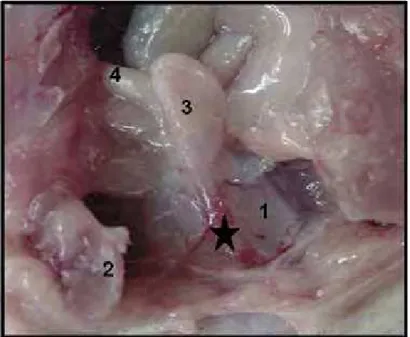
The transverse sections of the central part of the urethra in the virgin group showed different layers from the lumen to the periphery: stratified squamous epithelium (arrow), lamina propria () spongy vascular plexus (P), smooth muscle: longitudinal (1) and circular (2) fibers and striated muscle (3) (Figure 3).
Morphological and semi-quantitative analysis of striated muscle
fiber composition in the rat urethra
Virgin group
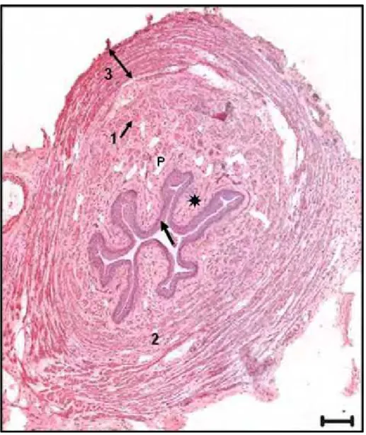
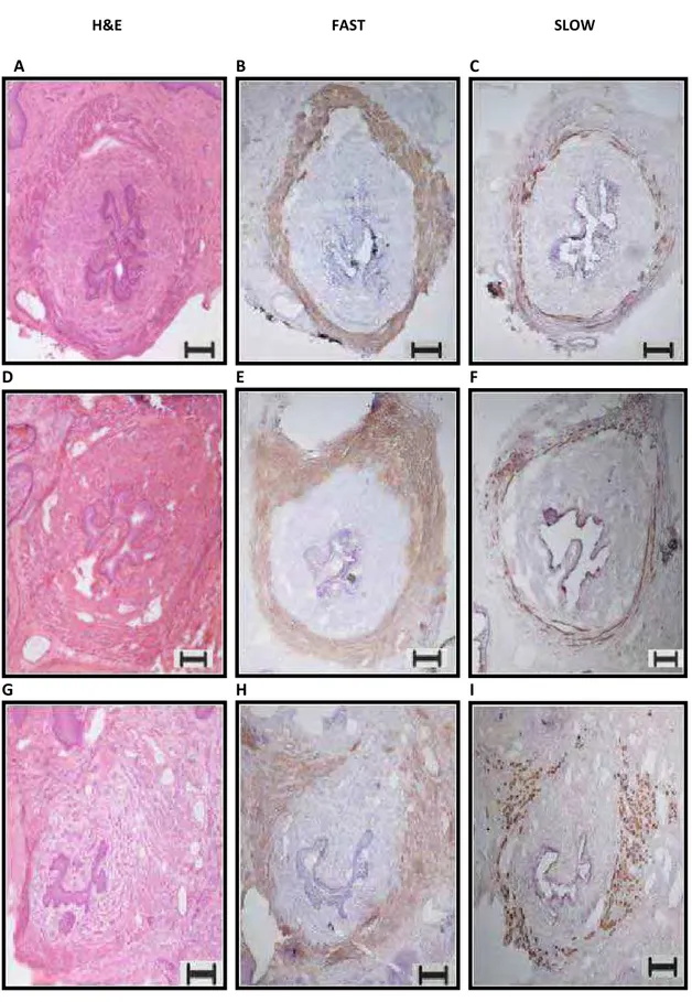
H&E-stained transverse cross-sections of the striated muscle fiber revealed many layers and compact outer circular layers. The fibers were long, with similar thickness throughout the circumference (Figure 4-A).
VtÑ•àâÄÉ E
FL
each type of fibers, with fast fibers being outermost and slow fibers innermost.
Pregnant group
H&E-stained transverse cross-section revealed that the appearance of the striated muscle layer was similar to that of the control group. An increase in connective tissue separated the fibers from one another. The most important finding in this group was the great interstitial spaces found between fibers (Figure 5-A).
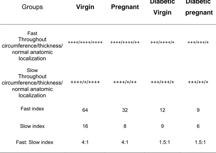
Immunohistochemical staining revealed that the distribution of fast and slow fibers and the proportion between them were similar to those of the virgin group (4:1) (Figure 4-E-F) (Table 1).
Diabetic virgin group
H&E-stained transverse cross-section showed that the circular annulus was lost. There was obvious fiber thinning and atrophy, and the striated muscle was disrupted. There were few complete layers of striated muscle (Figure 5-B).
VtÑ•àâÄÉ E
GC
Diabetic pregnant group
H&E-stained transverse cross-section showed that the circular annulus was lost. The fiber layers were thin, atrophic, and disorganized, and the striated muscle was disrupted. The findings were similar to those of the pregnant group in relation to the increase in connective tissue separating the fibers from one another and the great interstitial spaces (Figure 5-C).
Immunohistochemical staining here also revealed a loss of specific localization for each type of fiber, with co-localization of fast and slow fibers and a decrease in the proportion of fast to slow fibers for 1.5:1 (Figure 6-B-C) (Table 1).
Maternal and perinatal results
Mean maternal weight gain and offspring weights from the pregnant group showed no significant statistically differences as compared to those from diabetic pregnant rats (Table 2).
VtÑ•àâÄÉ E
GD
Discussion
The goal of the present study was to gain a more comprehensive understanding of striated muscle fiber composition in the urethra of the pregnant diabetic rat and the proportion between two basic types of urethral striated muscle fibers: type I (slow-twitch) and type II (fast-twitch). It is of paramount importance not only to understand the effects of DM and pregnancy on striated muscle but also to develop new therapeutic strategies. Human studies are often limited due to ethical concerns, to the challenges of obtaining large tissue samples, and to the use of strictly-managed control groups. To better understand how different risk factors for UI affect the morphological properties of striated muscle, animal models are useful as the experiments are conducted under controlled conditions [17].
VtÑ•àâÄÉ E
GE
circular annulus associated with co-localization of fast and slow fibers, and a steady decrease in the proportion of fast to slow fibers (fast:slow 1.5:1). An increase in connective tissue separating the fibers from one another, as in the interstitial spaces between fibers, occurred as an effect of pregnancy on urethral muscle.
Thinning and atrophy, disorganization, and disruption of the circular annulus of striated muscle were extensive damages caused by diabetes [18, 19]. DM was related to accumulation of reactive oxygen species and tissue ischemia can interactively or independently contribute to the myopathy causes of skeletal muscle dysfunctions [20-21]. In our research laboratory, the relationship between oxidative stress and diabetes in pregnant rats was confirmed by Damasceno et al., [22].
In analyzing this data, we were able to explain that extensive damage to striated muscle fibers characterized by reduced skeletal muscle mass and altered myofiber composition in diabetic pregnant rats links diabetes and pregnancy to UI. This specific loss of skeletal muscle mass is referred to as diabetic myopathy [23]. The present study confirms previous findings that diabetic myopathy and pregnancy are involved in the pathogenesis of urinary incontinence.
VtÑ•àâÄÉ E
GF
revealed two main differences. First, fast fibers lost their great predominance in relation to slow fibers. Second, the fast fibers lost their typical architecture, and the tissue was transformed into a mixture of slow and fast fibers. To the best of our knowledge, the findings are here described for the first time and may be labeled as a diabetic pregnant myopathy. Studies in animal models have shown a strong relationship between muscle fiber type and the development of diabetes [24].
Skeletal muscle is recognized not only for being responsible for movement but also for its function as the largest organ for glucose utilization. Our finding of increased type I slow-type fibers could be related to the abundant availability of lipids [25]. It is well established that changes in muscle fiber composition are often associated with glucose metabolism, diabetes and obesity [26]. Since muscle is a main site of glucose uptake, reduced muscle mass and changes in fiber type composition may directly impair acute glucose utilization. Skeletal muscle can adapt to functional and metabolic demands by remodeling with fiber-type switches to maintain a normal energy balance and utilization of nutrients.
Chen et al., [14] confirmed a higher proportion of type I fibers and
VtÑ•àâÄÉ E
GG
striated muscle of diabetic pregnant rats and that an eventual fiber-type switching could be present. The nature of the mechanism related to this altered fiber type in our model requires further investigation.
As the primary function of the lower urinary tract is the storage and expulsion of urine at the appropriate times, changes in the striated muscle composition could be related to the loss of type II fibers [27] or to the transformation of most type II fibers into type I fibers [28]. Given the limitations of this study, its results could represent muscle changes according to glucose levels.
The damages revealed by morphological studies demonstrate the impact of the association between diabetes and pregnancy on urethral striated muscle fibers, as three of the factors related to altered urethral striated muscle in diabetes and pregnancy – maternal weight gain, offspring weight and trauma related to vaginal delivery – were controlled. However, the results of our study should be interpreted with awareness of the following limitations: rats are quadrupeds; they have tails with associated musculature; and their bladders are abdominal rather than pelvic organs [18].
VtÑ•àâÄÉ E
GH
muscle and alter its fast and slow fiber composition. These data suggest that diabetic pregnant rats may present altered contractility of urethral striated muscle, supporting the high UI prevalence in women with previous GDM, two years after cesarean section [1].
The importance of this study is support of the previous hypothesis that diabetes and pregnancy detrimentally affect the normal function of urethral striated muscles in rats, providing a model for further studies.
Conclusion
VtÑ•àâÄÉ E
GI
Acknowledgements
Financial support was provided by a fellowship from Fundação de Amparo à Pesquisa do Estado de São Paulo (FAPESP), at Gabriela Marini (Grant/Process Number 2008/00989-4). The authors thank Research Center in Neurology (University of São Paulo) by technical assistance.
Competing interests
VtÑ•àâÄÉ E
GJ
References
1. Barbosa AMP (2006) Prevalência e fator de risco para incontinência urinária e disfunção muscular do assoalho pélvico dois anos após Diabete Melito gestacional. Botucatu-SP: Faculdade de Medicina de Botucatu, Universidade Estadual Paulista UNESP
2. Ebbesen MH, Hannestad YS, Midthjell K, Hunskaar S (2009) Diabetes related risk factors did not explain the increased risk for urinary incontinence among women with diabetes. The Norwegian HUNT/EPINCONT study. BMC Urology 9:11
3. Danforth KN, Townsend MK, Curhan GC, Resnick NM, Grodstein F (2009) Type 2 Diabetes mellitus and risk of stress, urge and mixed urinary incontinence. J
Urol 181(1):193-7
4. Pimenta WP, Calderon IMP, Cruz NS, Santos ML, Aragon FF, Padovani CR (2004) Subclinical anormalities o glucose metabolism in Brazilian women with a history of gestational Diabetes mellitus. Acta Obstet Gynecol Scand 83:1152-8
5. Luber KM, Boero S, Choe JY (2001) The demographics of pelvic floor disorders: current observations and future projctions. Am J Obstet Gynecol 184:1496-501
6. Bump RC, Norton PA (1998) Epidemiology and natural history of pelvic floor dysfunction. Obstet Gynecol Clin North Am 25(4):723-46
7. Liu G, Daneshgari F (2005) Temporal diabetes-and diuresis-induced
remodeling of the urinary bladder in the rat Am J Physiol Regul Integr 291(3):R837-43.
8. Daneshgari F, Moore C (2006) Diabetic uropathy. Semin Nephrol 26:182-5
9. Rocha MA, Sartori MG, De Jesus Simões M, Herrmann V, Baracat EC, Girão MJBC, et al (2007) The impact of pregnancy and childbirth in the urethra of female
rats. Int Urogynecol J Pelvic Floor Dysfunct 18(6):645-51
10. Barbosa A, Rudge M, Rudge C, Assis L, Marini G, Modotte W, et al (2008)
Does the elective cesarean protect the woman from the occurrence of urinary incontinence and from pelvic floor muscle dysfunction?. The FIEP Bulletin 78:202-4
11. Heesakkers JP, Gerretsen RR (2004) Urinary incontinence: sphincter functioning from a urological perspective. Digestion 69(2):93-101
VtÑ•àâÄÉ E
GK
13. Andersson PO, Malmgren A, Uvelius B (1990) Functional responses of different muscle types of the female rat urethra in vitro. Acta Physiol Scand 140:365-72
14. Chen M, Feng HZ, Gupta D, Kelleher J, Dickerson KE, Wang J, et al (2009)
G(s)alpha deficiency in skeletal muscle leads to reduced muscle mass, fiber-type switching, and glucose intolerance without insulin resistance or deficiency. Am J Physiol Cell Physiol 296(4):C930-40
15. Elbadawy A (1996) Functional anatomy of organs of micturition. Urol Clin North Am 23:177-210
16. Calderon IMP, Rudge MVC, Ramos MD, Peraçoli JC (1999) Estudo longitudinal, bioquímico e histoquímico de placentas de ratas diabéticas: relação com a macrossomia e o retardo de crescimento intra-uterino. Rev Bras Ginecol Obstet 21:91-8
17. Abramowitch SD, Feola A, Jallah Z, Moalli PA (2009) Tissue mechanics, animal models, and pelvic organ prolapse: a review. Eur J Obstet Gynecol Reprod Biol 144 Suppl 1:S146-58
18. Kim JH, Huang X, Liu G, Moore C, Bena J, Damaser MS, et al (2007)
Diabetes slows the recovery from urinary incontinence due to simulated childbirth in female rats. Am J Physiol Regul Integr Comp Physiol 293(2):R950-5
19. Gasbarro G, Lin DL, Vurbic D, Quisno A, Kinley B, Daneshgari F, et al (2009)
Voiding function in obese and type 2 diabetic female rats. Am J Physiol Renal Physiol 298(1):F72-7
20. Beshay E, Carrier S (2004) Oxidative stress plays a role in diabetes-induced bladder dysfunction in a rat model. Urology 64(5):1062-7
21. De Angelis KL, Cestari IA, Barp J, Dall'Ago P, Fernandes TG, de Bittencourt PI, et al (2000) Oxidative stress in the latissimus dorsi muscle of diabetic rats. Braz J
Med Biol Res 33(11):1363-8
22. Damasceno DC, Volpato GT, Calderon IMP, Rudge MVC (2002) Oxidative stress and diabetes in pregnant rats. Animal Reprod Science 72(3-4):235-44
23. Krause MP, Riddell MC, Gordon CS, Imam SA, Cafarelli E, Hawke TJ (2009) Diabetic myopathy differs between Ins2Akita+/- and streptozotocin-induced Type 1 diabetic models. J Appl Physiol 106(5):1650-9
24. Schuler M, Ali F, Chambon C, Duteil D, Bornert JM, Tardivel A, et al (2006)
VtÑ•àâÄÉ E
GL
25. de Wilde J, Mohren R, van den Berg S, Boekschoten M, Dijk KW, de Groot P,
et al (2008) Short-term high fat-feeding results in morphological and metabolic
adaptations in the skeletal muscle of C57BL/6J mice. Physiol Genomics 19;32(3):360-9
26. Oberbach A, Bossenz Y, Lehmann S, Niebauer J, Adams V, Paschke R, et al
(2006) Altered fiber distribution and fiber-specific glycolytic and oxidative enzyme activity in skeletal muscle of patients with type 2 diabetes. Diabetes Care 29(4):895-900
27. Pandit M, Delancey JOL, Ashton-Miller, Iyengar J, Blaivas M, Peruchini D (2000) Quantification of intramuscular nerves within the female striated urogenital sphincter muscle. Obstet Gynecol 95:797-800
28. Fujimoto S, Watanabe J, Ogawa R, Kanamura S (1994) Age related changes in fiber number, fiber size, fiber type composition and adenosine triphosphatase activity in rat soleus muscle. Ann Anat 176:429-35
29. Gregorevic P, Plant DR, Stupka N, Lynch GS (2004) Changes in contractile activation characteristics of rat fast and slow skeletal muscle fibers during
regeneration. J Physiol 15;558(Pt 2):549-60
VtÑ•àâÄÉ E
HC
Figure 1. Photograph of anesthetized female rat showing the uterine horns. Arrow points the place where the horns were opened and the fetuses withdraw.
VtÑ•àâÄÉ E
HD
VtÑ•àâÄÉ E
HE
H&E FAST SLOW
A B C
D E F
G H I
VtÑ•àâÄÉ E
HF
VtÑ•àâÄÉ E
HG
VtÑ•àâÄÉ E
HH
Table 1. Semi-quantitative analysis of slow and fast fibers according the
presence of each type of fiber throughout circumference of the layer; thickness of the muscle fiber layer; the degree to which the layers maintained a normal anatomic localization; fast index; slow index; and fast:slow index in each group.
Groups Virgin Pregnant Diabetic Virgin
Diabetic
pregnant
Fast Throughout
circumference/thickness/ normal anatomic
localization
++++/++++/++++ ++++/++++/++ +++/++++/+ +++/+++/+
Slow Throughout
circumference/thickness/ normal anatomic
localization
++++/+/++++ ++++/+/++ +++/+++/+ +++/++/+
Fast index 64 32 12 9
Slow index 16 8 9 6
VtÑ•àâÄÉ E
HI
Table 2. Maternal weight gain (g) and offspring weight (g) in pregnant
and diabetic pregnant groups. Maternal glycemia (mg/dL) from virgin, pregnant, diabetic virgin and diabetic pregnant groups at beginning and end of the experimental period.
Groups Virgin Pregnant Diabetic Virgin
Diabetic
pregnant
Maternal weight
gain (g) 115.4±16.3 72.2 ± 21.5
Offspring weight
(g) 82.6 ± 10.6 67.2 ± 14.3
Maternal glycemia (mg/dL) beginning
of experiment
81.0 ± 4.4 112.8 ± 4.0 568.4 ± 38.0* 544.0 ± 36.8*
Maternal glycemia (mg/dL) end of
experiment
81.6 ± 5.0 82.6 ± 7.7 584.8 ± 33.9* 497.8 ± 60.4*
Values are reported as mean ± SEM.
*p<0.05 – significant statistically difference compared to virgin and pregnant groups (Tukey’s Multiple Comparison Test).
VtÑ•àâÄÉ F
HJ
Vi,então,umnovocéueumanovaterra!“EisaquiotabernáculodeDeuscomoshomens.Habitará
comeleseserãoseupovo,eDeusmesmoestarácomeles.Enxugarátodalágrimadeseusolhosejá
nãohaverámorte,nemluto,nemgrito,nemdor,porquepassouaprimeiracondição”.“Eisquefaço
novatodasascoisas.”
Apocalipse21:1
VtÑ•àâÄÉ F
HK
Alterações morfológicas das fibras tipos I e II
do músculo estriado uretral de ratas prenhes
diabéticas
Autora:
Gabriela Marini
Orientadora:
Marilza Vieira Cunha Rudge
Co-orientadoras:
Angélica Mércia Pascon Barbosa, Selma
Maria Michelin Matheus
Banca:
Marilza Vieira Cunha Rudge, Débora Cristina
Damasceno, Manoel João Batista Castello Girão
Data da apresentação:
25 de fevereiro de 2010
Este capítulo foi redigido de acordo com as normas de publicação de resumos de Dissertação da Revista Brasileira de Ginecologia e Obstetrícia, para a qual será
VtÑ•àâÄÉ F
HL
RESUMO
Objetivos: avaliar as alterações morfológicas das fibras musculares
estriadas tipos I e II da uretra de ratas prenhes diabéticas submetidas à cesárea. Métodos: Foram avaliadas 20 ratas Wistar distribuídas em
quatro grupos: virgem, prenhe, diabético virgem e prenhe diabético. Os três primeiros grupos foram estudados para servir como controle do grupo principal, o prenhe diabético. O diabete foi induzido com streptozotocin na dose de 40mg/kg de peso corpóreo. O critério de inclusão foi uma glicemia acima de 200mg/dL. No final do experimento, as ratas foram anestesiadas e eutanasiadas para realização da laparotomia exploratória. A vagina e a uretra foram retiradas em monobloco, congeladas em nitrogênio líquido e mantidas a -80°C. O bloco foi submetido a cortes em criostato (6 μm de espessura). As lâminas foram coradas por H&E e utilizados anticorpos anti-miosina lenta e rápida para tipagem das fibras. Foi realizada análise morfológica e semi-quantitativa dos quatro grupos. Resultados: O músculo estriado
uretral do grupo prenhe diabético apresentou: adelgaçamento, atrofia, desorganização e rompimento associado à perda de localização anatômica normal das fibras rápidas e lentas e diminuição na proporção de fibras rápidas. Conclusões: Este estudo sugere que o binômio
diabete e prenhez danificou o músculo estriado uretral e alterou a composição e a distribuição das fibras tipo I e II .
Palavras-chave: diabete, fibra muscular estriada, prenhez, uretra
TÇxåÉá
IC
TÇxåÉá
TÇxåÉá
ID
TÇxåÉá
IE
TÇxåÉá
IF
ANEXO 3 – Ofício do envio para publicação do Capítulo 1
Article title: Diabetes in pregnancy and urinary incontinence: a little acknowledged association
MS ID : 1906283565344512
Authors : Gabriela Marini, Angelica MP Barbosa, Débora C Damasceno, Rodrigo A Castro, Selma MM Matheus and Marilza VC Rudge
Journal : Diabetology & Metabolic Syndrome Dear Dr Marini
Thank you for submitting your article. This acknowledgement and any queries below are for the contact author. This e-mail has also been copied to each author on the paper, as well as the person submitting. Please bear in mind that all queries regarding the paper should be made through the contact author.
Regards
The Diabetology & Metabolic Syndrome Editorial Team e-mail: editorial@dmsjournal.com
Web: http://www.dmsjournal.com/
Terçafeira,12deJaneirode201012:55