165
CASE REPORTRhabdomyomatous mesenchymal
hamartoma: a case report
Hamartoma mesenquimal rabdomiomatoso: um relato de caso
Fernanda Alves Luiz Rodrigues1; Maria Auxiliadora de Paula Carneiro Cysneiros2; Rubson Rodrigues Júnior3; Denis Masashi Sugita1
First submission on 03/02/14; last submission on 03/02/14; accepted for publication on 16/02/14; published on 20/02/14
1. Resident in Pathology at Hospital das Clínicas of Universidade Federal de Goiás (HC/UFG). 2. Master’s degree in Pathology from UFG; physician at the Pathology Service at HC/UFG. 3. Resident in Internal Medicine at HC/UFG.
ABSTRACT
The rhabdomyomatous mesenchymal hamartoma (RMH) is a rare type of hamartoma, composed of randomly arranged striated muscle ibers in dermis and subcutaneous tissue, associated with normal mesenchymal elements. Our objective is to report a case of this rare entity that occurred in the nasal dorsum of a 4-year-old child.
Key words: hamartoma; mesenchymal; rhabdomyomatous; striated muscle; skin.
J Bras Patol Med Lab, v. 50, n. 2, p. 165-166, abril 2014
INTRODUCTION
Originally described in 1986 by Hendrick et al. as striated
muscle hamartoma, the rhabdomyomatous mesenchymal hamartoma (RMH) of the skin is a rare congenital tumor that affects predominantly the face and neck of newborns, with rare cases reported in the literature(7, 10, 20).
The RMH occurs as a single or multiple lesions, generally polypoid, typically located in the midline(19), and is characterized
by the presence of mesenchymal elements (adipose, connective, vascular, and nervous tissues) and striated muscles, randomly arranged in dermis and subcutaneous tissues(7, 15, 19, 20).
CASE REPORT
A 4-year-old male child presented with a hardened well-delimited congenital solid tumoration in nasal dorsum. His mother reported slow growth in recent years, and denied istulization or nasal obstruction.
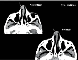
The computed tomography (CT) scan revealed a solid oval-shaped nodule enhanced after contrast, supericially located in the
nasal dorsum, in the right paramedian position, causing discrete adjacent bone erosion, and measuring approximately 1 cm in diameter, with a nonspeciic appearance (Figure 1).
Excision of the lesion was carried out, and followed by good scarring.
The patient has so far presented no symptoms and signs of recurrence.
FIGURE 1 – Post contrast CT image reveals an enhanced lesion in nasal dorsum
CT: computed tomography.
Contrast No contrast Axial sections
166
Anatomopathological study
Two irregularly-shaped brownish-gray ibroelastic tissue fragments were examined, with the largest measuring 0.8 cm in
its longer diameter.
By microscopy, a hamartomatous lesion composed of dense collagen bundles was observed, with randomly intermingled striated skeletal muscle ibers. Also, proliferated vessels, small foci of mature adipose tissue and a single lobule of adnexal gland
(Figures 2 and 3) were identiied. At the periphery of the lesion, there was well-structured mature cartilaginous tissue without atypia.
Immunohistochemistry (IHC) was positive for desmin
(Figure 4) and HHF-35 (Figure 5) in the striated muscle ibers.
The previously described histomorphological and IHC indings are compatible with the picture of RMH.
DISCUSSION
Hamartomas are malformations composed of tissue elements normally found at the lesion site, yet arranged in a disorganized
manner(2, 5, 12, 15).
RMHs are hamartomatous lesions more commonly reported in head and neck(4, 6, 17, 19), with rare cases in perineal regions(14, 18).
Lesions typically occur in children and are usually present since birth(8). They generally present as a small solitary
dome-shaped papule, or a polypoid pedunculated lesion, ranging in size from few millimeters to 1-2 cm(6). There are reports about
this entity in adults, but it is not known whether the lesions were present since birth(20).
The etiology is unknown, however, RMH is believed to
result from an abnormality in the migration of mesodermal FIGURE 2 – H&E stain, skeletal striated muscle ibers, intermingled with adipocytes
H&E: hematoxylin and eosin.
FIGURE 4 – IHC shows positivity for desmin in the skeletal muscle ibers
IHC: immunohistochemical.
FIGURE 3 – H&E stain, striations of skeletal muscle ibers are precisely observed
H&E: hematoxylin and eosin.
FIGURE 5 – IHC study shows positivity for HHF35 in the skeletal muscle ibers IHC: immunohistochemical.
167
tissue during embryogenesis or from genetic defects(3, 11, 18, 19),
as it is associated with other congenital defects like the amniotic band syndrome, Delleman syndrome, and Goldenhar
syndrome(19).
Almost all the lesions occur in males, with rare cases being described in females(6, 20).
Microscopically these lesions are constituted by striated muscle ibers randomly distributed, intermingled with mature adipose tissue, collagen bands, blood vessels, and elastic ibers(1, 16). Adnexal structures interwoven with the muscle bundles
are reported. However, it has been questioned if the presence of these structures is an incidental inding, as adnexal glands may normally be present in the reticular dermis(6).
IHC in RMH shows positivity for actin, desmin, and myoglobin in the skeletal muscle ibers.
Differential diagnoses include supericial lipomatous nevus, ibrous hamartoma of infancy, neuromuscular choristoma (benign Triton tumor), rhabdomyoma and cutaneous embryonal rhabdomyosarcoma. The supericial lipomatous nevus shows mature adipose tissue in dermis, but lacks elements of skeletal muscle. The ibrous hamartoma of infancy contains a mixture
of mature adipose tissue, collagen bands, but also lacks the ibers of skeletal muscle(20). The neuromuscular choristoma (benign
Triton tumor) is a subcutaneous lesion composed of skeletal
muscle ibers and neural tissue(13). The rhabdomyoma contains
variable amounts of myxoid and ibrous tissue, with mesenchymal undifferentiated cells and fetal striated muscle(8, 16). Lastly, the
cutaneous embryonal rhabdomyosarcoma represents a rare and much less differentiated entity(4, 20).
Surgery is the treatment of choice in these cases, with complete lesion resection. So far, recurrences have not been reported after surgical resection of these nodules(8, 12, 19, 20).
CONCLUSION
We report a case of RMH in a 4-year-old child, whose clinical, radiological, macroscopical, histopathological and immunohistochemical indings are compatible with those described in this rare entity.
Although RMHs are rare and benign lesions, their association with other congenital anomalies and embryological errors must always be evaluated in patients with this diagnosis.
RESUMO
O hamartoma mesenquimal rabdomiomatoso (HMR) representa um raro tipo de hamartoma composto por ibras musculares estriadas dispostas aleatoriamente em derme e tecido subcutâneo, associadas a elementos mesenquimais normais. O nosso objetivo é relatar um caso desta rara entidade que ocorreu no dorso nasal de uma criança de 4 anos.
Unitermos: hamartoma; mesenquimal; rabdomiomatoso; músculo estriado; pele.
REFERENCES
1. BALL, E. A. et al. Rhabdomyomatous mesenchymal hamartoma resembling scleroderma “en coup de sabre”: a case report and literature review. Br J Dermatol, v. 162, n. 1, p. 222-4, 2010.
2. BARNHILL, R. et al.Dermatopathology. 3 ed. N.Y: Mc Graw Hill, p. 857, 2010.
3. BERNAL-MAÑAS, C. M. et al. Hamartoma mesenquimal rabdomiomatoso. An Pediatr (Barc), v. 78, n. 4, p. 260-2, 2013. 4. BRINSTER, N. K.; FARMER, E. R. Rhabdomyomatous mesenchymal hamartoma presenting on a digit. J. Cutan Pathol, , v. 36, n. 1, p. 61-3, 2009.
5. DAL VECHIO, A. et al. Rhabdomyomatous (mesenchymal) hamartoma presenting as haemangioma on the upper lip: a case report with
immunohistochemical analysis and treatment with high-power lasers. Case Rep Dent, v. 2013; 2013.
6. DÍAZ- PEREZ, J. A. et al. [Rhabdomyomatous mesenchymal hamartoma]. Actas Dermosiiliogr, v. 99, n. 6, p. 474-6, 2008. 7. GNEPP, D. Diagnostic surgical pathology of the head and neck. 2 ed. Philadelphia: Saunders (Elsevier), 2009. p. 1004.
8. HAN, S. H. et al. Rhabdomyomatous mesenchymal hamartoma of the vagina. Pediatr Dermatol, v. 26, n. 6, p. 753-5, Nov/Dec 2009.
9. JOHNSTON, R. Weedon’s skin pathology essentials. 1. ed. Churchill Livingstone (Elsevier), 2012. p. 662.
10. KANG, J. W.; PARK, H. S.; KIM, J. H. Rhabdomyomatous mesenchymal hamartoma of nasal vestibule. J Craniofac Surg, v. 24, n. 5, p. e481-3, 2013. 11. KIM, H. S. et al. Rhabdomyomatous mesenchymal hamartoma. J Eur Acad Dermatol Venereol, v. 21, n. 4, p. 564-5, 2007.
168
12. LARA, B. W. et al. Hamartoma mesenquimal rabdomiomatoso. Rev Esp Patol, v. 37, n. 4, p. 429-32, 2004.13. READ, R. W. et al. Rhabdomyomatous mesenchymal hamartoma of the eyelid: report of a case and literature review. Ophthalmology, v. 108, n. 4, p. 798-804, April 2001.
14. RODRÍGUEZ, L. G.; RODRÍGUEZ, Á.; VARGAS, N. Hamartoma mesenquimal rabdomiomatoso. Rev Asoc Colomb Dermatol Cir Dermatol, v. 15, n. 3, p. 221-3, Sept. 2007.
15. ROSAI, J. Rosai and Ackerman’s Surgical Pathology. 10 ed. Missouri: Mosby (Elsevier), 2011. p.182.
16. ROSEMBERG, A. S.; KIRK, J.; MORGAN, M. B. Rhabdomyomatous mesenchymal hamartoma: an unusual dermal entity with a report
of two cases and a review of the literature. J Cutan Pathol, v. 29, n. 4, p. 238-43, 2002.
17. VAIDYNATHAN, M.; WILLIANS, C. E. C. S.; MORGAN, P. R. Rhabdomyomatous mesenchymal hamartoma of the tongue. BMJ Case Reports, 2011; 2011. p. 1-2.
18. WANG, J.-R. et al. Rhabdomyomatous mesenchymal hamartoma associated with congenital anomalies: report of an unusual perineal case. Dermatol Sinica, v. 26, p 93-8, 2008.
19. WEEDON, D. Weedon’s skin pathology. 3 ed. Mosby (Elsevier), 2012. p. 662.
20. WEISS, S.; GOLDBLUM, J. Enzinger& Weiss’s soft tissue tumors. 5. ed. Missouri: Elsevier, 2008. p. 591.
MAILING ADDRESS
Fernanda Alves Luiz Rodrigues
Rua Magda Perona Frossard, 155, ed. Monte das Oliveiras, apto 111, bairro Nova Aliança; CEP: 14026596; Ribeirão Preto-SP; Brazil; e-mail: [email protected]