FÁBIO DIAS LUNS
AVALIAÇÃO DA INTERAÇÃO ENTRE OS ISOLADOS FUNGICOS
Duddingtonia flagrans, Monacrosporium thaumasium E Arthrobotrys robusta NO
CONTROLE BIOLÓGICO DE NEMATÓIDES GASTRINTESTINAIS DE BOVINOS LEITEIROS A CAMPO
Dissertação apresentada à Universidade Federal de Viçosa, como parte das exigências do Programa de
Pós-Graduação em Medicina Veterinária, para obtenção do título de Magister Scientiae.
VIÇOSA
FÁBIO DIAS LUNS
AVALIAÇÃO DA INTERAÇÃO ENTRE OS ISOLADOS FUNGICOS
Duddingtonia flagrans, Monacrosporium thaumasium E Arthrobotrys robusta NO
CONTROLE BIOLÓGICO DE NEMATÓIDES GASTRINTESTINAIS DE BOVINOS LEITEIROS A CAMPO
Dissertação apresentada à Universidade Federal de Viçosa, como parte das exigências do Programa de Pós-Graduação em Medicina Veterinária, para obtenção do título de Magister Scientiae.
APROVADA: 25 de julho de 2013.
_______________________________ _______________________________
Profa. Márcia Cristina Cury Profa. Isabele da Costa Angelo
_______________________________ Prof. Jackson Victor de Araújo
ii A Deus, que me permitiu chegar até aqui.
A minha mãe, Lourdes, in memorian, que sempre fez de tudo para me dar a melhor educação.
A minha esposa, Rafaela.
iii
AGRADECIMENTOS
A Universidade Federal de Viçosa, por ter mudado a minha vida através da educação de qualidade.
A Coordenação de Aperfeiçoamento de Pessoal de Nível Superior CAPES e a Fundação de Amparo à Pesquisa do estado de Minas Gerais FAPEMIG pelo apoio financeiro. Ao orientador Jackson Victor de Araújo pelo apoio, a oportunidade e a confiança. A minha esposa Rafaela pelo apoio e compreensão pelas horas em que não estive junto a ela.
Aos estagiários Guilherme, Gustavo e Vitão que me ajudaram na realização da parte de campo.
Aos amigos André, Elton, Geraldinho, Luís, José e Luciano obrigado pela sua ajuda na realização desse projeto.
A equipe de trabalho do Laboratório de Parasitologia: ao Tuim, Ademir, Fábio Braga, Wendeo e Tavela, agradeço o apoio na confecção dos pellets e análises laboratoriais. Ao Fábio Ribeiro Braga, por ter coordenado e orientado toda a atividade do projeto em Viçosa. Obrigado pelo apoio, orientações e por sua presença na execução do projeto. Aos professores da UFV, em especial ao Pacífico pelas longas conversas e conselhos dados.
Aos amigos e funcionários da UFV, em especial as secretárias da Pós-graduação em Medicina Veterinária Rosi e Bete, sempre disponíveis e solicitas.
iv
SUMÁRIO
Lista de Figuras e Tabelas vi
Resumo viii
Abstract ix
Introdução Geral 01
Objetivos 04
Objetivo geral 04
Objetivos específicos 04
Referências 05
Capítulo 1. Association of the fungi Duddingtonia flagrans and Monacrosporium
thaumasium on biologic control of dairy cattle nematodiasis
1. Introduction 08
2. Materials and methods 08
2.1. Fungi and production of pellets 08
2.2. Animals and experimental site 09
3. Results 10
4. Discussion 13
5. Conclusion 15
6. References 15
Capítulo 2. Association of the fungi Duddingtonia flagrans and Arthrobothyrs robusta
on biologic control of dairy cattle nematodiasis
1. Introduction 20
2. Materials and methods 20
2.1. Fungi and production of mycelial pellets 20
2.2. Animals and experimental site 21
3. Results 22
4. Discussion 25
5. Conclusion 26
v
Capítulo 3. Association of the fungi Duddingtonia flagrans, Arthrobothyrs robusta and
Monacrosporium thaumasium on biologic control of dairy cattle nematodiasis
1. Introduction 30
2. Materials and methods 30
2.1. Fungi and production of mycelial pellets 30
2.2. Animals and experimental site 30
3. Results 32
4. Discussion 35
5. Conclusion 37
vi
LISTA DE FIGURAS E TABELAS
Capítulo 1
Fig. 1. Monthly means (±SD) of eggs per gram of feces of D.flagrans associated to M. thaumasium (Group 1) , D. flagrans only (control +) and control animals (control -) collected from May 2012 to
September 2012, in Ouro Branco, MG, southeastern Brazil………….………..10
Tab. 1. Percentage of reduction of infective larvae (L3) recovered from the coprocultures of the groups treated with the association of the nematophagous fungi D. flagrans and M. thaumasium (Group 1) and
the positive control group (D. flagrans only) in relation to the negative control from May 2012 to
September 2012, Ouro Branco, MG, Brazil………...………11
Tab. 2 Absolute values of L3 per kg of dry matter obtained from pastures grazed by the calves treated with the association of the nematophagous fungi D. flagrans and M. thaumasium (Group 1) , D. flagrans only (control +) and without fungus (control-) from May 2012 to September 2012, Ouro Branco,
MG, Brazil………...……….12
Fig. 2. Mean weight gains (kg/day) for calves treated with the association of the nematophagous fungi D. flagrans and M. thaumasium (Group 1) , D. flagrans only (control +) and without fungus (control-) from May 2012 to September 2012, Ouro Branco, MG, Brazil………..13
Capítulo 2
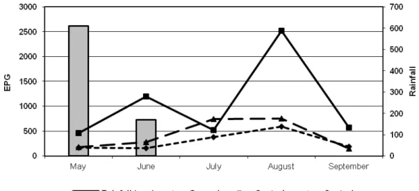
Fig. 1. Monthly averages of the countings of eggs per gram of feces (EPG) of the animals in the groups treated with the nematophagous fungi D. flagrans and A. robusta association (1 g of fungus/10 kg of b.
w.) (Group 1), the – control group (without treatment) and the + control group (D. flagrans only), collected from May 2012 to September 2012, Ouro Branco, Minas Gerais, Brazil. ……….……….22
Tab. 1. Percentage values corresponding to the infectant larvae recovered from the coprocultures of the groups treated with the nematophagous fungi D. flagrans and A. robusta association (Group 1) and
the + control group (D. flagrans only) in relation with the - control group (without treatment) collected
from May 2012 to September 2012, Ouro Branco, Minas Gerais, Brazil………...23
Tab. 2. Values of L3 per kg of dry matter obtained from pastures (0-20 cm and 20-40cm, respectively) grazed by the groups treated with the nematophagous fungi D. flagrans and A. robusta association (Group
Tested), the - control group (without treatment) and the + control group (D. flagrans only), collected from
vii
Fig 2. Mean weight gains (kg/day) of the groups treated with the nematophagous fungi D. flagrans and A. robusta association (Tested), the – control group (without treatment) and the + control group (D. flagrans
only), collected from May 2012 to September 2012, Ouro Branco, Minas Gerais, Brazil……….…24
Capítulo 3
Fig. 1. Monthly averages of the countings of eggs per gram of feces (EPG) of the animals in the groups treated with the nematophagous fungi D. flagrans, M. thaumasium and A. robusta association (1 g of
fungus/10 kg of b.w.) (Tested), the – control group (without treatment) and the + control group (D.
flagrans only), collected from May 2012 to September 2012, Ouro Branco, Minas Gerais, Brazil. ...…32
Tab. 1. Percentage values corresponding to the infectant larvae recovered from the coprocultures of the groups treated with the nematophagous fungi D. flagran, M thaumasium and A. robusta association
(Tested Group) and the + control group (D. flagrans only) in relation with the - control group (without
treatment) collected from May 2012 to September 2012, Ouro Branco, Minas Gerais, Brazil…………..33
Tab. 2. Values of L3 per kg of dry matter obtained from pastures (0-20 cm and 20-40cm, respectively) grazed by the groups treated with the nematophagous fungi D. flagrans and A. robusta association
(Tested Group), the - control group (without treatment) and the + control group (D. flagrans only), collected from May 2012 to September 2012, Ouro Branco, Minas Gerais, Brazil………...……….33
Fig 2. Mean weight gains (kg/day) of the groups treated with the nematophagous fungi D. flagrans, M thaumasium and A. robusta association (Tested), the – control group (without treatment) and the + control
group (D. flagrans only), collected from May 2012 to September 2012, Ouro Branco, Minas Gerais,
viii
RESUMO
LUNS, Fábio Dias, M.Sc., Universidade Federal de Viçosa, julho de 2013. Avaliação da interação entre os isolados fúngicos Duddingtonia flagrans, Monacrosporium thaumasium E Arthrobotrys robusta no controle biológico de nematóides gastrintestinais de bovinos leiteiros a campo. Orientador: Jackson Victor de Araújo. Coorientador: Fábio Ribeiro
Braga.
ix
ABSTRACT
LUNS, Fábio Dias. M.Sc., Universidade Federal de Viçosa, July, 2013. Evaluation of
the interaction between the fungal isolates Duddingtonia flagrans Monacrosporium thaumasium And Arthrobotrys robusta in biological control of gastrointestinal nematodes of dairy cattle in the field. Adviser: Jackson Victor de
Araújo. Co-adviser: Fábio Ribeiro Braga.
1
1. INTRODUÇÃO GERAL
A bovinocultura é um dos principais destaques do agronegócio brasileiro no cenário mundial. O rebanho bovino brasileiro proporciona o desenvolvimento de dois segmentos lucrativos: as cadeias produtivas da carne e leite. O valor bruto da produção desses dois segmentos, estimado em R$ 67 bilhões, aliado à presença da atividade em todos os estados brasileiros, evidenciam a importância econômica da bovinocultura em nosso país (MAPA, 2012).
O clima tropical e a extensão territorial do Brasil contribuem para esse resultado, uma vez que permitem a criação do gado em pastagens, mas, por outro lado, estas condições favorecem infecções por parasitos presentes nas pastagens, o que gera impacto na produção e altos custos nas medidas de controle, acarretando perda econômica estimada em milhões de dólares (ANUALPEC, 2003).
As helmintoses gastrintestinais de ruminantes produz impacto na bovinocultura de leite. Os prejuízos causados por essas infecções envolvem queda da produção, retardo no crescimento do animal, custos com tratamentos e até a morte dos animais (MOTA et al., 2003).
O alto custo do tratamento e o surgimento da resistência aos anti-helmínticos prevêem novas alternativas de controle dessas infecções (MOTA et al, 2003).
Segundo Campos et al. (1998), na bovinocultura leiteira, o sistema de cria e recria de bezerras não tem a importância merecida, visto que nessa fase não há lucro direto para o produtor. As novilhas representam cerca de 15 a 20% dos custos de produção da atividade leiteira e especial importância na recria deveria ser dada ao manejo sanitário e as instalações dos animais, já que um correto desenvolvimento das novilhas contribuiria significativamente na produção leiteira (SANTOS et al., 2002).
2 Dentre os mais variados antagonistas nematóides, encontram-se organismos como fungos, bactérias, protozoários, vírus, entre outros. Os fungos nematófagos são aqueles que apresentam melhores desempenhos em pesquisas de controle biológico de nematóides (MACIEL et al., 2006), destacando-se os fungos predadores dos gêneros Arthrobotrys, Duddingtonia e Monacrosporium (ARAÚJO et al., 2004).
Os fungos nematófagos são organismos saprófitas mundialmente estudados, com capacidade de predar nematóides, produzindo armadilhas ao longo das hifas e exibindo redução efetiva na população de nematóides em experimentos laboratoriais e a campo (ARAÚJO et al., 1998).
Esses fungos pertencem a um grupo heterogêneo que utilizam nematóides como fonte principal ou adicional de nutrientes. São encontrados em todo o mundo em diferentes habitats, sendo frequentemente encontrados em ambientes ricos em material orgânico a temperaturas que podem variar de 20 a 30°C (LARSEN, 2000).
São conhecidos como fungos destruidores de nematóides e estão catalogados em mais de 150 espécies. São divididos em três grupos: endoparasitas, predadores e oportunistas, mas a maioria das espécies é classificada como fungos predadores de nematóides (BARRON, 1977).
Esses fungos produzem estruturas em forma de anéis constritores e não constritores, hifas, botões e redes tridimensionais adesivas ao longo do micélio. Depois do nematóide aprisionado, segue-se a penetração das hifas na cutícula do nematóide, onde ocorre o crescimento das hifas e digestão dos conteúdos internos (LARSEN et al, 1999; ARAÚJO et al., 2004).
3 Em alguns laboratórios de pesquisa, formulações a base de alginato de sódio tem sido avaliadas, demonstrando bons resultados em condições laboratoriais e a campo (ARAÚJO et al., 2000).
Na Nova Zelândia, a combinação in vitro de fungos nematófagos dos gêneros Duddingtonia, Monacrosporium e Harposporium foi testada por Waghorn et al. (2006) sobre estádios de vida livre de Ostertagia circuncicta e apresentou resultados promissores.
4
2. OBJETIVOS
2.1. Objetivo geral
Avaliar a interação entre os fungos predadores de nematóides Duddingtonia flagrans, Monacrosporium thaumasium e Arthrobotrys robusta, em formulação de pellets de alginato de sódio, no controle das nematodioses gastrintestinais de bovinos de leite da raça Girolando criados a campo.
2.2. Objetivos específicos
Verificar a contagem de ovos por grama de fezes (OPG) e o número de larvas recuperadas em pastagem;
Comparar os resultados entre o grupo controle e os grupos tratados com diferentes associações fungicas;
Aferir o peso dos animais;
5
REFERÊNCIAS BIBLIOGRÁFICAS
ANUALPEC: Anuário estatístico da produção animal. São Paulo: FNP Consultoria &
Comércio. 380 p., 2003.
ARAÚJO, J.V.; GOMES, A.P.S.; GUIMARÃES, M.P. Biological control of bovine gastrointestinal nematode parasites in southern Brazil by the nematode-trapping fungus Arthrobotrys robusta. Revista Brasileira de Parasitologia Veterinária, 7, 117-122, 1998.
ARAÚJO, J.V.; STEPHANO, M.A.; SAMPAIO, W.M. Passage of nematode-trapping fungi through the gastrointestinal tract of calves. Veterinary Archive, 2, 69-78, 1999. ARAÚJO, J.V.; STEPHANO, M.A.; SAMPAIO, W.M. Effects of temperature and mineral salt in passage through the gastrointestinal tract of calves on sodium alginate formulation of Arthrobotrys robusta, a nematode-trapping fungus. Revista Brasileira
de Parasitologia Veterinária, 9, 55-59, 2000.
ARAÚJO, J.V.; RIBEIRO, R.R. Atividade predatória sobre larvas de tricostrongilídeos (nematoda: Trichostrongyloidea) de isolados fúngicos do gênero Monacrosporium após a passagem pelo trato gastrintestinal de bovinos. Revista Brasileira de Parasitologia
Veterinária, 12, 2, 76-81, 2003.
ARAÚJO, J.V.; ASSIS, R.C.L.; CAMPOS, A.K.; MOTA, M.A. Atividade in vitro dos fungos nematófagos dos gêneros Arthobotrys, Duddingtonia e Monacrosporium sobre nematóides parasitos gastrintestinais de bovinos. Revista Brasileira de Parasitologias
Veterinária, 13, 65-71, 2004.
BARRON, G.L. The nematode-destroying fungi. Topics in Mycobiology. Guelph: Canadian Biological Publications, 140 p., 1977.
CAMPOS, O.F.; LIZIERE, R.S. Estratégias para obtenção de fêmeas de reposição em rebanhos leiteiros. In: Simpósio sobre produção animal, 10, 1998, Anais... Piracicaba: FEALQ, 215-255, 1998.
GRONVOLD, J.; WOLSTRUP, J.; LARSEN, M.; HENRIKSEN, S.A.; NANSEN, P. Biological control of Ostertagia ostertagi by feeding selected nematode-trapping fungi to calves. Journal of Helminthology, 67, 31-36, 1993.
GRONVOLD, J.; WOLSTRUP, J.; LARSEN, M.; GILLESPIE, A.; GIACOMAZZI, F. Interspecific competition between the nematode-trapping fungus Duddingtonia flagrans and selected microorganisms and the effect of spore concentration on the efficacy of nematode trapping. Journal of Helminthology, 78, 41-46, 2004.
6 LARSEN, M.; NANSEN, P.; WOLSTRUP, J.; GRONVOLD, J.; HENRIKSEN, S.A.; ZORN, A. Biological control of trichostrongyles in calves by the fungus Duddingtonia flagrans fed to animals under natural grazing conditions. Veterinary Parasitology, 60, 321-330, 1995.
LARSEN, M.; NANSEN, P.; WOLSTRUP, J.; GRONVOLD, J.; HENRIKSEN, S.A.; ZORN, A. Biological control of helminthes. International Journal for Parasitology, 29, 139-146, 1999.
MACIEL, A.S.; ARAÚJO, J.V.; CECON, P.R. Atividade predatória in vitro dos fungos Arthobotrys robusta, Duddingtonia flagrans e Monacrosporium thaumasium sobre larvas infectantes de Ancylostoma spp. de cães. Revista Brasileira de Parasitologia
Veterinária, 15, 71-75, 2006.
MAPA. Ministério da Agricultura Pecuária e Abastecimento do Brasil. Disponível em
www.agricultura.gov.br. Acesso em 13/03/2012.
MOTA, M.A; CAMPOS, A.K; ARAÚJO, J.V. Controle biológico de helmintos parasitos de animais: estágio atual e perspectivas futuras. Pesquisa Veterinária
Brasileira, 23, 93-100, 2003.
RODRIGUES, M.L.A.; CASTRO, A.A.; OLIVEIRA, C.R.R.; ANJOS, D.H.S.; BITTENCOURT, V.R.E.P.; ARAÚJO, J.V. Trapping capability of Arthrobotrys sp. and Monacrosporium thaumasium on cyathostome larvae. Revista Brasileira de
Parasitologia Veterinária, 10, 51-54, 2001.
SANTOS, G.T.; DAMASCENO, J.C.; MASSUDA, E.M.; CAVALIERI, F.L. Importância do manejo e considerações econômicas na criação de bezerras e novilhas. In: Simpósio sobre Sustentabilidade da Pecuária Leiteira na Região Sul do Brasil, Maringá. Anais… Maringá: UEM/CCA/DZO – NUPEL, 239-267, 2002.
7
CAPÍTULO 1 – Veterinary Parasitology (submetido e em revisão)
F. D. LUNS a *; R. C. L. ASSIS a; J. V. ARAÚJO a; F. R. BRAGA a
Association of the fungi Duddingtonia flagrans and Monacrosporium thaumasium on biologic control of dairy cattle nematodiasis
a Federal University of Viçosa, Department of Veterinary Medicine - Laboratory of Parasitology, Av.
P.H. Rolphs - Viçosa Campus. Zip code 36570-000. Viçosa - Minas Gerais - Brazil.
* Corresponding author: P.H. Rolphs Avenue, Viçosa Campus, Department of Veterinary Medicine -
Laboratory of Parasitology, Viçosa, Minas Gerais, Brazil. Zip code: 36570-000, telephone: +55 31
3899-1458 Fax: +55 31 3899-1457, e-mail: fabio.luns@ufv.br
Abstract
The viability of the association of the fungi D. flagrans and M. thaumasium was tested for the biological control of dairy cattle nematodiasis in Brazil. Twenty-four 6-month-old female Girolando heifers were separated into three groups of 8 heifers each. The heifers were allocated to three 10ha paddocks of Brachiaria decumbens naturally infested with gastrointestinal parasitic helminths. Each animal of the group treated with the fungal association (group 1) received 1g of pellets (0.2g of fungal mycelium) for each 10kg of body weight (b.w.) containing D. flagrans and M. thaumasium mycelia. Animals of the positive control group received 1g of pellets containing the fungus D. flagrans, while the animals of the negative control group received 1g of fungus-free pellets for 5 months. The EPG reduction percentage in this study was 78% (group 1) and 73% (+ control). The reduction percentages of L3 in pasture in the distances up to 20 and 20-40 cm from the faecal pats were 39 and 34% (group 1) and 48 and
35% (+ control) respectively. There was no significant difference between the group treated with the fungal association and the group treated with D. flagrans only in the EPG and L3 recovered from the herbage samples, i.e., the use of the fungal association was not more efficient than the use of D. flagrans only. These results are promising because they show for the first time the passage of different fungal species associated in a pellet formulation containing D. flagrans and M. thaumasium through the gastrointestinal tract of dairy cattle monitoring the reduction of larvae in pasture.
KEYWORDS: Cattle-Nematoda; Biological Control; D. flagrans; M. thaumasium;
8
INTRODUCTION
Anthelmintic resistance in cattle nematode parasites has not been investigated as extensively as that in small ruminants (Coles et al., 2006). This scarcity of studies might be due to the almost always subclinical effects of nematodiasis in cattle. Until recently, anthelmintic resistance in cattle nematode was not considered a serious problem, but today it appears to be increasing (AHVLA, 2010). Moreover, the increasing demand by consumers for agricultural products that should not contain chemical residues and have less potential for environmental contamination has shown the need to invest in new alternative parasite control in livestock. The use of predatory nematophagous fungi has been described as an alternative control of gastrointestinal nematodes of domestic animals in natural and laboratory conditions (Larsen et al., 1992,1995; Paz-Silva et al., 2011;Tavela et al., 2012; Assis et al., 2013). Several studies have evaluated the effect of fungi of the genus Duddingtonia in vitro and in pasture, mainly on sheep (Waller et al., 2001; Waghorn et al., 2003), lambs (Githigia et al., 2001) and foals (Larsen et al., 1996) including the associated chemical compounds (Burke et al., 2005). But few studies have assessed the action of this fungus to control parasites of dairy cattle (Larsen et al., 1995). On the other hand, the fungus Monacrosporium sp. has been successfully used to combat nematodes of cattle in Brazil (Alves et al., 2004; Assis et al., 2004; Assis et al., 2013). The use of the association of nematode trapping fungi has also been investigated in vivo shortly. Tavela et al. (2012) evaluated the in vitro action of Brazilian isolates of nematophagous fungi D. flagrans and M. thaumasium on cyathostomin. This association provided a synergistic effect, achieving better results than when a single isolate was applied, however the increased competition between the fungi for nematode larvae need to be evaluated in these associations in vivo. This study aimed to evaluate the viability of the fungal association of D. flagrans and M. thaumasium in biologic control of dairy cattle nematodiasis in Brazil.
MATERIAL AND METHODS
Fungi and production of mycelial pellets
9 medium (glucose, sodium peptone and yeast extract), pH 6.5, under the agitation of 120 rpm, in the dark, 26°C, for 10 days. After this period, the mycelia were harvested with a platinum loop and weighed in an analytic scale for the future production of sodium alginate pellets, according to Walker and Connick (1983) and modified by Lackey et al. (1993).
Animals and experimental site
The experiment was carried out in the private farm located in the municipality of Ouro
Branco, state of Minas Gerais, southeast region of Brazil, 43°41’31” South latitude and
20°31’15” West longitude, from April to September 2012. The topography is hilly, with
average elevation of 1000m and the native vegetation is Atlantic rain forest-cerrado transition zone. The climate is tropical with a dry season (Rating Köppen-Geiger climate: Aw), annual average maximum temperature of 71.60°F and minimum of 44.60°F. At the beginning of the experiment, twenty-four 6-month old female Girolando calves, with average body weight of 130kg, were treated with oral application of 10% albendazole (Mogivet Lab®, Brazil), at the dose of 7.5ml/10kg of b.w. Fifteen days after the anthelmintic treatment, the animals were separated into three groups of 8 calves each, based on the average weight. The calves were allocated to three 10ha paddocks of Brachiaria decumbens, naturally infected with gastrointestinal parasitic helminths from the previous grazing by young and adult animals. Each group was allocated to only one paddock without rotational grazing between the groups during the experiment. Each animal of the treated group received 1g of pellets (0.2g of fungal mycelium) per 10kg of b.w. containing the associated fungi D. flagrans (AC001) and M. thaumasium (NF34a) in a single oral dose. In the positive control group, each animal received 1g of pellets (0.2g of fungal mycelium) per 10kg of b.w. containing the fungus D. flagrans (AC001). The animals of the negative control group received 1g of fungus-free pellets per 10kg of b.w. All animals received the pellets orally, twice a week, mixed in balanced dairy cattle ration (18% of total protein – Total®, Brazil), and water ad libitum during 6 months, starting from April 2012. Faecal samples were collected weekly directly from the rectum to determine egg account per gram of faeces (EPG) according Gordon and Whitlock (1939). Simultaneously to the EPG exam, coprocultures were carried out for each animal. The identification of the infective larvae in the coprocultures was performed according to Keith (1953).
10 the faecal pats, according to Amarante et al. (1996). Then, a 500g herbage sample was weighed and parasitic nematode larvae were recovered following the procedure of Lima (1989). The samples were incubated in a drying oven at 100°C for 3 days to determine the dry matter content. Data were transformed into larvae per kg of dry matter.
The data were submitted to analysis of variance (ANOVA) and means were compared using the Tukey test at the 5% level of probability.
RESULTS
In the first month of the experiment (April, 2012) no statistical difference was observed
(p<0.05) between the groups treated with fungi and the control group. The EPG was higher in the negative control group than in the treated animals of both groups during the months of May, June, August and September of 2012 (p<0.05). The reduction percentages obtained in this study were 78% (group 1) and 73% (+ control). In the last month of the study, there was a reduction of 67% and 74%. The largest absolute difference of EPG (2313 and 2417 eggs) was found in August 2012. The largest percentage difference also occurred in August 2012, reaching 92% and 96%. There was no significant difference between the group treated with pellets made with the two fungal isolates (G1) and the group treated with pellets of D. flagrans only (p<0.05). The monthly averages of the EPG are shown in Figure 1.
Fig. 1. Monthly means (±SD) of eggs per gram of feces of D. flagrans associated to M. thaumasium (Group 1), D. flagrans only (control +) and control animals (control -) collected from May 2012 to September 2012, in Ouro Branco, MG, southeastern Brazil.
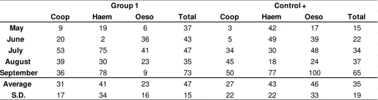
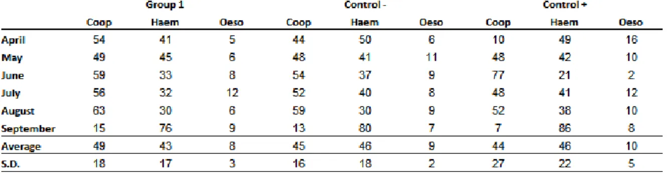
11 thaumasium) and D. flagrans only. In the end of five months of study, the percentage of nematodes found in groups 1, + control and - control were respectively: Cooperia sp. 49%, 45% and 44%, Haemonchus sp. 43%, 46% and 46% and Oesophagostomum sp. 8%, 9% and 10%. Other species found in smaller quantities in all groups were Bunostomum sp. and Strongyloides sp., whose percentages did not reach 1%.
Table 1 shows the reduction percentages corresponding to the infective larvae (L3)
recovered from the coprocultures of the groups treated with the association of the
nematophagous fungi D. flagrans and M. thaumasium (Group 1) and the positive
control group (D. flagrans only) in relation to the negative control. The larval numbers
of Cooperia, Haemonchus and Oesophagostomum of the group treated with the
association reduced by 31, 41 and 23%. In the group treated with D. flagrans the
reduction was 27, 43 and 46% respectively (p<0.05), in relation to the negative control.
The L3 total reduction was 47 and 35% for the Group 1 and positive control;
respectively. The association provided similar reduction percentage of infective larvae
in relation to D. flagrans and there was no significant difference in the L3 reduction
percentage between the groups.
Table 1. Percentage of reduction of infective larvae (L3) recovered from the coprocultures of the groups treated with the association of the nematophagous fungi D. flagrans and M. thaumasium (Group 1) and
the positive control group (D. flagrans only) in relation to the negative control from May 2012 to
September 2012, Ouro Branco, MG, Brazil.
Coop Haem Oeso Total Coop Haem Oeso Total
May 9 19 6 37 3 42 17 15
June 20 2 36 43 5 49 39 22
July 53 75 41 47 34 30 48 34
August 39 30 23 35 45 18 24 37
September 36 78 9 73 50 77 100 65
Average 31 41 23 47 27 43 46 35
S.D. 17 34 16 15 22 22 33 19
Group 1 Control +
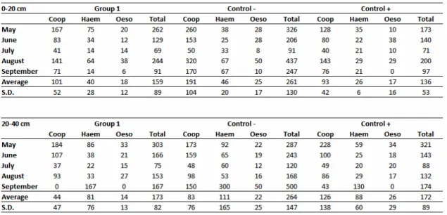
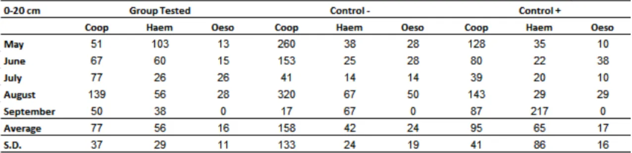
Table 2 shows the absolute values of L3 per kg of dry matter obtained from pastures
grazed by the three groups of calves. In pasture, larvae were found in the same
distribution pattern as the stool. The genus Cooperia was the most prevalent, followed
by Haemonchus and Oesophagostomum. The L3 reduction percentage in relation to
the - control in the distances up to 20 and 20-40 cm from the faecal pats was 39
12
35%, in the same distances. The largest reduction was found in the last month of the
study (September) with 65 and 64% for Group 1 and + control, respectively, however,
there was no significant difference between the group treated with the association and the group treated with D. flagrans only.
Table 2 Absolute values of L3 per kg of dry matter obtained from pastures grazed by the calves treated with the association of the nematophagous fungi D. flagrans and M. thaumasium (Group 1) , D. flagrans only (control +) and without fungus (control-) from May 2012 to September 2012, Ouro Branco,
MG, Brazil.
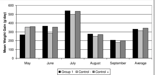
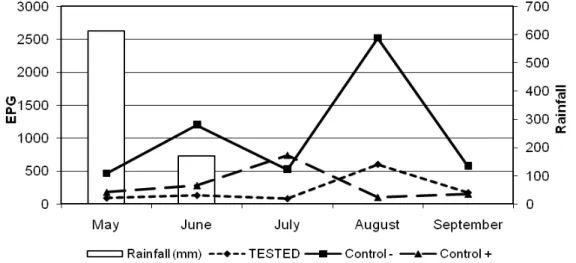
Figure 2 shows the mean weight gain of animals of the three groups. The weight gain
of the animals of the treated groups differed from those of the control group in the end of experiment (p<0.05). There was no significant difference for animal weight during the first 2 months of the experiment (May, June) between the three groups.
However, in the last three months of the experiment (July, August and September)
13 0 100 200 300 400 500 600
May June July August September Average
M e a n W e igh t G a in (g/ da y )
Group 1 Control - Control +
Fig. 2. Mean weight gain (kg/day) for calves treated with the association of the nematophagous fungi D. flagrans and M. thaumasium (Group 1) , D. flagrans only (control +) and without fungus (control-) from
May 2012 to September 2012, Ouro Branco, MG, Brazil.
DISCUSSION
Various reports have described the predatory activity of nematophagous fungi in laboratory and field conditions when used in sodium alginate pellets (Alves et al., 2004; Silva et al., 2009; Assis et al., 2012). However, there are no records of the use of these fungi in pellets containing associated formulations. Tavela et al. (2012)showed that the association of different nematophagous fungi in laboratory conditions was effective to control cyathostomin. The results of this work are the first report of in vivo association of D. flagrans and M. thaumasium in a pellet formulation through the gastrointestinal tract of dairy cattle with accompanying reduction of parasite load during five months of application of the formulation.
A number of studies on D. flagrans using horses and ruminants reported lower average monthly EPG counts for treated animals than for non-treated animals (Baudena et al., 2000; Knox and Faedo, 2001; Dimander et al., 2003; Fontenot et al., 2003; Araujo et al., 2004; Paraud et al., 2007). These findings are in agreement with results obtained in the
present work, confirming that the fungus acts on the infective forms in the fecal
14 fungi. Assis et al. (2013) in a comparative study of the action of the same fungal isolates in beef cattle found EPG reduction of 56.7% and 47.8% for isolates D. flagrans (AC001) and M. thaumasium (NF34A), separately. In this study, with dairy cattle, we found EPG reductions greater than 70% in both treated groups.
Regarding the percentage of infective larvae found in stool, the findings of this study concur with those of Dias et al. (2007) and Assis et al. (2012) who obtained similar results in studies with cattle in Southeastern Brazil. It is already known that the genera Cooperia sp., Haemonchus sp. and Oesophagostomum sp. are the most prevalent nematodes in southeastern Brazil (Lima, 1989).
Tavela et al. (2012) reported that the reduction of L3 recovery from contaminated faeces by the isolates AC001 and NF34, in association, was always over 80%. Araújo et al. (2004) found reduction of 59.3 % of L3 recovery from coprocultures in goat treated with pellets containing M. thaumasium.
Thus, these results corroborate the present work, in which the reduction of L3 in both treated groups was 47 and 35%. This finding confirmed that the association of pellets containing the fungi D. flagrans and M. thaumasium was able to pass through the gastrointestinal tract of dairy cattle, germinated in the faeces and effectively reduced L3 trichostrongylid larvae.
In this study, the results suggest that there was a direct action of D. flagrans on infective
trichostrongylid larvae present in the pasture and a consequent lower parasitic infection of fungus-treated animals. However, the association of the isolate of M. thaumasium to
D. flagrans did not enhance the reduction of larvae on pasture. Therefore, we concluded
that there was no synergism between the isolates tested.
The number of larvae recovered in the distances 0-20 and 20-40 cm from fecal pats is probably related with the use of nematophagous fungi that act directly on the L3 present
in pastures, confirming that D. flagrans accounted for the satisfactory reduction of environmental contamination (Araújo et al., 2004). Assis et al. (2013) reported reduction in L3 recovered from pasture in the distances 0-20 and 20–40 cm from the fecal pats of 64.5% and 73%, respectively for D. flagrans, while M. thaumasium showed percentage reductions of 47.3% and 58% in the same distances.
15 weight gain of cattle treated with pellets containing D. flagrans mycelia and Braga et al (2009) on weight gain of horses also treated with the fungus. Assis et al. (2013) also reported better weight gain in bulls treated with isolates of D. flagrans and M. thaumasium separately administered to animals.
The results of this work are promising because they show for the first time the passage of different fungal species associated in a pellet formulation containing D. flagrans and M. thaumasium through the gastrointestinal tract of dairy cattle monitoring the reduction of larvae in pasture.
CONCLUSION
The treatment of dairy cattle with sodium alginate pellets containing the mycelial mass of nematophagous fungi D. flagrans alone or in association with M. thamasium, twice a week for five months, decreased the EPG of the animals by more of 70%. However, the association of the fungi D. flagrans (AC001) and M. thaumasium (NF34A) showed no synergism, because there was no significant difference when compared with the group receiving the isolate D. flagrans alone.
Acknowledgements
CAPES, CNPq and FAPEMIG for financial support.
REFERENCES
Alves, P.H., Araújo, J.V., Guimarães, M.P., Assis, R.C.L., Campos, A.K., 2004. Aplicação de formulação do fungo predador de nematóides M. thaumasium (Drechsler, 1937) no controle de nematóides de bovinos. Arq. Bras. Med. Vet. Zootec. 55, 568-573. Amarante, A.F.T., Padovani, C.R., Barbosa, M.A., 1996. Contaminação de larvas de nematoides gastrintestinais parasitos de bovinos e ovinos em Botucatu-SP, Revista Brasileira de Parasitologia Veterinária, 5, 65-73.
Animal Health and Veterinary Laboratories Agency (AHVLA Parasitology Group). Annual Review of Literature & Horizon Scanning Report, 2010. Summary of papers published in 2010. In: http://www.defra.gov.uk/ahvla-en/files/pub-para-hor10.pdf Araújo, J.V., Assis, R.C.L., Campos, A.K., Mota, M.A., 2004. Atividade in vitro dos fungos nematófagos dos gêneros Arthobotrys, Duddingtonia e Monacrosporium sobre nematóides parasitos gastrintestinais de bovinos. Revista Brasileira de Parasitologias Veterinária, 13, 65-71.
16 bovinos pelo fungo Monacrosporium sinense. Arq. Bras. Med. Vet. Zootec. 56, 467-471.
Assis, R.C.L., Luns, F.D., Araújo, J.V., Braga, F.R., 2012. Biological control of trichostrongyles in beef cattle by the nematophagous fungus Duddingtonia flagrans in tropical southeastern Brazil. Exp. Parasitol. 132, 373–377.
Assis, R.C.L., Luns, F.D., Araújo, J.V., Braga, F.R., Assis, R.L., Marcelino, J.L., Freitas, P.C., Andrade, M.A.S., 2013. Comparison between the action of nematode predatory fungi D. flagrans and M. thaumasium in the biological control of bovine gastrointestinal nematodiasis in tropical southeastern Brazil. Vet. Parasitol. 193, 134- 140.
Baudena, M.A., Chapman, M.R., Larsen, M., Klei, R.R., 2000. Efficacy of the nematophagous fungus Duddingtonia flagrans in reducing equine cyathostome larvae on pasture in south Lousiana. Vet. Parasitol. 89, 219-230.
Burke, J.M., Miller, J.E., Larsen, M., Terrill, T.H., 2005. Interaction between copper oxide wire particles and Duddingtonia flagrans in lambs. Vet. Parasitol. 134, 141-146. Coles, G.C.; Jackson, F.; Powroy, H.E.; Prichard, R.K., 2006. The detection of anthelmintic resistance in nematodes of veterinary importance. Vet. Parasitol. 136, 167-185.
Dimander, S.O., Hoglund, J., Uggla, A., Sporndly, E., Waller, P.J., 2003. Evaluation of gastrointestinal nematode parasite control strategies for first-season grazing cattle in Sweden. Vet. Parasitol. 111, 192-209.
Fontenot, M.E., Miller, J.E., Pena, M.T., Larsen, M., Gillespie, A., 2003. Efficiency of feeding Duddingtonia flagrans chlamydospores to grazing ewes on reducing availability of parasitic nematode larvae on pasture. Vet. Parasitol. 118, 203-213.
Githigia, S.M., Thamsborg, S.M., Larsen, M., 2001. Effectiveness of grazing management in controlling gastrointestinal nematodes in lambs on pasture in Denmark. Vet. Parasitol. 99, 15-27.
Gordon, H.M., Whitlock, H.V., 1939. A new technique for counting nematode eggs in sheep faeces. Journal Council Science Industrial Research 12, 50-52.
Keith, R.K., 1953. The differentiation on the infective larvae of some common nematode parasites of cattle. Aust. J. Zool. 1, 223-235.
17 Lackey, B.A., Muldoon, A.E., Jaffe, B.A., 1993. Alginate pellet formulation of Hirsutella rossiliensis for biological control of plant-parasitic nematodes. Biologic Control, 3, 155-160.
Larsen, M., Wolstrup, J., Henriksen, S.A., Grønvold, J., Nansen, P., 1992. In vivo passage through calves of nematophagous fungi selected for biocontrol of parasitic nematodes. Journal of Helminthology 66, 137-141.
Larsen, M., Nansen, P., Wolstrup, J., Grønvold, J., Henriksen, S.A., Zorn, A., 1995. Biological control of trichostrongylosis in grazing calves by means of the fungus Duddingtonia flagrans. Vet. Parasitol. 60, 321-330.
Larsen, M., Nansen, P., Grøndahl, C., Thamsborg, S.M., Grønvold, J., Wolstrup, J., Henriksen, S.A., Monrad, J., 1996. The capacity of the fungus Duddingtonia flagrans to prevent strongyle infections in foals on pasture. Parasitology 113, 1-6.
Lima, W.S., 1989. Dinâmica das populações de nematóides parasitos gastrintestinais em bovinos de corte, alguns aspectos da relação parasito-hospedeiro e do comportamento dos estádios de vida livre na região do Vale do Rio Doce, MG, Brasil. PhD thesis, Instituto de Ciências Biológicas da Universidade Federal de Minas-Gerais, Belo Horizonte, Brasil.
Paraud, C., Pors, I., Chartier, C., 2007. Efficiency of feeding Duddingtonia flagrans chlamydospores to control nematode parasites of first-season grazing goats in France. Vet. Res. Commun. 31, 305-315.
Paz-Silva, A., Francisco, I., Valero-Coss, R.O., Cortiñas, F.J., Sánchez, J.A., Francisco, R., Arias, M., Suarez, J.L., López-Arellano, M.E., Mendoza de Gives, P., 2011. Ability of the fungus Duddingtonia flagrans to adapt to the cyathostomin egg-output by spreading chlamydospores. Vet. Parasitol. 179, 277-282.
Tavela, A.O., Araújo, J.V., Braga, F.R., Silveira, W.F., Silva, V.H.D., Júnior, M.C., Borges, L.A., Araújo, J.M., Benjamin, L.A., 2012. Coadministration of sodium alginate pellets containing the fungi Duddingtonia flagrans and Monacrosporium thaumasium on cyathostomin infective larvae after passing through the gastrointestinal tract of horses. Research in Veterinary Science.
18 Waller, P.J., Knox, M.R., Faedo, M., 2001. The potential of nematophagous fungi to control the free-living stages of nematode parasites of sheep: feeding and block studies with Duddingtonia flagrans. Vet. Parasitol. 102, 321-330.
19
CAPÍTULO 2 – Tropical Animal Health and Production (submetido)
F. D. LUNS a *; R. C. L. ASSIS a; J. V. ARAÚJO a; F. R. BRAGA a
Association of the fungi Duddingtonia flagrans and Arthrobotrys robusta on biologic
control of dairy cattle nematodiasis
a Federal University of Viçosa, Department of Veterinary Medicine - Laboratory of Parasitology, Av.
P.H. Rolphs - Viçosa Campus. Zip code 36570-000. Viçosa - Minas Gerais - Brazil.
* Corresponding author: P.H. Rolphs Avenue, Viçosa Campus, Department of Veterinary Medicine -
Laboratory of Parasitology, Viçosa, Minas Gerais, Brazil. Zip code: 36570-000, telephone: +55 31
3899-1458 Fax: +55 31 3899-1457, e-mail: fabio.luns@ufv.br
Abstract
The viability of the association the fungi D. flagrans and A. robusta was tested in biologic control of dairy cattle nematodiasis in Brazil. 24 6-month old female Girolando heifers were separated into three groups of 8 heifers each. The heifers were allocated to three 10ha paddocks of Brachiaria decumbens and each animal of group treated with association (group tested) received 1g of pellets (0.2g of fungal mycelium) for each 10kg of body weight (b.w.) containing the fungi D. flagrans and A. robusta. In group control positive, each animal received 1g of pellets containing the fungus D. flagrans. The animals of group control negative received 1g of fungus-free pellets, during 5 months. The EPG percentage of reduction in this study was 78% (group tested) and 73% (+ control). The percentage of reduction of L3 in pasture in the distances of up to 20 and 20-40 cm from the fecal pat was 43 and 55% (group tested) and 48
and 35% (control +). There was no significant difference between the group treated with association and the group treated with D. flagrans only in the EPG and L3 recovered of pasture. Concluded that the association has not been more effective than the isolate D. flagrans only. The results showed in this work are promising since it represents the first report of the passage of different fungal species of D. flagrans and A. robusta associated in a formulation at the same time through the gastrointestinal tract of cattle monitoring the reduction of larvae on pasture.
20
Introduction
Nematophagous fungi, especially the genera Duddingtonia, Monacrosporium and Arthrobotrys have predatory capacity on infective larvae (L3) of gastrointestinal nematode parasites of domestic animals (Araújo et al., 1998, 2004; Campos et al., 2007). Among predatory fungi, the genus Arthrobotrys has been proven to be a potential biological control agent of parasitic nematodes in domestic animals (Araújo et al., 1998; Braga et al., 2009). Arthrobotrys robusta is a nematode predator fungus which develops an adhesive network of tridimensional loops in which the nematodes are caught and then destroyed (Barron, 1977; Gives et al., 1992). The species A. robusta presents erect conidiophores, sometimes branched, about 300mm in length, having the extremity increased in size, usually carrying six ovoid-shaped conidia, hyaline, sectioned closely to the intermediate region, 18-27mm long and 8-12mm wide, and capable of producing chlamydospores and adhesive networks for preying on nematodes (Araújo et al., 1998, 2000). This fungus is considered a good predator of some plant-parasitic nematodes. In France it has been marketed as a commercial product (Royal 300 and Royal 350) to control the mycophagous nematode D. myceliophagus which destroys commercial mushrooms (Cayrol et al., 1978). According to Mota et al. (2003) this product out of the market due to problems with the formulation made grain rye.
This study aimed to test a formulation of nematophagous fungi to sodium alginate which has been highly promising in vivo tests (Araújo et al., 1998; Mota et al., 2003; Assis et al., 2012; Assis et al., 2013) the application of two different fungi in association. The use of association of nematode trapping fungi was also investigated in vivo shortly. This association can provide a synergistic effect, achieving better results than a single isolated applied, however the increased competition between the fungi for nematode larvae need to be evaluated in these associations in vivo. This study aimed to evaluate the viability of the association the fungi D. flagrans and A. robusta in biologic control of dairy cattle nematodiasis in Brazil.
2. Material and methods
2.1. Fungi and production of mycelial pellets
21 transferred to 250 mL Erlenmeyer flasks with 150 mL of liquid GPY medium (glucose, sodium peptone and yeast extract), pH 6.5, under the agitation of 120 rpm, in the dark, 26°C, for 10 days. After this period, the mycelia were harvested with a platinum loop, and weighed in an analytic scale for the future production of the pellets, which were made in a sodium alginate matrix, according to Walker and Connick (1983) and modified by Lackey et al. (1993).
2.2. Animals and experimental site
The experiment was carried out in a private farm located in the municipality of Ouro
22 Every 15 days, herbage samples were collected in each paddock of groups, in a zigzag pattern from alternated points, 20cm and 20–40cm far from the fecal pats, according to Amarante et al. (1996). Then, a 500g herbage sample was weighed, and parasitic nematode larvae were recovered following the procedure of Lima (1989). The samples were incubated in a drying oven at 100°C for 3 days to determine the dry matter content. Data were transformed into larvae per kg of dry matter.
The data were submitted to analysis of variance (ANOVA). Subsequently, the means were compared using the Tukey test at the 5% level of probability.
This study was approved by the Ethics Committee of the Federal University of Viçosa, protocol 66/2012.
3. Results
23
Fig. 1. Monthly averages of the countings of eggs per gram of feces (EPG) of the animals in the groups treated with the nematophagous fungi D. flagrans and A. robusta association (1 g of fungus/10 kg of b.
w.) (Group 1), the – control group (without treatment) and the + control group (D. flagrans only),
collected from May 2012 to September 2012, Ouro Branco, Minas Gerais, Brazil.
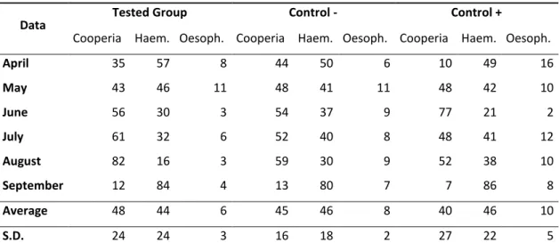
Table 1 shows the analysis of genera larvae recovered from coprocultures. There was no difference between the groups treated with association of fungi (D. flagrans and A. robusta) and only D. flagrans. In the end of five months of study, the percentage of nematodes found in tested group, control- and control+ were, respectively: Cooperia sp. 48%, 45% and 44%, Haemonchus sp. 44%, 46% and 46% and Oesophagostomum sp. 8%, 9% and 10%. Other species found in smaller quantities in all groups were Bunostomum sp. and Strongyloides sp. whose percentages did not reach 1%.
Tab. 1. Percentage values corresponding to the infectant larvae recovered from the coprocultures of the groups treated with the nematophagous fungi D. flagrans and A. robusta association (Group 1) and
the + control group (D. flagrans only) in relation with the - control group (without treatment) collected
from May 2012 to September 2012, Ouro Branco, Minas Gerais, Brazil.
24
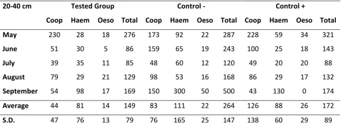
Tab. 2. Values of L3 per kg of dry matter obtained from pastures (0-20 cm and 20-40cm, respectively) grazed by the groups treated with the nematophagous fungi D. flagrans and A. robusta association (Group
Tested), the - control group (without treatment) and the + control group (D. flagrans only), collected from
May 2012 to September 2012, Ouro Branco, Minas Gerais, Brazil.
Fig. 2 shows the mean weight gain of animals of the three groups. The weight gain of the animals of the treated groups differed from those of the control group in the end of experiment (p<0.05). There was no significant difference for animal weight
during the first 2 months of the experiment (May, June) between the three groups.
However, in the last two months of the experiment (August and September) significant
25
Fig 2. Mean weight gains (kg/day) of the groups treated with the nematophagous fungi D. flagrans and A. robusta association (Tested), the – control group (without treatment) and the + control group (D. flagrans
only), collected from May 2012 to September 2012, Ouro Branco, Minas Gerais, Brazil.
4. Discussion
The results showed in this work are the first report of in vivo association of D. flagrans and A. robusta in a pellet formulation through the gastrointestinal tract of cattle with accompanying reduction of parasite load during five months of application of the formulation. A number of studies on only D. flagrans using bovines recorded average monthly EPG counts lower for treated animals than for non-treated groups (Assis et al,
2012; Assis et al., 2013). These findings are in agreement with results obtained in the present work, confirming that the fungus acts on the infective forms in the fecal
environment, with consequently decrease in EPG. The fungus D. flagrans is most widely used in biologic control because it produces chlamydospores, structures highly resistant to the passage intestinal tract of animals (Mota et al., 2003). Araujo et al. (1998) tested the only isolated A. robusta (I31) in the biological control of bovine gastrointestinal nematodes parasites with two million of conidias and obtained reduction of 54% in the EPG. However, new data found in this study, was that the EPG counts of animals treated with D. flagrans and A. robusta in association were not significantly lower than those of the animals treated with D. flagrans alone. This finding suggests that there was no synergism between these fungi.
26 able to pass through the gastrointestinal tract in cattle, and then these fungi were germinated in the faeces and after, were effective in reducing L3 trichostrongyles. However, the association of the isolate of A. robusta to D. flagrans not enhanced the reduction of larvae on pasture. So we concluded that there was no synergism between the isolates tested. The number of larvae recovered in the distances 0-20 and 20-40 cm from fecal pats is likely to be directly related with the use of nematophagous fungi that
act directly on the L3 present in pastures, confirming that nematophagous fungi was responsible for the satisfactory reduction of environmental contamination (Araújo et al., 2004). Assis et al. (2013) related a reduction of L3 recovered by pasture in the distances of up to 20 and 20–40 cm from the fecal pat for isolated AC001 corroborate this study. The difference found in weight gain of treated animals compared to the control group may have been caused by a lower parasite load in animals that received pellets containing fungus mycelia, which may have contributed to a better food conversion of treated animals. These results are similar to those found by Dias et al. (2007) and Assis et al. (2013) on weight gain of cattle treated with pellets containing D. flagrans mycelia. The results showed in this work are promising since it represents the first report of the passage of different fungal species associated in a formulation of pellets containing D. flagrans and M. thaumasium at the same time through the gastrointestinal tract of cattle monitoring the reduction of larvae on pasture.
Conclusion
The treatment of dairy cattle with sodium alginate pellets containing the mycelial mass of nematophagous fungi D. flagrans alone or in association with A. robusta, twice a week for five months, decreased the EPG of the animals in more of 70%. However, the association of the fungi D. flagrans (AC001) and A. robusta (I31) showed no synergism, because there was no significant difference when compared with the group receiving the isolated D. flagrans alone.
References
Amarante, A.F.T., Padovani, C.R., Barbosa, M.A., 1996. Contaminação de larvas de nematoides gastrintestinais parasitos de bovinos e ovinos em Botucatu-SP, Revista Brasileira de Parasitologia Veterinária, 5, 65-73.
27 Araújo, J.V., Sampaio, W.M., Vasconcelos, R.S., Campos, A.K., 2000. Effects of different temperatures and mineral salt on pellets of M. thaumasium - a nematode-trapping fungus. Veterinary Archive, 80, 181-190.
Araújo, J.V., Guimarães, M.P., Campos, A.K., Sá, N.C., Sarti, P., Assis, R.C.L., 2004. Control of bovine gastrointestinal nematode parasites using pellets of nematode trapping fungus M. thaumasium, Ciência Rural, 34, 457-463.
Assis, R.C.L., Luns, F.D., Araújo, J.V., Braga, F.R., 2012. Biological control of
trichostrongyles in beef cattle by the nematophagous fungus Duddingtonia flagrans in
tropical southeastern Brazil. Experimental Parasitology 132, 373-377.
Assis, R.C.L., Luns, F.D., Araújo, J.V., Braga, F.R., Assis, R.L., Marcelino, J.L., Freitas, P.C., Andrade, M.A.S., 2013. Comparison between the action of nematode predatory fungi D. flagrans and M. thaumasium in the biological control of bovine gastrointestinal nematodiasis in tropical southeastern Brazil, Veterinary Parasitology, 193, 134- 140.
Barron, G.L., 1977. The Nematode-destroying Fungi. Topics in Mycobiology, No. 1. Canadian Biological Publications, Guelph, Canada. 140 p.
Braga, F.R., Carvalho, R.O., Araujo, J.M., Araujo, J.V., 2009. Predatory activity of the fungi D. flagrans, M. thaumasium, M. sinense and A. robusta on A. vasorum first-stage larvae. Journal of Helminthology, 83, 303-308.
Campos, A.K., Araujo, J.V., Assis, R.C.L., Gandra, J.R., Guimarães, M.P., 2007. Viabilidade de formulação peletizada do fungo nematófago M. sinense no controle biológico de nematóides parasitos gastrintestinais de bezerros, Arquivo Brasileiro de Medicina Veterinária e Zootecnia, 59, 14-20.
Castro, A.A., Oliveira, C.R.C., Anjos, D.H.S., Ornelas, E.I., Bittencourt, V.R., 2003. Potencial dos fungos nematófagos Arthrobotrys sp. e M. thaumasium para o controle de larvas de ciatostomíneos de equinos (Nematoda: Cyathostominae). Revista Brasileira de Parasitologia Veterinária, 12, 2, 53-57.
Cayrol, J.C., Frankowsky, J.P., Laniece, A., D’Hardemare, G., Talon, J.P., 1978. Contre
les nematódes en champignoniére. Mise au point d’une méthode de lute biologique a l’aide d’un hyphomicete prédateur: Arthrobotrys robustus souche antipolis (Royal 300). Revue Horticole, 184, 23-30.
28 Gordon, H.M., Whitlock, H.V., 1939. A new technique for counting nematode eggs in sheep faeces. Journal Council Science Industrial Research, 12, 50-52.
Keith, R.K., 1953. The differentiation on the infective larvae of some common nematode parasites of cattle. Aust. J. Zool. 1, 223-235.
Lackey, B.A., Muldoon, A.E., Jaffe, B.A., 1993. Alginate pellet formulation of Hirsutella rossiliensis for biological control of plant-parasitic nematodes. Biologic Control, 3, 155-160.
Lima, W.S., 1989. Dinâmica das populações de nematóides parasitos gastrintestinais em bovinos de corte, alguns aspectos da relação parasito-hospedeiro e do comportamento dos estádios de vida livre na região do Vale do Rio Doce, MG, Brasil. PhD thesis, Instituto de Ciências Biológicas da Universidade Federal de Minas-Gerais, Belo Horizonte, Brasil.
Mendoza-de Gives, P.; Zavaleta-Mejia, E.; Quiroz-Romero, H., 1992. Interaction between the nematode-destroying fungus Arthrobotrys robusta (Hyphomycetales) and Haemonchus contortus infective larvae in vitro. Veterinary Parasitology, 41, 101-107. Mota, MA, Campos, AK, Araújo, JV, 2003. Controle biológico de helmintos parasitos de animais: estágio atual e perspectivas futuras, Pesquisa Veterinária Brasileira, 23, 93-100.
29 CAPITULO 3
F. D. LUNS a *; R. C. L. ASSIS a; J. V. ARAÚJO a; F. R. BRAGA a
Association of the fungi Arthrobotrys robusta, Duddingtonia flagrans and
Monacrosporium thaumasium on biologic control of dairy cattle nematodiasis
a Federal University of Viçosa, Department of Veterinary Medicine - Laboratory of Parasitology, Av.
P.H. Rolphs - Viçosa Campus. Zip code 36570-000. Viçosa - Minas Gerais - Brazil.
* Corresponding author: P.H. Rolphs Avenue, Viçosa Campus, Department of Veterinary Medicine - Laboratory of Parasitology, Viçosa, Minas Gerais, Brazil. Zip code: 36570-000, telephone: +55 31 3899- 11458 Fax: +55 31 3899-1457, e-mail: fabio.luns@ufv.br
Abstract
The viability of the association the fungi D. flagrans, M. thaumasium and A. robusta was tested in biologic control of dairy cattle nematodiasis in Brazil. 24 6-month old female Girolando heifers were separated into three groups of 8 heifers each. The heifers were allocated to three 10ha paddocks of Brachiaria decumbens, naturally infested with gastrointestinal parasite helminths. Each animal of group treated with association (group tested) received 1g of pellets (0.2g of fungal mycelium) for each 10kg of body weight (b.w.) containing the fungi D. flagrans, M. thaumasium and A. robusta in association. In group control positive, each animal received 1g of pellets/10kg of b.w. containing the fungus D. flagrans only. The heifers of group control negative received 1g of fungus-free pellets, during 5 months. The EPG percentage of reduction in this study was 80% (group tested) and 73% (+ control). The percentage of reduction of L3 in pasture in the distances of up to 20 and 20-40 cm from the fecal pat was 57 and 43% (group tested)
30
1. Introduction
Among the nematophagous fungi, those of the genera Duddingtonia, Monacrosporium and Arthrobotrys have been widely studied in the control of gastrointestinal nematode parasites of domestic animals (Araújo et al., 2004, Braga et al., 2009). The genus Duddingtonia was evaluated in vitro and in pasture, on sheep (Waller et al., 2001; Waghorn et al., 2003), lambs (Githigia et al., 2001) and bulls (Assis et al., 2012). The Monacrosporium sp. also has been successfully used to combat nematodes of cattle in Brazil (Alves et al., 2004; Assis et al., 2013). As the genus Duddingtonia and Monacroscoporium, the Arthrobotrys has been proven to be a potential biological control agent of parasitic nematodes in domestic animals (Castro et al., 2003; Braga et al., 2009). Thus, it is observed that there are several studies showing the effectiveness of these fungi to pass through the gastrointestinal tract of animals and kill infective larvae when used alone, but no study has examined the association of the three fungi. This association can provide a synergistic effect, achieving better results than a single isolated applied, however can also the increased competition between the fungi for nematode larvae need to be evaluated in these associations in vivo. This study aimed to evaluate the viability of the association the fungi D. flagrans, M. thaumasium and A. robusta in biologic control of dairy cattle nematodiasis in Brazil.
2. Material and methods
2.1. Fungi and production of mycelial pellets
Three isolates of the predatory fungi D. flagrans (AC001), M. thaumasium (NF34) and A. robusta (I31) were kept in test tubes containing corn meal agar 2% (2% CMA, Difco®), at 4°C in the dark. These isolates came from a Brazilian soil and belonged to the mycology collection of the Federal University of Viçosa, Brazil. To induce the formation of the fungal mycelium, culture discs of 5 mm in diameter in 2% water-agar (2% WA) were transferred to 250 mL Erlenmeyer flasks with 150 mL of liquid GPY medium (glucose, sodium peptone and yeast extract), pH 6.5, under the agitation of 120 rpm, in the dark, 26°C, for 10 days. After this period, the mycelia were harvested with a platinum loop, and weighed in an analytic scale for the future production of the pellets, which were made in a sodium alginate matrix, according to Walker and Connick (1983) and modified by Lackey et al. (1993).
31 The experiment was carried out in a private farm located in the municipality of Ouro
Branco, state of Minas Gerais, southeast region of Brazil, 43°41’31” South latitude and 20°31’15” West longitude, from April to September 2012. The topography is hilly, with
an average elevation height of 1000m and the native vegetation is Atlantic rain forest-cerrado transition zone. The climate is tropical with a dry season (Rating Köppen-Geiger climate: Aw), annual average maximum temperature of 71.60°F and minimum of 44.60°F. In the beginning of the experiment, 24 6-month old female Girolando calves, with average body weight of 130kg were previously treated with 10% albendazole at an oral dose of 7.5mL/10kg of b.w. Fifteen days after the anthelmintic treatment, the animals were separated into three groups of 8 calves each, based on the average weight. The calves were allocated to three 10ha paddocks of Brachiaria decumbens, naturally infested with gastrointestinal parasite helminths, due to the previous grazing by young and adult animals. Each group was allocated to only one paddock without rotational grazing between the groups during the experimental period. Each animal of group treated received 1g of pellets (0.2g of fungal mycelium) for each 10kg of b.w. containing the fungi D. flagrans (AC001), M. thaumasium (NF34) and A. robusta (I31) associated and in a single oral dose. In group control positive, each animal received 1g of pellets (0.2g of fungal mycelium) for each 10kg of b.w. containing the fungus D. flagrans (AC001). The animals of group control negative received 1g of fungus-free pellets for each 10kg of b.w. All the animals received the pellets orally, twice a week, mixed in concentrated and balanced ration provided for dairy cattle (18% of total protein – Total®, Brazil), and water ad libitum during 6 months, starting from April 2012. Once time for week, stool samples were collected directly from the rectum to be performed the egg account per gram of faeces (EPG) according Gordon and Whitlock (1939). Simultaneously to the EPG exam, coprocultures were carried out, for each animal, according to the methodology described by Roberts et al. (1952). The identification of the infectant larvae in the coprocultures was performed according to Keith (1953).
32 The data were submitted to analysis of variance (ANOVA). Subsequently, the means were compared using the Tukey test at the 5% level of probability.
3. Results
In the first month of the experiment (April, 2012) no statistical difference was observed
(p<0.05) between the groups treated with fungi and the control group. The EPG was higher in the negative control group than in the treated animals of both groups during the months of May, June, August and September of 2012 (p<0.05). In July the tested group (three fungi) presented EPG lower than control groups, but no significant difference. The percentage of reduction in this study was 80% (group tested) and 73% (+ control). In the last month of the study was a reduction of 70% and 74%. The largest absolute difference of EPG (1917 and 2417 eggs) was found in August 2012. The largest percentage difference occurred in June (89%) for tested group and August 2012, (96%) for the group treated with only D. flagrans (Control +). There was no significant difference between the group treated with pellets made with the three fungal isolates (G1) and the group treated with pellets of D. flagrans only (p<0.05). The monthly averages of the EPG are shown in Figure 1.
Fig. 1. Monthly averages of the countings of eggs per gram of feces (EPG) of the animals in the groups treated with the nematophagous fungi D. flagrans, M. thaumasium and A. robusta association (1 g of fungus/10 kg of b.w.) (Tested), the – control group (without treatment) and the + control group (D. flagrans only), collected from May 2012mto September 2012, Ouro Branco, Minas Gerais, Brazil.