Bloodstream-To-Eye Infections Are
Facilitated by Outer Blood-Retinal Barrier
Dysfunction
Phillip S. Coburn1, Brandt J. Wiskur2, Frederick C. Miller3,4, Austin L. LaGrow1, Roger A. Astley1, Michael H. Elliott1,2,5, Michelle C. Callegan1,2,6*
1Department of Ophthalmology, The University of Oklahoma Health Sciences Center, Oklahoma City, Oklahoma, United States of America,2Oklahoma Center for Neuroscience, The University of Oklahoma Health Sciences Center, Oklahoma City, Oklahoma, United States of America,3Department of Cell Biology, The University of Oklahoma Health Sciences Center, Oklahoma City, Oklahoma, United States of America, 4Department of Family and Preventative Medicine, The University of Oklahoma Health Sciences Center, Oklahoma City, Oklahoma, United States of America,5Department of Physiology, The University of Oklahoma Health Sciences Center, Oklahoma City, Oklahoma, United States of America,6Department of Microbiology and Immunology, The University of Oklahoma Health Sciences Center, Oklahoma City, Oklahoma, United States of America
Abstract
The blood-retinal barrier (BRB) functions to maintain the immune privilege of the eye, which is necessary for normal vision. The outer BRB is formed by tightly-associated retinal pig-ment epithelial (RPE) cells which limit transport within the retinal environpig-ment, maintaining retinal function and viability. Retinal microvascular complications and RPE dysfunction resulting from diabetes and diabetic retinopathy cause permeability changes in the BRB that compromise barrier function. Diabetes is the major predisposing condition underlying endogenous bacterial endophthalmitis (EBE), a blinding intraocular infection resulting from bacterial invasion of the eye from the bloodstream. However, significant numbers of EBE cases occur in non-diabetics. In this work, we hypothesized that dysfunction of the outer BRB may be associated with EBE development. To disrupt the RPE component of the outer BRBin vivo, sodium iodate (NaIO3) was administered to C57BL/6J mice. NaIO3-treated and untreated mice were intravenously injected with 108colony forming units (cfu) of Staphylo-coccus aureusorKlebsiella pneumoniae. At 4 and 6 days postinfection, EBE was observed in NaIO3-treated mice after infection withK.pneumoniaeandS.aureus, although the inci-dence was higher followingS.aureusinfection. Invasion of the eye was observed in control mice followingS.aureus infection, but not in control mice followingK.pneumoniaeinfection. Immunohistochemistry and FITC-dextran conjugate transmigration assays of human RPE barriers after infection with an exoprotein-deficientagr/sarmutant ofS.aureussuggested thatS.aureusexoproteins may be required for the loss of the tight junction protein, ZO-1, and for permeability of thisin vitrobarrier. Our results support the clinical findings that for both pathogens, complications which result in BRB permeability increase the likelihood of bacterial transmigration from the bloodstream into the eye. ForS.aureus, however, BRB a11111
OPEN ACCESS
Citation:Coburn PS, Wiskur BJ, Miller FC, LaGrow AL, Astley RA, Elliott MH, et al. (2016) Bloodstream-To-Eye Infections Are Facilitated by Outer Blood-Retinal Barrier Dysfunction. PLoS ONE 11(5): e0154560. doi:10.1371/journal.pone.0154560
Editor:Fu-Shin Yu, Wayne State University, UNITED STATES
Received:March 7, 2016
Accepted:April 15, 2016
Published:May 19, 2016
Copyright:© 2016 Coburn et al. This is an open access article distributed under the terms of the
Creative Commons Attribution License, which permits unrestricted use, distribution, and reproduction in any medium, provided the original author and source are credited.
Data Availability Statement:Data are available from FigShare. The DOI is10.6084/m9.figshare.3187533.
permeability is not required for the development of EBE, but toxin production may facilitate EBE pathogenesis.
Introduction
The blood-retinal barrier (BRB) is a component of ocular immune privilege and serves to pro-tect the delicate, nonregenerative neural retina from the immune system and bloodborne path-ogens. The BRB consists of inner (endothelial cells, pericytes, and astrocytes) and outer (retinal pigment epithelial cells) components. The retinal pigment epithelium (RPE) consists of a single layer of cuboidal pigmented cells whose specific functions are critical for neural retina homeo-stasis. The RPE maintains the retinal environment by limiting transport across the retina, thus, maintaining a tight barrier to choroidal bloodborne substances [1,2]. The endothelial cells lin-ing the capillaries supplylin-ing the retina with oxygen and nutrients form the inner BRB, which exhibits selective permeability to small molecules, and is virtually impermeable to large macro-molecules [3]. During the development of diabetes and its ocular complication diabetic reti-nopathy, changes occur in the BRB which result in greater vascular permeability and loss of RPE function [4–20].
Diabetes is the leading predisposing condition for the development of endogenous bacterial endophthalmitis (EBE) [21], a severe, often blinding intraocular infection emanating from the bloodstream [21–27]. In 60% of cases of EBE, an underlying condition is present, and diabetes is present in 33% of those cases [21]. EBE occurs at a frequency of approximately 2% to 8% of all cases of endophthalmitis. Patients with EBE typically present with ocular pain, blurring or loss of vision, a hypopyon, an insufficient fundus view, and photophobia. Infection of the eye via this route can result in vision loss, and in the worst-case scenario, enucleation or eviscera-tion of the globe. EBE can also affect both eyes at the same time, causing bilateral blindness. Jacksonet al. [21] reported in a recent review of 342 EBE cases from 1986 to 2012 that the median final visual acuity after EBE was 20/100. In 44% of these cases, visual acuities were worse than 20/200. In approximately 24% of all cases examined, patients required evisceration or enucleation of the globe. Associated mortality in these EBE cases was 4% [21]. The leading causes of Gram-negative and Gram-positive EBE areKlebsiella pneumoniaeandStaphylococcus aureus, respectively [21–27]. Despite antibiotic and surgical intervention, the clinical outcome for patients with EBE continues to be poor [21].
Our previous studies suggest that during diabetes, a compromised BRB serves as a portal for bacteria to gain access to the eye from the bloodstream [28,29]. We reported that an increased incidence ofK.pneumonaieandS.aureusEBE in a diabetic murine model correlated with the length of time following diabetes induction with streptozotocin (STZ) [28,29]. This increased EBE incidence also correlated with greater vascular permeability in the eyes of STZ-induced diabetic mice [28,29]. Our results supported clinical reports that diabetes is a predisposing risk factor for the development of EBE [28,29]. However, diabetes progression results in a myriad of other host changes, including immunological deficits such as the inability of inflammatory cells to phagocytizeK.pneumoniaeandS.aureus[30,31]. To dissect the specific mechanisms that underlie EBE development, we sought to divorce BRB permeability from the immunologi-cal changes that occur during diabetes progression. Specifiimmunologi-cally, we hypothesized that dysfunc-tion of the RPE, a component of the outer BRB which is altered during the development of diabetes, facilitates the development of EBE. To test this hypothesis, we selectively induced RPE degeneration using sodium iodate (NaIO3), an oxidizing agent that exerts toxicity
specifi-cally towards the RPE [32] and is a neurodegenerative insult [33]. In the present study, EBE Competing Interests:The authors have declared
incidence after infection withS.aureusandK.pneumoniaein NaIO3-treated mice was
compa-rable to the incidence observed in the diabetic EBE models [28,29]. In control mice,S.aureus infection resulted in EBE, butK.pneumoniaeinfection did not. Furthermore, we observed that S.aureusexoprotein production was associated with a disruption in ZO-1 staining and increased permeability of anin vitroRPE barrier. Our results therefore suggest that alterations in the RPE component of the outer BRB may serve as a mechanism by whichK.pneumoniae andS.aureusEBE develops, but these alterations are not required forS.aureusEBE to occur.
Results
Permeabilization of the RPE with sodium iodate
To determine whether alterations specifically in the RPE resulted in an increased incidence of EBE, we first disrupted the RPE of C57BL/6J mice with sodium iodate (NaIO3) [1,2,32]. The
extent of damage to and resulting permeability of the RPE barrier was visualizedin vivoby fluorescein angiography. After 24 hours, significant changes in RPE pigmentation were observed in NaIO3-treated mice (Fig 1C), but not in PBS-treated mice (Fig 1A). The retinal
vasculature and optic nerve tissue in these eyes appeared normal. Wanget al. observed similar effects in retinal tissue following treatment of C57BL/6J mice with 20 and 30 mg/kg NaIO3
from 1 to 8 days after injection [32]. NaIO3treatment resulted in extensive leakage of
fluores-cein dye into the vitreous relative to PBS-injected mice. In PBS-treated mice, the fluorescence from the AK-FLUOR dye (Fig 1B) demarcated the retinal and/or choroidal vasculature, distin-guishing it from adjacent areas and structures. InFig 1D, diffuse fluorescence resulting from increased permeability and leakage of the dye was observed in NaIO3-treated mice.
To quantify the extent of permeability of the RPE in NaIO3-treated and untreated mice, a
modified Evans Blue dye assay was employed to measure albumin leakage into the retina [34]. Eyes from mice treated with sodium iodate allowed a greater concentration of albumin into the retina compared to that of untreated mice (Fig 1E, P = 0.01). Together, these results demon-strated that NaIO3disrupted the barrier properties of the RPE and rendered mouse eyes
per-meable to AK-FLUOR and albumin 24 hours following treatment.
RPE dysfunction and incidence of
S
.
aureus
and
K
.
pneumoniae
EBE
To establish a link between RPE dysfunction and the development of EBE, groups of mice were infected 24 hours after intraperitoneal injection of either PBS or NaIO3. These data are
Fig 1. Blood-retinal Barrier Breakdown in NaIO3-treated mice. (A-D)Funduscopic imaging of mouse eyes
24 hours after injection of either PBS or NaIO3. In PBS-injected mice (A and B), the fluorescence from the
AK-FLUOR dye demarcates the retinal and/or choroidal vasculature and distinguishes it from adjacent areas/ structures. In NaIO3-injected mice (C and D), note the diffuse fluorescence resulting from increased outer
BRB permeability and leakage of the dye.(E)Albumin leakage into the retina after injection of either PBS or NaIO3was quantified using a modified Evans Blue protocol. Bars represent mean±standard deviation (SD)
for N5 animals for all groups. A two-tailed t-test was used to assess significance between PBS-injected and NaIO3-injected mice (P = 0.01).
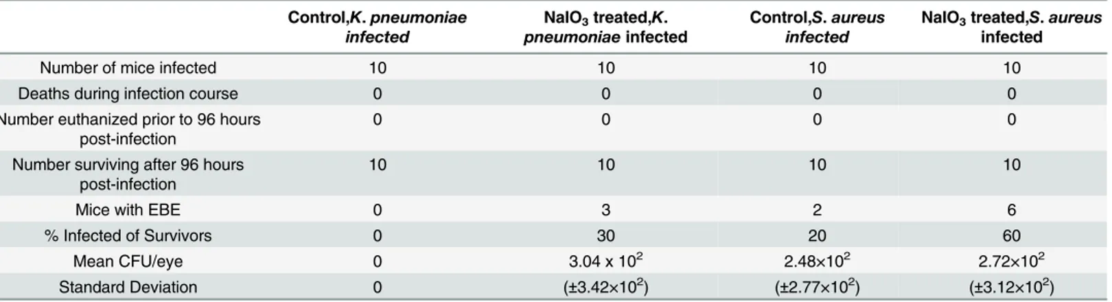
summarized inTable 1. At 96 hours postinfection withK.pneumoniae, 3 out of 10 NaIO3
-treated mice developed EBE. One eye from each mouse was infected. The meanK.pneumoniae cfu per eye among the NaIO3-treated mice was 3.04 x 10². None of the control mice infected
withK.pneumoniaedeveloped EBE. At 96 hours postinfection withS.aureus, 6 out of 10 NaIO3-treated mice (6 eyes) developed EBE and 2 out of 10 control mice (2 eyes) developed
EBE. The meanS.aureuscfu per eye was 2.72×10²for the NaIO3-treated mice, and 2.48×10²
for the control mice. NaIO3-induced RPE permeabilization resulted in a 30%K.pneumoniae
EBE incidence, similar to the 27%K.pneumoniaeEBE incidence that we previously reported for mice rendered diabetic for 5 months [28]. NaIO3-induced RPE permeabilization also
resulted in a 60%S.aureusEBE incidence, similar to the 58%S.aureusEBE incidence we observed in our 3-month diabetic mice [29].
Assessment of EBE incidence in NaIO3-treated mice 6 days after infection with both
patho-gens (Table 2) revealed a 20% incidence ofK.pneumoniaeEBE and a 50% incidence ofS. aureusEBE, but no infections in the control mice. After 6 days postinfection, the mean cfu per eye for theK.pneumoniae-infected mice was 1.16 x 10², and forS.aureus-infected mice was 2.58×10². These results indicated that intraocular infection with eitherK.pneumoniaeorS. aureuscan occur after specific disruption of the RPE component of the outer BRB in nondia-betic mice at incidences similar to that reported in dianondia-betic mice [28,29]. Similar to what we observed previously [29],S.aureuscaused EBE even when the BRB was intact in control mice not treated with NaIO3, whileK.pneumoniaedid not cause infections in these mice.
Table 1. Incidence ofK.pneumoniaeandS.aureusEBE at 4 days postinfection in control and NaIO3-treated mice.
Control,K.pneumoniae infected
NaIO3treated,K.
pneumoniaeinfected
Control,S.aureus infected
NaIO3treated,S.aureus
infected
Number of mice infected 10 10 10 10
Deaths during infection course 0 0 0 0
Number euthanized prior to 96 hours post-infection
0 0 0 0
Number surviving after 96 hours post-infection
10 10 10 10
Mice with EBE 0 3 2 6
% Infected of Survivors 0 30 20 60
Mean CFU/eye 0 3.04 x 10² 2.48×10² 2.72×10²
Standard Deviation 0 (±3.42×10²) (±2.77×10²) (±3.12×10²)
doi:10.1371/journal.pone.0154560.t001
Table 2. Incidence ofK.pneumoniaeandS.aureusEBE at 6 days postinfection in control and NaIO3-treated mice.
Control,K.pneumoniae infected
NaIO3treated,K.pneumoniae
infected
Control,S.aureus infected
NaIO3treated,S.aureus
infected
Number of mice infected 5 5 5 5
Deaths during infection course 0 0 0 1
Number euthanized prior to 96 hours post-infection
0 0 0 0
Number surviving after 96 hours post-infection
5 5 5 4
Mice with EBE 0 1 0 2
% Infected of Survivors 0 20 0 50
Mean CFU/eye 0 1.16 x 10² 0 2.58×10²
Standard Deviation 0 0 0 (±14)
Wanget al. reported significant effects of NaIO3treatment on the scotopic and photopic
b-wave ERG responses, showing almost complete elimination of ERG responses at 8 days follow-ing treatment [32]. In our previous study [29], we reported no changes in ERG responses 4 days following infection withS.aureusin diabetic animals, likely due to the low numbers of bacteria detected in those infected eyes [28,29]. In the current study, ERGs were not performed on infected mice because of the anticipated low numbers of bacteria in these eyes and because the interpretation of any observed ERG decrease could be potentially confounded by the effects of NaIO3treatment.
S
.
aureus
-induced alterations in an
in vitro
human outer BRB are
exoprotein-dependent
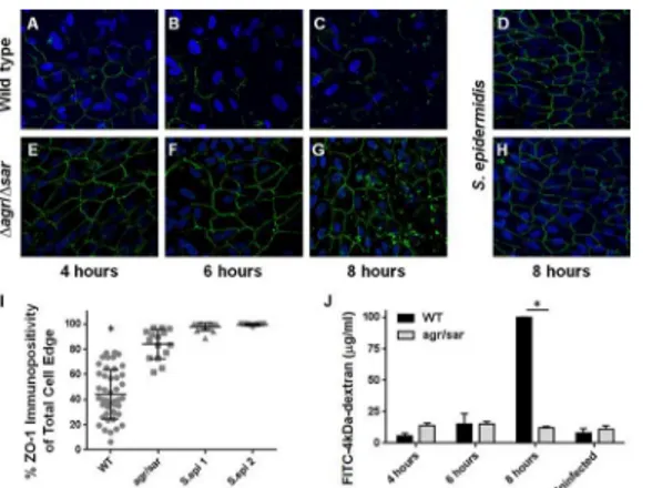
We previously reported thatS.aureus, but notK.pneumoniae, caused a significant reduction in immunoreactivity of the tight junction protein ZO-1 between cultured human RPE cells in ourin vitromodel of the outer BRB, suggesting thatS.aureusis able to directly disrupt the expression and/or organization of tight junctions between RPE cells [29]. Disruption of ZO-1 immunostaining correlated with changes in the permeability of ourin vitroouter BRB model to both FITC-dextran conjugate molecules and to liveS.aureus[29]. Because RPE dysfunction was necessary for invasion ofK.pneumoniaebut notS.aureusinto the eye from the blood-stream, the question arose whether RPE alterations were the direct result ofS.aureus exopro-tein production. We therefore examined the ability of anagr/sarquorum sensing-deficient, exoprotein-deficient mutant ofS.aureus[35] to alter ZO-1 immunoreactivity. Immunofluores-cence microscopy revealed that infection with wild typeS.aureuscaused progressive disruption in ZO-1 staining over time (Fig 2, panels A-C), while infection with theagr/sar-deficient mutant (Fig 2, panels E-G) or two ocular isolates ofS.epidermidis(Fig 2, panels D and H) did not. The percent of ZO-1 immunopositivity of RPE monolayers infected with wild typeS. aureuswas significantly less than in RPE monolayers infected with theagr/sar-deficient mutant or two ocular isolates ofS.epidermidis(P<0.0001,Fig 2, panel I). These results suggested that the observed changes in ZO-1 immunostaining after infection with wild typeS.aureuswere dependent on exoprotein production. RPE viability was greater than 95% at all time points, as determined by trypan blue staining [29].
Intact monolayers of human RPE cells in 0.4 micron transwells were infected with wild type S.aureusor theagr/sar-deficient mutant and diffusion of FITC-4-kDa-dextran across the bar-rier and into the bottom chamber was assessed by fluorescence spectrometry (Fig 2, panel J). No significant differences in RPE monolayer permeability were observed after 4 or 6 hours of infection with the two strains. However, after 8 hours, the fluorescence intensity was significantly greater in the bottom chamber of RPE monolayers infected with wild typeS.aureus(p<0.0001) compared with that of RPE monolayers infected with the exoprotein-deficient mutant. These results indicated thatS.aureuscould disrupt ZO-1 and the permeability of ourin vitrohuman outer BRB in an exoprotein-dependent manner. Taken together, these findings substantiate our observation that the greater incidence ofS.aureusEBE compared toK.pneumoniaeEBE corre-lates with the capacity ofS.aureusto directly disrupt RPE tight junctions, and suggests thatS. aureusmigration across the outer BRB may be facilitated by its toxic exoproteins.
Discussion
previously reported that the environment created by a compromised BRB promoted the entry of bloodborne pathogens into the eye. In mice with STZ-induced diabetes, we previously observed 24% (3-month) and 27% (5-month) incidences ofK.pneumoniaeEBE [28]. We also observedS.aureusEBE among 3-month (58% incidence) and 5-month (33% incidence) dia-betic mice. Although the frequency ofS.aureusEBE in 5-month diabetic mice was comparable to previous observations forK.pneumoniaeEBE, the incidence ofS.aureusEBE in 3-month diabetic mice was 2.5-fold greater. We found noK.pneumoniaeEBE in control (nondiabetic) or 1-month diabetic mice, but observed a 7% and a 12% incidence ofS.aureusEBE in these same groups, respectively. These data implied thatS.aureuswas capable of invading the eye regardless of the degree of BRB integrity, and raised the possibility thatS.aureusmight directly affect the outer BRB, resulting in infection of the eye. Junget al. recently reported that 9% ofS. aureus bacteremia patients developed ocular infections [36], but only 30% of those had diabe-tes as an underlying condition [36]. TheS.aureusEBE cases in that study were primarily asso-ciated with infective endocarditis, providing clinical support for our hypothesis thatS.aureus can cross the BRB and invade the eye in the absence of diabetes-related changes to the BRB. Our hypothesis is also supported by findings in a murine model of hematogenousS.aureus meningitis [37]. Sheenet al. reported thatS.aureuswas capable of crossing the blood-brain barrier (BBB) and infecting the brains of normal CD-1 mice [37]. After tail vein injection of 2 x 10⁸cfu ofS.aureus, bacteria were detected in the brains of 7 out of 9 infected animals at con-centrations ranging from approximately 102to greater than 106cfu per gram of brain tissue at 96 hours postinfection [37]. Although no eyes were analyzed in that study, these findings dem-onstrate thatS.aureuscan infiltrate intact barriers of the central nervous system.
Fig 2. Exoprotein-dependent alterations in ZO-1 immunoreactivity of cultured human RPE cells infected withS.aureus. (A-H)Human ARPE-19 monolayers were infected with wild typeS.aureus8325–4 (A-C), anagr/sar-deficient mutant (E-G), or two ocular isolates ofS.epidermidis(D and H), each at a concentration of 104cfu/ml, MOI = 0.02. After 4, 6, or 8 hours postinfection, monolayers were stained with
anti-ZO-1 and analyzed by immunofluorescence microscopy (10x magnification).(I)Quantitative analysis of ZO-1 staining demonstrates the exoprotein-dependency of ZO-1 disruption duringS.aureusinfection. The y-axes represent percent immunopositivity for anti-ZO-1 from 5 randomly-selected cells from each of N10 separate fields (S.aureus8325–4 infected RPE cells versusS.aureusRN6390agr/sarinfected at 8 hours postinfection,*P<0.0001).(J)Alterations in the permeability of a cultured RPE barrier are dependent onS.
aureusexoprotein production. Intact monolayers of human RPE cells in 0.4 micron transwells were infected withS.aureus8325–4 or RN6390agr/sarat a concentration of 104cfu/ml (MOI = 0.01). After 4–8 hours of infection, diffusion of FITC-4-kDa-dextran across the monolayer was assessed by fluorescence spectrometry of media from the bottom chamber. After 8 hours, the fluorescence intensity in the bottom chamber media was significantly greater after infection with 8325–4 than after infection with theagr/sar-deficient strain (*P<0.0001). Values represent the mean concentration of the conjugate in the bottom chamber±the SD (N3 at each time point) based on extrapolation from a standard curve of the fluorimetry of known FITC-dextran concentrations.
NaIO3treatment has been utilized as a model for RPE degeneration of the retina [38].
NaIO3may increase the ability of melanin to convert glycine into toxic glucoxylate. NaIO3also
inhibits the activities of various enzymes which contribute to cellular energy production (triose phosphate, lactate, and succinyl dehydrogenases). [1,2]. NaIO3has been shown to exert toxicity
to the RPE in a number of mammalian species, including mice [39]. Toxicity to other organs or tissues has not been observed [2,32]. NaIO3injection of 20 to 30 mg/kg in C57BL6/J mice
caused loss of retinal pigmentation and atrophy as early as 3 days after treatment [32]. Histo-logical analysis revealed swelling of the RPE and migration of pigmented cells into the outer segment during this time frame. No acute inflammation was reported [32]. Immunostaining revealed loss of RPE65 8 days following treatment and reductions in scotopic and photopic b-wave amplitudes that reached zero by day 8 [32]. These results showed that functional deficits occurred as early as 1 day post-NaIO3treatment, with significant morphological changes
occurring thereafter. Based on these results, we chose a concentration of 50 mg/kg of NaIO3
and infection at 24 hours after treatment as a sufficient dosage and length of time to affect func-tional changes in the RPE.
In the current study, we established that direct disruption of the RPE component of the outer BRB by NaIO3led to increased RPE permeability and an increase in EBE incidence with
K.pneumoniaeandS.aureus. The incidence of EBE due to each pathogen after NaIO3
treat-ment was similar to what we observed in our diabetic mouse model, suggesting that disruption of the RPE barrier facilitated the initiation and development of EBE. Our current results sug-gest that an intact and functional RPE is critical for preventing infection withK.pneumoniae, as evidenced by the lack of infection in control nondiabetic mice [28] and control mice not treated with NaIO3. In contrast, our observations ofS.aureusEBE in control nondiabetic and
untreated animals, albeit at a lower frequency than in diabetic and treated animals, suggests thatS.aureusis able induce outer BRB barrier dysfunction on its own.
In addition to its barrier function [40], the RPE provides the retina with a number of essen-tial functions, including nutrient transport and waste removal, regeneration of the visual pig-ment, and removal of photoreceptor outer segments. The RPE contributes to the normal immune privilege of the eye and restricts bloodstream access to the sensitive neuroretina. Pit-kanenet al. conducted a systematic study of the permeability of isolated bovine RPE [40]. This group showed that the bovine RPE-choroid was 10 to 100 times less permeable to a series of fluorescent probes of differing molecular masses (ranging from 376 to 77,000 Da) than the sclera, and that the permeability of the RPE exponentially decreased with an increase in the molecular radius of the fluorescent compounds [40]. These experiments demonstrated that the RPE functions as a major permeability barrier to the choroidal vasculature due to the intracel-lular tight junctions.
disruption of anin vitroRPE barrier byB.cereus[41].S.aureuselaborates a number of exopro-teins that are regulated by theagrquorum sensing system and include theα-,β-,γ-, andδ -tox-ins, the Panton-Valentine leukocidin (PVL), enterotoxins B-D, exfoliative toxins A and B, toxic-shock syndrome toxin-1, V8 protease, serine and cysteine proteases, phospholipase, sta-phylokinase, and hyaluronidases [42–47,48]. Thesar-regulated factors include theδ-toxin, coagulase, and the surface fibronectin binding proteins A and B [48]. Previous analysis of experimental exogenous endophthalmitis initiated by toxin-deficientS.aureusdemonstrated that toxin production is very important to pathogenesis [49–51]. These toxins may directly damage intraocular tissues and may factor into EBE pathogenesis by interacting with and dis-rupting the RPE barrier, resulting inS.aureusinvasion into the retinal vasculature. However, enterotoxin A, that is regulated independently ofagr, and enterotoxins B-D that can be elabo-rated at higher levels independently of theagrsystem [59] could all potentially contribute to this process. Sheenet al. [37] reported a correlation betweenS.aureusinvasion across anin vitroBBB model of human brain microvascular endothelial cells and the presence of cell-asso-ciated liptotechoic acid (LTA) [37]. Deletion ofypfP, the gene encoding the glycosyltransferase responsible for synthesizing the glycolipid moiety that anchors LTA to the cytoplasmic mem-brane, resulted in decreased invasion in thein vitroBBB model and infection in the mouse meningitis model [37]. These results suggested thatS.aureusmight utilize factors other than toxins to invade the central nervous system or, in our case, the eye via the outer BRB.
In summary, our models support the clinical findings that for both pathogens, complica-tions which result in BRB permeability increase the likelihood of transmigration ofK. pneumo-niaeandS.aureusfrom the bloodstream into the eye. RPE compromise is a key element of EBE pathogenesis in this model, but it is clear that the mechanisms by which different patho-gens cause EBE are unique to each species. Identifying the critical host and pathogen factors that contribute to this blinding infection is critical when devising improved therapeutic strate-gies for treating a disease that has experienced only incremental therapeutic success over sev-eral decades.
Materials and Methods
Animals and Ethics Statement
This study was carried out in strict accordance with the recommendations in the Guide for the Care and Use of Laboratory Animals of the National Institutes of Health. The protocol was approved by the Institutional Animal Care and Use Committee of the University of Oklahoma Health Sciences Center (Protocol numbers 12–100 and 13–086). Six week old C57BL/6J mice were acquired from the Jackson Laboratory (Catalog 000664, Bar Harbor ME). Mice were allowed to adjust to conventional housing two weeks prior to PBS/NaIO3injection. Mice were
anesthetized with a cocktail of 85 mg ketamine/kg and 14 mg xylazine/kg prior to tail-vein injections of bacteria.
RPE Permeabilization
Male C57BL/6J mice were intraperitoneally injected with sodium iodate (NaIO3, 50 mg/kg) to
induce RPE permeabilization [1,2,32]. Controls consisted of mice intraperitoneally injected with PBS (pH 7.4).
Fluorescent Angiography
Male C57BL/6J control and NaIO3-injected mice were imaged at 24 hours postinjection using
Following general anesthesia, 0.002 mL of AK-FLUOR110% (100mg/mL) was injected intra-peritoneally two minutes prior to fluorescein angiography [1,2,32].
Evans Blue Dye Vascular Permeability Assay
Albumin leakage from blood vessels into the retina was quantified using a modified Evans Blue protocol [34]. NaIO3-treated or PBS-treated mice were anesthetized and 15 mg Evans Blue dye
(Sigma-Aldrich, St. Louis, MO) per kg was intraperitoneally injected. The Evans Blue dye leak-age assay was performed as previously described [28]. The OD620of the supernatants was
mea-sured and the concentration of Evans Blue was calculated from a standard curve. Pellets were then solubilized in 0.2% SDS in PBS and protein concentrations measured using a BCA protein assay. The concentration of Evans Blue in each sample was then normalized to the total protein per sample. Results were expressed in micrograms of Evans Blue/mg total protein content.
Endogenous Bacterial Endophthalmitis (EBE) Model
A hypermucoviscosity (HMV)-negativeK.pneumoniaeendophthalmitis isolate (KLP02) [52] andS.aureusstrain 8325–4, a well-characterized prophage and plasmid-free strain derived from the clinical ocular isolate 8325 [53], were utilized for our studies [28,29,52]. Both strains were grown for 18 hours in brain heart infusion media (BHI; Difco Laboratories, Detroit, MI) and subcultured in pre-warmed BHI to logarithmic phase. Bacteria were then centrifuged and resuspended in phosphate buffered saline (PBS). EBE was established by injecting mice via the tail vein with 108colony forming units (cfu) in 100μl PBS, as previously described [28,29]. At
4 and 6 days postinfection, both eyes from each mouse were harvested for bacterial quantitation.
Bacterial Quantitation
Both eyes from each mouse were enucleated, placed into separate tubes of sterile PBS and 1.0 mm sterile glass beads and homogenized for 60 seconds at 5,000 rpm in a Mini-BeadBeater (Biospec Products, Inc., Bartlesville, OK). Eye homogenates were serially diluted and plated in triplicate on BHI agar plates forK.pneumoniae-infected mice, and tryptic soy agar (TSA) + 5% sheep erythrocyte and mannitol salt agar plates forS.aureus-infected mice. After overnight incubation at 37°C, the cfu per eye was determined as previously described [28,29,54,55].
In Vitro
Human Outer BRB Model
Following infection, bacterial growth was assessed at 2 hour intervals. Mock, uninfected cover-slips or transwells were incubated with RPE cell culture medium only.
Immunocytochemistry
Infected RPE monolayers on coverslips were fixed in 100% methanol at -80°C for 30 minutes. Coverslips were incubated once in TBS + 0.25% Triton-X100 for 10 minutes, followed by Protein Block (DakoCytomation, Carpinteria CA) for 10 minutes at room temperature. Anti-ZO-1 anti-body (Invitrogen, Carlsbad CA) was added to a final concentration of 15μg/mL. The anti-ZO-1
antibody was removed and the coverslips were washed 3 times with PBS + 0.001% Tween 20. Alexa Fluor 488 goat anti-mouse IgG (1:200 dilution) (Life Technologies, Eugene, OR) was added and coverslips were incubated for 30 minutes at room temperature. After 3 washes with PBS + 0.001% Tween 20, coverslips were mounted on glass slides with Vectashield Hard Set, with DAPI (Vector, Burlingame, CA), and imaged by confocal microscopy (Olympus Confocal FV500, Waltham, MA). The fluorescence intensity of ZO-1 staining at the periphery of individ-ual RPE cells was quantified using Image J [29,57–59]. Briefly, N10 random confocal images were taken per group and N = 5 cells were chosen at random from each confocal image. The edge of each cell was traced and a plot profile of intensity for each trace was generated (approxi-mately 500–1000 points per trace). The percent ZO-1 immunopositivity for each cell was calcu-lated as the fraction of points greater than 25% of the maximum intensity for each cell.
Percentages for each group were averaged and are presented as the mean ± SD for N10 images per group (S.aureus8325–4,S.aureusRN6390agr/sarand twoStaphylococcus epidermidis ocu-lar isolates [negative controls at equivalent concentrations] at 8 hours postinfection).
FITC-Dextran Conjugate Diffusion Assay
To measure the degree of permeability of human RPE cell culture monolayers after infection withS.aureus8325–4 and RN6390agr/sar, the diffusion of a 4 kDa fluorescein isothiocyanate (FITC)-dextran conjugate across the monolayer was assessed by fluorescent spectrophotometry as previously described [29]. Monolayers were cultured on 0.4μm transwells and infected with
104cfu/mL in RPE cell culture medium or medium alone for 4, 6, or 8 hours postinfection. Addi-tion of hydrogen peroxide (H2O2, Sigma-Aldrich) to a final concentration of 30% for 30 minutes
served to permeabilize the monolayers and functioned as a positive control. The 4 kDa FITC-dextran conjugate at 1 mg/mL were added to the transwells at each time point and incubated for 1 h at 37°C. Fluorescence was measured in the lower chamber by fluorescence spectroscopy, and the concentration of the 4 kDa FITC-dextran conjugate that diffused across the monolayer was calculated from a standard curve of known concentrations. Values are expressed as the mean FITC-dextran conjugate concentration ± SD of N = 3 measurements per time point.
Statistics
All values represent the mean ± standard deviation (SD) of the bacterial counts in infected eyes, Evans Blue dye values, and FITC-dextran conjugate concentrations. Two-tailed, 2-sample t-tests were used for statistical comparisons between groups for the Evans Blue dye leakage and FITC-dextran conjugate diffusion assays. The Mann-Whitney U test was used to assess levels of signifi-cance in the ZO-1 immunopositivity assay. A P value of<0.05 was considered significant.
Acknowledgments
also funded in part by NIH Core Grant P30EY021725 (to Robert E. Anderson, OUHSC) and an unrestricted grant from Research to Prevent Blindness Inc. (http://www.rpbusa.org) to the Dean A. McGee Eye Institute. The funders had no role in study design, data collection and analysis, decision to publish, or preparation of the manuscript.
We thank Bolanle Adebayo (Cameron University, Lawton OK), Craig Land (Oklahoma State University, Stillwater OK), Nathan Jia (Oklahoma Christian University, Edmond OK), Kobbe Wiafe (Oklahoma School of Science and Mathematics, Oklahoma City OK), and Amanda Roehrkasse and Madhu Parkunan (OUHSC) for intellectual discussions and technical assistance. The authors also acknowledge thank Nanette Wheatley, Dr. Feng Li, and Mark Ditt-mar (OUHSC Live Animal Imaging Core, P30EY021725) for their invaluable technical assistance.
This work was presented in part at the 2014 Association for Research in Vision and Oph-thalmology Annual Conference in Orlando FL.
Author Contributions
Conceived and designed the experiments: MCC PSC MHE. Performed the experiments: PSC BJW FCM ALL RAA. Analyzed the data: PSC RAA MCC. Contributed reagents/materials/ analysis tools: MCC MHE. Wrote the paper: PSC MCC.
References
1. Enzmann V, Row BW, Yamauchi Y, Kheirandish L, Gozal D, Kaplan HJ, McCall MA. Behavioral and anatomical abnormalities in a sodium iodate-induced model of retinal pigment epithelium degeneration. Exp Eye Res. 2006; 82: 441–448. PMID:16171805
2. Kiuchi K, Yoshizawa K, Shikata N, Moriguchi K, Tsubura A. Morphologic characteristics of retinal degeneration induced by sodium iodate in mice. Curr Eye Res. 2002; 25: 373–379. PMID:12789545 3. Campbell M, Humphries P. The blood-retina barrier: tight junctions and barrier modulation. Adv Exp
Med Biol. 2012; 763: 70–84. PMID:23397619
4. Schroder S, Palinski W, Schmid-Schonbein GW. Activated monocytes and granulocytes, capillary non-perfusion, and neovascularization in diabetic retinopathy. Am J Pathol. 1991; 139: 81–100. PMID:
1713023
5. Miyamoto K, Hiroshiba N, Tsujikawa A, Ogura Y.In vivodemonstration of increased leukocyte entrap-ment in retinal microcirculation of diabetic rats. Invest Opthalmol Vis Sci. 1998; 39: 2190–2194. 6. Miyamoto K, Khosrof S, Bursell S–E, et al. Prevention of leukostasis and vascular leakage in
streptozo-tocin-induced diabetic retinopathy via intercellular adhesion molecule-1 inhibition. Proc Natl Acad Sci USA. 1999; 96: 10836–10841. PMID:10485912
7. Funatsu H, Yamashita H, Sakata K, Noma H, Mimura T, Suzuki M, et al. Vitreous levels of vascular endothelial growth factor and intercellular adhesion molecule 1 are related to diabetic macular edema. Ophthalmology. 2005; 112: 806–816. PMID:15878060
8. Fong DS, Aiello L, Gardner TW, King GL, Blankenship G, Cavallerano JD, Ferris FL 3rd, Klein R. Amer-ican Diabetes Association: Diabetic retinopathy. Diabetes Care. 2003; 26: 226–229. PMID:12502685 9. Neely KA, Gardner TW. Ocular neovascularization: clarifying complex interactions. Am J Pathol. 1998;
153: 665–670. PMID:9736014
10. Qaum T, Xu Q, Joussen AM, Clemens MW, Qin W, Miyamoto K, et al. VEGF-initiated blood-retinal bar-rier breakdown in early diabetes. Invest Ophthalmol Vis Sci. 2001; 42: 2408–2413. PMID:11527957
11. Takeda M, Mori F, Yoshida A, Takamiya A, Nakagomi S, Sato E, et al. Constitutive nitric oxide synthase is associated with retinal vascular permeability in early diabetic rats. Diabetologia. 200; 44: 1043–1050. PMID:11484083
12. Asnaghi V, Gerhardinger C, Hoehn T, Adeboje A, Lorenzi M. A role for the polyol pathway in the early neuroretinal apoptosis and glial changes induced by diabetes in the rat. Diabetes. 2003; 52: 506–511. PMID:12540628
13. Martin PM, Roon P, Van Ells TK, Ganapathy V, Smith SB. Death of retinal neurons in streptozotocin induced diabetic mice. Invest Ophthalmol Vis Sci. 2004; 45: 3330–3336. PMID:15326158
15. Vinores SA, Gadegbeku C, Campochiaro PA, Green WR. Immunohistochemical localization of blood-retinal barrier breakdown in human diabetics. Am J Pathol. 1989; 134: 231–235. PMID:2916645
16. Weinberger D, Fink-Cohen S, Gaton DD, Priel E, Yassur Y. Non-retinovascular leakage in diabetic maculopathy. Br J Ophthalmol. 1995; 79: 728–731. PMID:7547782
17. Xu HZ, Le YZ. Significance of outer blood-retina barrier breakdown in diabetes and ischemia.Invest Ophthalmol Vis Sci. 2011; 52: 2160–2164. doi:10.1167/iovs.10-6518PMID:21178141
18. Dahrouj M, Desjardins DM, Liu Y, Crosson CE, Ablonczy Z. Receptor mediated disruption of retinal pig-ment epithelium function in acute glycated-albumin exposure. Exp Eye Res. 2015; 137: 50–56. doi:10.
1016/j.exer.2015.06.004PMID:26070987
19. Garcia-Ramírez M, Hernández C, Palomer X, Vázquez-Carrera M, Simó R. Fenofibrate prevents the disruption of the outer blood retinal barrier through downregulation of NF-κB activity. Acta Diabetol.
2016; 53: 109–118. doi:10.1007/s00592-015-0759-3PMID:25936740
20. Wang S, Du S, Wu Q, Hu J, Li T. Decorin prevents retinal pigment epithelial barrier breakdown under diabetic conditions by suppressing p38 MAPK activation. Invest Ophthalmol Vis Sci. 2015; 56: 2971– 2979. doi:10.1167/iovs.14-15874PMID:25829413
21. Jackson TL, Paraskevopoulos T, Georgalas I. Systematic review of 342 cases of endogenous bacterial endophthalmitis. Surv Ophthalmol. 2014; 59: 627–635. doi:10.1016/j.survophthal.2014.06.002PMID:
25113611
22. Ho V, Ho LY, Ranchod TM, Drenser KA, Williams GA, Garretson BR. Endogenous methicillin-resistant
Staphylococcus aureusendophthalmitis. Retina. 2011; 31: 596–601. doi:10.1097/IAE.
0b013e3181ecccf0PMID:21343874
23. Durand ML. Endophthalmitis. Clin Microbiol Infect. 2013; 19: 227–234. doi:10.1111/1469-0691.12118
PMID:23438028
24. Arevalo J, Jap A, Chee S, Zeballos D. (2010). Endogenous endophthalmitis in the developing world. Int Ophthalmol Clin. 2010; 50: 173–187. doi:10.1097/IIO.0b013e3181d26dfcPMID:20375870
25. Greenwald MJ, Wohl LG, Sell CH. Metastatic bacterial endophthalmitis: a contemporary reappraisal. Surv Ophthalmol. 1986; 31: 81–101. PMID:3541265
26. Jackson TL, Eykyn SJ, Graham EM, Stanford MR. Endogenous bacterial endophthalmitis: a 17-year prospective series and review of 267 reported cases. Surv Ophthalmol. 2003; 48: 403–423. PMID:
12850229
27. Okada AA, Johnson RP, Liles WC, D'Amico DJ, Baker AS. Endogenous bacterial endophthalmitis. Report of a ten-year retrospective study. Ophthalmology. 1994; 101: 832–838. PMID:8190467 28. Coburn PS, Wiskur BJ, Christy E, Callegan MC. The diabetic ocular environment facilitates the
devel-opment of endogenous bacterial endophthalmitis. Invest Ophthalmol Vis Sci. 2012; 53: 7426–7431. doi:10.1167/iovs.12-10661PMID:23036996
29. Coburn PS, Wiskur BJ, Astley RA, Callegan MC. Blood-retinal barrier compromise and endogenous
Staphylococcus aureusendophthalmitis. Invest Ophthalmol Vis Sci. 2015; 56: 7303–7311. doi:10.
1167/iovs.15-17488PMID:26559476
30. Lin J, Siu L, Fung C, Tsou H, Wang J, Chen C, et al. Impaired phagocytosis of capsular serotypes K1 or K2Klebsiella pneumoniaein type 2 diabetes mellitus patients with poor glycemic control. J Clin Endocri-nol Metab. 2006; 91: 3084–3087. PMID:16720670
31. Park S, Rich J, Hanses F, Lee J. Defects in innate immunity predispose C57BL/6J-Leprdb/Leprdb mice to infection byStaphylococcus aureus. Infect Immun. 2009; 77: 1008–1014. doi:10.1128/IAI.00976-08 PMID:19103772
32. Wang J, Iacovelli J, Spencer C, Saint-Geniez M. Direct effect of sodium iodate on neurosensory retina. Invest Ophthalmol Vis Sci. 2014; 55: 1941–1953. doi:10.1167/iovs.13-13075PMID:24481259 33. Reagan A, Gu X, Hauck SM, Ash JD, Cao G, Thompson TC, et al. Retinal Caveolin-1 Modulates
Neuro-protective Signaling. Adv Exp Med Biol. 2016; 854: 411–418. doi:10.1007/978-3-319-17121-0_54
PMID:26427439
34. Moyer AL, Ramadan RT, Novosad BD, Astley R, Callegan MC.Bacillus cereus-induced permeability of the blood-ocular barrier during experimental endophthalmitis. Invest Ophthalmol Vis Sci. 2009; 50: 3783–3793. doi:10.1167/iovs.08-3051PMID:19264886
35. Cheung AL, Eberhardt KJ, Chung E, Yeaman MR, Sullam PM, Ramos M, Bayer AS. Diminished viru-lence of a sar-/agr- mutant ofStaphylococcus aureusin the rabbit model of endocarditis. J Clin Invest. 1994; 94: 1815–1822. PMID:7962526
36. Jung J, Lee J, Yu SN, Kim YK, Lee JY, Sung H, et al. Ocular involvement ofStaphylococcus aureus
37. Sheen TR, Ebrahimi CM, Hiemstra IH, Barlow SB, Peschel A, Doran KS. Penetration of the blood-brain barrier byStaphylococcus aureus: contribution of membrane-anchored lipoteichoic acid. J Mol Med (Berl). 2010; 88(6): 633–639.
38. Machalińska A, Lubiński W, Kłos P, Kawa M, Baumert B, Penkala K, et al. Sodium iodate selectively injuries the posterior pole of the retina in a dose-dependent manner: morphological and electrophysio-logical study. Neurochem Res. 2010; 35: 1819–1827. doi:10.1007/s11064-010-0248-6PMID:
20725778
39. Xia H, Krebs MP, Kaushal S, Scott EW. Enhanced retinal pigment epithelium regeneration after injury in MRL/MpJ mice. Exp Eye Res. 2011; 93: 862–872. doi:10.1016/j.exer.2011.09.020PMID:21989111 40. Pitkänen L, Ranta VP, Moilanen H, Urtti A. Permeability of retinal pigment epithelium: effects of
per-meant molecular weight and lipophilicity. Invest Ophthalmol Vis Sci. 2005; 46: 641–646. PMID:
15671294
41. Moyer AL1, Ramadan RT, Thurman J, Burroughs A, Callegan MC.Bacillus cereusinduces permeability of an in vitro blood-retina barrier. Infect Immun. 2008; 76:1358–1367. doi:10.1128/IAI.01330-07PMID:
18268029
42. Herbert S, Ziebandt AK, Ohlsen K, Schäfer T, Hecker M, Albrecht D, et al. Repair of global regulators in
Staphylococcus aureus8325 and comparative analysis with other clinical isolates. Infect Immun. 2010; 78: 2877–2889. doi:10.1128/IAI.00088-10PMID:20212089
43. Plata K, Rosato A, Wegrzyn G.Staphylococcus aureusas an infectious agent: overview of biochemis-try and molecular genetics of its pathogenicity. Acta Biochim Pol. 2009; 56:597–612. PMID:20011685
44. Wirtz C, Witte W, Wolz C, Goerke C. Transcription of the phage-encoded Panton-Valentine leukocidin ofStaphylococcus aureusis dependent on the phage life-cycle and on the host background. Microbiol-ogy. 2009; 155: 3491–3499. doi:10.1099/mic.0.032466-0PMID:19661179
45. Otto M. (2010). Basis of virulence in community-associated methicillin-resistantStaphylococcus aureus. Annu Rev Microbiol. 2010; 64: 143–162. doi:10.1146/annurev.micro.112408.134309PMID:
20825344
46. Löffler B, Hussain M, Grundmeier M, Brück M, Holzinger D, Varga G, et al.Staphylococcus aureus pan-ton-valentine leukocidin is a very potent cytotoxic factor for human neutrophils. PLoS Pathog. 2010; 6: e1000715. doi:10.1371/journal.ppat.1000715PMID:20072612
47. Ibberson CB, Jones CL, Singh S, Wise MC, Hart ME, Zurawski DV, et al.Staphylococcus aureus hyal-uronidase is a CodY-regulated virulence factor. Infect Immun. 2014; 82: 4253–4264. doi:10.1128/IAI. 01710-14PMID:25069977
48. Novick RP. Autoinduction and signal transduction in the regulation of staphylococcal virulence. Mol Microbiol. 2003; 48: 1429–1449. PMID:12791129
49. Booth MC, Cheung AL, Hatter KL, Jett BD, Callegan MC, Gilmore MS. Staphylococcal accessory regu-lator (sar) in conjunction with agr contributes toStaphylococcus aureusvirulence in endophthalmitis. Infect Immun. 1997; 65: 1550–1556. PMID:9119503
50. Booth MC, Hatter KL, Miller D, Davis J, Kowalski R, Parke DW, et al. Molecular epidemiology of Staphy-lococcus aureusandEnterococcus faecalisin endophthalmitis. Infect Immun. 1998; 66: 356–360. PMID:9423880
51. Callegan M, Booth M, Jett B, Gilmore M. Pathogenesis of gram-positive bacterial endophthalmitis. Infect Immun. 1999; 67: 3348–3356. PMID:10377112
52. Wiskur BJ, Hunt JJ, Callegan MC. Hypermucoviscosity as a virulence factor in experimentalKlebsiella pneumoniaeendophthalmitis. Invest Ophthalmol Vis Sci. 2008; 49: 4931–4938. doi:
10.1167/iovs.08-2276PMID:18586871
53. Novick R. Properties of a cryptic high-frequency transducing phage inStaphylococcus aureus. Virol-ogy. 1967; 33: 155–166. PMID:4227577
54. Ramadan RT, Ramirez R, Novosad BD, Callegan MC. Acute inflammation and loss of retinal architec-ture and function during experimentalBacillusendophthalmitis. Curr Eye Res. 2006; 31: 955–965. PMID:17114121
55. Ramadan RT, Moyer AL, Callegan MC. A role for tumor necrosis factor-alpha in experimentalBacillus cereusendophthalmitis pathogenesis. Invest Ophthalmol Vis Sci. 2008; 49:4482–4489. doi:10.1167/ iovs.08-2085PMID:18586878
56. Bæk KT, Frees D, Renzoni A, et al. Genetic Variation in theStaphylococcus aureus8325 Strain Line-age Revealed by Whole-Genome Sequencing. Otto M, ed. PLoS ONE. 2013; 8: e77122. doi:10.1371/ journal.pone.0077122PMID:24098817
58. Schneider CA, Rasband WS, Eliceiri KW. NIH Image to ImageJ: 25 years of image analysis. Nature Methods. 2012; 9: 671–675. PMID:22930834