THE OCCURRENCE OF TEMPOROMANDIBULAR
DISORDERS IN SUBJECTS PRESENTED
WITH DENTOFACIAL DEFORMITIES
Ocorrência de disfunção temporomandibular
em indivíduos com deformidade dentofacial
Dannyelle Christinny Bezerra de Oliveira Freitas Passos (1),
Paulo César Rodrigues Conti (2), Hugo Nary Filho (3), Giédre Berretin-Felix (2)
(1) Santa Inês Clinica Médica, Rio Branco, AC, Brasil. (2) Faculdade de Odontologia de Bauru da Universidade de
São Paulo. Bauru, São Paulo, Brasil.
(3) Instituto HNary; Universidade do Sagrado Coração Bauru, São Paulo, Brasil.
Source: Fundação de Amparo à Pesquisa do Estado de São Paulo (FAPESP).
Conlict of interest: non-existent
treatment promotes alignment and leveling of the teeth within their bone bases, correcting all possible dental compensation, aiming at the future balance between the mandible and maxilla2,3, as well as
facial and dental harmony, with functional occlusion and stability of the orofacial structures, following the orthognathic surgery 4.
Cases of temporomandibular dysfunction (TMD) can be found, associated with the presence of dental occlusal imbalance5-7, corresponding to the
generic term of a clinical set of signs and symptoms involving the masticatory muscles, the temporo
-mandibular joint (TMJ) and associated structures8-10.
The etiology of this dysfunction is multifactorial, however, the literature has stated that the presence or absence of occlusal changes, necessarily, does not cause signs and symptoms of TMD11-14.
INTRODUCTION
Dentofacial deformity (DFD) is the result of alterations in the growth and development of facial bones, leading to changes in the position of the teeth and the occlusion, as well as in facial aesthetics, oral functions and function of other structures such as articulations , muscles, teeth and
periodontal ligament 1. The pre-surgical orthodontic ABSTRACT
Purpose: to investigate the occurrence of temporomandibular dysfunction in subjects with dentofacial
deformity. Methods: 60 subjects of both sexes and aged between 18 and 40 years (mean = 27
years) and formed two groups, one composed of 30 subjects with dentofacial deformity undergoing presurgical orthodontic treatment were evaluated and a control group consisted of 30 individuals with dentofacial balance, paired to dentofacial group, according to gender and age. Anamnestic questionnaire of temporomandibular dysfunction and Axis 1 of Research Diagnostic Criteria for Temporomandibular Disorders were applied, respectively, so as to verify and rate the degree and type of temporomandibular disorders. Results: the results of the questionnaire demonstrated that
dentofacial group presented a greater dysfunction degree and score than control group (p <0.01). From the Research Diagnostic Criteria for Temporomandibular Disorders, a greater occurrence of diagnoses involving disc displacement (p = 0.02) and arthritis, arthrosis and arthralgia (p <0.01) for dentofacial group, in relation to control group, was veriied. Conclusion: individuals with dentofacial
deformity had increased incidence of temporomandibular dysfunction, compared with individuals with dentofacial equilibrium, in the sample studied.
10 questions asked by a single researcher, in order to rate TMD in terms of presence and degree, was applied. Three answers are ofered to the questions:
“yes”, “no” or “sometimes”, and a value is assigned.
The sum of the values (scores) allows sample classiication by TMD (TMD index). Values from 0 to 3 indicate the absence of TMD (0); values from 4 to 8, the presence of mild TMD; 9 to 14, moderate TMD; and severe TMD, when the sum of the values of the responses was between 15 and 2326. For the
results of this questionnaire, the subjects in both groups were classiied according to TMD index (no TMD, mild, moderate or severe TMD) and individual scores, i.e., the values obtained from the sum of the
responses.
The subjects underwent clinical exam as well – Axis 1 of the Research Diagnostic Criteria for
Temporomandibular Disorders (RDC/TMD), for
classiication based on the signs and symptoms of TMD, this examination being performed by a single researcher. Based on the speciications of the RDC / TMD, the clinical evaluation assessed the following aspects: opening pattern, extent of mandibular movement (opening, laterality, protrusion), overbite, noise and pain in the TMJs during mandibular extension movements, muscular and articular palpation. From the data collected, the individuals were diagnosed and classiied according to the examination criteria, into: GI – muscular diagnoses, GII – disc displacement and GIII – arthralgia, arthritis, arthrosis. The data were tabulated in a database and analyzed, statistically, using Mann-Whitney and Chi-square. The Mann-Whitney test was used for comparison, between the two groups (DFDG and CG), of the procedure with ordinal qualitative variable (anamnesis questionnaire), using the TMD index rating (no TMD, mild, moderate or severe TMD) and the individual scores.
On the other hand, the Chi-square test was used for comparison, between the two groups (DFDG and CG), of items with nominal variable (RDC / TMD), using the classiication, according to the diagnosis according to the examination criteria: GI – muscular diagnosis, GII – disc displacement, and GIII – arthralgia, arthritis, arthrosis. Data analysis was performed by using the Statistica V.5.3 software, Statsoft Inc., Tulsa, USA, with a signiicance level of 5% (p <0.05).
RESULTS
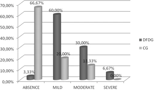
Figure 1 shows the results of using the anamnesis questionnaire, in which the predomi
-nance of the absence of TMD (66,67%) and the mild degree (20,00%) of this dysfunction when present for the CG, is observed. Mild (57.00%), moderate The presence of TMD has been largely discussed
in all stages of DFD treatment, and the literature shows that subjects present a high rate of TMD signs and symptoms in the pre-surgical orthodontic treatment phase as compared to post-surgical
one15-22. Nevertheless, although some authors have
veriied a reduction in the occurrence of TMD in individuals, following the orthognathic surgery, they report no relationship between dentofacial defor
-mities and TMD23,24.
Studies relating TMD and DFD abound in the literature, however, most compare their indings between preoperative and postoperative periods, some being contradictory, therefore, impairing the understanding of professionals regarding the treatment of TMD in subjects with or without occlusal changes. Thus, this study aimed at verifying the occurrence of temporomandibular dysfunction in individuals presented with dentofacial deformity.
METHODS
This observational cross-sectional study was approved by the research ethics committee of the Bauru School of Dentistry, University of São Paulo (process 049/2009) and all participants signed the informed consent.
Sixty subjects in the age range 18-40 years (mean = 27 years), participated in the study, taking into account the following exclusion criteria: intel
-lectual deicits, neurological, psychiatric disorders and / or syndromes, arthritis, history of facial trauma and prior orthognathic surgery. The participants were divided into two groups:
The group with DFD (GDFD), which consisted of 30 subjects, 19 females and 11 males, in their inal stage of the preparatory orthodontic treatment for the orthognathic surgery, 19 female and 11 male. Out of these individuals, 18 presented facial pattern III (12 females and six males) and 12, facial pattern II (seven females and ive males). The inclusion criteria were: being in pre-surgical orthodontic treatment and present DFD, as diagnosed by clinical and radiographic exams.
· The group with no deformity, control group (CG), comprised 30 individuals paired according to gender and age with the DFDG. They underwent Interviews and orofacial myofunctional assessment through the MBGR – orofacial myofunctional exami -nation25, so as to see if they met the following inclusion
signiicant diference (p = 0.00) was observed between the groups, based on the results, showing that the DFD group presented a higher degree and score of TMD than the CG.
(30.00%) and severe (6.67%) degrees were seen in the DFDG.
Table 1 shows the average and median values of the results obtained with an anamnesis question
-naire, when comparing DFDG and CG. A statistically
Figure 1 – Percentage of individuals, according to the presence and severity of temporomandibular dysfunction, for the groups with dentonfacial deformity and control group
Table 1 – Comparison between group of dentofacial deformity (DFDG) and control group (CG), in relation to the degree and scores of TM, obtained the aplicattion of anamnestic questionnaire
Group Mean Median Value of “p”
Degree of TMD CG 0.47 0.00 0.00*
DFDG 1.47 1.00
Scores CG 3.17 2.50 0.00*
DFDG 8.50 7.50
Subtitles: CG – control group; DFDG – group of dentofacial deformity; TMD – temporomandibular dysfunction. *Statistically signiicant diference. Mann-Whitney Test.
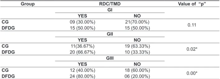
Table 2 shows the results obtained by applying the clinical examination of the RDC / TMD. It was found that A higher occurrence of diferent TMD diagnoses was found for the DFD group in relation to
compared to controls18,19,20,29. It is worth noting the
methodological diferences between the studies, as in the literature, other instruments have been used, diferent from the anamnesis questionnaire applied in this research, nevertheless, they used the clinical exam of TMJs, analogue visual scale, Helkimo´s anamnesis and dysfunction index or applied the RDC / TMD protocol. As for the diagnosis of TMD, held from the application of the clinical examination (axis I) of the RDC / TMD protocol, a higher occur
-rence of articular diagnoses GII (disc displacement) was revealed and GIII (arthritis, arthralgia and arthrosis), for the DFD group, as compared to the control group. Corroborating the results of this
study, Farella et al., Farella et al.18, using RDC/
TMD only in individuals with DFD, found that 50% of the subjects presented articular diagnosis (GII). However, according to Abrahamsson et
al. 29, a diferent result was seen, using the same
test protocol, with a prevalence of muscular TMD diagnosis (GI) in individuals presented with DFD, as compared to the control group with normal occlusion, with statistical signiicance among the three types of diagnoses (GI, GII and GIII), when the groups were compared. The diferences in relation to the present study may be justiied by the fact that, in the study by Abrahamsson et al.29, despite the
larger sample, the control group was not matched by gender and age. The prevalence of articular diagnoses (GII and GIII) in subjects with DFD, in this study, can be attributed to the skeletal and dental imbalance in these individuals, which is not present in the controls, since internal changes in the TMJs of
DISCUSSION
The importance of occlusion and its relationship between the cause or maintenance of TMD cases, compared with other factors, speciically in individuals presented with DFD, has been widely discussed in the literature15, 27-29. The present study
found that 97% of subjects with DFD showed some degree of TMD, while in the CG, the percentage of this disorder was 33%, which may be linked to the fact that TMD symptoms such as pain, are often the main complaint of patients presented with
malocclusion30.
According to the literature, individuals who have certain occlusal problems, mainly the most severe ones, as that studied here, have an expressively higher prevalence of TMD signs and symptoms than subjects with normal occlusion normal29 , owing to
an interdependence between the conditions of the teeth and bones, since a normally functioning TMJ
relies on good occlusion31.
Furthermore, a statistically signiicant result has been observed, when comparing the severity of the dysfunction between DFDG and CG, for the degree of TMD and scores obtained by applying the anamnesis questionnaire, showing a greater occurrence of TMD, to some degree, in the DFDG, as compared to the CG. Thus, the indings of this study are consistent with the data in the literature, since diferent authors have observed a prevalence of TMD signs and symptoms in subjects presented with DFD, in the pre-surgical orthodontic period, as
Table 2 – Comparison between group of dentofacial deformity (DFDG) and control group (CG), in
relation to the diagnosis of TMD, considering the classiication of the clinical exam of Research Diagnostic Criteria for Temporomandibular Disorders
Group RDC/TMD Value of “p”
GI
YES NO
CG 09 (30.00%) 21(70.00%)
0.11
DFDG 15 (50.00%) 15 (50.00%)
GII
YES NO
CG 11(36.67%) 19 (63.33%)
0.02*
DFDG 20 (66.67%) 10 (33.33%)
GIII
YES NO
CG 12 (40.00%) 18 (60.00%)
0.00*
DFDG 24 (80.00%) 06 (20.00%)
Subtitles: control group; DFDG – group of dentofacial deformity; RDC/TMD – Research Diagnostic Criteria for Temporomandibular
Disorders; GI: Muscular diagnoses; GII: Disc displacement; GIII: Arthralgia, arthritis, arthrosis. *Statistically signiicant diference.
Thus, interdisciplinary assistance, in all phases of the orthodontic-surgical treatment is necessary, since the etiology of TMD has multi-factorial origins and needs continuous monitoring of all causal factors for a better prognosis, aiming at the best approach to treat these patients.
CONCLUSION
This study showed that individuals presented with dentofacial deformity had an increased incidence of temporomandibular dysfunction in relation to the degree and classiication of the dysfunction, as compared to patients with dentofacial balance, in the sample.
ACKNOWLEDGEMENT
This study was supported by Fundação de
Amparo a Pesquisa do Estado de São
Paulo-FAPESP, Process Number 2009/06562-5. individuals presented with DFD are reported in the
literature15,16,32.
Thus, the disharmony caused by DFD can inluence the correct position and function of the TMJ, in the period prior to the orthognathic surgery, since occlusal interferences or severe malocclusion may be involved in the multifactorial etiology of TMD33.
However, special attention should be taken into account in the assessment of the results, for this research is part of a cross-sectional study, thus, a cause and efect relationship between the variables (dentofacial deformity and temporomandibular dysfunction) studied cannot be assumed.
It is known that many of the TMD events, including the articular ones (present in a good portion of the sample) can lead to secondary occlusal changes because they cause postural changes of the mandible, with a signiicant impact
on dental occlusion.
RESUMO
Objetivo: veriicar a ocorrência de disfunção temporomandibular em indivíduos com deformidade
dentofacial. Métodos: foram avaliados 60 indivíduos de ambos os gêneros e idade entre 18 e 40 anos
(média=27 anos), sendo formados dois grupos, um composto por 30 sujeitos com deformidade den
-tofacial, em tratamento ortodôntico pré-cirúrgico e um grupo controle constituído por 30 indivíduos com equilíbrio dentofacial, pareados segundo o gênero e a idade com o grupo deformidade. Para avaliação da articulação temporomandibular, foram aplicados o questionário anamnésico de disfunção temporoman
-dibular e o Eixo 1 do Research Diagnostic Criteria for Temporomandibular Disorders para veriicar e
classiicar o grau e o tipo da disfunção temporomandibular, respectivamente. Resultados: os
resul-tados da aplicação do questionário demonstraram que o grupo com deformidade apresentou maior grau e escore da disfunção que o grupo controle (p<0,01). A partir do Research Diagnostic Criteria
for Temporomandibular Disorders veriicou-se maior ocorrência de diagnósticos de deslocamento de
disco (p=0,02) e de artrite, artralgia e artrose (p<0,01) no grupo com deformidade em relação ao
grupo controle. Conclusão: indivíduos com deformidade dentofacial apresentaram maior ocorrência
de disfunção temporomandibular, quando comparados aos indivíduos com equilíbrio dentofacial, na
amostra estudada.
DESCRITORES: Transtornos da Articulação Temporomandibular; Desenvolvimento Maxilofacial; Má
Oclusão
REFERENCES
1. Gonçales ES. Cirurgia ortognática: guia de orientação para portadores de deformidades faciais
esqueléticas. São Paulo: Editora Santos; 2010. 2. Manganello LCS, Silveira ME, Cappellette
M, Garducci M, Lino AP. Cirurgia ortognática e ortodontia. São Paulo: Santos; 1998.
3. Hall B, Jamsa T, Soukka T, Peltomaki T. Duration of surgical-orthodontic treatment. Acta Odontol
Scand. 2008;66(5):274-7.
4. Okasaki LK. Quando indicar uma cirurgia
18. Farella M, Michelotti A, Bocchino T, Cimino R, Laino A, Steenks MH. Efects of orthognathic surgery for class III maloclusion on signs and symptoms of temporomandibular disorders and on pressure pain thresholds of the jaw muscles. Int J Oral Maxillofac
Surg. 2007;36(7):583-7.
19. Pahkala RH, Kellokoski JK. Surgical-orthodontic treatment and patients’functional and psychosocial well-being. Am J Orthod Dentofacial Orthop.
2007;132(2):158-64.
20. Oland J, Jensen J, Melsen B. Factors of importance for the functional outcome in orthognathic surgery patients: a prospective study of 118 patients. J Oral Maxillofac Surg. 2010;68(9):2221-31.
21. Ramieri G, Piancino MG, Frongia G, Gerbino G, Fontana PA, Debernardi C, et al. Clinical and instrumental evaluation of temporomandibular joint before and after surgical correction of asymptomatic skeletal class III patients. J Craniofac Surg.
2011;22(2):527-31.
22. Silva MMA, Ferreira AT, Migliorucci RR, Nary Filho H, Berretin-Felix G. Inluência do tratamento ortodôntico-cirúrgico nos sinais e sintomas de disfunção temporomandibular em indivíduos com deformidades dentofaciais. Rev Soc Bras
Fonoaudiol. 2011;16(1):80-4.
23. Panula K, Somppi M, Finne K, Oikarinen K. Efects of orthognathic surgery on temporomandibular joint dysfunction. A controlled prospective 4-year follow-up study.Int J Oral Maxillofac Surg. 2000;29(3):183-7. 24. Dervis E. Tuncer E. Long-term evaluations of temporomandibular disorders in patients undergoing orthognathic surgery compared with a control group. Oral Surg Oral Med Oral Pathol Oral Radiol Endod. 2002;94(5):554-60.
25. Genaro KF, Berretin-Felix G, Rehder MIBC, Marchesan IQ. Avaliação miofuncional orofacial – protocolo MBGR. Rev CEFAC. 2009;11(2):237-55.
26. Conti PC, Ferreira PM, Pegoraro LF, Conti
JV, Salvador MC. A cross-sectional study of prevalence and etiology of signs and symptoms of temporomandibular disorders in high school and university students. J Orofac Pain. 1996;10(3):254-62.
27. Egermark I, Blomqvist JE, Cromvik U, Isaksson S.
Temporomandibular dysfunction in patients treated with orthodontics in combination with orthognathic surgery. Eur J Orthod. 2000;22(5):537-44.
28. Felício CM de, Braga APG. Sinais e sintomas de desordem temporomandibular em pacientes orto-cirúrgicos. J Bras Ortodon Ortop Facial. 2005;10(56):187-94.
29. Abrahamsson C, Ekberg E, Henrikson T, Nilmer M, Sunzel B, Bondemark L. TMD in consecutive patients referred for orthognathic surgery. Angle Orthod. 2009;79(4):621-7.
studies on prevalence and etiology of functional disturbances of the masticatory system. J Prosthet Dent. 1979;41(1):76-82.
6. Gesch D, Bernhardt O, Mack F, John U, Kocher T, Alte D. Association of malocclusion and functional occlusion with subjective symptoms of TMD in adults: results of the Study of Health in Pomerania (SHIP). Angle Orthod. 2005;75(2):183-90.
7. Conti ACCF, Freitas MR, Conti PCR. Avaliação da
posição condilar e disfunção temporomandibular em pacientes com má oclusão de classe III submetidos à protrusão mandibular ortopédica. R Dental Press Ortodon Ortop Facial. 2008;13(2):49-60.
8. Thilander B, Rubio G, Pena L, de Mayorga C. Prevalence of temporomandibular dysfunction and its association with malocclusion in children
and adolescents: an epidemiologic study related
to speciied stages of dental development. Angle Orthod. 2002;72(2):146-54.
9. Oliveira AS, Dias EM, Contato RG, Berzin F. Prevalence study signs and symptoms of temporomandibular disorder in Brazilian college
students. Braz Oral Res. 2006;20 (1):3-7.
10. Okeson JP. Management of temporomandibular disorders and occlusion. 6th ed. St.Louis: Elsevier;
2008.
11. Valle-Corotti KM, Pinzan A, Conti PCR, Janson G. A oclusão e a sua relação com as disfunções temporomandibulares (DTM) em jovens com e sem tratamento ortodôntico: um estudo comparativo. Revista Dental Press Ortodon. 2003;8(6):79-87. 12. Al-Ani MZ, Davies SJ, Gray RJ, Sloan P, Glenny AM. Stabilisation splint therapy for temporomandibular pain dysfunction syndrome. Cochrane Database Syst Rev. 2004;(1):CD002778. Review.
13. Koh H, Robinson PG. Occlusal adjustment for treating and preventing temporomandibular joint disorders. J Oral Rehabil. 2004;31(4):287-92. Review.
14. Mohlin B, Axelsson S, Paulin G, Terttu P, Bondemark L, Brattstrom V, et al. TMD in relation to malocclusion and orthodontic treatment. Angle Orthodontist. 2007;77(3):542-8.
15. Wolford LM, Karras S, Mehra P. Concomitant temporomandibular joint and orthognathic surgery: a preliminary report. J Oral Maxillofac Surg.
2002;60(4):356-62.
16. Wolford LM, Reiche-Fischel O, Mehra P. Changes in temporomandibular joint dysfunction after orthognathic surgery. J Oral Maxillofac Surg.
2003;61(6):655-60.
17. Aoyama S, Kino K, Kobayashi J, Yoshimasu H, Amagasa T. Clinical evaluation of the temporomandibular joint following orthognathic
surgery – multiple logistic regression analysis. J
32. Toll DE, Popovic N, Drinkuth N. The use of MRI dignostics in orthognathic surgery: prevalence of TMJ pathologies in Angle Class I, II, III patients. J Orofac Orthop. 2010;71(1):68-80.
33. Bourzgui F, Sebbar M, Nadour A, Hamza M. Prevalence of temporomandibular dysfunction in orthodontic treatment. Int Orthod. 2010;8(4):386-98. 30. Mazzone N, Matteini C, Incisivo V, Belli
E. Temporomandibular joint disorders and maxillomandibular malformations: role of condylar “repositionin” plat. J Craniofc Surg. 2009;20(3):909-15.
31. Madeira MC. Anatomia da Face – Bases
Anátomo-Funcionais para a Prática Odontológica. São Paulo: Sarvier; 1998.
Received on: September 07, 2014
Accepted on: January 08, 2015
Mailing address:
Giédre Berretin-Felix
Faculdade de Odontologia de Bauru da Universidade de São Paulo
Departamento de Fonoaudiologia Al. Octávio Pinheiro Brisola, 9-75
Bauru – São Paulo – SP