Methicillin-Resistant
Staphylococcus aureus
Clone
ST93-IV [2B] Is Highly Virulent and Genetically Distinct
Kyra Y. L. Chua1,2,4, Torsten Seemann3, Paul F. Harrison3, Shaun Monagle6, Tony M. Korman7, Paul D. R. Johnson4, Geoffrey W. Coombs8, Brian O. Howden9, John K. Davies1, Benjamin P. Howden1,2,4,5*., Timothy P. Stinear1,2*.
1Department of Microbiology, Monash University, Clayton, Australia,2Department of Microbiology and Immunology, University of Melbourne, Parkville, Australia, 3Victorian Bioinformatics Consortium, Monash University, Clayton, Australia,4Department of Infectious Diseases, Austin Centre for Infection Research, Austin Health, Heidelberg, Australia,5Department of Microbiology, Austin Health, Heidelberg, Australia,6Department of Anatomical Pathology, Eastern Health, Box Hill, Australia, 7Department of Infectious Diseases, Monash Medical Centre, Clayton, Australia,8Department of Microbiology and Infectious Diseases, PathWest Laboratory Medicine WA, Royal Perth Hospital, Perth, Australia,9Microsurgical Consultants, Blackburn, Australia
Abstract
Community-associated methicillin-resistant Staphylococcus aureus (CA-MRSA) USA300 has spread rapidly across North America, and CA-MRSA is also increasing in Australia. However, the dominant Australian CA-MRSA strain, ST93-IV [2B] appears distantly related to USA300 despite strikingly similar clinical and epidemiological profiles. Here, we compared the virulence of a recent Australian ST93 isolate (JKD6159) to other MRSA, including USA300, and found that JKD6159 was the most virulent in a mouse skin infection model. We fully sequenced the genome of JKD6159 and confirmed that JKD6159 is a distinct clone with 7616 single nucleotide polymorphisms (SNPs) distinguishing this strain from all otherS. aureusgenomes. Despite its high virulence there were surprisingly few virulence determinants. However, genes encodinga-hemolysin, Panton-Valentine leukocidin (PVL) anda-type phenol soluble modulins were present. Genome comparisons revealed 32 additional CDS in JKD6159 but none appeared to encode new virulence factors, suggesting that this clone’s enhanced pathogenicity could lie within subtler genome changes, such as SNPs within regulatory genes. To investigate the role of accessory genome elements in CA-MRSA epidemiology, we next sequenced three additional Australian non-ST93 CA-MRSA strains and compared them with JKD6159, 19 completedS. aureusgenomes and 59 additionalS. aureusgenomes for which unassembled genome sequence data was publicly available (82 genomes in total). These comparisons showed that despite its distinctive genotype, JKD6159 and other CA-MRSA clones (including USA300) share a conserved repertoire of three notable accessory elements (SSCmecIV, PVL prophage, and pMW2). This study demonstrates that the genetically distinct ST93 CA-MRSA from Australia is highly virulent. Our comparisons of geographically and genetically diverse CA-MRSA genomes suggest that apparent convergent evolution in CA-MRSA may be better explained by the rapid dissemination of a highly conserved accessory genome from a common source.
Citation:Chua KYL, Seemann T, Harrison PF, Monagle S, Korman TM, et al. (2011) The Dominant Australian Community-Acquired Methicillin-Resistant Staphylococcus aureusClone ST93-IV [2B] Is Highly Virulent and Genetically Distinct. PLoS ONE 6(10): e25887. doi:10.1371/journal.pone.0025887
Editor:Malcolm James Horsburgh, University of Liverpool, United Kingdom
ReceivedJune 28, 2011;AcceptedSeptember 12, 2011;PublishedOctober 3, 2011
Copyright:ß2011 Chua et al. This is an open-access article distributed under the terms of the Creative Commons Attribution License, which permits unrestricted use, distribution, and reproduction in any medium, provided the original author and source are credited.
Funding:This research was supported by the National Health and Medical Research Council of Australia Project Grant 1008656, the CASS Foundation and the Austin Hospital Medical Research Foundation. The funders had no role in study design, data collection and analysis, decision to publish, or preparation of the manuscript.
Competing Interests:The authors have declared that no competing interests exist.
* E-mail: [email protected] (TPS); [email protected] (BPH)
.These authors contributed equally to this work.
Introduction
Infections with community-associated methicillin-resistant Staphylococcus aureus(CA-MRSA) in patients with no contact with the hospital environment are now well described worldwide [1]. In some regions such as North America, the CA-MRSA phenomenon has become particularly well defined. Here, the epidemic CA-MRSA strain USA300 is the most common cause of skin infections in patients presenting to the emergency department [2]. Much of the work performed thus far to understand the CA-MRSA phenomenon has focused on USA300 and these studies have demonstrated the importance of virulence determinants in both the staphylococcal accessory and core
genome such as Panton-Valentine leukocidin (PVL),a-hemolysin (Hla) and a-type phenol soluble modulins (PSMs) [3,4,5,6,7, 8,9,10].
clones of CA-MRSA has the potential to provide new and important insights into staphylococcal pathogenesis.
In Australia,S. aureus ST93-IV [2B] has recently emerged to become the dominant CA-MRSA clone. First described in the early 2000s, it is colloquially known as the ‘‘Queensland’’ clone [13]. However, since this initial description, ST93-IV [2B] has been reported across Australia and is responsible for the increasing prevalence of all CA-MRSA nationwide [14]. ST93 is associated with skin infection and severe invasive infection including necrotizing pneumonia, deep-seated abscesses, and septicemia [14,15]. This PVL positive clone is a singleton by MLST eBURST analysis and is therefore distinct to otherS. aureusclones, including previously sequenced strains [16]. While predominantly an Australian strain, it has recently also been reported in the United Kingdom, with many cases having epidemiological links to Australia [16,17]. Other less frequent clones of CA-MRSA in Australia include PVL negative ST1-IV [2B] (WA-1) and PVL positive ST30-IV [2B] (SouthWest Pacific, SWP) [14]. The dominant health-care associated clone of MRSA in Australia is multi-resistant ST239-III [3A] [14].
To investigate the apparent enhanced clinical virulence of ST93-IV [2B], we examined a recent clinical isolate (JKD6159) that caused an outbreak of both severe and minor staphylococcal infections within a household, representative of the typical spectrum of disease associated with this clone. We compared the virulence of JKD6159 to three other well-characterized and representative Australian MRSA strains and the epidemic North American strain, USA300 and found that JKD6159 was the most virulent clone in twoin vivomodels.
We next sequenced, fully assembled and annotated the genome of S. aureus ST93-IV [2B] (JKD6159) and then sequenced three non-ST93 Australian CA-MRSA clones. We employed compar-ative genomics against these strains, the 19 published, completeS. aureusgenomes and 59 unassembled, publically availableS. aureus genomes to try and identify genetic factors that might explain the recent emergence, and increased virulence of ST93-IV [2B].
Materials and Methods
Bacterial strains and culture
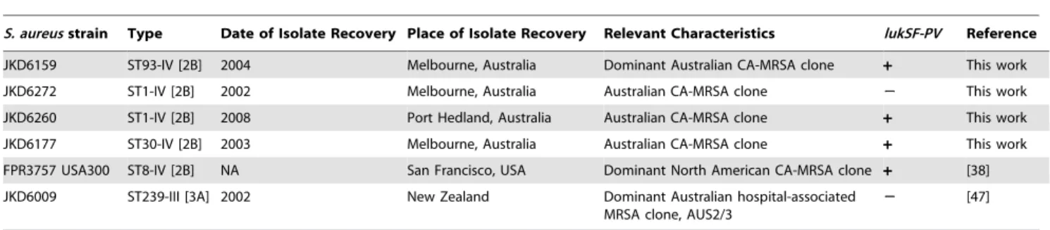
The bacterial strains used in this study are described in Table 1. For all experiments, bacteria were grown in brain heart infusion broth (BHI, Oxoid). For growth kinetic experiments, overnight cultures ofS. aureuswere diluted 1:100 and incubated at 37uC with shaking (180 rpm). Optical density at 600 nm (OD600) and viable counts were performed hourly until stationary phase was achieved and at 24 hours after commencement of the experiment. These experiments were performed in triplicate. For theGalleria mellonella
killing assay and the mouse skin infection assay, S. aureus were harvested at the stationary phase of growth after 18 hours incubation (OD600approx. 2.0), washed, diluted and resuspended in PBS. The bacterial inoculum (CFU) and viable counts were determined by plating onto BHI agar and colony enumeration.
DNA Manipulation and Molecular Typing
DNA was extracted using the GenElute kit according to the manufacturer’s instructions (Sigma-Aldrich). Detection oflukSF-PV was performed as described by Lina et al. [18]. MLST was performed as described by Enrightet al.[19], and Pulsed Field Gel Electrophoresis (PFGE) performed as previously described [20]. SCCmec typing was performed as previously described [21]. SCCmecnomenclature was used as proposed by the International Working Group on the Classification of Staphylococcal Cassette Chromosome Elements (IWG-SCC) [22]. A Roman numeral indicates the SCCmectype with a lowercase letter indicating the subtype. Theccrcomplex and themeccomplex are indicated by an Arabic numeral and an uppercase letter respectively.
Wax Moth (Galleria mellonella)virulence assay
Final stage instarG. mellonellalarvae were infected withS. aureus and time to death measured as described previously [23]. Approximately 106CFU ofS. aureussuspended in 10mL of PBS was injected into the first left proleg of each larva. Each experimental group consisted of at least 16 larvae. All experiments included biological triplicates. The larvae were incubated at 37uC and assessed daily for six days. Larvae were considered dead when there was no movement in response to stimuli. The control groups were PBS injected and non-injected larvae.
Mouse skin infection assay
Mice were housed in individually ventilated cages and received food and water ad libitum. Six-week-old female BALB/c mice were anesthetized with intraperitoneal ketamine/xylazine, and inoculated in the right flank by intradermal injection, with 108CFU ofS. aureus(in 50mL of PBS suspension). This dose ofS. aureuswas determined in preliminary experiments as the lower limit of inoculum, which produced consistent dermonecrosis for all strains ofS. aureustested. Mice were assessed and weighed daily for five days. On the 5thday, the mice were culled. The infected area of skin and muscle was harvested and these tissues were homogenized and CFU measured by plating onto BHI and colony enumeration. Skin lesion area was quantitated by obtaining a digital image of the lesion and processed using ImageJ [24]. For eachS. aureusstrain, at least 15 mice were assessed – the tissues for four of these mice were used for histological analysis and data for these mice were not available for CFU enumeration.
Table 1.Bacterial strains used in this study.
S. aureusstrain Type Date of Isolate Recovery Place of Isolate Recovery Relevant Characteristics lukSF-PV Reference JKD6159 ST93-IV [2B] 2004 Melbourne, Australia Dominant Australian CA-MRSA clone + This work
JKD6272 ST1-IV [2B] 2002 Melbourne, Australia Australian CA-MRSA clone 2 This work
JKD6260 ST1-IV [2B] 2008 Port Hedland, Australia Australian CA-MRSA clone + This work
JKD6177 ST30-IV [2B] 2003 Melbourne, Australia Australian CA-MRSA clone + This work
FPR3757 USA300 ST8-IV [2B] NA San Francisco, USA Dominant North American CA-MRSA clone + [38]
JKD6009 ST239-III [3A] 2002 New Zealand Dominant Australian hospital-associated MRSA clone, AUS2/3
2 [47]
NA: not available.
Ethics Statement
All studies were reviewed and approved by the Animal Ethics Committee at the University of Melbourne approval number 0911248.2.
Whole Genome sequencing of Australian CA-MRSA strains JKD6159 (ST93-IV [2B]), JKD6272 (ST1-IV [2B]), JKD6260 (ST1-IV [2B]) and JKD6177 (ST30-IV [2B])
We have previously described the sequencing, complete assembly and annotation of the genome of JKD6159, however the comparative genomics have not previously been reported [25]. Here, we also sequenced and partially assembled the genomes of three Australian CA-MRSA strains, using Illumina GAIIx 36 bp paired-end chemistry, yielding an average of 555 Mbp of reads per strain (,2006coverage). These genomes werede novoassembled to draft form using Velvet 1.0 [26]. The reads from whole genome sequencing of these strains were also included in defining the core and accessory genome of JKD6159. The sequences are accessible from the NCBI Sequence Read Archive under accession SRA026511.1.
Analysis of the JKD6159 genome sequence
A read mapping approach was developed to define aS. aureus core genome and the accessory genome for a given strain. Sequence reads from eachS. aureusstrain were aligned separately to the completed JKD6159 reference genome using SHRiMP 2.0 [27]. Publically available completed genomes (Table 2) were shredded into 75 bp reads at 256coverage to produce a set of synthetic sequence reads. Those positions in JKD6159 that were covered by at least one read from each and every genome defined aS. aureuscore genome. Reads from JKD6159 that did not map to any S. aureus genome (Table 2) were used to define JKD6159 regions of difference. Reads from any strain that did not map to the core genome represented that strain’s accessory genome. SNPs were identified using Nesoni v0.35, which used the sequence reads for each genome aligned to the above-defined core genome to construct a tally of putative differences at each nucleotide position, including substitutions, insertions, and deletions (www.bioinfor-matics.net.au). This tally was then employed in a simple Bayesian model to decide whether a base (or deletion) could be called for the position, and if so, whether it differed from the reference sequence. A similar procedure was used to determine the presence or absence of insertions between positions in the reference. NCBI BLASTn+, Artemis and BRIG were also used for comparisons of JKD6159 with selectedS. aureusgenomes [28] (http://sourceforge. net/projects/brig/). SNPs were identified in the accessory genome elements pMW2,wSA2, and SCCmecIV for strains carrying them by read mapping using Nesoni v0.35 against reference sequences of each element taken fromS. aureusMW2. Phylogenetic analyses were performed using a distance method, based on pairwise nucleotide sequence alignments for the S. aureus core genome among all strains or selected accessory elements among some strains. Split decomposition analysis was employed using uncor-rected p distances with bootstrapping as implemented in SplitsTree4 [29].
Statistical analysis
Kaplan Meier plots ofG. mellonellakilling results were analysed using the log rank test. Percentage mouse weight change at day 5, viable counts ofS. aureusin mouse tissues and skin lesion area of each isolate versus JKD6159 were analyzed using the Mann Whitney test. All analyses were performed using Prism 5 for Macintosh v5.0b (GraphPad Software Inc.).
Results
Clinical Details
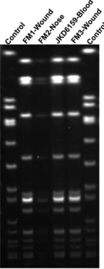
Staphylococcus aureusJKD6159 was isolated from the blood of a young male intravenous drug user with no prior health care exposure, who presented with a severe sepsis syndrome in 2004. He had cavitating pulmonary lesions, polyarticular septic arthritis and multiple deep-seated muscle abscesses. The isolate was resistant to methicillin, but susceptible to erythromycin, clinda-mycin, tetracycline, ciprofloxacin, rifampicin, fusidic acid and vancomycin. The patient required extensive surgical debridement and prolonged antimicrobial therapy with vancomycin and subsequently, linezolid, but eventually made a full recovery. In addition, the patient reported multiple household family members with recurrent skin infections. The SmaI PFGE pattern was identical between JKD6159 and theS. aureus isolates from three other family members, indicating an intra-familial outbreak with the same clone (Figure 1). Further typing demonstrated that JKD6159 was ST93-IV [2B]. This isolate was representative of the Australian CA-MRSA Queensland clone.
S. aureus strain JKD6272 was isolated from the blood of a woman with pneumonia and septic shock who had no prior hospitalizations. The isolate was resistant to methicillin and fusidic acid but susceptible to erythromycin, clindamycin, tetracycline, ciprofloxacin, rifampicin, trimethoprim and vancomycin. Further typing demonstrated that JKD6272 was ST1-IV [2B], and PVL negative. This isolate was representative of the Australian CA-MRSA clone WA-1.
S. aureusstrain JKD6260 was isolated from a wound swab from a patient with a skin infection. The isolate was resistant to methicillin, fusidic acid and ciprofloxacin, but susceptible to erythromycin, clindamycin, tetracycline, rifampicin, mupirocin, Table 2.Staphylococcus aureuscompleted genomes used in this study.
Genome
Multilocus Sequence Type (Clonal
Complex) Reference
1 NCTC8325 8 (8) [48]
2 N315 5 (5) [49]
3 Mu50 5 (5) [49]
4 Mu3 5 (5) [50]
5 MW2 1 (1) [37]
6 USA300 FPR3757 8 (8) [38]
7 USA300 TCH1516 8 (8) [39]
8 MRSA252 36 (30) [51]
9 MSSA476 1 (1) [51]
10 RF122 151 [52]
11 Newman 8 (8) [53]
12 COL 250 (8) [54]
13 JH1 105 (5) [55]
14 JH9 105 (5) [55]
15 ED98 5 (5) [56]
16 TW20 239 (8) [57]
17 04-02981 225 (5) [58]
18 S0385 398 [59]
19 JKD6008 239 (8) [60]
trimethoprim and vancomycin. Typing demonstrated that JKD6272 was ST1-IV [2B], and PVL positive.
S. aureusstrain JKD6177 was isolated from the blood of a patient with mitral valve endocarditis. He had recurrent skin infections with MRSA prior to presentation. The isolate was resistant to methicillin, but susceptible to erythromycin, clindamycin, tetracy-cline, ciprofloxacin, rifampicin, fusidic acid and vancomycin. Typing demonstrated that JKD6177 was ST30-IV [2B], and PVL positive. This isolate was representative of the SouthWest Pacific clone of CA-MRSA.
JKD6159 exhibits high virulence in two animal models The reports of severe infections associated with ST93-IV [2B], and the severity of disease in our patient led us to compare the virulence of JKD6159 with other well-characterized clones ofS. aureus (Table 1). AllS. aureus strains tested had the same growth curve characteristics, indicating that any differences in virulence were not due to differential growth kinetics (Figure S1). However, JKD6159 was significantly more virulent than other community and healthcare-associated MRSA strains, including the USA300 strain FPR3757, in both an invertebrate and mouse infection model ofS. aureusinfection. In theGalleria mellonellalarvae assay, JKD6159 caused significantly more rapid time-to-death than all otherS. aureusstrains (p,0.001, Fig. 2A). The differential virulence seen in the larvae assay was mirrored by the results of a mouse skin infection assay. Mice infected with JKD6159 developed large skin lesions and lost significantly more weight over a 5-day experi-mental period than mice infected with the other strains (p,0.05, Figure 2B-C). In line with these findings, the bacterial load in
mouse tissues was highest in JKD6159-infected mice (p,0.05, Figure 2D). Histopathological analysis of mouse tissues showed that all CA-MRSA strains caused extensive epidermal and fat necrosis with myositis including in some cases myonecrosis, whilst infection with the hospital-associated MRSA strain JKD6009 caused less severe inflammatory changes with only mild myositis. Representative sections are shown in Figure 2E-I.
Comparative genome analysis of JKD6159
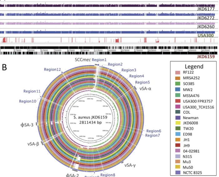
To gain insight into the high virulence ofS. aureusST93 strain JKD6159 we fully sequenced, assembled and annotated its genome and used it as a reference genome for comprehensive comparative genomics [25]. The JKD6159 genome consisted of a single 2,811,434 bp circular chromosome and a 20,730 bp circular, pMW2-like plasmid. For comparison, we also sequenced the genomes of three other Australian CA-MRSA isolates (JKD6272, JKD6260 & JKD6177) that were representative of the commonly occurring ST1 and ST30 clones (Table 1, Table S1, Figure 3A). Whole genome DNA:DNA comparisons showed that the architec-ture of the JKD6159 chromosome was conserved compared to otherS. aureusstrains (Figure 3B). Using a read-mapping approach to objectively and comprehensively define the core and accessory genome of JKD6159, we found that 2,302,644 bp of JKD6159 (81.9%) was absolutely conserved amongst all 20S. aureusstrains with fully assembled genomes and 62 strains for which only unassembled sequence data was available (Table 2, Table S1, Figure 3). Pairwise comparisons of the concatenated 2,302,644 bp JKD6159 core genome with the core genomes of otherS. aureus strains uncovered 78,967 variable nucleotide positions among all strains and permitted the construction of a high-resolution phylogeny by split decomposition analysis that confirmed the divergent genotype of ST93 (Figure 4A). ST93 was clearly divergent from otherS. aureusstrains with 7616 single nucleotide polymor-phisms (SNPs) distinguishing this strain from other genomes. In comparison, the USA300 genome contained only 167 SNPs that varied from other strains. The JKD6159 core genome, like otherS. aureus genomes, contained the genes encoding the important virulence factors Hla and a-type PSMs. The genes encoding gamma-hemolysin were also present.
Using the read mapping approach, we determined that 18.1% of the JKD6159 chromosome constituted its accessory genome (Figure 3A), a proportion that is consistent across all S. aureus genomes (range 13.2% [RF122]218.1% [JH1]) and is also consistent with previous assessments based on microarray analyses [30,31]. For a strain associated with severe clinical disease and high virulence in animal models, the accessory genome of JKD6159 was notable for the absence of common virulence-associated factors such as toxic shock syndrome toxin, exfoliative toxins and most of the staphylococcal enterotoxins; a finding consistent with an earlier report [32]. There were no recognized pathogenicity islands in JKD6159 although it contained the S. aureusgenomic islands,nSAa,nSAbandnSAc(Figure 4B). Within nSAathere was a complement of tandem lipoprotein genes and eleven staphylococcal superantigen-like (ssl) genes, however ssl9 appeared to be a pseudogene. Two of the 11 ssl CDS, ssl3 (SAA6159_00377) and ssl4 (SAA6159_00378), encoded proteins with predicted N-terminal sequences that demonstrated significant variation from orthologs in other strains. Although the exact role of SSLs is still being defined, they play a role in modulating the host immune system; in particular neutrophil and complement activation, both of which are key to human defense against S. aureus[33]. SSLs have been shown to be upregulated in some CA-MRSA strains [34]. We postulate that these variant SSLs may in part contribute to the strain’s increased virulence.
Figure 1.SmaI pulsed field gel electrophoresis analysis.PFGE of index isolate, S. aureus JKD6159 and isolates from three family members, FM1, FM2, FM3.
Figure 2. Virulence characteristics ofS. aureusisolates.S. aureusJKD6159 (ST93-IV [2B], CA-MRSA) compared to four other MRSA strains. (A)Galleria mellonellavirulence assay. Kaplan-Meier plot showing the percent survival of larvae injected withS. aureusstrains JKD6159, JKD6009 (ST239-III [3A], hospital-associated MRSA), JKD6272 (ST1-IV [2B], CA-MRSA), JKD6177 (ST30-IV [2B], CA-MRSA), FPR3757 USA300 (ST8-IV [2B], CA-MRSA) up to 6 days post injection. The reduced survival time for larvae infected with JKD6159 compared to all other strains was significant (p,0.0001). (B) BALB/c mouse skin infection assay. Weight loss induced by intradermal infection withS. aureusstrains is demonstrated as percentage loss of weight over 5 days. The difference in percentage weight loss between JKD6159 and all other strains was significant (p,0.05). At least 15 mice were used for each bacterial strain. Data shown are mean weight loss and SEM. (C) BALB/c mouse skin infection assay. Skin lesion area (mm2) at 5 days after infection was greatest with JKD6159 infected mice (p,0.05). Data shown are mean area and SEM. (D) BALB/c mouse skin infection assay. Recovery ofS. aureus(log CFU) from infected tissues at 5 days after infection was greatest with JKD6159 infected mice (p,0.05). Data shown are mean CFU and SEM. (E-I) BALB/c mouse skin infection assay. Representative H&E stained histology sections from infected tissues at 1006magnification from JKD6009, JKD6272, JKD6177, USA300 and JKD6159 respectively. There was no significant difference in degree of inflammation observed in the CA-MRSA strain infections. However, mice infected with JKD6009, the hospital-associated MRSA strain had less marked inflammation and only mild myositis. (J-N) BALB/c mouse skin infection assay with representative macroscopic appearance of lesions resulting from infection with JKD6009, JKD6272, JKD6177, USA300 and JKD6159 respectively.
InnSAb, like otherS. aureusgenomes, JKD6159 had a cluster of serine protease genes and lukDE. However, unlike MW2 and FPR3757 USA300, the bacteriocin gene cluster was located elsewhere. In each ofnSAa andnSAbthere were loci harboring distinctive type I restriction-modification systems. Restriction-modification systems attack foreign DNA, restricting DNA transfer [35,36]. Type I systems consists of enzyme subunits which are encoded by the geneshsdR, hsdM and hsdS.The HsdS (specificity) subunit contains target recognition domains (TRD1 and TRD2) that allow for target sequence specificity. In JKD6159 the hsdS genes in each of these regions (SAA6159_00387 and SAA6159_01737) contained previously unreported TDR1 and TDR2 domains. Additionally, there was a third restriction modification system with a distinct hsdS gene (SAA6159_00055) in a novel 10 kb locus adjacent to SCCmec (Table S2). These
differences in the restriction-modification system of JKD6159 may explain its relative resistance to the uptake of foreign DNA that we have observed during laboratory manipulation of this strain (unpublished observations).
JKD6159 contained two prophageswSA2 andwSA3. As in the other major CA-MRSA clones,wSA2 habourslukSF-PV [1], the genes that encode the two component pore-forming Panton-Valentine leukocidin (PVL). JKD6159 also contained wSA3, which is present in many other S. aureus genomes including MW2 (Figure 3B). This bacteriophage inserts into and disrupts the beta-hemolysin gene. In JKD6159, wSA3 contained genes encoding staphylokinase, and chemotaxis inhibitory protein but unlike MW2, did not harbor enterotoxin genes. A gene encoding an enterotoxin-like protein similar to that in RF122 (SAB0026) was also present in JKD6159.
Figure 3. Whole genome sequence analysis and comparison of JKD6159 with otherS. aureusstrains.(A) Artemis linear view of JKD6159 chromosome, with vertical red bars identifying the position of accessory genome elements as determined by read mapping against 19 publicly available completed genomes and 62 unpublished, partially assembled genome sequences. Increasing height of vertical red lines indicates increasing specificity for JKD6159. Shown above the accessory genome analysis are the mapped positions of the short-reads obtained from Illumina sequencing CA-MRSA strains JKD6177, JKD6272 and JKD6260. Depicted also is an example of the synthetic reads obtained from completed whole genome sequences (USA300 shown here) to facilitate the comprehensive read mapping approach described in the methods. (B) Circular diagram of the JKD6159 chromosome showing (from inner to outer), % G+C, GC skew and the homology based on BLASTn+analysis of JKD6159 to 19 completedS. aureusgenomes (refer color-coded legend). Outer circle shows the location of accessory elements (grey) and the 12 regions of difference (blue) not present in the otherS. aureusgenomes examined.
The accessory genome of JKD6159 also contained 12 regions not present in the 19 other published, completeS. aureusgenomes. These regions spanned 40,776 bp of the chromosome. The 32 CDS contained therein are listed in Table S2 and lie within segments identified as Regions 1–12 (Figure 3B, Table S2). JKD6159 also contained two copies of a Group II intron (Regions 6 & 11) that to our knowledge is the first example of this type of mobile DNA element inS. aureus.
As expected from its antibiotic phenotype, JKD6159 had few antibiotic resistance elements. It contained the SCCmecIV element found in other CA-MRSA strains but it did not have the adjacent arginine catabolic mobile element (ACME) seen in USA300. JKD6159 contained a bla operon located on a plasmid. This plasmid was identical to pMW2 and pSAS (from MSSA476). This plasmid was also highly similar to that carried on other community S. aureus strains including pWBG757 (NCBI Accession GQ900397.1), pSAP073A (NCBI Accession GQ900425.1), pWBG763 (NCBI Accession GQ900467.1), and pWBG750 (NCBI Accession GQ900392.1). Other mobile DNA present in JKD6159 included six intact copies and four partial copies of the insertion sequence element ISSau3.
Comparison of JKD6159 with other CA-MRSA
Comparisons of JKD6159 with the three other complete, publicly available genomes of CA-MRSA (MW2, USA300 FPR3757, USA300 TCH1516) [37,38,39], and the unassembled sequences for CA-MRSA strains JKD6272, JKD6260 & JKD6177, highlightedwSA2, pMW2 and SCCmecIV as elements common to these CA-MRSA strains, although JKD6272 did not containwSA2, the phage bearinglukSF-PVrequired for expression
of Panton Valentine Leukocidin (PVL). While high-resolution phylogenomic analysis based on SNP comparisons of the 2,302,644 bp S. aureus core genome demonstrated that some of these strains were distantly related to each other (Figure 4A), the same analysis based on SNP comparisons of only the accessory elements identified more limited sequence differences and produced individual tree topologies that suggested all strains except JKD6177 had recently acquired these elements (Figure 4B-C). Most striking was the limited sequence diversity among SCCmec, with 0.4 SNPs/100 bp for all CA-MRSA strains compared with 3.4 SNPs/100 bp across the entire chromosome for the same set of strains. These data are consistent with the contemporary acquisition of SSCmecby these diverse strains from a common source and the recent emergence of the global CA-MRSA phenomenon.
Discussion
ST93 MRSA is now the most common CA-MRSA clone in Australia [14]. The reasons for its recent emergence are unknown but this increase is consistent with a worldwide spread of other global CA-MRSA clones. ST93 MRSA causes severe clinical disease that appears to mimic USA300. Little is known about ST93 although MLST and other typing methods have shown that ST93 is distinct to otherS. aureusclones [16]. To understand the genetic basis for its recent emergence, we have compared both the virulence and genome of a recent clinical ST93 MRSA isolate (JKD6159) with other CA-MRSA including USA300.
We have demonstrated that ST93-IV [2B] was highly virulent in both theGallerialarvae virulence assay and mouse skin infection Figure 4. Phylogenetic analysis of S. aureusstrains.Phylogenetic analysis based on the core genomes ofS. aureusstrains and selected accessory elements from CA-MRSA strains. (A)S. aureusphylogeny inferred by split decomposition analysis from pairwise comparisons of the 78,967 variable nucleotide positions identified from the core chromosome sequences of 20 completedS. aureuschromosomes and the 62S. aureusstrains for which unassembled reads were available, including the CA-MRSA strains sequenced in this study, JKD6177, JKD6260, JKD6272. CA-MRSA strains are indicated in colour (red JKD6159, pink JKD6177, orange FPR3757 USA300, blue MW2, light green JKD6260, dark green JKD6272) (B-D), Phylogenetic relationship ofwSA2, pMW2 and SCCmecIV inferred by split decomposition analysis of nucleotide differences for each of these accessory elements between CA-MRSA strains. Scale bar indicates the number of nucleotide substitutions per site. All nodes have 100% bootstrap support except those indicated by asterisk (*) which have.60% support (1000 replicates). CC: clonal complex. ST: sequence type.
model. We used the Galleria model as a screening assay and validated our results in the mouse skin infection model. TheGalleria killing assay has been previously used to assess the virulence ofS. aureus [23,40]. Galleria mellonella larvae are easily reared to large numbers, they can be maintained at 37uC and the assay gives consistently reproducible results. Furthermore, whilst the precise mechanisms by whichS. aureuskillsGallerialarvae remain uncertain, ourGalleriaassay time-to-death rates were comparable to the more conventional BALB/c mouse skin infection results. The skin infection assay was chosen because it approximates the most common disease presentation associated with the ST93 clone. The mouse skin infection model is a well-validated model and the histological lesions that result from staphylococcal infection are comparable to human staphylococcal skin infection [3,4,7,8,9]. The murine model for staphylococcal infections has some limitations in that mouse neutrophils are less susceptible to the effects of PVL than human and rabbit neutrophils [5,10,41]. Nonetheless, the pathology of murine skin infection caused byS. aureusis similar to that seen in rabbit staphylococcal skin infection [42].
While the whole genome sequence of JKD6159 demonstrated a relative paucity of recognizable virulence determinants, this ST93 strain did contain genes encoding the three most important CA-MRSA virulence factors, namely Hla, PVL and a-type PSMs [4,5,6,7,8,10]. Given that we found few novel genes and no apparent candidates within these to explain the observed heightened virulence of JKD6159, it is possible that there are small genetic changes such as SNPs which lead to regulatory changes and therefore altered gene expression that are the reason for the observed increased virulence of JKD6159 compared to other CA-MRSA including USA300. These subtle mutations could impact known global regulators such as agr, or even other regulatory systems including sRNAs. The importance of SNPs that alter gene expression has been previously demonstrated in USA300 [12]. Investigations are underway to determine the relative contribution of SNPs to the enhanced virulence of JKD6159 using large-scale comparative genomics and virulence assessment approaches. Experiments are also in progress to determine the relative expression of Hla, PVL anda-type PSMs in ST93 compared to other CA-MRSA clones.
Because recent studies by others usingspagene high-resolution melt curve profiles have demonstrated genetic diversity within ST93 [43], we are currently examining additional methicillin-susceptible and methicillin-resistant ST93 isolates to determine whether the observations reported here in JKD6159 are representative of ST93 as a whole. In addition to assessing the generalizability of our findings, these studies will also be used to exploit any phenotypic differences we find through more extensive comparative and functional genomics to further enhance our understanding of the molecular basis for the emergence and enhanced virulence of ST93 CA-MRSA.
Our implementation of DNA sequence read mapping enabled us to accurately define at the nucleotide level, the core and accessory genome of S. aureus JKD6159. This approach is not biased by decisions based on orthology of protein coding sequences (CDS) and enables more precise quantification of differences and similarities between genomes. In this manner we demonstrated that the accessory genome constitutes between 13.2% [RF122] to 18.1% [JH1 and JKD6159] of the S. aureus chromosome. Read mapping also uncovered that JKD6159
shared elements with other CA-MRSA namely the PVL phage wSA2, pMW2 and SCCmecIV.
Comparative genomics supports the association previously reported between PVL and genetically disparate CA-MRSA S. aureus clones, and hence suggests a possible role for the PVL-containing phage in the emergence of community S. aureus infections. The PVL toxin has been associated epidemiologically with severe clinical presentations such as necrotizing pneumonia [44]. The laboratory evidence for its importance has focused on its role in staphylococcal virulence and the results of these studies are seemingly contradictory [3,4,5,9,45,46]. We found that JKD6159 (PVL positive) showed the highest comparative virulence in both animal models (Figure 2). Interestingly, the PVL negative clone JKD6272 was as virulent as the other non-ST93 PVL strains including USA300, suggesting that the enhanced virulence of JKD6159 in our animal models may be PVL independent. The role of PVL and other genes encoded onwSA2 in dissemination and spread ofS. aureusremains frustratingly uncertain.
Overall, the close relationship among distinct accessory genome elements including wSA2, pMW2 and SCCmecIV, in otherwise distantly related globalS. aureus strains (which are separated by large distances in space and time) is an intriguing finding, and indicates the very recent acquisition of these elements from a common source by horizontal gene transfer. This association infers, but does not prove, a possible role for these elements in the fitness-for-spread and virulence of these strains in the community. These observations also suggest there are common strong selective pressures acting on genetically diverseS. aureus populations that are driving these specific gene acquisition events. These factors merit further investigation as they may hold clues for control of this global public health problem.
Supporting Information
Figure S1 Growth rates ofS. aureusstrains.Comparative
growth rates of S. aureus strains used in this study, showing no significant differences in growth characteristics between the strains. (DOC)
Table S1 Published, incomplete S. aureus genome
sequences used in this study and mean read coverage againstS. aureusJKD6159.
(DOC)
Table S2 Genomic Regions of Difference (1) - (12) inS. aureus strain ST93 JKD6159 compared to other
non-ST93S. aureusstrains.
(DOC)
Acknowledgments
Aspects of this research were reported at the 14th International Symposium on Staphylococci and Staphylococcal Infections, Bath, UK, September 2010. We thank Elizabeth Grabsch, Daniel Martin and Russell Goodman for technical assistance in the preparation of this manuscript.
Author Contributions
Conceived and designed the experiments: TPS BPH. Performed the experiments: KYLC TPS TS PFH SM. Analyzed the data: KYLC BPH TPS TS PFH. Contributed reagents/materials/analysis tools: TMK PDRJ GWC BOH JKD. Wrote the paper: KYLC BPH TPS TS JKD GWC.
References
1. David MZ, Daum RS (2010) Community-associated methicillin-resistant Staphylococcus aureus: epidemiology and clinical consequences of an emerging epidemic. Clin Microbiol Rev 23: 616–687.
3. Voyich JM, Otto M, Mathema B, Braughton KR, Whitney AR, et al. (2006) Is Panton-Valentine leukocidin the major virulence determinant in community-associated methicillin-resistantStaphylococcus aureus disease? J Infect Dis 194: 1761–1770.
4. Labandeira-Rey M, Couzon F, Boisset S, Brown EL, Bes M, et al. (2007) Staphylococcus aureusPanton-Valentine leukocidin causes necrotizing pneumonia. Science 315: 1130–1133.
5. Diep BA, Chan L, Tattevin P, Kajikawa O, Martin TR, et al. (2010) Polymorphonuclear leukocytes mediate Staphylococcus aureusPanton-Valentine leukocidin-induced lung inflammation and injury. Proc Natl Acad Sci U S A 107: 5587–5592.
6. Bubeck Wardenburg J, Bae T, Otto M, Deleo FR, Schneewind O (2007) Poring over pores: alpha-hemolysin and Panton-Valentine leukocidin inStaphylococcus aureuspneumonia. Nat Med 13: 1405–1406.
7. Wang R, Braughton KR, Kretschmer D, Bach TH, Queck SY, et al. (2007) Identification of novel cytolytic peptides as key virulence determinants for community-associated MRSA. Nat Med 13: 1510–1514.
8. Li M, Diep BA, Villaruz AE, Braughton KR, Jiang X, et al. (2009) Evolution of virulence in epidemic community-associated methicillin-resistant Staphylococcus aureus. Proc Natl Acad Sci U S A 106: 5883–5888.
9. Brown EL, Dumitrescu O, Thomas D, Badiou C, Koers EM, et al. (2009) The Panton-Valentine leukocidin vaccine protects mice against lung and skin infections caused byStaphylococcus aureus USA300. Clin Microbiol Infect 15: 156–164.
10. Li M, Cheung GY, Hu J, Wang D, Joo HS, et al. (2010) Comparative analysis of virulence and toxin expression of global community-associated methicillin-resistantStaphylococcus aureusstrains. J Infect Dis 202: 1866–1876.
11. Chambers HF, Deleo F (2009) Waves of resistance:Staphylococcus aureusin the antibiotic era. Nat Rev Micro 7: 629–641.
12. Kennedy AD, Otto M, Braughton KR, Whitney AR, Chen L, et al. (2008) Epidemic community-associated methicillin-resistantStaphylococcus aureus:recent clonal expansion and diversification. Proc Natl Acad Sci U S A 105: 1327–1332. 13. Munckhof WJ, Schooneveldt J, Coombs GW, Hoare J, Nimmo GR (2003) Emergence of community-acquired methicillin-resistant Staphylococcus aureus (MRSA) infection in Queensland, Australia. Int J Infect Dis 7: 259–264. 14. Coombs GW, Nimmo GR, Pearson JC, Christiansen KJ, Bell JM, et al. (2009)
Prevalence of MRSA strains among Staphylococcus aureus isolated from outpatients, 2006. Commun Dis Intell 33: 10–20.
15. Peleg AY, Munckhof WJ, Kleinschmidt SL, Stephens AJ, Huygens F (2005) Life-threatening community-acquired methicillin-resistantStaphylococcus aureus infec-tion in Australia. Eur J Clin Microbiol Infect Dis 24: 384–387.
16. Coombs GW, Nimmo GR, Bell JM, Huygens F, O’Brien FG, et al. (2004) Genetic diversity among community methicillin-resistant Staphylococcus aureus strains causing outpatient infections in Australia. J Clin Microbiol 42: 4735–4743.
17. Ellington MJ, Ganner M, Warner M, Boakes E, Cookson BD, et al. (2010) First international spread and dissemination of the virulent Queensland community-associated methicillin-resistantStaphylococcus aureusstrain. Clin Microbiol Infect 16: 1009–1012.
18. Lina G, Piemont Y, Godail-Gamot F, Bes M, Peter MO, et al. (1999) Involvement of Panton-Valentine leukocidin-producingStaphylococcus aureus in primary skin infections and pneumonia. Clin Infect Dis 29: 1128–1132. 19. Enright MC, Day NP, Davies CE, Peacock SJ, Spratt BG (2000) Multilocus
sequence typing for characterization of resistant and methicillin-susceptible clones ofStaphylococcus aureus. Journal of Clinical Microbiology 38: 1008–1015.
20. Horne KC, Howden BP, Grabsch EA, Graham M, Ward PB, et al. (2009) Prospective comparison of the clinical impacts of heterogeneous vancomycin-intermediate methicillin-resistantStaphylococcus aureus(MRSA) and vancomycin-susceptible MRSA. Antimicrob Agents Chemother 53: 3447–3452.
21. Coombs GW, Monecke S, Ehricht R, Slickers P, Pearson JC, et al. (2010) Differentiation of clonal complex 59 community-associated methicillin-resistant Staphylococcus aureusin Western Australia. Antimicrob Agents Chemother 54: 1914–1921.
22. International Working Group on the Classification of Staphylococcal Cassette Chromosome Elements (IWG-SCC) (2009) Classification of staphylococcal cassette chromosome mec (SCCmec): guidelines for reporting novel SCCmec elements. Antimicrob Agents Chemother 53: 4961–4967.
23. Gao W, Chua K, Davies JK, Newton HJ, Seemann T, et al. (2010) Two novel point mutations in clinicalStaphylococcus aureusreduce linezolid susceptibility and switch on the stringent response to promote persistent infection. PLoS Pathog 6: e1000944.
24. Rasband WS (1997–2011) ImageJ. U S National Institutes of Health, Bethesda, Maryland, USA, available at http:/imagejnihgov/ij/, accessed 9 December 2009.
25. Chua K, Seemann T, Harrison PF, Davies JK, Coutts SJ, et al. (2010) Complete genome sequence ofStaphylococcus aureusstrain JKD6159, a unique Australian clone of ST93-IV community methicillin-resistantStaphylococcus aureus. J Bacteriol 192: 5556–5557.
26. Zerbino DR, Birney E (2008) Velvet: algorithms for de novo short read assembly using de Bruijn graphs. Genome Res 18: 821–829.
27. Rumble SM, Lacroute P, Dalca AV, Fiume M, Sidow A, et al. (2009) SHRiMP: accurate mapping of short color-space reads. PLoS Comput Biol 5: e1000386.
28. Carver TJ, Rutherford KM, Berriman M, Rajandream MA, Barrell BG, et al. (2005) ACT: the Artemis Comparison Tool. Bioinformatics 21: 3422–3423. 29. Huson DH, Bryant D (2006) Application of phylogenetic networks in
evolutionary studies. Mol Biol Evol 23: 254–267.
30. Fitzgerald JR, Sturdevant DE, Mackie SM, Gill SR, Musser JM (2001) Evolutionary genomics of Staphylococcus aureus: insights into the origin of methicillin-resistant strains and the toxic shock syndrome epidemic. Proc Natl Acad Sci U S A 98: 8821–8826.
31. Lindsay JA, Holden MT (2004)Staphylococcus aureus: superbug, super genome? Trends Microbiol 12: 378–385.
32. Vandenesch F, Naimi T, Enright MC, Lina G, Nimmo GR, et al. (2003) Community-acquired methicillin-resistantStaphylococcus aureuscarrying Panton-Valentine leukocidin genes: worldwide emergence. Emerg Infect Dis 9: 978–984. 33. Foster TJ (2005) Immune evasion by staphylococci. Nat Rev Microbiol 3:
948–958.
34. Voyich JM, Braughton KR, Sturdevant DE, Whitney AR, Said-Salim B, et al. (2005) Insights into mechanisms used byStaphylococcus aureusto avoid destruction by human neutrophils. J Immunol 175: 3907–3919.
35. Murray NE (2000) Type I restriction systems: sophisticated molecular machines (a legacy of Bertani and Weigle). Microbiol Mol Biol Rev 64: 412–434. 36. Corvaglia AR, Francois P, Hernandez D, Perron K, Linder P, et al. (2010) A
type III-like restriction endonuclease functions as a major barrier to horizontal gene transfer in clinicalStaphylococcus aureusstrains. Proc Natl Acad Sci U S A 107: 11954–11958.
37. Baba T, Takeuchi F, Kuroda M, Yuzawa H, Aoki K, et al. (2002) Genome and virulence determinants of high virulence community-acquired MRSA. Lancet 359: 1819–1827.
38. Diep BA, Gill SR, Chang RF, Phan TH, Chen JH, et al. (2006) Complete genome sequence of USA300, an epidemic clone of community-acquired meticillin-resistantStaphylococcus aureus. Lancet 367: 731–739.
39. Highlander SK, Hulten KG, Qin X, Jiang H, Yerrapragada S, et al. (2007) Subtle genetic changes enhance virulence of methicillin resistant and sensitive Staphylococcus aureus. BMC Microbiol 7: 99.
40. Peleg AY, Monga D, Pillai S, Mylonakis E, Moellering RC, Jr., et al. (2009) Reduced susceptibility to vancomycin influences pathogenicity inStaphylococcus aureusinfection. J Infect Dis 199: 532–536.
41. Loffler B, Hussain M, Grundmeier M, Bruck M, Holzinger D, et al. (2010) Staphylococcus aureuspanton-valentine leukocidin is a very potent cytotoxic factor for human neutrophils. PLoS Pathog 6: e1000715.
42. Kobayashi SD, Malachowa N, Whitney AR, Braughton KR, Gardner DJ, et al. (2011) Comparative Analysis of USA300 Virulence Determinants in a Rabbit Model of Skin and Soft Tissue Infection. J Infect Dis 204: 937–941. 43. Tong SY, Lilliebridge RA, Holt DC, McDonald MI, Currie BJ, et al. (2009)
High-resolution melting analysis of the spa locus reveals significant diversity within sequence type 93 methicillin-resistantStaphylococcus aureusfrom northern Australia. Clin Microbiol Infect 15: 1126–1131.
44. Gillet Y, Issartel B, Vanhems P, Fournet JC, Lina G, et al. (2002) Association between Staphylococcus aureus strains carrying gene for Panton-Valentine leukocidin and highly lethal necrotising pneumonia in young immunocompetent patients. Lancet 359: 753–759.
45. Bubeck Wardenburg J, Palazzolo-Ballance AM, Otto M, Schneewind O, DeLeo FR (2008) Panton-Valentine leukocidin is not a virulence determinant in murine models of community-associated methicillin-resistantStaphylococcus aureus disease. J Infect Dis 198: 1166–1170.
46. Diep BA, Palazzolo-Ballance AM, Tattevin P, Basuino L, Braughton KR, et al. (2008) Contribution of Panton-Valentine leukocidin in community-associated methicillin-resistantStaphylococcus aureuspathogenesis. PLoS One 3: e3198. 47. Howden BP, Stinear TP, Allen DL, Johnson PD, Ward PB, et al. (2008)
Genomic analysis reveals a point mutation in the two-component sensor gene graSthat leads to intermediate vancomycin resistance in clinicalStaphylococcus aureus. Antimicrob Agents Chemother 52: 3755–3762.
48. Gillaspy AF, Worrell V, Orvis J, Roe BA, Dyer DW, et al. (2006) The Staphylococcus aureus NCTC 8325 Genome. In: Fischetti VA, Novick RP, Ferretti JJ, Portnoy DA, Rood JI, eds. Gram Postive Pathogens: ASM Press. pp 381–412.
49. Kuroda M, Ohta T, Uchiyama I, Baba T, Yuzawa H, et al. (2001) Whole genome sequencing of meticillin-resistant Staphylococcus aureus. Lancet 357: 1225–1240.
50. Neoh HM, Cui L, Yuzawa H, Takeuchi F, Matsuo M, et al. (2008) Mutated response regulator graR is responsible for phenotypic conversion ofStaphylococcus aureusfrom heterogeneous intermediate resistance to vancomycin-intermediate resistance. Antimicrob Agents Chemother 52: 45–53.
51. Holden MT, Feil EJ, Lindsay JA, Peacock SJ, Day NP, et al. (2004) Complete genomes of two clinical Staphylococcus aureus strains: evidence for the rapid evolution of virulence and drug resistance. Proc Natl Acad Sci U S A 101: 9786–9791.
52. Herron-Olson L, Fitzgerald JR, Musser JM, Kapur V (2007) Molecular correlates of host specialization inStaphylococcus aureus. PLoS One 2: e1120. 53. Baba T, Bae T, Schneewind O, Takeuchi F, Hiramatsu K (2008) Genome
sequence of Staphylococcus aureusstrain Newman and comparative analysis of staphylococcal genomes: polymorphism and evolution of two major pathoge-nicity islands. J Bacteriol 190: 300–310.
an early methicillin-resistantStaphylococcus aureusstrain and a biofilm-producing methicillin-resistantStaphylococcus epidermidisstrain. J Bacteriol 187: 2426–2438. 55. Mwangi MM, Wu SW, Zhou Y, Sieradzki K, de Lencastre H, et al. (2007)
Tracking the in vivo evolution of multidrug resistance inStaphylococcus aureusby whole-genome sequencing. Proc Natl Acad Sci U S A 104: 9451–9456. 56. Lowder BV, Guinane CM, Ben Zakour NL, Weinert LA, Conway-Morris A,
et al. (2009) Recent human-to-poultry host jump, adaptation, and pandemic spread ofStaphylococcus aureus. Proc Natl Acad Sci U S A 106: 19545–19550. 57. Holden MT, Lindsay JA, Corton C, Quail MA, Cockfield JD, et al. (2010)
Genome sequence of a recently emerged, highly transmissible, multi-antibiotic-and antiseptic-resistant variant of methicillin-resistant Staphylococcus aureus, sequence type 239 (TW). J Bacteriol 192: 888–892.
58. Nubel U, Dordel J, Kurt K, Strommenger B, Westh H, et al. (2010) A timescale for evolution, population expansion, and spatial spread of an emerging clone of methicillin-resistantStaphylococcus aureus. PLoS Pathog 6: e1000855.
59. Schijffelen MJ, Boel CH, van Strijp JA, Fluit AC (2010) Whole genome analysis of a livestock-associated methicillin-resistantStaphylococcus aureusST398 isolate from a case of human endocarditis. BMC Genomics 11: 376.
![Figure 2. Virulence characteristics of S. aureus isolates. S. aureus JKD6159 (ST93-IV [2B], CA-MRSA) compared to four other MRSA strains](https://thumb-eu.123doks.com/thumbv2/123dok_br/16433562.196161/5.918.93.690.86.891/figure-virulence-characteristics-aureus-isolates-aureus-compared-strains.webp)