427
MIR-139-3P IS RELATED TO LEFT VENTRICULAR
HYPERTROPHY AND CARDIOMYOCYTE APOPTOSIS
IN TWO-KIDNEY ONE-CLIP HYPERTENSIVE RATS
Xiaomin Yang1, †Hai Hu2,†, Hui Yu3, Jianwei Yue1, Xuyang Tian1, Cong Wang1,
Xulong Yan1, Gang Sun1,
*
and Zhanli Wang4,*
1Research Institute of Hypertension, Department of Cardiovascular Medicine, The Second Affiliated Hospital,
Baotou Medical College, Baotou, Inner Mongolia Autonomous Region 014030, P.R. China
2Department of Pathophysiology, Baotou Medical College, Baotou, Inner Mongolia Autonomous Region 014060, P.R. China
3 Clinical Laboratory, Second Affiliated Hospital, Baotou Medical College, Baotou, Inner Mongolia Autonomous Region 014030, P.R. China
4Clinical Laboratory, First Affiliated Hospital, Baotou Medical College, Baotou, Inner Mongolia Autonomous Region 014010, P.R. China
*Corresponding authors: [email protected]; [email protected]
†These authors contributed equally to this work.
Abstract:MicroRNAs (miRNAs) are important post-transcriptional regulators of gene expression in many physi-ological and pathphysi-ological processes. Previous studies have reported the role of miR-139-3p in cancer. However, its specific roles and functions in the heart undergoing hypertrophy have yet to be fully elucidated. In the present study, a significant upregulation of miR-139-3p expression was demonstrated in the left ventricular myocardium of two-kidney one-clip (2K1C) hypertensive rats using microarray and quantitative real-time PCR (qRT-PCR). Based on computational analysis, we observed that miR-139-3p can control the expression of mitogen-activated protein kinase 1 (MAPK1) as a target gene, which is essential for the induction of cardiac hypertrophy and cardiomyocyte apoptosis. This study provides first information that the highly expressed miR-139-3p might be closely involved in MAPK1-mediated cardiac hypertrophy and cardiomyocyte apoptotic processes in 2K1C rat.
Key words: hypertension; MiR-139-3p; cardiac hypertrophy; cardiomyocyte apoptosis
INTRODUCTION
Systemic hypertension increases cardiac work-load and subsequently induces a hypertrophic response of cardiomyocytes (Barnes et al., 2012). Initially, hypertrophic growth is an adaptive re-sponse of the heart to increased workload. In the long term, however, the response of cardiomyo-cytes may trigger maladaptive growth of the heart muscle and is characterized by various cellular events that are associated with apoptosis, includ-ing cytoskeletal reorganization, morphological change and altered expression of apoptosis-reg-ulatory genes (Putinski et al., 2013). Cardiomyo-cyte apoptosis is common in cardiac hypertrophy and is related to ventricular wall thinning and chamber dilation (Chien et al., 1999).
MicroRNAs (miRNAs) are endogenous, small, non-coding RNAs that have emerged as a new set of modulators of gene expression (Lecellier et al., 2005). Recently, miRNAs have been identified as critical regulators of cardiac hypertrophy (Wang et al., 2005). Additionally, increasing evidence indicates that miRNAs have been implicated in the regulation of apoptosis in cardiovascular disease (Li et al., 2010). It is worth noting that the miRNAs 1, 21, miR-133, miR-195 and miR-208 that have been shown to regulate hypertrophy also have the potential to regulate apoptosis (Kirchhoff et al., 2002; Cheng et al., 2010; Zhu et al., 2012)
The miR-139 family plays a critical role not only in the regulation of the proliferation and in-vasion of tumor cells but also in inducing apop-tosis (Li et al., 2013; Gu et al., 2014). MiR-139-3p belongs to the miR-139 family. The expression profile of miR-139-3p in tumors has been ex-plored. The results of previous studies showed that miR-139-3p was aberrantly expressed in several tumors, indicating that miR-139-3p is
in-volved in the pathogenesis of tumors (Lin et al., 2011). However, little information is available on the role of the expression of miR-139-3p during the development of left ventricular hypertrophy. Moreover, despite all recent advances in under-standing the mechanisms of apoptosis induced by miR-139-3p, the roles of miR-139-3p in myocar-dial apoptosis are currently not well understood.
Here, we performed miRNA microarray tech-nology to analyze miR-139-3p expression in hy-pertrophied left ventricular tissue of two-kidney one-clip (2K1C) hypertensive rats, followed by quantitative real-time PCR (qRT-PCR)-based validation. The target genes and biological func-tions of miR-139-3p were also identified using in-genuity pathway analysis (IPA) for the first time. Finally, we identified the signaling pathways that miR-139-3p and its target gene influences using GeneGo, which helps understand the possible mechanisms of miR-139-3p-mediated cardiac hypertrophy and cardiomyocyte apoptosis.
Table 1. The list of top five relevant diseases and biological func-tions with their respective p-value obtained fro m IPA.
Terms p-value Molecular
Number
Cell Death and Survival 7.88E-8-1.75E-2 4 Tumor Morphology 7.88E-8-6.45E-4 3 Cell-To-Cell Signaling and
Interaction
4.41E-7-6.45E-4 3
Tissue Development 4.41E-7-6.45E-4 3 Embryonic Development 4.75E-7-6.45E-4 3
Table 2. The list of top 5 functions relevant to cell death and sur-vival with their respective p-value obtained from IPA.
Terms p-value Molecular
Number
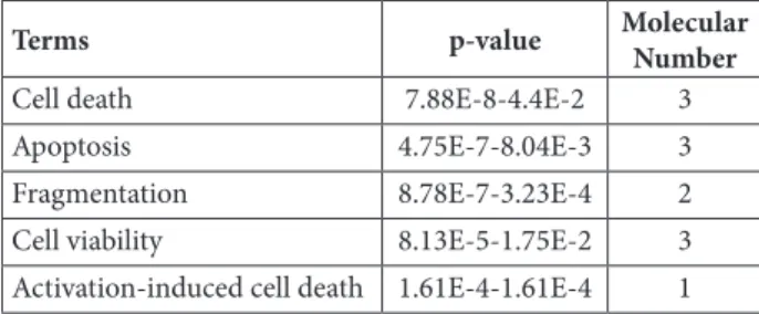
Cell death 7.88E-8-4.4E-2 3
Apoptosis 4.75E-7-8.04E-3 3
Fragmentation 8.78E-7-3.23E-4 2
Cell viability 8.13E-5-1.75E-2 3
MATERIALS AND METHODS
Animals, tissues samples and RNA isolation
Male Wistar rats (weight 120-150 g) were pur-chased from Vital River Lab Animal Technology Co., Ltd (Beijing, China). All rats were randomly divided into 2 groups: 2K1C group (n=14) and sham group (n=12). The 2K1C hypertension model was then established as described in detail by Sun et al. (2013). Eight weeks after this initial surgery, the rats wereanesthetized with sodium pentobarbital after overnight starvation. The left ventricle was immediately removed, blotted dry, weighed and stored at -80°C until RNA extrac-tion. For miRNA microarray, total RNA was ex-tracted as previously described (Sun et al., 2013). For real-time PCR, miRNA extraction was per-formed with an mirVana miRNA Isolation Kit (Ambion, Austin, TX, USA), according to the manufacturer’s instructions. RNA quality and quantity were determined by a NanoDrop 1000
Spectrophotometer (Thermo Fisher Scientific Inc., Waltham, MA, USA). Animal studies were performed under conditions approved by the Lo-cal Animal Care and Use Committee.
miRNA microarray
miRNA expression profiling was performed with 8×15K Agilent Rat miRNA Microarray V12.0 (Agilent Technologies, Santa Clara, CA, USA) containing probes for 350 miRNAs. 100 ng total RNA was dephosphorylated, denatured, Cyanine3-pCp labeled and hybridized, using the Agilent rat miRNA microarray according to the manufacturer’s instructions. After wash-ing, array scanning was performed at a resolu-tion of 5 µm using an Agilent DNA Microarray scanner (Agilent Technologies, Santa Clara, CA, USA). Feature extraction and gene signal normalization were performed with Feature Ex-traction Software 9.5.3 and GeneSpring soft-ware 10.1 (Agilent Technologies, Santa Clara, CA, USA).
qRT-PCR validation
miRNAs with the highest fold change revealed by miRNA microarray were selected for fur-ther validation by qRT-PCR. TaqMan MicroR-NA Assays were used as follows: miR-139-3p (002546), miR-21 (000397) and U6 snRNA (001973), which were purchased from Ap-plied Subsystems (Foster City, CA, USA). To-tal RNA was reverse transcribed by TaqMan MicroRNA Reverse Transcription Kit (Applied Biosystems, Foster City, CA, USA). qRT-PCR was performed using TaqMan Universal PCR Master Mix II on a 7500 Fast Real-Time PCR System according to the manufacturer’s proto-col. A 5-fold serial dilution of pooled cDNA samples was also generated for each assay. All samples were run in triplicate. Based on results of the serial dilution curve and melting curve, a technical quality assessment was performed. Relative expression was then performed using the ΔΔCt method. Data were standardized by log2 transformation.
Bioinformatics analysis
The target genes of the differentially expressed miR-139-3p were identified using the IPA pro-gram (http://www.ingenuity.com). Meanwhile,
the target genes of rno-mir-139-3p were collected, and subjected to GeneGo pathway annotation.
Statistical analysis
Statistical analysis was performed using SPSS15.0. Differences between groups were compared using the Student’s t-test. A P value less than 0.05 was considered as statistically significant. All values are expressed as mean ± SEM.
RESULTS
miRNA expression in left ventricular myocardium from sham-operated and 2K1C rats
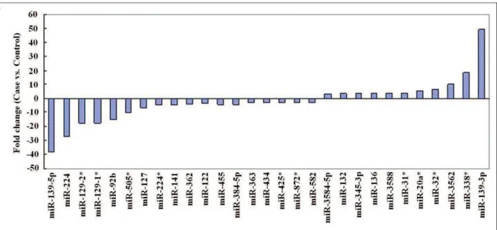
We developed a rat model of 2K1C hyperten-sion as previously described (Sun et al., 2013). An miRNA microarray platform containing 350 probes was used to assess miRNA expression profiles in the left ventricular myocardium. Our results demonstrated that miRNAs were differen-tially expressed between the sham-operated group and 2K1C groups (Fig. 1). The analysis identified that 11 miRNAs were upregulated and 18 were downregulated in the 2K1C group compared to the sham-operated group (P<0.05). A ±3.0-fold change cut-off was applied to all array datasets.
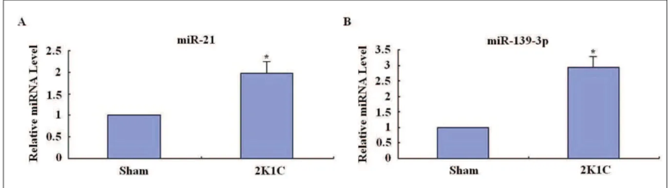
Validation of the microarray data by miRNA qRT-PCR analysis
To confirm the results from the microarray analysis, the highest expression miRNA miR-139-3p was selected for further confirmation using qRT-PCR. As expected, a statistically sig-nificant upregulation in the expression of miR-139-3p was observed in the 2K1C group com-pared to the sham-operated group (P<0.05), which was found to be in accordance with the microarray analysis results (Fig. 2). Fig. 2 further revealed that the hypertrophic cardio-myocytes showed a significantly higher expres-sion level of miR-21 compared to the controls
(P<0.05), which was in agreement with other
studies (Cheng et al., 2010). These results indi-cated that the results of qRT-PCR analysis were reliable.
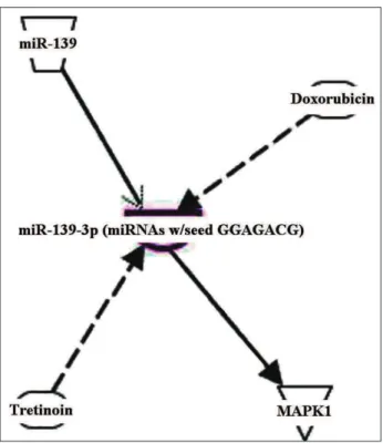
The target genes and biological functions of miR-139-3p
The target genes of the differentially expressed miR-139-3p were analyzed using the IPA tool. One target gene, MAPK1, was identified after analysis of published data, validated by biologi-cal experiments (Fig. 3). In addition, the relevant diseases and biological functions of miR-139-3p were analyzed and the results revealed 5 signifi-cant functions (Table 1). Of these functions, Cell
Death and Survival was the highest rated
func-tion with the p-value of 7.88E-8-1.75E-2. Among
Cell Death and Survival, cell death and apoptosis
were identified as the top 2 significant functions (Table 2).
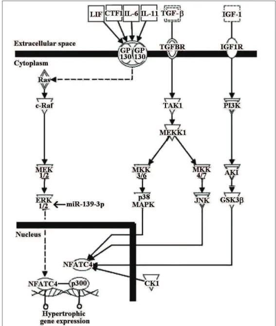
Potential mechanisms of miR-139-3p-mediated cardiac hypertrophy and cardiomyocyte apoptosis
Signal transduction pathways associated with the target genes of miR-139-3p were next inves-tigated. IPA analysis showed that the target gene
MAPK1 participates in NFAT-mediated cardiac
hypertrophy (Fig. 4). Moreover, GeneGo analysis revealed that MAPK1 mainly participates in 90 signaling pathways, of which 4 signaling pathways are linked to apoptosis and survival (Table 3). Among these, MEK/Erk/Bad signaling plays an integral role in apoptosis.
DISCUSSION
In the present study, we identified 29 miRNAs that were deregulated in the left ventricular myo-cardium of 2K1C rats compared with normal tis-sues, of which 11 miRNAs were upregulated and 18 were downregulated. Additionally, miRNA ex-pressions were further analyzed by transforming
to “fold change”. Out of the 11 miRNAs that were upregulated in our study, 6 miRNAs (miR-3584-5p, miR-132, miR-345-3p, rno-miR-136, rno-miR-3588, and rno-miR-31*) were expressed at least 3-fold, and 5 miRNAs (miR-20a*, miR-32*, miR-3562, rno-miR-338*, and rno-miR-139-3p) were expressed at least 5-fold. Out of 18 miRNAs that showed decreased expressions in our study, 11 miRNAs (rno-miR-582, rno-miR-872*, rno-miR-425*, miR-434, miR-363, miR-122, miR-362, miR-384-5p, miR-455, rno-miR-224*, and rno-miR-141) were downregulat-ed at least 3-fold, and 7 miRNAs (rno-miR-127, rno-miR-505*, rno-miR-92b, rno-miR-129-1*, 129-2*, 224, and rno-miR-139-5p) were downregulated at least 6-fold. These results demonstrated that aberrant expres-sion of miRNAs is involved in cardiac remodel-ing. The expression profiles of miRNAs in our study were not entirely consistent with previously
reported results focusing on aberrant miRNAs in 2K1C rats (Sun et al., 2013). In our previous work, a ±2-fold change cut-off was applied to all array datasets. However, a ±3-fold change cut-off was applied in our current work. We consider this to be due to differences in the screening criteria.
In addition to identifying the aberrant miR-NAs in 2K1C rats, certain miRmiR-NAs that might have potential clinical significance as reported elsewhere were found to be differentially ex-pressed in the left ventricular myocardium from 2K1C rats. For example, it has been reported that miR-139-3p was differentially expressed in can-cers of different stages and grades (Chen et al., 2012). In this study, miR-139-3p was found to be highly expressed in 2K1C rats. These findings indicate that deregulation of miR-139-3p might affect the molecular events in the progression of cardiac hypertrophy.
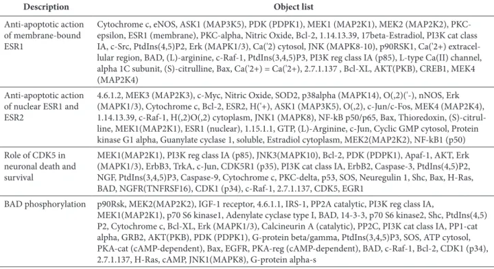
Table 3. MAPK1-mediated apoptosis and survival pathways obtained from GeneGo.
Description Object list
Anti-apoptotic action of membrane-bound ESR1
Cytochrome c, eNOS, ASK1 (MAP3K5), PDK (PDPK1), MEK1 (MAP2K1), MEK2 (MAP2K2), PKC-epsilon, ESR1 (membrane), PKC-alpha, Nitric Oxide, Bcl-2, 1.14.13.39, 17beta-Estradiol, PI3K cat class IA, c-Src, PtdIns(4,5)P2, Erk (MAPK1/3), Ca('2) cytosol, JNK (MAPK8-10), p90RSK1, Ca('2+) extracel-lular region, BAD, (L)-arginine, c-Raf-1, PtdIns(3,4,5)P3, PI3K reg class IA (p85), L-type Ca(II) channel, alpha 1C subunit, (S)-citrulline, Bax, Ca('2+) = Ca('2+), 2.7.1.137 , Bcl-XL, AKT(PKB), CREB1, MEK4 (MAP2K4)
Anti-apoptotic action of nuclear ESR1 and ESR2
4.6.1.2, MEK3 (MAP2K3), c-Myc, Nitric Oxide, SOD2, p38alpha (MAPK14), O(,2)('-), nNOS, Erk (MAPK1/3), Cytochrome c, Bcl-2, ESR2, H('+), ASK1 (MAP3K5), O(,2), c-Jun/c-Fos, MEK4 (MAP2K4), 1.14.13.39, c-Raf-1, H(,2)O(,2) cytoplasm, JNK1 (MAPK8), NF-kB p50/p65, Bax, Thioredoxin, (S)-citrul-line, MEK1(MAP2K1), ESR1 (nuclear), 1.15.1.1, GTP, (L)-Arginine, c-Jun, Cyclic GMP cytosol, Protein kinase G1 alpha, Guanylate cyclase 1, soluble, Estradiol cytoplasm, MEK2(MAP2K2), NF-kB1 (p50)
Role of CDK5 in neuronal death and survival
MEK1(MAP2K1), PI3K reg class IA (p85), JNK3(MAPK10), Bcl-2, PDK (PDPK1), Apaf-1, AKT, Erk (MAPK1/3), ErbB3, TrkA, c-Jun, CDK5R1 (p35), PI3K cat class IA, ErbB2, Caspase-3, PtdIns(4,5)P2, NGF, PtdIns(3,4,5)P3, Caspase-9, Cytochrome c, PKC-delta, p53, SOS, Neuregulin 1, Shc, Bax, H-Ras, BAD, NGFR(TNFRSF16), CDK1 (p34), c-Raf-1, 2.7.1.137, CDK5, EGR1
BAD phosphorylation p90Rsk, MEK2(MAP2K2), IGF-1 receptor, 4.6.1.1, IRS-1, PP2A catalytic, PI3K reg class IA,
In order to understand the roles of miR-139-3p in cardiac hypertrophy of hypertensive rats, the target genes and mechanism of regulation were further exploration. With IPA we identified that MAPK1 was the validated target gene of miR-139-3p. Additionally, miR-139-3p and its target gene MAPK1 were involved in “Cell Death and
Survival” as suggested by IPA functional analysis. Furthermore, IPA analysis revealedcell death and apoptosis to be the most favored associated func-tions in cell death and survival. Therefore, miR-139-3p might play a key role in cell death and survival depending on which gene it is targeting during cardiac hypertrophy in hypertensive rats.
Moreover, signal transduction pathways as-sociated with miR-139-3p and its target gene
MAPK1 were investigated. Results from the IPA
analysis revealed that the target gene participat-ed in the NFATC4-mparticipat-ediatparticipat-ed signaling pathway, which is crucial for cardiac hypertrophy as de-scribed previously (Mathew et al., 2004). Results from the GeneGo analysis revealed that MAPK1 could participate in 90 signaling pathways. Since miR-139-3p and its validated target gene MAPK1 were involved in cell death and survival as sug-gested by IPA functional analysis, one result to arouse great interest was that miR-139-3p modu-lated the apoptosis and survival pathways (Table 3). The results showed that MEK/Erk/Bad sig-naling was a component of these four sigsig-naling pathways linked to apoptosis and survival, and regulated apoptosis through anti-apoptotic Bcl-2 family members (Eisenmann et al., 2003). It has been reported that the development of left ven-tricular hypertrophy and the level of apoptosis in spontaneously hypertensive rats could be modu-lated by Bcl-2 family proteins (Jiang et al., 2013). Therefore, we have reason to consider that miR-139-3p might modulate cardiomyocyte apoptosis in 2K1C rats through the affected MEK/Erk/Bad pathway.
In conclusion, the results of the present study suggest that miR-139-3p may play a key role in the left ventricular remodeling process. Analysis of its target gene and signaling pathways may add a new mechanism to its roles. However, further studies are required to validate this. The findings of the present study have expanded our knowl-edge of the molecular alterations involved in heart pathogenesis and the role of miR-139-3p in left ventricular hypertrophy and cardiomyo-cyte apoptosis.
Acknowledgments: This work was supported by the National Natural Science Foundation of China (No.
81160033). We thank Mr. J.C. Lin, CloudScientific Tech-nology Co. Ltd., for performing the ingenious pathway analysis.
Authors’ contributions: X.Y. conceived and conducted the experiment and performed data collection; H.H. and H.Y. performed the experiments, collected the data and wrote the manuscript; J.Y. and X.T. performed the experiments and collected the data; C.W. and X.Y. − collected the data; G.S. conceived and designed the experiments, collected and interpreted the data; Z.W. conceived and designed the experiments, conducted the experiments, collected the data, constructed the figures and wrote the manuscript.
Conflict of interest disclosure: The authors declare no conflicts of interest.
REFERENCES
Barnes, V.A., Kapuku, G.K. and F.A Treiber (2012). Impact of tran-scendental meditation on left ventricular mass in African American adolescents. Evid. Based Complement. Alternat.
Med. 2012: 923153.
Chen, W.C., Lin, M.S., Ye, Y.L., Gao, H.J., Song, Z.Y. and X.Y. Shen
(2012). microRNA expression pattern and its alteration fol-lowing celecoxib intervention in human colorectal cancer.
Exp. Ther. Med. 3:1039-1048.
Cheng, Y. and C. Zhang (2010). MicroRNA-21 in cardiovascular disease. J. Cardiovasc. Transl. Res. 3: 251-255.
Chien, K.R. (1999). Stress pathways and heart failure. Cell. 98: 555-558.
Eisenmann, K.M., VanBrocklin, M.W., Staffend, N.A., Kitchen, S.M. and H.M. Koo (2003). Mitogen-activated protein kinase pathway-dependent tumor-specific survival signal-ing in melanoma cells through inactivation of the proapop-totic protein bad. Cancer Res.63: 8330-8337.
Gu, W., Li, X. and J. Wang (2014). miR-139 regulates the prolifera-tion and invasion of hepatocellular carcinoma through the WNT/TCF-4 pathway. Oncol. Rep. 31: 397-404.
Jiang, F.L., Leo, S., Wang, X.G., Li, H., Gong, L.Y., Kuang, Y. and
X.F. Xu (2013). Effect of tanshinone IIA on cardiomyocyte hypertrophy and apoptosis in spontaneously hypertensive rats. Exp. Ther. Med. 6: 1517-1521.
Kirchhoff, S.R., Gupta, S. and A.A. Knowlton (2002). Cytosolic HSP60, apoptosis, and myocardial injury. Circulation. 105:
2899–2904.
Li, P. (2010). MicroRNAs in cardiac apoptosis. J. Cardiovasc. Transl. Res. 3: 219-224.
Li, R.Y., Chen, L.C., Zhang, H.Y., Du, W.Z., Feng, Y., Wang, H.B., Wen, J.Q., Liu, X., Li, X.F., Sun, Y., Yang, D.B., Jiang, T., Li, Y.L. and C.L. Jiang (2013). MiR-139 inhibits Mcl-1 expres-sion and potentiates TMZ-induced apoptosis in glioma.
CNS Neurosci. Ther. 19: 477-483.
Lin, M., Chen, W., Huang, J., Gao, H., Ye, Y., Song, Z. and X. Shen
(2011). MicroRNA expression profiles in human colorectal cancers with liver metastases. Oncol. Rep. 25: 739-747.
Mathew, S., Mascareno, E. and M.A. Siddiqui (2004). A ternary complex of transcription factors, Nishéd and NFATc4, and co-activator p300 bound to an intronic sequence, intronic regulatory element, is pivotal for the up-regulation of
myosin light chain-2v gene in cardiac hypertrophy. J. Biol. Chem. 279: 41018-41027.
Putinski, C., Abdul-Ghani, M., Stiles, R., Brunette, S., Dick, S.A., Fernando, P. and L.A. Megeney (2013). Intrinsic-mediated caspase activation is essential for cardiomyocyte hypertro-phy. Proc. Natl. Acad. Sci. U S A. 110: E4079-E4087.
Sun, G., Hu, H., Tian, X., Yue, J., Yu, H., Yang, X. and Z. Wang
(2013). Identification and analysis of microRNAs in the left ventricular myocardium of two-kidney one-clip hyperten-sive rats. Mol. Med. Rep. 8: 339-344.
Wang, Z., Luo, X., Lu, Y. and B. Yang (2008). miRNAs at the heart of the matter. J. Mol. Med. (Berl). 86: 771-783.