Identification of Differential MicroRNAs in
Cerebrospinal Fluid and Serum of Patients
with Major Depressive Disorder
Yunqiang Wan1☯, Yuanhui Liu1☯, Xiaobin Wang1*, Jiali Wu1, Kezhi Liu2, Jun Zhou1, Li Liu1, Chunxiang Zhang3
1Department of Anesthesiology and Psychiatry, Affiliated Hospital of Luzhou Medical College, Luzhou, Sichuan Province, China,2Department of Psychiatry, Affiliated Hospital of Luzhou Medical College, Luzhou, Sichuan Province, China,3Department of Pharmacology, Rush Medical College, Rush University, Chicago, IL, United States of America
☯These authors contributed equally to this work.
*wangxiaobin67@163.com
Abstract
Major depression is a debilitating disease. To date, the development of biomarkers of major depressive disorder (MDD) remains a challenge. Recently, alterations in the expression of microRNAs (miRNAs) from post-mortem brain tissue and peripheral blood have been linked to MDD. The goals of this study were to detect the differential miRNAs in cerebrospinal fluid (CSF) and serum of MDD patients. First, the relative expression levels of 179 miRNAs (rela-tive high levels in serum) were analyzed by miRNA PCR Panel in the CSF of MDD patients. Then, the differentially altered miRNAs from CSF were further assessed by qRT-PCR in the serum of the same patients. Finally, the serum differentially altered miRNAs were further validated by qRT-PCR in the serum of another MDD patients. The CSF-results indicated that 11 miRNAs in MDD patients were significantly higher than these in control subjects, and 5 miRNAs were significantly lower than these in control subjects. The serum-results from the same patients showed that 3 miRNAs (miR-221-3p, miR-34a-5p, and let-7d-3p) of the 11 miRNAs were significantly higher than these in control subjects, and 1 miRNA (miR-451a) of 5 miRNAs was significantly lower than these in control subjects. The up-regulation of miR-221-3p, miR-34a-5p, let-7d-3p and down-regulation of miR-451a was further validat-ed in another 32 MDD patients. ROC analysis showvalidat-ed that the area under curve of let-7d-3p, miR-34a-5p, miR-221-3p and miR-451a was 0.94, 0.98, 0.97 and 0.94, with specificity of 90.48%, 95.24%, 90.48% and 90.48%, and sensitivity of 93.75%, 96.88%, 90.63% and 84.85%, respectively. In addition, target gene prediction found that the altered miRNAs are involved in affecting some important genes and pathway related to MDD. Our results sug-gested that differentially altered miRNAs in CSF might be involved in MDD, and serum miR-221-3p, miR-34a-5p, let-7d-3p, and miR-451a might be able to serve as biomarkers for MDD.
a11111
OPEN ACCESS
Citation:Wan Y, Liu Y, Wang X, Wu J, Liu K, Zhou J, et al. (2015) Identification of Differential MicroRNAs in Cerebrospinal Fluid and Serum of Patients with Major Depressive Disorder. PLoS ONE 10(3): e0121975. doi:10.1371/journal.pone.0121975
Academic Editor:Kandiah Jeyaseelan, National University of Singapore, SINGAPORE
Received:October 9, 2014
Accepted:February 5, 2015
Published:March 12, 2015
Copyright:© 2015 Wan et al. This is an open access article distributed under the terms of the
Creative Commons Attribution License, which permits unrestricted use, distribution, and reproduction in any medium, provided the original author and source are credited.
Data Availability Statement:All relevant data are within the paper.
Introduction
Major depressive disorder (MDD) is a common chronic mood illness, resulting in heavy social
and economic burdens [1,2]. However, current diagnosis of MDD is mainly based on the
pa-tients' description of symptoms, assessment of mental status and evaluation of clinical behav-iors, which increase the chance of misdiagnosis. There is no reliable biological marker from blood or cerebrospinal fluid that can be used for the diagnosis of MDD.
MicroRNAs (miRNAs) are small noncoding RNA molecules that regulate the stability and/
or the translational efficiency of target messenger RNAs [3] and can influence the activity of
approximately 50% or more of all protein-coding genes in mammals [4,5], and several studies
have reported that miRNAs are very important regulatory factors in the normal developmental, physiological and disease states, including cancer, mental disorders and cardiovascular diseases
[6–8]. Recently, alterations in miRNAs expression from post-mortem brain tissues and
periph-eral blood samples have been linked to MDD [9,10].
However, unlike tumor tissue, some tissues such as from the heart and brain are not easy to obtain in clinical practice. Interestingly, recent studies have found that the diseased tissues of heart or brain can release miRNAs into the circulatory blood or CSF, and these miRNAs may
be new biological markers of cardiovascular and cerebral diseases [11,12].
CSF is separated from the blood circulation by the blood brain barrier; therefore, the change of miRNAs in CSF might better reflect the alteration of miRNAs from the diseased tissues of
brain than blood [13]. Unfortunately, the change in CSF miRNAs in MDD is unclear, and it is
very difficult to draw CSF from MDD patients via lumber puncture. Therefore, the detection of serum miRNAs is easier and convenient than CSF. The goals of this study were to detect the candidate miRNAs in cerebrospinal fluid of MDD patients and to investigate these candidate miRNAs in serum from MDD patients further to provide information about miRNAs as specif-ic biomarkers for the diagnosis of MDD.
Methods
Patients
The protocols of this study and the procedures employed for sample collection were approved by the Ethical Committee of Luzhou Medical College (Ky2012044). Informed written consent was obtained from all participants. Subjects with major depression were diagnosed according to either the ICD-10 or DSM IV criteria with a total score of 15 or higher on the 24-item HDRS. The exclusion criteria were as follows: a personal history of bipolar affective disorder, schizophrenia, mood incongruent psychotic symptoms, primary substance abuse or primary organic disease, current treatment with antidepressants or contraindication of lumbar punc-ture. The inclusion criteria of control subjects were as follows: no psychiatric disorders accord-ing to either the ICD-10 or DSM IV criteria, no serious systemic diseases, no complications and tumor, and these control patients also matched the above the exclusion criteria.
Samples preparation and RNA Extraction
First, CSF samples of 6 depressed and 6 control patients were collected by diagnostic lumbar puncture or spinal anesthesia due to elective surgery (renal calculus, bladder stones), the blood samples of these patients were collected at the same time. Subsequently, the blood samples of another 32 MDD patients and 21 healthy individuals were collected. CSF samples were
centri-fuged (500×g, 10 min, room temperature) and blood samples were centrifuged (3000×g,
10 min, room temperature) within 60 min after collection to remove cells and debris and were stored at -80°C until further analysis. Hemorrhagic CSF samples were not included. Total RNA Competing Interests:The authors have declared
was extracted using with Trizol (Invitrogen, Carlsbad, CA, USA) based isolation kit (Shanghai
Kangchen Biotechnology Company, Shanghai, China) according to the manufacturer’s
proto-col. The concentration and purity of RNA were analyzed by the NanoDrop-1000 (Thermo Sci-entific, Waltham, MA, USA).
Real-time PCR
Approximately 20–25 ng RNA was retro-transcribed into cDNA with the MicroRNA Reverse
Transcription Kit and the RT Primer Pools (Exiqon, Danmark) according to the manufacturer’s
protocol. First, qRT-PCR was conducted in ABI PRISM7900 system (Applied Biosystems, Foster, CA, USA) with microRNA PCR Panel (V3.M) (Exiqon, Danmark), which detected 179 miRNAs
in the CSF of 6 depressed and 6 control patients according to the manufacturer’s protocol. Then,
the differentially expressed miRNAs from CSF (p<0.05, Fold change>1.5) were further
ana-lyzed in serum samples from these patients using qRT- PCR. Finally, the serum differentially
al-tered miRNAs (p<0.05, Fold change>1.5) were further validated by the serum from another 32
depressed patients and 21 healthy individuals using qRT-PCR. Cycling conditions were as fol-lows: 95°C for 10 min and 40 cycles of 15 s at 95°C and 60 s at 60°C. The fluorescence signal was
normalized to unify the internal reference level, and the threshold cycle (CT) was set in the
expo-nential amplification phase of the PCR. The relative expression levels of miRNAs were calculated
based on the number of PCR cycles of miRNAs. Undetectable data were assigned a default CT
value of 40. The CTvalues were normalized according to the delta CTmethod with the
endoge-nous controls (miR-423–5p) because miR-423–5p was identified as suitable serum-based
refer-ence genes of MDD[14], and our results also showed that the CTvalue of miR-423–5p in both
the CSF and the serum between depressed and control subjects are very stable(data no shown).
To determine the correspondingΔCTvalue, the CTvalue of the target gene miRNAs was
sub-tracted from the miR-423–5p CTvalue so to normalize the values. Among the different
experi-mental treatments, the relative expression levels (RELs) of miRNAs were calculated using the
following formula: relative gene expression = 2-(ΔCTof experimental sample -ΔCTof control sample).
The data obtained from the miRNA PCR array were analyzed with the GenEx qPCR analysis software.
Statistics
All statistical analyses were performed using SPSS (version 19; SPSS) and GraphPad Prism (version 5.0, GraphPad Software). Data are presented as mean±SD. Differences in miRNA
con-centrations between control and MDD subjects were compared using Student’s t-test or
Welch’s t-test for equal or unequal variances; the Mann-Whitney U test was used when the
dis-tribution was skewed. A difference was considered significant at p<0.05 or p<0.01.
Results
Patient characteristics
As shown inTable 1, there were 38 depressed patients that had never been treated with
antide-pressant drugs and 27 control subjects that had no psychiatric disorders. Age and HAMD scores of control and depressed patients in the analysis group were 32.5±9.2 vs 32.0±10.8
(p>0.05) and 4.5±1.2 vs 23.7±4. 8 (t = 9.396, p<0.0001), respectively, there were 2 patients
with suicidal ideations among the depressed patients of the analysis group, the control patients of the analysis group there were 4 with renal calculus and 2 with bladder stones. Age and HAMD scores of the control and depressed subjects in the validated group were 35.34±9.5 vs
patients with suicidal ideations among the depressed patients of the validated group, and the control subjects of the validated group consisted of 21 healthy individuals.
Measure of RNA concentration and purity about CSF and serum
samples
We determined whether miRNAs were detectable in frozen CSF and serum samples collected from depressed and control subjects. For direct measurement of total RNA we used the Nano-Drop-1000. RNA concentrations were detected in the CSF specimens (control subjects: 38.39
±17.78 ng/μL, depressed patients: 25.05±2.72 ng/μL) and the serum specimens (control
sub-jects: 21.83±5.21ng/μL, depressed patients: 23.73±7.82 ng/μL). The ratio of A 260 to A 280
val-ues (a measure of RNA purity) of all specimens fell in the range of 1.8 to 2.1 (data no shown).
Detection of differential miRNAs in the CSF and serum of 6 MDD
patients
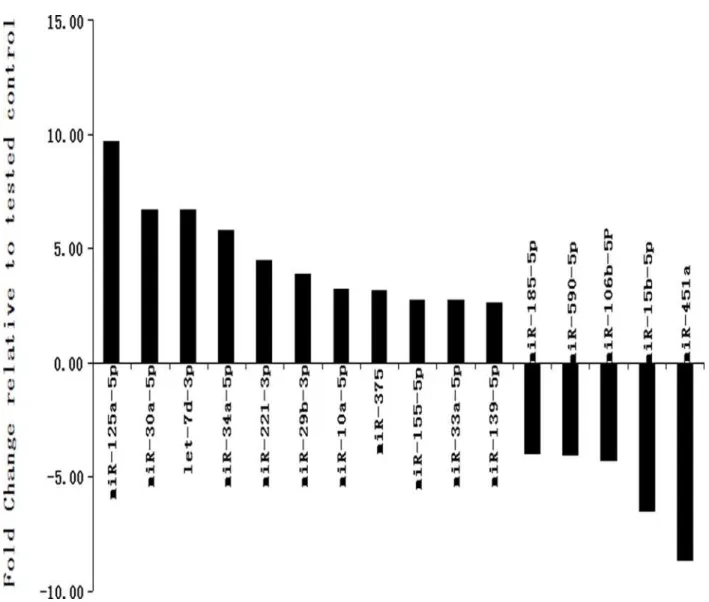
First, the 179 miRNAs in CSF from MDD and control patients were analyzed by microRNA
PCR panel analysis. As shown inTable 2,Fig. 1, the results indicated that 16 miRNAs were
dif-ferentially expressed between control and depressed patients (p<0.05, fold change>1.5), the
mean RELs of miR-125a-5p, let-7d-3p, miR-30a-5p, miR-34a-5p, miR-221-3p, miR-29b-3p,
miR-10a-5p, miR-375, miR-155–5p, miR-33a-5p and miR-139–5p in the depressed patients
were obviously higher than these in the control patients (p<0.05, fold change from 9.75 to
2.63), the mean RELs of miR-451a, miR-15b-5p miR-106b-5p, miR-590–5p and miR-185–5p
in the depressed patients were obviously lower than these in the control patients (P<0.05, fold
change from-8.64 to-3.99).
Then, 16 CSF significantly altered miRNAs were further analyzed in the serum from the
same MDD patients using qRT-PCR. As shown inTable 3,Fig. 2, the results indicated that
only 4 miRNAs were differentially expressed between control and depressed patients (p<0.05,
fold change>1.5), the mean RELs of let-7d-3p, miR-34a-5p and miR-221-3p in the depressed
patients were obviously higher than these in the control patients (p<0.01, fold change: 1.67,
2.14, 2.23), and the mean RELs of miR-451a in the depressed patients were obviously lower
than that in the control patients (P<0.001, fold change: -1.85). The differences in mean RELs
of miR-125a-5p, miR-30a-5p, miR-29b-3p and miR-15b-5p were not statistically significant
between control and depressed patients (p>0.05).
Table 1. Characteristics of depressed patients and control subjects.
Variable Analysis group Validated group
C(n = 6) T(n = 6) p-value C(n = 21) T(n = 32) p-value
Sex(M/F) 2/4 3/3 0.925a 10/11 15/17 0.958a
Age(years) 32.5±9.2 32.0±9.10.8 0.837b 35.34±9.5 34.33±10.4 0.720b
HAMD-scores 4.5±1.2 23.7±4.8 <0.001b 4.4±1.6 25.7±4.1 <0.001b
Idea of suicide 0/6 2/6 N/A 0/21 9/23 N/A
Relevant diseases Renal calculus N/A N/A N/A N/A N/A
Bladder stones
Age and HAMD scores were presented as mean±standard deviation
C group: control subjects with no psychiatric disorders, T group: MDD patients, M: male, F: female
aPearson Chi-Square; bIndependent sample-t test.
Validation of miR-34a-5p, miR-221-3p, let-7d-3p and miR-451a in the
serum of another 32 MDD patients
Four miRNAs (miR-34a-5p, miR-221-3p, let-7d-3p and miR-451a) were significantly altered in serum and were further analyzed in the serum from another 32 MDD patients using
qRT-PCR. As shown inFig. 3, the results indicated that the mean RELs of let-7d-3p, miR-34a-5p
and miR-221-3p in the depressed patients were also obviously higher than these in the control
subjects (p<0.0001, fold change: 1.79, 2.86, 2.29), the mean RELs of miR-451a in the depressed
patients were obviously lower than that in the control subjects (p<0.0001, fold change: -1.48).
Receiver operating characteristic (ROC) analysis of let-7d-3p,
miR-34a-5p, miR-221-3p and miR-451a
To observe the relationship between the four microRNAs and MDD, we conducted ROC anal-ysis to estimate the sensitivity and specificity of the diagnostic ability of let-7d-3p, miR-34a-5p,
miR-221-3p and miR-451a. As shown inFig. 4, the ROC curves of let-7d-3p, miR-34a-5p,
miR-221-3p and miR-451a reflected obvious separation between MDD and control groups,
with an AUC of 0.94[95% confidence interval (CI) = 0.87~1.01, P<0.0001], 0.98 (95% CI =
0.97~1.00, P<0.0001), 0.97 (95% CI = 0.94~1.01, P<0.0001) and 0.94(95% CI = 0.88~1.00,
P<0.0001). The specificity of let-7d-3p, miR-34a-5p, miR-221-3p and miR-451a were 90.48%,
95.24%, 90.48% and 90.48%, respectively. The sensitivity of let-7d-3p, miR-34a-5p, miR-221-3p and miR-451a were 93.75%, 96.88%, 90.63% and 84.85%, respectively. let-7d-miR-221-3p, miR-34a-5p, miR-221-3p and miR-451a displayed a very high sensitivity and specificity for diagnosis of MDD.
Table 2. Differentially expressed miRNAs in the CSF between depressed and control subjects.
miRNA ID MDD patients(n = 6) Control patients(n = 6) p valuea
Mean ofΔCt SD Mean ofΔCt SD
miR-125a-5p -1.31 1.02 1.96 1.49 0.0012
miR-30a-5p 0.75 1.16 3.69 2.25 0.0143
let-7d-3p -0.75 1.23 2.19 2.96 0.0422
miR-34a-5p 0.85 1.48 3.38 1.74 0.0411
miR-221-3p -0.27 0.89 1.89 1.37 0.0092
miR-29b-3p 1.38 1.22 3.52 1.73 0.0223
miR-10a-5p 3.87 0.75 5.94 0.83 0.0249
miR-375 3.92 1.48 5.57 1.06 0.0160
miR-155-5p 3.61 1.29 5.07 0.78 0.0185
miR-33a-5p 3.42 1.39 4.87 0.86 0.0191
miR-139-5p 4.18 1.21 5.57 1.06 0.0375
miR-185-5p 4.15 1.46 2.25 1.75 0.0276
miR-590-5p 3.61 1.29 5.07 1.65 0.0018
miR-106b-5p 1.54 1.12 -0.58 1.99 0.0451
miR-15Bb-5p 2.83 1.16 0.12 0.78 0.0008
miR-451a -4.3 2.14 -7.42 2.58 0.0455
ΔCt = Ct of target miRNAs-Ct of miR-423-5p(endogenous reference gene) SD = standard deviation
aIndependent sample t test.
Prediction of target genes and pathway of let-7d-3p, 34a-5p,
miR-221-3p and miR-451a
To investigate the target genes and pathway of let-7d-3p, 34a-5p, 221-3p and miR-451a in MDD, target genes were predicted by using miRNA target prediction databases(Target Scan, miRanda and PicTar). The possible regulatory pathway of the target genes were analyzed
based on functional annotation according to KEGG pathway terms [15]. The analysis found
that some important genes related to MDD, such as serine protein kinases(AKT)[16],
seroto-nin receptors(5HT2A, HTR2C)[17], corticotrophin-releasing hormone receptor (Crhr1)[18],
glutamate transporters (SCL1A2)[19], were targeted by 451a, 221-3p or/ and
miR-34a-5p(data no shown). The analysis of regulatory pathway of these target genes indicated a significant enrichment in several pathways related to neuronal brain function of the depression such as PI3K-Akt signaling pathway, Axon guidance, Wnt signaling pathway, Neurotrophin signaling pathway, Hippo signaling pathway, mTOR signaling pathway, ErbB signaling path-way, Cell cycle, Apoptosis, Long-term depression(data no shown).
Fig 1. miRNAs that are significantly up-or down-regulated in the CSF from MDD vs. control subjects.
Discussion
Our results showed that CSF miRNAs from MDD patients could be detected. Unfortunately, it is very difficult to obtain CSF from MDD patients by lumber puncture. It is more convenient and easier to use serum specific miRNAs from MDD patients for diagnosis. Therefore, we should seriously consider how to look for serum specific miRNAs as biomarker for MDD be-cause the change of CSF miRNAs was poorly related to the alteration of serum miRNAs for
CNS diseases[13]. Serum miRNAs that are consistent with CSF changes of miRNAs should be
identified in MDD patients, and these miRNAs from CSF should have relative high serum lev-els and be easily detected in serum. Therefore, Focus 179 microRNA PCR Panlev-els were chosen
for the analysis of CSF miRNAs from MDD patients (
http://www.Exiqon.com/serum-plasma-miRNA-qPCR). The reasons why we only measured 179 miRNAs instead of performing a mi-croarray for miRNome profiling analysis are as follows: (1) The amount of samples such as serum or CSF is really small, which means that a more sensitive and accurate platform is need-ed. (2) Not all microRNAs could be detected in serum, and we cared about those which may exist in serum. Exiqon has extensive in-house knowledge of microRNA profiling in serum. All
179 miRNA assays have been carefully selected based on Exiqon’s vast number of in-house
analyses of miRNA expression in blood serum and plasma samples as well as on the limited number of peer-reviewed published papers available.
Emerging evidence has demonstrated that dysregulation of miRNAs may contribute to the
etiology and pathophysiology of MDD [20]. miRNAs in diseased cerebral tissues are able to be
released into CSF, and CSF miRNAs can be used as novel biomarkers for the diagnosis of gliomas
[21], but the change in CSF miRNAs from MDD patients is unclear. To our knowledge, this is
the first comparison of deregulated miRNAs expression levels in CSF samples from MDD
Table 3. Differentially expressed miRNAs in the serum between depressed and control subjects.
miRNA ID MDD patients(n = 6) Control patients(n = 6) p valuea
Mean ofΔCt SD Mean ofΔCt SD
miR-125a-5p 0.27 0.88 0.58 0.92 0.5614
miR-30a-5p 7.33 0.81 7.63 0.42 0.4367
let-7d-3p -0.95 0.46 -0.28 0.28 0.0094
miR-34a-5p 5.61 0.49 6.71 0.52 0.0038
miR-221-3p -1.73 0.26 -0.55 0.40 0.0001
miR-29b-3p -2.23 0.17 -2.08 0.27 0.2685
miR-10a-5p 5.11 0.38 5.44 0.52 0.2475
miR-375 4.62 0.63 4.94 0.58 0.3760
miR-155–5p 5.78 0.82 6.61 0.96 0.1427
miR-33a-5p 6.44 0.74 6.94 0.41 0.1857
miR-139–5p 2.95 0.42 3.10 0.51 0.5698
miR-185–5p 3.61 0.42 3.77 0.30 0.4685
miR-590–5p 3.15 0.55 3.24 0.51 0.7703
miR-106b-5p 4.61 0.76 4.88 0.47 0.4741
miR-15Bb-5p -1.14 0.26 -0.85 0.47 0.2101
miR-451a -9.51 0.21 -10.18 0.17 0.0001
ΔCt = Ct of target miRNAs-Ct of miR-423–5p(endogenous reference gene) SD = standard deviation
aIndependent sample t test.
Fig 2. miRNAs that are significantly up- or down-regulated in the serum from the same 6 MDD vs. control subjects.C: control subjects; T: MDD patients; FC: fold change;**p<0.01 vs control.
doi:10.1371/journal.pone.0121975.g002
Fig 3. miRNAs that are significantly up-or down-regulated in the serum from another 32 MDD vs. 21 control subjects.C: control subjects, T: MDD patients; FC: fold change;**p<0.0001 vs control.
patients who are not being treated with antidepressants compared to that of control patients. The quantitative expression analysis indicated that different expression levels of 16 miRNAs in the CSF were modulated by the MDD patients, and included 11 up-regulated miRNAs (miR-125a-5p, let-7d-3p, miR-30a-5p, miR-34a-5p, miR-221-3p, miR-29b-3p, miR-10a-5p, miR-375,
miR-155–5p, miR-33a-5p and miR-139–5p) and 5 down-regulated miRNAs (451a,
miR-15b-5p, miR-106b-5p, miR-590–5p and miR-185–5p). After we further analyzed the REL of CSF
16 differential miRNAs in the each patient, we found that there was a obvious high level in the REL of let-7d-3p, miR-29a-3p, miR-30a-5p, miR-34a-5p, miR125a-5p, miR-221-3p. Interesting, the high value came from a patient with suicidal ideation and high HAMD(32 score), however, there were no impact on the the REL of the other 10 differential miRNAs in the same patient(no data shown), and the phenomenon were not found in the addition a patient with suicidal idea-tion (no data shown) and the serum of MDD patients. Due to the small ample size, whether sui-cidal ideations with depressed patients have an impact on the CSF results should be investigated in future study. Other studies have shown that the expression of 30a-5p, 34a-5p,
miR-221 and miR-34a were downregulated by antidepressant medications [22–25]. The expression of
BDNF was downregulated by the overexpression of miR-34a, miR-30a-5p, let-7d, miR-10a-5p,
miR-375 and miR-155 in neurons or brain tissues [26–31], and the low expression levels of
Fig 4. Receiver operating characteristics curve evaluation of serum microRNAs for the diagnosis of MDD.Let-7d-3p (A), miR-34a-5p (B), miR-221-3p (C) and miR-451a (D). AUC: the area under curve.
BDNF in brain tissue were thought to be part of the main pathogenesis of MDD [32]. The low
expression of SIRT1 gene plays an important role in the pathogenesis of MDD[33], studies have
found that the overexpression of miR-10a-5p and miR-34a can downregulate the expression of
SIRT1 gene [28,34]. miR-185 has also been suggested to participate in the pathogenesis of major
depression and/or suicide[35]. Therefore, we believe that the differential change of miRNAs in
CSF from MDD patients was involved in the onset and development of MDD. The hypothesis needs to be further validated in depression models in future studies.
We further analyzed serum relative levels of the 16 differentially altered miRNAs from CSF in the same MDD patients. The results showed that the serum relative levels of only 3 miRNAs (miR-34a-5p, miR-221-3p and let-7d-3p) in MDD patients were obviously higher than these in control patients, and the serum relative levels of only miR-451a in MDD patients were obvious-ly lower than that in control patients. miRNAs levels in CSF and serum samples were poorobvious-ly
correlated, and our results are consistent with a recent study by Gallego et al[13], who found
that miRNAs from CSF were not strongly correlated to miRNAs from whole blood in psychotic disorders. The mechanisms involved in the obvious difference between CSF and serum are still unclear, and the mechanisms should be investigated in future study. In addition, the serum changes of miR-34a-5p, miR-221-3p, let-7d-3p and miR-451a have further been demonstrated in serum from another 32 MDD patients. By receiver operating characteristic (ROC) curve analysis, let-7d-3p, miR-34a-5p, miR-221-3p and miR-451a displayed a very high sensitivity and specificity for diagnosis of MDD. In addition, our analysis of target genes and pathway of the altered miRNAs showed some important genes related to MDD, such as serine protein
ki-nases(AKT)[16], serotonin receptors(HTR2C)[17], corticotrophin-releasing hormone receptor
(CRHR1)[18], glutamate transporters (SCL1A2)[19], were targeted by miR-451a, miR-221-3p
or/ and miR-34a-5p (data no shown) and several pathway related to neuronal brain function of the depression, such as PI3K-Akt signaling pathway, Axon guidance, Wnt signaling pathway, Neurotrophin signaling pathway, Hippo signaling pathway, mTOR signaling pathway, ErbB signaling pathway, Cell cycle, Apoptosis, Long-term depression, were significantly enriched by miR-451a, let-7d-3p, miR-221-3p or/ and miR-34a-5p (data no shown). We plan to investigate these target genes or proteins regulated by these altered miRNA and analyze the relationship between these target proteins and MDD in future study. Therefore, our results indicated that the four serum miRNAs might be able to serve as candidate biomarkers for MDD diagnosis, and some researches found that serum miR-221 and miR-34a levels were decreased by
antide-pressant treatment [15,19]. Although the sample size of our study was not very large, and we
did not include other neurodegenerative disorders, such as schizophrenia and anxiety disorder, which may cause restriction in developing MDD biomarkers, our present results could provide the rationale for future investigations of serum and CSF miRNAs for diagnostic and prognostic purposes in MDD. Therefore, a larger sample size and comprehensive study in human should be conducted to further demonstrate the diagnostic ability of the four miRNAs for MDD in future study.
In summary, in this study we have identified that differentially altered miRNAs in CSF might be related to MDD, and serum miR-221-3p, miR-34a-5p, let-7d-3p, and miR-451a might be able to serve as biomarkers for MDD.
Author Contributions
References
1. Ghourchian S, Zamani B, Poorkosary K, Malakouti SK, Rohani M. Raphe nuclei echogenicity changes in major depression. Med J Islam Repub Iran. 2014; 28:9. PMID:25250254
2. Vos T, Flaxman AD, Naghavi M, Lozano R, Michaud C. Years lived with disability (YLDs) for 1160 se-quelae of 289 diseases and injuries 1990–2010: a systematic analysis for the Global Burden of Disease Study. Lancet. 2012; 380:2163–2196. doi:10.1016/S0140-6736(12)61729-2PMID:23245607 3. Flynt AS, Lai EC. Biological principles of microRNA-mediated regulation: shared themes amid diversity.
Nat Rev Genet. 2008; 9:831–842. doi:10.1038/nrg2455PMID:18852696
4. Krol J, Loedige I, Filipowicz W. The widespread regulation of microRNA biogenesis, function and decay. Nat Rev Genet. 2010; 11: 597–610. doi:10.1038/nrg2843PMID:20661255
5. Bartel DP. MicroRNAs: target recognition and regulatory functions. Cell. 2009; 136:215–233. doi:10.
1016/j.cell.2009.01.002PMID:19167326
6. Cappuzzo F, Sacconi A, Landi L, Ludovini V, Biagioni F, D'Incecco A, et al. MicroRNA signature in met-astatic colorectal cancer patients treated with anti-EGFR monoclonal antibodies. Clin Colorectal Can-cer. 2014; 13:37–45. doi:10.1016/j.clcc.2013.11.006PMID:24503111
7. Wang Z, Zhang C, Huang J, Yuan C, Hong W, Chen J, et al. MiRNA-206 and BDNF genes interacted in bipolar I disorder. J Affect Disord. 2014; 162:116–119. doi:10.1016/j.jad.2014.03.047PMID:24767015 8. Choi E, Cha MJ, Hwang KC. Roles of calcium regulating microRNAs in cardiac ischemia-reperfusion
in-jury. Cell. 2014; 3:899–913.
9. Smalheiser NR, Lugli G, Rizavi HS, Torvik VI, Turecki G, Dwivedi Y. MicroRNA expression is down-regulated and reorganized in prefrontal cortex of depressed suicide subjects. PLoS One. 2014; 7:e33201. 10. Fan HM, Sun XY, Guo W, Zhong AF, Niu W, Zhao L, et al. Differential expression of microRNA in
periph-eral blood mononuclear cells as specific biomarker for major depressive disorder patients. J Psychiatr Res. 2014; 59:45–52. doi:10.1016/j.jpsychires.2014.08.007PMID:25201637
11. Zhou X, Mao AQ, Wang XB, Duan XX, Yao Y, Zhang C. Urine and serum microRNA-1 as novel bio-markers for myocardial injury in open-heart surgeries with cardiopulmonary Bypass. PLoS One. 2013; 8:e62245. doi:10.1371/journal.pone.0062245PMID:23630629
12. Müller M, Kuiperij HB, Claassen JA, Küsters B, Verbeek MM. MicroRNAs in Alzheimer's disease: differ-ential expression in hippocampus and cell-free cerebrospinal fluid. Neurobiol Aging. 2014; 35:152–158. doi:10.1016/j.neurobiolaging.2013.07.005PMID:23962497
13. Gallego JA, Gordon ML, Claycomb K, Bhatt M, Lencz T, Malhotra AK. In vivo microRNA detection and quantitation in cerebrospinal fluid. J Mol Neurosci. 2012; 47: 243–248. doi:
10.1007/s12031-012-9731-7PMID:22402993
14. Liu X, Zhang L, Cheng K, Wang X, Ren G, Xie P. Identification of suitable plasma-based reference genes for miRNAome analysis of major depressive disorder. J Affect Disord. 2014; 163:133–139. doi:
10.1016/j.jad.2013.12.035PMID:24479999
15. Bocchio-Chiavetto L, Maffioletti E, Bettinsoli P, Giovannini C, Bignotti S, Tardito D, et al. Blood micro-RNA changes in depressed patients during antidepressant treatment. Eur Neuropsychopharmacol. 2013; 23:602–611. doi:10.1016/j.euroneuro.2012.06.013PMID:22925464
16. Leibrock C, Ackermann TF, Hierlmeier M, Lang F, Borgwardt S, Lang UE. Akt2 deficiency is associated with anxiety and depressive behavior in mice. Cell Physiol Biochem. 2013; 32:766–777. doi:10.1159/
000354478PMID:24080829
17. Golimbet V E, Volel BA, Dolzhikov AV, Korovaitseva GI, Isaeva MI. Association of HTR2A and 5-HTR2C serotonin receptor gene polymorphisms with depression risk in patients with coronary heart dis-ease. Bulletin of Experimental Biology and Medicine. 2014; 156: 680–683. doi: 10.1007/s10517-014-2424-1PMID:24770757
18. Wan Q, Gao K, Rong H, Wu M, Wang H, Wang X, et al. Histone modifications of the Crhr1 gene in a rat model of depression following chronic stress. Behavioural Brain Research. 2014; 271: 1–6. doi:10. 1016/j.bbr.2014.05.031PMID:24867333
19. Oh DH, Oh D, Son H, Webster MJ, Weickert CS, Kim SH. An association between the reduced levels of SLC1A2 and GAD1 in the dorsolateral prefrontal cortex in major depressive disorder: possible involve-ment of an attenuated RAF/MEK/ERK signaling pathway. J Neural Transm. 2014; 121:783–792. doi:
10.1007/s00702-014-1189-zPMID:24652383
20. Dwivedi Y. Evidence demonstrating role of microRNAs in the etiopathology of major depression. Chem Neuroanat. 2011; 42: 142–156. doi:10.1016/j.jchemneu.2011.04.002PMID:21515361
22. Angelucci F, Croce N, Spalletta G, Dinallo V, Gravina P, Bossù P, et al. Paroxetine rapidly modulates the expression of brain-derived neurotrophic factor mRNA and protein in a human glioblastoma-astrocytoma cell line. Pharmacology. 2011; 87:5–10. doi:10.1159/000322528PMID:21178384 23. Liu BB, Luo L, Liu XL, Geng D, Liu Q, Yi LT. 7-Chlorokynurenic acid (7-CTKA) produces rapid
antide-pressant-like effects: through regulating hippocampal microRNA expressions involved in TrkB-ERK/ Akt signaling pathways in mice exposed to chronic unpredictable mild stress. Psychopharmacology (Berl). 2015; 232:541–550. doi:10.1007/s00213-014-3690-3PMID:25034119
24. Oved K, Morag A, Pasmanik-Chor M, Rehavi M, Shomron N, Gurwitz D. Genome-wide expression pro-filing of human lymphoblastoid cell lines implicates integrin beta-3 in the mode of action of antidepres-sants. Transl Psychiatry. 2013; 3:e313. doi:10.1038/tp.2013.86PMID:24129413
25. Zhou R, Yuan P, Wang Y, Hunsberger JG, Elkahloun A, Wei Y, et al. Evidence for selective microRNAs and their effectors as common long-term targets for the actions of mood stabilizers. Neuropsychophar-macology. 2009; 34: 1395–1405. doi:10.1038/npp.2008.131PMID:18704095
26. Wibrand K, Pai B, Siripornmongcolchai T, Bittins M, Berentsen B, Ofte ML,et al. MicroRNA regulation of the synaptic plasticity-related gene Arc. PLoS One. 2012; 7:e41688. doi:10.1371/journal.pone. 0041688PMID:22844515
27. Croce N, Gelfo F, Ciotti MT, Federici G, Caltagirone C, Bernardini S, et al. NPY modulates miR-30a-5p and BDNF in opposite direction in an in vitro model of Alzheimer disease: a possible role in neuropro-tection?. Mol Cell Biochem. 2013; 376:189–195. doi:10.1007/s11010-013-1567-0PMID:23358924 28. Chandrasekar V, Dreyer JL. MicroRNAs miR-124, let-7d and miR-181a regulate cocaine-induced
plas-ticity. Mol Cell Neurosci. 2009; 42:350–362. doi:10.1016/j.mcn.2009.08.009PMID:19703567 29. Prins SA, Przybycien-Szymanska MM, Rao YS, Pak TR. Long-term effects of peripubertal binge EtOH
exposure on hippocampal microRNA expression in the rat. PLoS One. 2014; 9:e83166. doi:10.1371/ journal.pone.0083166PMID:24416161
30. Abdelmohsen K, Hutchison ER, Lee EK, Kuwano Y, Kim MM, Masuda K, et al. MiR-375 inhibits differ-entiation of neurites by lowering HuD levels. Mol Cell Biol. 2010; 30:4197–4210. doi:10.1128/MCB. 00316-10PMID:20584986
31. Varendi K, Kumar A, Härma MA, Andressoo JO. MiR-1, miR-10b, miR-155, and miR-191 are novel reg-ulators of BDNF. Cell Mol Life Sci. 2014; 71:4443–4456. doi:10.1007/s00018-014-1628-xPMID:
24804980
32. Ray MT, Shannon Weickert C, Webster MJ. Decreased BDNF and TrkB mRNA expression in multiple cortical areas of patients with schizophrenia and mood disorders. Transl Psychiatry. 2014; 4:e389. doi:
10.1038/tp.2014.26PMID:24802307
33. Kishi T, Yoshimura R, Kitajima T, Okochi T, Okumura T, Tsunoka T, et al. SIRT1 gene is associated with major depressive disorder in the Japanese population. J Affect Disord. 2010; 126:167–173. doi:
10.1016/j.jad.2010.04.003PMID:20451257
34. Li L, Yuan L, Luo J, Gao J, Guo J, Xie X. MiR-34a inhibits proliferation and migration of breast cancer through down-regulation of Bcl-2 and SIRT1.Clin Exp Med. 2013; 13:109–117. doi: 10.1007/s10238-012-0186-5PMID:22623155