RESEARCH ARTICLE
Biotype Characterization, Developmental
Profiling, Insecticide Response and Binding
Property of
Bemisia tabaci
Chemosensory
Proteins: Role of CSP in Insect Defense
Guoxia Liu1, Hongmei Ma1, Hongyan Xie1, Ning Xuan1, Xia Guo1, Zhongxue Fan1, Balaji Rajashekar2, Philippe Arnaud3, Bernard Offmann3, Jean-François Picimbon1¤*
1Shandong Academy of Agricultural Sciences, Biotechnology Research Center, Jinan, China,2University of Tartu, Institute of Computer Science, 2 Liivi, Tartu, Estonia,3University of Nantes, Protein Engineering and Functionality Unit, UMR CNRS 6286, 2 La Houssinière, Nantes, France
¤ Current address: QILU University of Technology, College of Bioengineering, Institute of Agricultural Microbiology, Changqing District, Jinan, Shandong Province, P.R. China
Abstract
Chemosensory proteins (CSPs) are believed to play a key role in the chemosensory pro-cess in insects. Sequencing genomic DNA and RNA encoding CSP1, CSP2 and CSP3 in the sweet potato whiteflyBemisia tabacishowed strong variation between B and Q bio-types. Analyzing CSP-RNA levels showed not only biotype, but also age and developmen-tal stage-specific expression. Interestingly, applying neonicotinoid thiamethoxam
insecticide using twenty-five different dose/time treatments in B and Q young adults showed thatBemisiaCSP1, CSP2 and CSP3 were also differentially regulated over insecticide exposure. In our study one of the adult-specific gene (CSP1) was shown to be significantly up-regulated by the insecticide in Q, the most highly resistant form ofB.tabaci. Correla-tively, competitive binding assays using tryptophan fluorescence spectroscopy and molecu-lar docking demonstrated that CSP1 protein preferentially bound to linoleic acid, while CSP2 and CSP3 proteins rather associated to another completely different type of chemi-cal, i.e.α-pentyl-cinnamaldehyde (jasminaldehyde). This might indicate that some CSPs in whiteflies are crucial to facilitate the transport of fatty acids thus regulating some metabolic pathways of the insect immune response, while some others are tuned to much more vola-tile chemicals known not only for their pleasant odor scent, but also for their potent toxic insecticide activity.
Introduction
Whiteflies are well known as severe agricultural pests, devastating all sorts of green ornamental and vegetable plants around the world. Whitefly species such as the sweet potatoBemisia tabaci
Gennadius pierce and suck the sap, causing direct and indirect damages to various plant a11111
OPEN ACCESS
Citation:Liu G, Ma H, Xie H, Xuan N, Guo X, Fan Z, et al. (2016) Biotype Characterization, Developmental Profiling, Insecticide Response and Binding Property ofBemisia tabaciChemosensory Proteins: Role of CSP in Insect Defense. PLoS ONE 11(5): e0154706. doi:10.1371/journal.pone.0154706
Editor:Xiao-Wei Wang, Zhejiang University, CHINA
Received:November 9, 2014
Accepted:April 18, 2016
Published:May 11, 2016
Copyright:© 2016 Liu et al. This is an open access article distributed under the terms of theCreative Commons Attribution License, which permits unrestricted use, distribution, and reproduction in any medium, provided the original author and source are credited.
Data Availability Statement:All relevant data are within the paper and its Supporting Information files.
Funding:This work was supported by the Youth Scientific Research Foundation of SAAS (2014QNM54), Natural Sciences Foundation of Shandong Province (ZR2010CM033 and ZR2011CM046) and Shandong Province Overseas High-Level Talents Program (Taishan scholar, NO. tshw20091015).
species.Bemisiadamages the plants at all various moments of their life cycle. Females lay eggs on the inner surface of young leaves. Eggs will give birth to nymphs and adults that feed on the original leaf, releasing a sweet liquid (honeydew), which makes both the leaf and the fruit very sticky. Then a black fungus develops (fumago) and ultimately alters leaf photosynthesis and fruit cosmetic value. Among more than twenty bioforms, the biotype B ofB.tabaciis known to cause squash silverleaf; this is how it received its most common name,“silverleaf whitefly”[1]. B expansion now worryingly shifts to anotherBemisiabiotype (Q), which turns out to become even more and extremely invasive throughout the whole world [2].
Hence, multiple studies have been initiated inBemisiato understand better the biology of the different biotypes. The B and Q-biotypes ofB.tabacihave the same development pattern, but are rather different in many other aspects including bacterial endosymbiont composition, dispersal behavior, fecundity, insecticide resistance and plant-host preference, among others [3]. They are morphologically indistinguishable share all common characteristics of the devel-opmental processes from eggs to adults through four nymphal instar stages that are usually parasitized by wasps [4–5]. The mechanisms of defense in Q whiteflies have received immedi-ate attention particularly since the biotype is known to develop a strong resistance to a high and large variety of insecticides, including mainly the neonicotinoids [6–10]. Still, we know so far very little about complete molecular mechanisms involved in this process.
Meanwhile, a large protein family called Chemo-Sensory Proteins (CSPs) has been described in various physiological systems of insects including Hemiptera [11–16]. CSPs have been originally described in regenerating legs of the cockroachPeriplaneta americanaand mature olfactory organs ofDrosophila melanogaster, suggesting a function in development and olfaction depending on species [17–20]. CSPs have been proposed to play a role in olfaction as a sort of odor binding protein by delivering hydrophobic sensory molecules to sensory neurons in locusts and ants [21–22]. However, a role in relation with development has been seriously brought up in bees following the result of CSP knock out experiments. InApis mellifera, the lack of functional CSP has been shown to cause abnormal head development [23]. This is in agreement with the occurrence of CSPs during various stages of the insect development as found in moths [24–25]. This is also in agreement with the very broad tissue expression profile characteristic of this very peculiar protein family [26–32].
In addition to olfaction and development, numerous other physiological functions have been suggested by various studies of the CSP family. The fact that a CSP protein (Mp10) is pro-duced by the saliva of the green peach aphidMyzus persicaeand triggers plant physiological defenses has suggested a role for CSP as effector protein rather than in the transportation of hydrophobic odorant chemical molecules [33]. Other similarly fancy experiments in moths have suggested a role of CSP as wetting agent to reduce the surface tension of aqueous sugar solutions and thereby the pressure involved in sucking nectar [34]. In contrast, the observation that expression of two CSP genes is up regulated during the response to bacterial infection in
D.melanogasterhas suggested some immunological function for this protein family [35]. In agreement with this observation, we have shown in a totally independent study that fourteen CSP genes in the silkworm mothBombyx moriare significantly up regulated in various non-chemosensory tissues in response to insecticide avermectins [36]. These two studies together fromBombyxtoDrosophilaare in strong agreement with a role of CSPs in xenobiotic degrada-tion and insect defense [35,36].
CSPs from aleyrods have a role in relation with insecticide chemicals, what is the mode of action for those who have the ability to counteract insecticide, and what is the mode of action of others. Here, we described genes and RNAs encoding three different chemosensory proteins in the B and Q biotypes of the aleyrodid whiteflyB.tabaci(BtabCSP1,BtabCSP2and
BtabCSP3). We linked these threeBemisia CSPswith other functionally characterizedCSPs
from other insects and tested their behavior during the insect development and in response to different time-doses of insecticide thiamethoxam. We found in B and Q thatCSP1andCSP2
mainly expressed in adults, whileCSP3expressed in third-instar nymphs. We then observed an extreme fluctuation of theBtabCSP1gene expression over time points after insecticide treat-ments specifically in biotype Q, whileCSP2andCSP3expression levels did not change very sig-nificantly. Curiously, however,BtabCSP2expression fluctuated over time when insects were exposed to the plant leaf; it switched on in B when it switched off in Q. We thus tried to link theBemisia CSPswith the expression of possibly related genes. Finally, we attempted to find interaction of CSP1, CSP2 and CSP3 with a true volatile or non-volatile cognate chemical ligand.
Results
BtabCSP1, BtabCSP2 and BtabCSP3 characterize
Bemisia tabaci
Q
and B biotypes
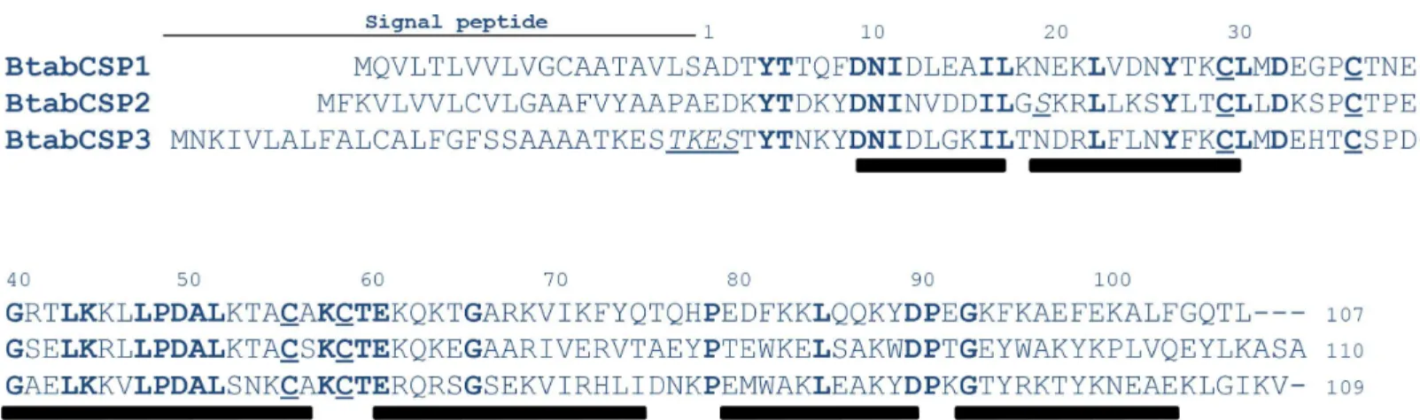
Analyzing the EST database from the whiteflyB.tabaci[4], three different sequences signifi-cantly related to CSPs were identified and called BtabCSP1, BtabCSP2 and BtabCSP3, respec-tively (Table 1). BtabCSP1, BtabCSP2 and BtabCSP3 are proteins of 107–110 amino acids with an isoelectric point of 6.61–8.84. Totally, thirty-four amino acids are strictly conserved between the three CSPs including the four cysteines characteristic of the CSP family. The middle part of the BtabCSP proteins (amino acid motif 40–70) is particularly well conserved. BtabCSP1, BtabCSP2 and BtabCSP3 rather differ in the N and C-terminal sequences (Fig 1).
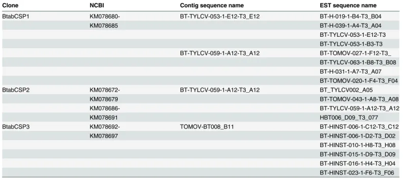
Table 1. EST database ofB.tabaciCSP clones.
Clone NCBI Contig sequence name EST sequence name
BtabCSP1 KM078680- BT-TYLCV-053-1-E12-T3_E12 BT-H-019-1-B4-T3_B04
KM078685 BT-H-039-1-A4-T3_A04
BT-TYLCV-053-1-E12-T3 BT-TYLCV-053-1-B3-T3 BT-TYLCV-059-1-A12-T3_A12 BT-TOMOV-027-1-F12-T3_
BT-TYLCV-063-1-B8-T3_B08 BT-H-031-1-A7-T3_A07 BT-TOMOV-020-1-F4-T3_F04
BtabCSP2 KM078672- BT-TYLCV-059-1-A12-T3_A12 BT_TYLCV002_A05
KM078679 BT-TOMOV-043-1-A8-T3_A08
KM078686- BT-TYLCV-059-1-A12-T3_A12
KM078691 HBT006_D09_T3_077
BtabCSP3 KM078692- TOMOV-BT008_B11 BT-HINST-006-1-C12-T3_C12
KM078697 BT-HINST-006-1-D2-T3_D02
BT-HINST-010-1-H8-T3_H08 BT-HINST-015-1-D9-T3_D09 BT-HINST-016-1-H4-T3_H04 BT-HINST-023-1-F6-T3_F06
doi:10.1371/journal.pone.0154706.t001
The hydropathy plots of the three proteins are also very similar, showing high levels of hydrophobicity. However, the relative flexibility plots are rather different. Two flexible domains are predicted to occur in the protein BtabCSP1. The flexible domain in BtabCSP2 and BtabCSP3 is predicted to span over different regions (S1A Fig). The protein structure homol-ogy models built using MbraCSPA6 as template are very similar, except for C-terminalα-helix (S1B Fig). This was seen as a first indication that BtabCSP1 have different functions than CSP2 and CSP3.
We previously identified numerous substitution sites on the RNA encoding CSP1 in the B and Q biotypes ofB.tabaci[37]. In this study, we found a high degree of RNA variance for CSP2, but not for CSP3 (S2 Fig). Some of these substitution sites in CSP2-RNA sequences (KM078671-KM078679) led to crucial changes in amino acid sequences, particularly in the motif between N-terminalα1 andα2-helices (Figs1andS1andS1andS2Tables). However, CSP2 clones mostly differed by the C-terminal tail. In clones from B and Q (I74 and I80), an early stop codon led to the truncation of the three last residues ASA (S2 FigandS1andS2
Tables). Comparatively, only a few nucleotide replacements occurred on the RNA encoding CSP3 (KM078694-KM078697) although we cloned a mutation (I62-BtabCSP3) in Q that repeated three times the motif TKES in the N-terminal region (S2 Fig and S2 Table).
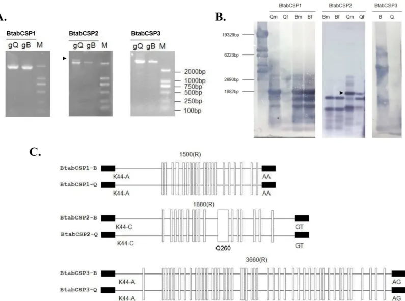
We thus aimed to identify the three CSP gene structures in selective PCR experiments. Using specific CSP1, CSP2 and CSP3 primers generated genomic DNA (gDNA) fragments of about 1800, 2000 and 5000 bps, respectively. Two gDNA fragments were amplified in Q for CSP2 gene (Fig 2A). Two 2 000 bps-gDNA bands for CSP2 were also detected in Q samples by Southern blot and DIG-labeling of PCR products. Gene diversity was observed also forCSP1. CSP1 probe hybridized to three main gDNA bands (Fig 2B). Using gDNA from ten individuals amplified repeatedly two CSP1 PCR bands for Q, suggesting that the number of CSP genes is different between Q and B biotypes of the sweet potato whiteflyB.tabaci(S3 Fig). In contrast, PCR and blot using CSP3 probe identified only one major gDNA band of about 5 Kbs (Fig 2A and 2B).
Despite differences in the number of gene copies, sequencing gDNA encoding for CSP1, CSP2 and CSP3 in B and Q showed that the exon-intron structures were strictly conserved in the two biotypes. CSP1, CSP2 and CSP3 are single-intron genes of about 1500, 1880 and 3360 bps length, respectively (KM078680-KM078693). Intron differs in size, but it is always located at the same position (after the first base of codon for amino acid residue at position 45).
Fig 1. Identification ofB.tabaciCSP1, CSP2 and CSP3.Conserved amino acids are shown in bold. The four Cys residues characteristic of CSPs are underlined. The residues or motifs in italic indicate specific mutations sites leading to amino acid replacement. Ala and Glu residues at position 1 are based on N-terminal sequencing of cockroach and moth CSPs [11,13]. Bars in black indicate the position of putative alpha-helical domains (http://npsa-pbil.ibcp.fr).
Interestingly, we found widespread biotype B and Q sequence differences not only in gDNA encoding CSP1 and CSP2, but also in gDNA encoding CSP3 in the aleyrodid sweet potato whiteflyB.tabaci(Fig 2C). The comparison between B and Q revealed a high number of sites
where the gDNA sequence encoding for CSP1 from B differs from the corresponding gDNA sequence of the Q-biotype. The same observation was made comparing between B and Q the gDNA sequences encoding for CSP2 and CSP3, respectively (S4 Fig). Q even showed a very peculiar sequence (Q260) in the mid-region of theCSP2gene [38], demonstrating the occur-rence of biotype-specific landmarks inB.tabaci CSPs.
Fig 2. Identification ofB.tabaciCSP1, CSP2 and CSP3 gene structures in B and Q biotypes.(A) Agarose gel electrophoretic analysis of genomic DNA PCR products from B and Q biotype using specific CSP1, CSP2 and CSP3 primers. (B) Southern blot analysis of genomic DNA PCR products isolated from
B.tabaciin B and Q-biotype. Qm: Q males, Qf: Q females, Bm: B males; Bf: B females. A mixed pool of males and females were used for blot hybridized with
CSP3 probe. Size markers (bps) are from HindIII-digested lambda DNA. The arrow tip shows CSP2-DNA band specific to Q. (C) Comparative analysis of gene structures encoding CSP1, CSP2 and CSP3 in B and Q biotypes ofB.tabaci. Exon/intron boundaries (the first base of codon for K/R at position 45) were determined by comparing complementary and genomic DNA encoding CSP1, CSP2 and CSP3, respectively (KM078671-KM78697). Exons are represented by small black boxes, introns by straight black lines. The number above indicates the intron size. Mutation sites between B and Q for each CSP gene are shown by white vertical rectangles. Q260 indicates the position of intron motif specific to Q [38].
doi:10.1371/journal.pone.0154706.g002
BtabCSP1
,
BtabCSP2
and
BtabCSP3
represent three different
orthology groups of
CSPs
Analyzing amino acid sequences, a particularly high degree of similarity was found not only between whitefly and aphid CSPs, but also between CSPs from Hemiptera and various counterparts mainly from coleopteran, dipteran, lepidopteran and phthirapteran species (S5 Fig).BemisiaCSP1 was mainly related to aphid, fly and mosquito CSPs (about 50% identity, 70% similarity).Bemisia
CSP2 found orthologs not only in aphids and flies, but also in beetles, mealy bugs and moths. Bemi-siaCSP3 was similar (about 70%) to CSPs fromAedes,Culex,Ceratitis,Delia,Drosophila,Musca,
Stomoxysand many bark beetle, flour beetle,Pediculusand true bug species (S5 Fig). A phylogenetic analysis (RAxMLGUI) based on amino acid sequences of BtabCSP1, BtabCSP2, BtabCSP3 and CSPs from other insect species confirmed a clear relationship between BtabCSPs and CSPs from hemipteran and dipteran species (Figs3andS5). Using crustacean CSPs as outgroups, all of the CSPs used for the analysis fell on a common“insect” branch with high bootstrap values (94%). Interestingly, we noted that BtabCSP1 fell outside the main group of CSPs similarly to developmentalAmelGB10389gene from honeybees, very distantly related to CSPs described in the olfactory system (Fig 3). In addition, on the phyloge-netic amino acid tree, BtabCSP2 clearly grouped with effector protein Mp10 and developmen-tal genes such asAgamCSP5andMvicOSD, suggesting that these four CSPs play a similar role eventually as effector proteins in both developmental and immunological systems (Fig 3). In contrast to BtabCSP1 and BtabCSP2, BtabCSP3 fell much closer to a large group including in particular locust olfactory CSPs/OSDs, the bee pheromone olfactory ASP3c gene (CAJ01448) and two known developmentally regulated CSP genes, p10 and HvirCSP1 (Fig 3). However, BtabCSP3 did not make a clear orthology group with any of the otherCSPs. It even made a sin-gleton despite close relatedness to TcitOSD (CAJ01484), a CSP from the brown citrus aphid,
Toxoptera citricida(Fig 3). This strongly suggested a mechanism very specific to whiteflies for this peculiar type of CSP.
BtabCSP1
,
BtabCSP2
and
BtabCSP3
differentially express across
various developmental stages of biotypes B and Q
We then compared the two biotypes B and Q ofB.tabacion expression of the three CSP genes identified (BtabCSP1,BtabCSP2andBtabCSP3).
Under control conditions, CSP1 and CSP2 showed preferential expression in biotype Q, while CSP3 gene expression was preferentially expressed in the biotype B. Slightly higher expression in males was detected for CSP1 and CSP2. No significant differences in CSP3 expression were found comparing males and females (S6 Fig).
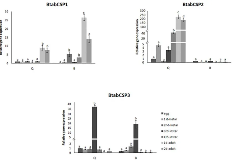
Detailed comparison of the gene expression profiles at seven developmental stages of the whitefly showed that CSP1 gene expression significantly increased by a factor of 10–30 at the adult stage in the two biotypes (Fig 4). CSP2 gene expression gradually increased starting at the third-instar stage to reach a 300-fold increase at the adult stage specifically in the biotype Q. Such an increase in CSP2 gene expression across the different developmental stages ofB.tabaci
was not observed in B biotype (Fig 4). In the two biotypes B and Q, CSP3 gene expression remarkably increased (by a factor of 20–40) in third-instar nymphs (Fig 4).
BtabCSP1
,
BtabCSP2
and
BtabCSP3
differentially express in response
to age and thiamethoxam insecticide exposure
expressed in nymphs. This observation suggested different functions forBemisiaCSPs in a good agreement with such diversity in RNA variance, gene copy number and predicted structure.
We focused about a function in relation with insecticides and xenobiotics on the basis of the results from Sabatier et al. [35], Xuan et al. [36] and Liu et al. [37,38].
Fig 3. Phylogenetic analysis of amino acid sequences fromB.tabaciCSP1, CSP2, CSP3 and orthologs in other insect species (RAxMLGUI).Aech:Acromyrmex echinatior, Apis:Acyrthosiphon pisum, Alin:Adelphocoris lineolatus,Aaeg:Aedes aegypti, Adar:
Anopheles darlingi, Agam:Anophales gambiae, Amel:Apis mellifera, Aluc:Apolygus lucorum, Blun:Biphyllus lunatus, Bter:Bombus
terrestris, Bimp:Bombus impatiens, Bmor:Bombyx mori, Cflo:Camponotus floridanus, Cson:Culicoides sonorensis, Dper/Dvir/Dyak:
Drosophila persimilis/virilis/yakuba, Dpon:Dendroctonus ponderosae, Gmor:Glossina morsitans morsitans, Hsal:Harpegnatus saltator,
Harm:Heliothis armigera, Lmig:Locusta migratoria, Mrot:Megachile rotundata, Mvic:Megoura viciae, Mdir:Metopolophium dirhodum, Mper:
Myzus persicae, Npub:Nylanderia pubens, Nlug:Nilaparvata lugens, Nrib:Nasonovia ribis nigri, Phum:Pediculus humanus corporis, Sgre:
Schistocerca gregaria, Scal:Stomoxys calcitrans, Tpit:Thaumetopoea pityocampa, Tcit:Toxoptera citricida, Tcas:Tribolium castaneum.
Sequences are all published in NCBI.B.tabaciCSP1, CSP2 and CSP3 sequences are from JQ678989-ADG56568, KM078671- KM078679 and KM078694- KM078697, respectively. CSP sequences fromDaphnia pulex(DpulCSP1 and DpulCSP2; ABH88167, ABH88166) are used as an outgroup. The triangles indicate the CSP proteins that have been functionally characterized. Black triangles: developmental genes, grey triangles: effector genes, white triangles: olfactory genes.
doi:10.1371/journal.pone.0154706.g003
Fig 4. Differential expression ofB.tabaciCSP1, CSP2 and CSP3 during development in biotypes B and Q.Relative gene expression levels for CSP1, CSP2 and CSP3 in B and Q biotypeB.tabacieggs, 1st, 2nd, 3rdand 4thinstar nymphs and 1 to 2-days-old mixed adults (reference gene =β-actin). The
relative expression levels observed in eggs were used for calibration (egg calibration value = 1). Data are means±standard deviation (n = 9). Different letters indicate significant differences atα= 0.05 by one-way Anova. The symbol“═”on the Y-axis indicates very high expression of gene.
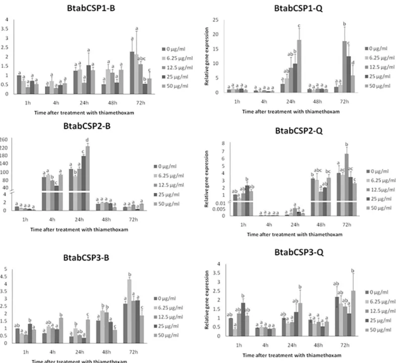
Comparison of the gene expression profiles at five exposure times (1, 4, 24, 48 and 72 h) for five doses (0, 6.25, 12.5, 25 and 50μg/ml) of insecticide thiamethoxam showed that CSP1 gene expression significantly increased in a time- and dose-dependent manner specifically in the biotype Q (Fig 5). After 1 and 4 h of insecticide treatment, no changes in CSP1 gene expression were noted for all five doses tested in the two biotypes. However, we observed in Q gradual Fig 5. Modulation ofB.tabaciCSP1, CSP2 and CSP3 expression over different time-dose treatments of thiamethoxam in biotypes B and Q.
Relative gene expression levels for CSP1, CSP2 and CSP3 in B and Q biotypeB.tabacimixed adult individuals exposed to 0, 6.25, 12.5, 25 and 50μg/ml dose of thiamethoxam (reference gene =β-actin). CSP1, CSP2 and CSP3 gene expression were measured in B and Q biotypes after 1, 4, 24, 48 and 72 h exposure. The relative expression levels observed in control individuals (0μg/ml concentration of thiamethoxam) after 1 h were used for calibration
(value = 1). Data are means±standard deviation (n = 9). Different letters indicate significant differences atα= 0.05 by one-way Anova. The symbol“═”on the Y-axis indicates very high expression of gene.
doi:10.1371/journal.pone.0154706.g005
increase in CSP1 gene expression using gradual concentrations of thiamethoxam after 24 h (Fig 5). No effects were seen in Q-whiteflies after 48 h, but stimulatory effects were observed after 72 h exposure to thiamethoxam dosed at 12.5 and 50μg/ml, triggering an increase by a factor of about 10–25 in CSP1 gene expression (Fig 5). We did not observe such a time/dose effect of thiamethoxam on CSP1 expression in B (Fig 5). We observed no time/dose effect of thiamethoxam on CSP2 and CSP3 expression, neither in B nor Q (Fig 5).
However, we observed in the two biotypes an effect of age on CSP2 expression (100–200 folds increase), while no age-related effects were seen for CSP1 and CSP3 (Fig 5). Age-related effects on CSP2 were clearly opposite in B and Q; CSP2 up-regulated at 4 and 24 h in B, while it clearly up-regulated at 1, 48 and 72 h in Q (Fig 5), clearly illustrating biotype-specific changes with age in CSP2 expression in the sweet potato whiteflyB.tabaci.
In order to understand the reason behind the modulation ofCSP1,CSP2andCSP3, we sub-sequently analyzed gene expression for some possibly related genes such ascytochrome P450 oxidase CYP6CM1(GQ214539),CYP6CX1(GQ292715),ecdysone receptor EcR(EF174329),
trehalase(JX024261) andjuvenile hormone binding protein JHBP(seeS7 Fig). We identifiedB.
tabacijuvenile hormone binding protein (BtabJHBP) through the analysis of the whitefly EST database [4]. The EST clone BT-TYLCV-014-1-G6-T3_G06 showed between 30 and 60% iden-tity with members of the JHBP superfamily from various insect species including mainly
Acyrthosiphon pisum(XP_001949424) andReticulitermes flavipes(ADM18966) in a blastx analysis (not shown). We saw thatBtabCSP3andBtabJHBPhad coordinate expression in nymphs (see Figs4andS7). In addition, we saw no correlation betweenBtabCSPexpression and expression oftrehalaseandEcR, but we saw increasedCYP6CM1expression in Q and reducedCYP6CX1expression in B in response to sublethal dose of insecticide thiamethoxam molecule as found forCSP1(see Figs5andS7) [37].
BtabCSP1, BtabCSP2 and BtabCSP3 proteins differentially bind linoleic
acid and cinnamaldehyde
To complete our systemic investigation of CSPs of whitefly, we did a full comprehensive study analyzing not just CSP1, but also functions of CSP2 and CSP3 (Figs6and7andS8–S11, Tables
2and3andS3 Table).
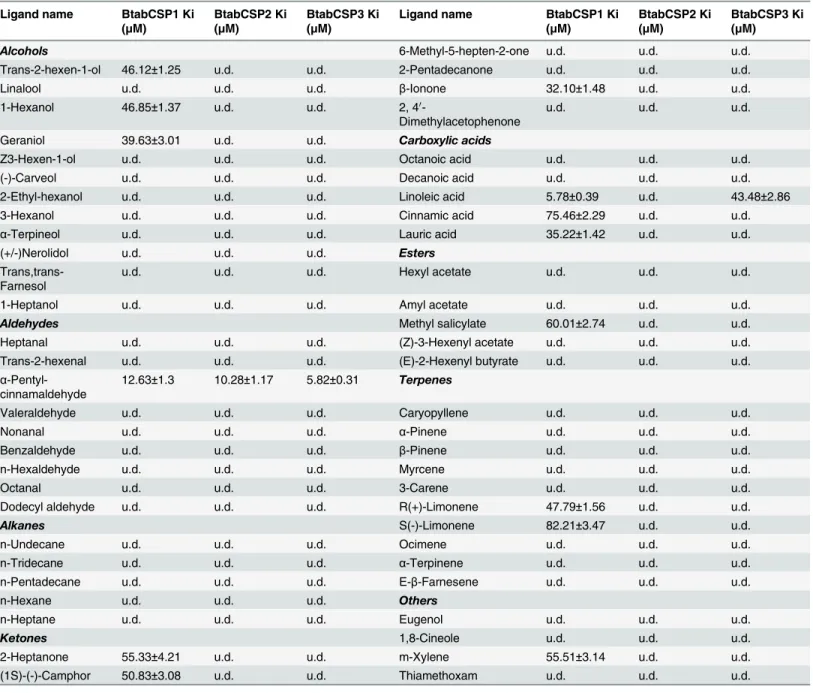
We first used pure recombinant CSP protein samples, a selected repertoire of ligands and binding assay fluorescence spectroscopy (1-NPN) to analyze the functional properties of BtabCSP1, for which we found increased expression in Q following exposure to thiamethoxam (seeFig 5). In a preliminary binding assay, BtabCSP1 did not display certain binding ability with 25% pure thiamethoxam. Binding of thiamethoxam to CSP1 was found to be rather weak (IC50 = 326.26μM, Ki = 285.43μM), so there was not obvious relevance between insecticide binding to CSP protein and thiamethoxam induction (S9 FigandTable 2).
To CSP1 protein, chemicals such asα-pentyl-cinnamaldehyde and S(-)-limonene showed a binding constant of 12.63±1.3 and 82.21±3.47μM, respectively (Figs6andS9andTable 2). The IC50 values obtained with other chemicals such as trans-2-hexen-1-ol, linalool, 1-hexanol, geraniol, Z3-hexen-1-ol,α-terpineol, nerolidol, n-hexaldehyde, n-heptane, 2-heptanone, (1S)-(-)-camphor, 6-methyl-5-hepten-2-one,β-ionone, octanoic acid, decanoic acid, cinnamic acid, lauric acid, amyl acetate, methyl salicylate, caryopyllene, myrcene, R(+)-limonene, ocimene, α-terpinene and m-xylene were comprised between 14.59±1.75 and 95.08±3.72μM (Figs6and
S9andTable 2). No binding or IC50>100μM was observed for other ligands including plant
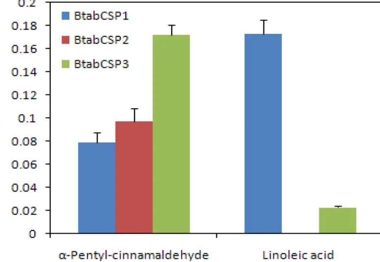
We then set out to know the binding ability of BtabCSP2 and BtabCSP3 to most of all chem-icals, but more precisely with linoleic acid andα-pentyl-cinnamaldehyde, respectively (Figs6
andS8–S10andTable 2). Importantly, no or low binding affinity of CSP2 and CSP3 was found for linoleic acid chemical (LA-CSP2: IC50>100μM; LA-CSP3: IC50 = 48.45±3.42, Ki = 43.48
±2.86μM; Figs6andS10andTable 2). Even more importantly, both CSP2 and CSP3 proteins displayed significantly much higher binding affinity for the common direct contact toxic chem-ical of plant oil,α-pentyl-cinnamaldehyde (IC50 = 6.50–11.13±0.37–1.24, Ki = 5.82–10.28 ±0.31–1.17μM; Figs6andS10,Table 2andS3 Table), showing that CSP1 and CSP2 (and CSP3) fromB.tabaciwhiteflies have two completely different functions, although both related to detoxification and insect defense.
Finally, we studied the molecular structure of bound CSP1, CSP2 and CSP3 fromB.tabaci
using docking molecular techniques (Figs7andS11andTable 3). Molecular modelling using multiple alignments that contained all seven CSP templates (1K19, 1KX8, 1KX9, 1N8U, 1N8V, 2JNT, 2GVS) failed to yield molecular models with the large cavity as observed in templates 1N8U and 1N8V. Molecular docking of linoleic acid andα-pentyl-cinnamaldehyde on these models gave binding modes where the ligands bound exclusively on the surface of the proteins Fig 6. Fluorescence binding assay for the binding affinity ofB.tabaciCSP1, CSP2 and CSP3.Binding constants (Ki as presented as 1/Ki) of
BtabCSP1, BtabCSP2 and BtabCSP3 to linoleic acid (LA) andα-pentyl-cinnamaldehyde. The Ki values to various chemical ligands are given inTable 2. The highest binding constant value of BtabCSP1 (presented as 1/Ki) is found to LA (about 5.8μM). Ki value of BtabCSP2 to LA is null, while Ki of BtabCSP3 to LA is only of about 48.5μM. Higher binding constant values of BtabCSP2 and BtabCSP3 are toα-pentyl-cinnamaldehyde (5.8–10.3μM).
doi:10.1371/journal.pone.0154706.g006
with poor energies. Molecular modelling of BtabCSP1, BtabCSP2 and BtabCSP3 with the holo forms of the CSP as templates yielded models that displayed the presence of a large cavity that Fig 7. Docking analysis for the functional structure ofB.tabaciCSP1, CSP2 and CSP3.Docking results showing (A) binding of linoleic acid to BtabCSP1, (B) binding ofα-pentyl-cinnamaldehyde to BtabCSP2 and (C) binding ofα
-pentyl-cinnamaldehyde to BtabCSP3. Shown on the left are the cartoon representations of the bound ligands to the CSP models and on the right the Ligplot charts showing the residues that interact with the ligands. Amino acid sequences of mature peptides (after removal of peptide signal) andα-helical profiles for each CSP are provided below each cartoon representation. Residue numberings in these profiles are those that are featured in the LigPlot charts.
doi:10.1371/journal.pone.0154706.g007
Table 2. Binding affinities of ligand chemicals to BtabCSP1, BtabCSP2 and BtabCSP3.
Ligand name BtabCSP1 Ki
(μM)
BtabCSP2 Ki (μM)
BtabCSP3 Ki (μM)
Ligand name BtabCSP1 Ki
(μM)
BtabCSP2 Ki (μM)
BtabCSP3 Ki (μM)
Alcohols 6-Methyl-5-hepten-2-one u.d. u.d. u.d.
Trans-2-hexen-1-ol 46.12±1.25 u.d. u.d. 2-Pentadecanone u.d. u.d. u.d.
Linalool u.d. u.d. u.d. β-Ionone 32.10±1.48 u.d. u.d.
1-Hexanol 46.85±1.37 u.d. u.d. 2, 40
-Dimethylacetophenone
u.d. u.d. u.d.
Geraniol 39.63±3.01 u.d. u.d. Carboxylic acids
Z3-Hexen-1-ol u.d. u.d. u.d. Octanoic acid u.d. u.d. u.d.
(-)-Carveol u.d. u.d. u.d. Decanoic acid u.d. u.d. u.d.
2-Ethyl-hexanol u.d. u.d. u.d. Linoleic acid 5.78±0.39 u.d. 43.48±2.86
3-Hexanol u.d. u.d. u.d. Cinnamic acid 75.46±2.29 u.d. u.d.
α-Terpineol u.d. u.d. u.d. Lauric acid 35.22±1.42 u.d. u.d.
(+/-)Nerolidol u.d. u.d. u.d. Esters
Trans,trans-Farnesol
u.d. u.d. u.d. Hexyl acetate u.d. u.d. u.d.
1-Heptanol u.d. u.d. u.d. Amyl acetate u.d. u.d. u.d.
Aldehydes Methyl salicylate 60.01±2.74 u.d. u.d.
Heptanal u.d. u.d. u.d. (Z)-3-Hexenyl acetate u.d. u.d. u.d.
Trans-2-hexenal u.d. u.d. u.d. (E)-2-Hexenyl butyrate u.d. u.d. u.d.
α -Pentyl-cinnamaldehyde
12.63±1.3 10.28±1.17 5.82±0.31 Terpenes
Valeraldehyde u.d. u.d. u.d. Caryopyllene u.d. u.d. u.d.
Nonanal u.d. u.d. u.d. α-Pinene u.d. u.d. u.d.
Benzaldehyde u.d. u.d. u.d. β-Pinene u.d. u.d. u.d.
n-Hexaldehyde u.d. u.d. u.d. Myrcene u.d. u.d. u.d.
Octanal u.d. u.d. u.d. 3-Carene u.d. u.d. u.d.
Dodecyl aldehyde u.d. u.d. u.d. R(+)-Limonene 47.79±1.56 u.d. u.d.
Alkanes S(-)-Limonene 82.21±3.47 u.d. u.d.
n-Undecane u.d. u.d. u.d. Ocimene u.d. u.d. u.d.
n-Tridecane u.d. u.d. u.d. α-Terpinene u.d. u.d. u.d.
n-Pentadecane u.d. u.d. u.d. E-β-Farnesene u.d. u.d. u.d.
n-Hexane u.d. u.d. u.d. Others
n-Heptane u.d. u.d. u.d. Eugenol u.d. u.d. u.d.
Ketones 1,8-Cineole u.d. u.d. u.d.
2-Heptanone 55.33±4.21 u.d. u.d. m-Xylene 55.51±3.14 u.d. u.d.
(1S)-(-)-Camphor 50.83±3.08 u.d. u.d. Thiamethoxam u.d. u.d. u.d.
u.d.: undetermined (binding constant was not calculated; IC50>100μM)
Data are averages of three replicates (±SD).
doi:10.1371/journal.pone.0154706.t002
could accommodate linoleic acid andα-pentyl-cinnamaldehyde with much improved binding energies (Figs7andS11).
The docking showed that linoleic acid could be accommodated into the large cavity of BtabCSP1 after the ligand adopted a U shape (Fig 7). Indeed the aliphatic chain is too long to be accommodated in the cavity if it were to remain straight. The ligand establishes mainly hydrophobic contacts with cavity residues. Noteworthy, a hydrogen bond between the carbox-ylic group of linoleic acid and the C = O of the backbone of Ala50 was observed in this pre-ferred binding mode (Fig 7). The predicted Kdvalue was 14.9±8.6μM (Table 3). We observed several possible orientations of the same U-shaped linoleic acid in this large cavity with binding energy levels that were grossly equivalent. The same kind of binding modes were observed for linoleic acid in BtabCSP2 and BtabCSP3, but the predicted Kdvalues showed between three fold to six fold weaker binding affinities (Table 3).
Docking results forα-pentyl-cinnamaldehyde showed that all three CSPs could bind the ligand in the large cavity. The interactions are mainly hydrophobic, but clearly involved differ-ent sets of residues as shown for BtabCSP2 and BtabCSP3 inFig 7. BtabCSP3 was predicted to have a higher affinity to the ligand with predicted Kd of 6.45±5.7μM, which is about two fold higher than for BtabCSP2 and BtabCSP1 (Table 3).
Discussion
We identified and analyzed the gene structures of three chemosensory proteins from the white-flyBemisia tabaci(BtabCSPs). Four to twenty-oneCSPsexist in the various insect species investigated so far [39–41]. Similarly, our PCR and Southern blot analysis of genomic DNA in the sweet potato whiteflyB.tabaciis indicative of a high diversity inCSPs(undiscovered homo-logs, recently duplicated genes and/or pseudogenes). Therefore,BtabCSP1,BtabCSP2and
BtabCSP3most probably do not represent the full complement of the CSP gene family in Bemi-sia. They represent, however, homologous genes sharing 37–43% identity with each other and probably the most abundant CSP-encoding RNAs expressed in the sequence tag database from adults and nymphs inB.tabaci(see Figs1and2andS1–S4andTable 1) [4,37,38,42].
Interestingly, the analysis of genomic DNA encoding CSP1, CSP2 and CSP3 allows us to clearly distinguish between the biotypes B and Q in theBemisiacomplex (seeFig 2). The Q-biotype is known to have a higher adaptive capacity and in particular a stronger insecticide resistance capacity than the B-biotype [43–44]. Correlatively, we find thatBtabCSP1,BtabCSP2
andBtabCSP3are all three single-intron genes similarly to most of allCSPs, but clearly display biotype-specific hallmarks, particularly BtabCSP2 (see Figs2andS2andS3) [38]. This suggests perhaps a link between the genetic repertoire of CSPs and the insecticide resistance capacities of the insect. Q-whiteflies retain a very specific genetic sequence (Q260) originating from bac-teria that may be crucial for this process [38]. Furthermore, Q apparently contains more CSP genes than B (see Figs2andS3).
Also interestingly, we show a lot of biotype-specific variants of CSP1 and CSP2 transcripts (seeS2 FigandS1andS2Tables) [37]. This probably represents variation at the RNA level. A recent study from Xuan et al. in the silkworm mothB.morihave clearly demonstrated that var-iants of CSP transcripts do not represent variation at the genomic level, but rather tissue-Table 3. Molecular docking results for linoleic acid andα-pentyl-cinnamaldehyde on 3D models of BtabCSP1, BtabCSP2 and BtabCSP3.
Ligand BtabCSP1(μM) BtabCSP2 (μM) BtabCSP3 (μM)
Linoleic acid 14.9±8.6 82±9.2 37.17±4.41
α-pentyl-cinnamaldehyde 12.36±4.73 10.68±6.7 6.45±5.7
specific RNA mutations resulting in the synthesis of multiple abundant CSP protein isoforms mainly differing by the terminal tails [45–46]. In addition, in the sweet potato whiteflyB.
tabaci, we show a high number of base substitution sites on the genomic DNA comparing CSP1, CSP2 and CSP3 between the two biotypes. We also show a different PCR profile analyz-ing CSP-encodanalyz-ing gDNA in Q and B individuals (seeS3 Fig). Finally, we show that intron has been inserted or deleted in specific CSP genes during the evolutionary process of the two spe-cificBemisiabiotypes (seeS4 Fig). Confirming the results from Liu et al. [38], all these observa-tions strongly indicate that B and QB.tabacibiotypes are not truly unique species, subspecies or twin species but rather represent two very distinct species, each governed by very specific physiological systems [47–48].
Curiously, we find a lower degree of RNA and gDNA mutations for whitefly CSP3 com-pared to CSP1 and CSP2 (seeS2–S4Figs andS1andS2Tables). This is in agreement with the hypothesis that the degree of mutation depends on the function the protein has in a specific tis-sue of the insect [49]. A not striking but significant amino acid sequence similarity is found between these whitefly CSPs and some other CSPs from many various insect species (see Figs3andS5). A phylogenetic analysis based on amino acid sequences and implemented in RAxMLGUI shows insertion of BtabCSP1, BtabCSP2 and BtabCSP3 in a very large group of CSPs referring to many various functions. A BtabCSP1 ortholog in Hymenoptera (ASP3c, CAJ01448) found to be abundantly expressed in the antennae of adult honeybees and wasps is also found in high amounts in the hemolymph of drones [37,50–52]. Another ortholog in bees (AmelGB10389) is involved in tegument development [23]. Orthologs in locusts are not only up-regulated during phase transition, but also highly expressed in antennae and legs at the adult stage [12,53]. Other orthologs in flies are up-regulated in olfactory organs in adult alate swarmers [54–55], similarly to some orthologs from the alfalfa plant bugAdelphocoris lineola-tus(AlinCSP2) [56]. Still, the same such CSP orthologs (pherokines) are found in hemolymph of virus-infected flies [35], suggesting a very general function for all of these CSPs. However, in our exhaustive study of two whiteflyB.tabacibiotypes, we show at first glance that CSP1 and CSP2 preferentially express in Q, while CSP3 preferentially expresses in B (seeS6 Fig), suggest-ing that at least these three CSPs inBemisiamight possibly be involved in some very different biotype-specific functions.
Interestingly, in our analysis of CSP functions inB.tabaci, we show that CSP1 and CSP2 are specific to the adult stage, suggesting a function in reproduction and/or plant odor detection at least for these two CSPs. In contrast, the extremely high levels of CSP3 expression at the third instar stage rather suggests a function in the preparation into adult stages, in particular in the development of sensory organs such as the antennae, head, eyes and various legs (seeFig 4). Many CSPs in other insect species have been shown to be associated with developing sensory organs [17,23–25]. In addition, specifically inB.tabaci, a CSP from nymphs such as CSP3 could also play a function in wax lipid secretion and/or insect defense against parasitoids [57–
58]. A function of CSPs in insect defense is strongly suggested by the analysis of genes abun-dantly expressed in the whiteflyB.tabaciEST database from infected tissues [4]. No CSP-ESTs are found in the cDNA library of salivary glands in agreement with transcriptomic studies [59]. CSP3-ESTs are found in cDNAs from the instar library, but CSP1 and CSP2-ESTs are specifi-cally found in the libraries of mature adults fed on tomato plants infected by the geminiviruses TYLCV (tomato yellow leaf curl virus) and TOMOV (tomato mottle virus) (seeTable 1).
Hence, we set out to correlate the function ofBemisiaCSPs to insect defense and in particu-lar to whitefly resistance to neonicotinoid insecticide thiamethoxam. We measured transcript levels of CSP1, CSP2 and CSP3 genes with the treatments of five time points and five doses as well as the expression of genes involved in insecticide resistance [60]. The lifetime of adult whiteflies is of one month in the laboratory conditions. We have only used a population of
young adults for our experiments, rejecting the possibility of biological mortal ratio of insects. There are probably variations due to various inter-individual physiological conditions, so we calibrated quantitative measurements of gene expression to a negative control (0μg/ml) for each time point and for each CSP. The results confirmed previous data from Liu et al. [37].
There might be that CSP transcripts are regulated by many internal and external factors such as age, biotype, temperature and humidity, background odor, plant chemicals, light, circa-dian rhythm and season rhythm, etc., but for two time points, we show a clear dose effect of thiamethoxam on CSP1, particularly in biotype Q. CSP1 expression up-regulated 10–20 fold following exposure to sublethal doses of thiamethoxam at 24 and 72 h, but not at 48 h (seeFig 4). Perhaps it takes 48 h after involving CSP1 for full-system recovery to occur; perhaps it takes different durations of exposition to insecticide to recruit multiple resistance genes in a variety of defense mechanisms [61–62]. Time and dose dependent induction of detoxifying genes is usually observed at 24 h [63]. Hundreds genes are differentially expressed at 48 and 72 h post-infectedBemisiain response to fungal infection [64]. Similarly, we show only late responses to exposition of thiamethoxam molecule for CSP1 (seeFig 4). This delay could be due to the time needed for the thiamethoxam molecule to penetrate through the cuticle of adult whiteflies. However, it could also simply be that CSP1 is a late-response gene enrolled during prolonged exposure to xenobiotics. Sublethal doses of insecticide (6.25–50μg/ml) are known to have vari-ously delayed effects on key biological traits of the insect such as feeding behavior, honeydew excretion and fecundity [65–66]. Therefore, different CSPs could well be involved at different exposure time to protect various tissue-specific physiological pathways.
Nevertheless, in contrast to Q-CSP1, we note only little if any effects of thiamethoxam mole-cule on CSP2 and CSP3 fromBemisia(seeFig 4), suggesting that these two genes are not typi-cally involved in the insect neonicotinoid response. BtabCSP2 and BtabCSP3 could be involved in the immunological response to another class of insecticides and/or simply have other func-tions than immunological defense.
It is noticeable in the sweetpotato whiteflyB.tabacithat relative expression levels ofCSP1
andCSP3were stable over time in series of negative controls (0μg/ml). Intriguingly, however, we found thatCSP2was turned on in biotype B when it was turned off in biotype Q (seeFig 4). The phenomenon behind such an odd variation in CSP2 gene expression was rather mysteri-ous. The vials were capped with an absorber cotton stopper, so there could not be an air con-taminant or any signal released by B to inhibit Q. It could be instead a hormone or some toxic plant chemical that has differential effects on B and Q [67–68]. This was a very interesting point to characterize the CSP2 function by using same methods with CSP1 and CSP3.
In our study inBemisiawhiteflies we see some coordinate expression between CSPs and other gene families such as CYPs and JHBPs over development and/or insecticide exposure (see Figs4and5andS7). Coordinate expression ofCSP1andCYP6CM1inBemisiaQ is in agreement with coordinate expression ofCSPsandCYPsover insecticide exposure shown to be tissue-specific inBombyxmoths [36]. TwoCSPs,BmorCSP7andBmorCSP10, andJH esterase
had coordinate expression [36]. Taken together, the two studies suggest some relationships between CSP, CYP and JH related genes. The interactions among the CSP, CYP and JHBP genes probably need to be cautiously addressed by recombinant proteins interactionin vitro, RNAi, in situ hybridization and/or immunohistochemistry in moths, whiteflies and other insect species. It may be that some CSPs work with CYPs to mediate xenobiotics response, while some others play a key role in JH signaling pathways.
We have checked for the mode of action of BtabCSP1, BtabCSP2 and BtabCSP3 using pro-tein expression, purification and fluorescence spectroscopic binding assay (see Figs6andS8–
for volatile odorant molecules including terpenoids (see Figs6andS8andS9,Table 2andS3 Table), contrasting results obtained using various CSPs from mosquitoes and bugs [55,69]. Using a similar binding assay, we find that BtabCSP1 rather involves in the binding of long fatty acid chains such as C18-linoleic acid as other proteins in the odorant binding protein and Niemann-Pick type C2 protein families [70–71]. The three 3D structures and binding data available for CSPs are in agreement with binding to various hydrophobic ligands including fatty acid molecules of various sizes and shapes [72–76]. Linoleic acid (delta 9,12 double bonds) is an unsaturated long carbon chain fatty acid molecule present in most of all insects in all tissues for lipid metabolism and fatty acid biosynthesis [77–78]. In the pheromone gland tis-sue, it is a key derivative of the fatty acyl intermediates from biosynthetic pathways for specific sex pheromone chemicals [79]. In addition, it is known to be involved in immunological response. Genes involved in linoleic acid metabolism are up regulated upon insecticide expo-sure along with CYP genes [80–81]. One function for linoleic acid is to help produce lipids and phospholipid-derived hormones that are directly involved in anti-xenobiotic response [82]. The molecule even can retain by itself an anti-xenobiotic activity [83]. Fatty acids such as lino-leic acid molecule are therefore certainly crucial not only for various processes such as phero-mone biosynthesis, horphero-mone synthesis, neural sensory development and/or adult tissue growth, but also for immunological defense including general responses against insecticide or some other toxic xenobiotic chemicals.
Importantly, along our work onBemisiaCSP1 we show that two additional BtabCSPs do not bind fatty acid, but rather interact directly with specific toxic xenobiotic chemical. BtabCSP2 and BtabCSP3 proteins have a completely different functional task than CSP1. Using same binding assay method of CSP1, we clearly show that linoleic acid binds neither to CSP2 nor CSP3. CSP2 and CSP3 attach smaller, more volatile cyclic component structures such asα-pentyl-cinnamaldehyde with a much more higher affinity (see Figs6,S8–S10,
Table 2andS3 Table). Interestingly,α-pentyl-cinnamaldehyde present in large amounts in plant essential oils is known not only for its pleasant cinnamon fragrance, but also for its very high insecticide activity [84–86]. This strongly suggests that, while CSP1s might be involved in the transport of fatty acids necessary to counteract neonicotinoid infection, CSP2s and CSP3s might rather be involved in the transport and direct degradation of much more volatile contact insecticide chemicals such as cinnamaldehydes and structurally related compounds.
We report molecular data in support of speciation and Q/B biotype-specific physiological pathways in the sweet potato/silver leaf whiteflyB.tabaci. Our study shows that a particular CSP gene (BtabCSP1) expresses at the adult stage and responds to thiamethoxam intoxication specifically in the Q-biotype whitefly known to be highly resistant to neonicotinoid insecti-cides. Our study also shows that the BtabCSP1 gene encodes for a protein that binds particular long chain fatty acid, linoleic acid, among a large set of volatile and non-volatile ligands. The docking and 3D analysis of CSP1 with linoleic acid is supplied (see Figs7andS11andTable 3), illustrating a profile of sevenα-helices and detailed binding mechanism in the lipid solubiliza-tion process. BtabCSP1 bound to linoleic acid in a cavity formed byα3,α4 andα5 helices (see Figs7andS11). Residue numberings hereafter are as per the indications given in Figs7and
S11. In the front of Cys55, linoleic acid molecule is sandwiched between Leu51-Ile 70-Phe81 and Ala66-Phe92-Lys95-Phe98 with the acid group anchored by Ala50 in the core of the pro-tein. The extremity of the hydrophobic lipid chain is in close contact with the side chains of Phe81 and Phe98 and backbone of Lys95 (seeFig 7). This structural data is in good agreement with general distribution and behavior of most of all CSPs [20,24–32,36]. ParticularlyCSP1
expression profiling strongly supports a general function in the transport of elongated lipids and fatty acids. InBemisia,CSP1ubiquitously expressed in adult tissues from antennae to wing tips [20,24–32,36]. We note thatBtabCSP1is particularly highly expressed in the wings (see
S12 Fig). In the wings, fatty acids such as linoleic acid and proteins such as CSP1 may be able to determine the wettability and the ability to fly. In most other tissues including antennae and legs, the CSP-C18 complex may be essential for immunological response under xenobiotic stress such as exposure to insecticide (see Figs5–7). Additionally, in most of all tissues, specific CSP-xenobiotic complexes are certainly crucial for specific responses of insect defense. While results from Sabatier et al. in theDrosophilafruit fly are in favor of an important role of CSPs in toxin degradation [35], our results in theBemisiawhitefly are in favor of a very important role of CSPs not only in the innate immune response, but also in adaptive (or acquired) immu-nity to a specific infectious agent such as a highly toxic host plant oil chemical molecule.
In this study, we also supply the docking and 3D analysis of CSP2 and CSP3 bound to cin-namaldehyde (seeFig 7andS11andTable 3). Similarly to CSP1, CSP2 and CSP3 are made of sevenα-helices. The ring of the insecticide sits in the middle of Ile14-Ile19-Tyr29-Leu49 and Leu60-Ile76-Glu94-Tyr97 in CSP2 and CSP3, respectively. The aldehyde group interacts with Ile14 in CSP2, while it rather connects with Leu56 and Trp90 in CSP3. The extremity of the short aliphatic chain curves in the two proteins, folding in a net of amino acid residues includ-ing Tyr11-Glu42-Glu45-Leu46 (CSP2) and Phe32-Phe36-Leu39-Trp90-Leu93 (CSP3). BtabCSP2 and BtabCSP3 bound to the aromatic aldehyde in the central cavity of the prism, the N- and C-terminalα-helical tails remaining free (see Figs7andS11) [74]. This can be perhaps helpful to envision new strategies to interfere with fatty acid metabolism, cell signaling and/or insecticide resistance in an insect pest. Most of all BmorCSP genes are up regulated in the gut and other various tissues of the female silkworm infected by insecticide [36]. Our study pres-ently shows that additional CSPs inBemisia(BtabCSP2 and BtabCSP3) are able to directly interact with a strong potent contact insecticide volatile molecule, strongly suggesting the importance of CSPs at many different levels of the insect defense mechanism. It seems that this mechanism has reached an extremely high level of sophistication in insects, particularly in sweet potato whiteflyB.tabacibiotype Q.
Materials and Methods
Ethics statement
B.tabaciB- and Q-biotypes were collected from eggplant fields in Shandong province (North/ East-Penninsula, China). Shandong Academy of Agricultural Sciences gave permission to con-duct field study on site (permit number SAAS 20110720). The field study did not involve endangered or protected species, but rather a very serious pest of agricultural crops worldwide.
Insect rearing and tissue collection
Molecular cloning of genomic DNA and cDNA
Antennal, abdominal, thoracic, leg, head and wing RNAs were extracted using Trizol for one-step Reverse Transcriptase PCR experiments. Whole body was used for RNA extraction in fur-ther molecular studies. Genomic DNA (gDNA) was extracted using DNeasy blood and tissue kit (Qiagen) for pools of thirty individuals in each biotype. Total RNA was extracted from the whole body of pooled individuals using the TrizolTMmethod (Life Technologies) and treated with DNase I (Fermentas). Complementary DNA (cDNA) was synthesized using RevertAidTM First Strand cDNA Synthesis Kit (Fermentas). Primers used to clone cDNA and gDNA of BtabCSP1, BtabCSP2 and BtabCSP3 were as follows:
BtabCSP1s: 5’-atgcaggttttgactttagtt-3’(MQVLTLV), BtabCSP1as: 5’-ttacagggtttgtccgaag-3’(FGQTL), BtabCSP2s: 5’-atgttcaaagttctcgtggt-3’(MFKVLVV), BtabCSP2as: 5’-ttaggcggatgccttgag-3’(LKASA), BtabCSP3s: 5’-atgaacaagattgtattggc-3 (MNKIVLA), BtabSCP3as: 5’-ctagactttgatacccaatt-3’(KLGIKV).
Additional primers tuned to specific intron fragments were used to sequence full-length gDNA clones:
CSP1-1f: 5’-GACTGTGGCAACCCTGAACGT-3’, CSP1-1r: 5’-CGGTGAAATTAGATGTAGCCA-3’, CSP2-1f: 5’-CAGAGGCCCAGACATTGCACC-3’, CSP2-1r: 5’-GTAAATATCGGTGTTCTGA-3’, CSP3-1f: 5’-TTTGGAAGAGTTTAGTGA-3’, CSP3-1r: 5’-CTTGGCGCACTTGTTGGA-3’, CSP3-2f: 5’-AGTCCACCCCAAAAACGATG-3’, CSP3-2r: 5’-CTGGTTCTGGAAAAAGGTTGGT-3’, CSP3-3f: 5’-AGGGTTGGTGCTTCTGAC-3’, CSP3-4f: 5’-TTTAGAAAGACAAAGGACAA-3’.
PCR was done in a TaKaRa PCR Thermal Cycler Dice, programmed as follows: 94°C for 5 min; 35 cycles of 94°C for 30 s, 52°C for 30 s and 72°C for 1 min; final extension at 72°C for 7 min. PCR products were separated by electrophoresis in a 1% agarose gel stained with ethidium bromide. After staining, cDNA and gDNA PCR products of interest were purified using Easy-Pure Quick Gel Extraction Kit (TransGen Biotech) and cloned into pEasy T1 vector (TransGen Biotech). For each biotype-specific cDNA or gDNA PCR product, ten clones were subjected to sequencing on an ABI3700 sequencer instrument using the RR Dye Deoxy terminator cycle sequencing kit (Perkin Elmer) and Sp6/T7 sequencing primers.
Southern blot
PCR products were amplified from specific BtabCSP1, BtabCSP2 and BtabCSP3 plasmids as tem-plates, purified and labeled with Digoxigenin-11-dUTP using DIG-High Prime (DIG High Prime DNA Labeling and Detection Starter Kit I; Roche). Genomic DNA sequences corresponding to BtabCSP1, BtabCSP2 and BtabCSP3 were amplifed in biotypes B and Q using HiFi Taq (TransGen Biotech). PCR products and labeledλ-EcoR T14 digest DNA markers were separated using 0.8% agarose gel electrophoresis at low voltage for 10 h. After denaturation and neutralization, PCR prod-ucts and markers were transferred from gel to nylon membrane by capillary transfer using 20× SSC for 20 h. Following transfer, nylon membrane was backed in a vacuum oven maintained at 80°C for 2 h. Membrane DNA was then prehybridized in DIG Easy Hyb buffer at 42°C for 2 h and hybrid-ized with specific DIG BtabCSP probe in DIG Easy Hybridization buffer (12 rpm, 39°C, overnight). After hybridization, membrane was incubated in 2× SSC/0.1%SDS (2 × 5min) and stringency wash solution (0.5× SSC/0.1% SDS) at 65°C for 2 × 15min on a shaker agitator. Immunological detection was done according to DIG High Prime DNA Labeling and Detection Starter Kit I (Roche).
Bioinformatic analysis
The 9110 Sequences from the EST whitefly database were downloaded from GenBank (www. ncbi.nlm.nih.gov) and analyzed using the blastx algorithm of the NCBI database. In this sequence similarity study, eighteen clones showed specific hits to OS-D superfamily (Table 1). Sequences orthologous to BtabCSP1, BtabCSP2 and BtabCSP3 in other insect species were searched by screening the NCBI database using the BLASTP tool (http://blast.ncbi.nlm.nih. gov/). Only the sequences having total score above 120, query coverage above 90%, e-value below 1e-33 and a maximum identity superior to 50% were selected for phylogenetic analysis.
For phylogenetic analysis, BtabCSP and orthologous sequences were aligned in MUSCLE Version 3.8.31. The resulting PHYLIP file (SEAVIEW;http://pbil.univ-lyon1.fr/software/ seaview3) was used as template for further processing in RAxMLGUI [88]. In RAxMLGUI, the phylogenetic tree was built using parsimony with the inclusion of maximum likelihood (PROTGAMMA model, BLOSUM62, 100 boostraps). The two CSP sequences (ABH88167, ABH88166) from the water fleaDaphnia pulex(Arthropoda, Crustacea, Branchiopoda, Clado-cera, Daphniidae) were used as outgroup.
Quantitative real-time PCR (qRT-PCR)
One-step Reverse transcriptase PCR (TAKARA Bio. Inc) was used to check for tissue-distribu-tion of CSP1 following the method described in Liu et al. [37]. qRT-PCR was used to measure the changes of CSP1, CSP2 and CSP3 gene expression during development and over insecticide exposure. In a typical qRT-PCR experiment, cDNA templates (20 ng/μl) were used in a total volume of 20μl containing SYBR Green I PCR Master Mix (Roche) and 200 nM of each primer, using 96-well optical-grade PCR plates and matched optical-grade membrane of Ste-pOne Plus ABI system. qPCR primers were specifically designed to generate an amplicon of about 100–150 bps:
qPCR-BtabCSP1s: 5’-gacaactacaccaagtgcc-3’(DNYTKC), qPCR-BtabCSP1as: 5’-gtagaacttgatgaccttacg-3’(RKVIKFY), qPCR-BtabCSP2s: 5’-caacgtcgacgatattttgg-3’(NVDDIL), qPCR-BtabCSP2as: 5’-cttttctgtgcatttggagc-3’(CSKCTE), qPCR-BtabCSP3s: 5’-tgatggatgaacacacatgc-3’(LMDEHTC), qPCR-BtabCSP3as: 5’-gaatgaccttttcggatcca-3’(SGSEKVIR).
We used actin (housekeeping gene) as an internal invariant endogenous control for
qRT-PCR (BtabActin-sense: 5’-tcttccagccatccttcttg-3’, BtabActin-antisense: 5’-cggtgatttccttctg-catt-3’; BT-H-019-1-C5-T3_C05). We also compared in each body sample the expression val-ues of CYP6M1, CYP6X1, JHBP, EcR, USP and trehalase genes referred toBtabActinas control (= 1). Primers were as follows (GQ214539, GQ292715, G6-T3_G06, EF174329, EF174330, JX024261):
qPCR-BtabCYP6CM1s: 5’-tccacgagggaattttagct-3’, qPCR-BtabCYP6CM1as: 5’-gcagcgtctcatcaataactt-3’, qPCR-BtabCYP6CX1V1s: 5’-gtgccctacatctcgcctatca-3’, pPCR-BtabCYP6CX1V1as: 5’-ccagccactctttcaccattcc-3’, pPCR-BtabEcRs: 5’-actcaacgacgagttgttctcc-3’,
pPCR-BtabEcRas: 5’-gctacattttcttcttctggtgc-3’, pPCR-BtabTres: 5’-caacaatgggacgcacaaa-3’, pPCR-BtabTreas: 5’-ctcacctccgcctccaatag-3’, pPCR-BtabJHBPs: 5’-aacggaaaccaaagactgag-3’, pPCR-BtabJHBPas: 5’-ggaacacttggttgagtaca-3’.
genes (see below). Reactions were run in a StepOne Plus system with initial denaturing step consisting of 2 min at 50°C and 10 min at 95°C, followed by 40 cycles of 15 s at 95°C-30 s at 60°C and terminated by a dissociation curve program from 60 to 95°C with a heating rate of 0.1°C and a continuous fluorescence acquisition. Expression levels were determined using the relative 2(-Delta Delta C(T)) method [89]. Data were statistically analyzed by one-way Anova in SPSS.
Thiamethoxam assay
Thiamethoxam is one major neonicotinoid insecticide, known to act on the nicotinic acetyl-choline receptors (nAChR) within the insect central nervous system. To exposeB.tabacito the neonicotinoid insecticide thiamethoxam (25% WG, Syngenta, China), we chose the cotton leaf-dip method [37,90]. One week<-old adult forms from the laboratory colonies of B- and
Q-biotypes ofB.tabaciwere collected separately and exposed in the laboratory conditions (26°C temperature, 60% humidity, 16/8 h day/night) to 0, 6.25, 12.5, 25 and 50μg/ml of thia-methoxam, respectively. Significant mortality rate was observed only for higher doses (>100μg/ml) at different exposure time. A 100μg/ml dose of insecticide killed most of the
whiteflies after 72 h exposure.
The experiment started (time 0) when insecticide treated and non-treated plant discs (2.5 cm diameter) were individually placed on agar base in 20-ml glass vials.Twenty-eight whiteflies belonging either to B or Q-biotype were aspirated into each vial. Vials were capped with an absorbent cotton stopper and inverted. Insecticide-treated whiteflies fed in treated vials on cot-ton plant leaf discs dipped for 10 s in formulated thiamethoxam diluted with distilled water. Whiteflies kept in vials containing cotton plant leaf discs of the same diameter dipped for 10 s in distilled water only were used as controls (0μg/ml of thiamethoxam). After 1, 4, 24, 48 and 72 h post-exposure, all individuals in each category were collected in an eppendorf tube in treated and untreated groups of whiteflies in each of the two biotypes, kept on ice for 10 min and freeze-dried in nitrogen before storage at -70°C.
Protein expression and purification
Recombinant CSP1, CSP2 and CSP3 proteins were expressed using ChampionTMpET SUMO protein expression system (Invitrogen) according to the Invitrogen protocol. The coding region was amplified by PCR using two specific primers:
BtabCSP1prots: 5’-GCCGATACCTACACGACCCA-3’, BtabCSP1protas: 5’-TTACAGGGTTTGTCCGAAGAGG-3’. BtabCSP2prots: 5’-GAAGACAAATACACGGACAAATATGA-3’
BtabCSP2protas: 5’-TTAGGCGGATGCCTTGAGGT-3’
BtabCSP3prots: 5’-GAAAGTACGTACACGAATAAATACCAC-3’
BtabCSP3protas: 5’-CTAGACTTTGATACCCAATTTTTCG-3’
For protein expression, specific PCR products were ligated into Pet SUMO vector, before to use the construct to transform competent One Shot Mach1-T1E.colicells. Sequencing a dozen of clones identified constructs with correct orientation and gene sequence. The CSP1, CSP2 and CSP3 constructs were all three sub-cloned into BL21 (DE3) One Shot cells. Single colonies were grown in 10 ml LB medium containing 50μg/ml kanamycin at 37°C with shaking (200 rpm) overnight. The next day, 50 ml LB medium containing 50μg/ml kanamycin with 1 ml of the overnight culture was grown at 37°C with shaking (200 rpm) until the optical density reached 0.4–0.6. CSP1 and CSP3 proteins were induced with 1mM isopropyl β-D-thiogalacto-side (IPTG) for 3 h at 37°C, while CSP2 protein was induced with IPTG for 3 h at 32°C. In the three experiments, the cells were harvested by centrifugation at 5000 rpm for 10 min, resus-pended in lysis buffer (20 mM sodium phosphate, 500 mM NaCl, 10 mM imidazole, pH 8.0),
lysed by sonication (10 s, 5 passes) and centrifuged again at 12 000 rpm for 15 min at 4°C. Pro-teins from the clear supernatant and the precipitated pellet were then collected separately and analysed by SDS-PAGE. Western blot was done to attest the identity of the expressed protein. The proteins separated by SDS-PAGE were transferred to nitrocellulose blotting membranes (Pall Corporation) at 1mA/cm2for 2 h using semi-dry transfer system JY-ZY3 (Beijing Junyi-Dongfang Electrophoresis Equipment CO. LTD). Blocking was done in TBST (10 mM Tris-HCl, 0.15 M NaCl, 0.05% Tween-20) overnight at 4°C.HeliothisCSP1b antibody was used as primary antibody at dilution of 1:1000 as described in Xuan et al. [45]. Anti-rabbit IgG labeled with HRP was used as secondary antibody at a dilution of 1:3000. Protein was detected using the HRP-DAB chromogenic substrate detection system (Tiangen) as described by the manufacturer.
SUMO-BtabCSP1, BtabCSP2 and BtabCSP3 fusion proteins were purified through Ni-NTA column and gel-filtration on a Superdax 75 column conditioned in the AKTA Purifier 10 sys-tem (GE Healthcare Biosciences). The N-terminal peptide containing the 6xHis tag and SUMO was cleaved using SUMO protease (Invitrogen) following the manufacturer’s recom-mendations and the cleaved protein purified using Ni-NTA column. Resulting peak protein fractions were desalted by HiTrap desalting column (GE Healthcare Biosciences) and concen-trated using 3-kDa cutoff filters (Millipore). The final concentration of pure recombinant CSP1, CSP2 and CSP3 protein samples was assessed by the Bradford method using BSA as standard.
Fluorescence binding assay
Protein-ligand binding affinity was determined for the three recombinant proteins. Emission fluorescence spectra were recorded on a Jasco FP-6500 Series fluorescence spectrofluorometer. To measure the affinity of the fluorescence ligand of 1-NPN to CSP1, CSP2 and CSP3, a 2μM solution of protein in 50 mM Tris buffer (pH 7.4) was titrated with aliquots of 1 mM methanol solutions of ligand to a final concentration of 2–30μM. Fluorescence was measured using an exciting wavelength of 337 nm. Emission spectrum was recorded between 350 and 500 nm. Ligand affinity was measured in competitive fluorescence binding assay using 2μM of 1-NPN as fluorescent reporter and 2–50μM of a competitor ligand solution. Binding constants of competitors were calculated from IC50 values using the equation: Ki = [IC50]/ (1+[1-NPN]/ K1-NPN), [1-NPN] and K1-NPNbeing the free concentration of 1-NPN and the dissociation con-stant of the complex CSP/1-NPN, respectively [91].
Molecular modeling of CSP1, CSP2 and CSP3
inherent flexibility of the protein, 100 models for each BtabCSP were built. These display struc-tural variations namely at the side chains levels hence mimicking protein flexibility. These were used in the subsequent docking detailed below.
Molecular docking
The binding of linoleic acid andα-pentyl-cinnamaldehyde to BtabCSP1, BtabCSP2 and BtabCSP3 was further assessed using molecular docking simulations. For each CSP, docking of the ligands were performed on the 100 models using AutodockVina 4.2 with standard docking parameters, a grid that englobed all the protein and ligands that were left completely flexible [93]. For each CSP, the most populated cluster of ligand poses across all the 100 protein models was selected as the most representative binding mode of the ligand. From these, average pre-dicted binding energy (in kcal/mol) were calculated and corresponding average prepre-dicted Kd values (inμM) were derived where Kd= exp(ΔG/RT) with T = 300 K. In order to take into account the flexibility of the protein, this Kdvalued was further weighted by the fractionfree/
boundwherefreeis the amount of models among 100 where the most populated ligand pose was not observed and bound is the amount of models among 100 where the most populated ligand pose was observed. Qualitative analysis of the residues involved in the interaction with the ligands was performed using the LigPlot+ software [94].
Supporting Information
S1 Fig. Predicted secondary and tertiary structure of threeB.tabaciCSPs.Hydropathy and
relative flexibility plots (http://web.expasy.org/cg-bin/protscale). Bars in black indicate the position of putative flexible protein domains (A). Homology modeling usingM.brassicae
CSPA6 (1n8v.1.A; X-ray, 1.4 Å) as template (http://swissmodel.expasy.org) (B). (EPS)
S2 Fig. Comparison ofB.tabaciCSP-encoding cDNA nucleotide sequences between B and
Q.CSP2 (A), CSP3 (B). CSP1-RNA mutations are reported in Liu et al. [37]. (DOC)
S3 Fig. Agarose gel electrophoretic analysis ofB.tabaciCSP1-genomic DNA PCR products
from B and Q individuals ofB.tabaci.Qm: Q males, Qf: Q females, Bm: B males, Bf: B
females. Ten individuals were tested in each category (1–10). The arrow tip indicates the occur-rence of two CSP1-gDNA products in Q-biotype.
(EPS)
S4 Fig. Comparison of B.tabaciCSP-encoding genomic DNA nucleotide sequences
between B and Q.(A) CSP1, (B) CSP2 and (C) CSP3.
(DOCX)
S5 Fig. Amino acid sequence alignment ofB.tabaciCSP1, CSP2 and CSP3 with orthologs
from other insect species.(A) CSP1, (B) CSP2 and (C) CSP3.
(DOCX)
S6 Fig. ComparativeB.tabaciCSP1, CSP2 and CSP3 gene expression between B and Q
bio-types.Relative gene expression levels for CSP1, CSP2 and CSP3 in B and Q biotypeB.tabaci
males and females (reference gene =β-actin). The relative expression levels observed in Q bio-type males (Qm) were used for calibration (Qm calibration value = 1). Data are
means ± standard deviation (n = 9). Different letters indicate significant differences atα= 0.05 by one-way Anova.
(EPS)
S7 Fig. Developmental profiling and insecticide response of related CYP, Trehalase, EcR
and JHBP genes in B and Q.Relative gene expression levels for CYP6CM1, CYP6CX1,
Treha-lase, EcR and JHBP gene expression levels are compared between 3rdinstar nymphs and one day-old (1d) adults and between thiamethoxam-treated and untreated mixed adults. CYP6CM1, CYP6CX1, Trehalase, EcR and JHBP gene expression was measured in thia-methoxam-treated individuals in B and Q biotypes after 24 h exposure to a 50μM dose of thia-methoxam (24h-50) and compared to untreated control individuals (24h-0). The relative expression levels observed at the third instar nymphal stage in Q were used for calibration (cal-ibration value = 1). Data are means ± standard deviation (n = 9). Different letters indicate sig-nificant differences atα= 0.05 by one-way Anova.
(EPS)
S8 Fig. Expression, purification and 1-NPN binding ofB.tabaciCSP1, CSP2 and CSP3
proteins.(A) SDS-PAGE analysis of the SUMO BtabCSP1-, BtabCSP2- and BtabCSP3- tag
protein expression inE.coli. Lane 1: Protein molecular weight marker (from top: 100, 62, 40, 30, 24, 12 Kda), Lane 2: non-induced protein, Lane 3: induced protein, Lane 4: purified protein by Ni-NTA, Lane 5: pure protein after cutting His-tag tail, Lane 6: protein immunoblot using HarmCSP1b antibody. (B) Binding of 1-NPN to BtabCSP1, BtabCSP2 and BtabCSP3. The scatchart plots (inserted ones) indicate that the binding constants were 11.66, 20.77 and 15.32μM for CSP1, CSP2 and CSP3, respectively.
(EPS)
S9 Fig. Competitive binding of different classes of chemical messengers to BtabCSP1
pro-tein.Alcohols, aldehydes, alkanes, carboxylic acids, esters, ketones, terpenes and other
chemi-cal structures including insecticide thiamethoxam were added stepwise to a 1μM solution ofB.
tabaciCSP1 protein and 1-NPN (2μM) in 0.5–5μl aliquots from a 10 mM stock solution of the ligand. Values of fluorescence (wavelength scan: 350–500 nM) were calculated as percent of the total fluorescence in absence of competitor and plotted against chemical ligand concen-trations. (A) Alcohols, (B) Aldehydes, (C) Alkanes, (D) Carboxylic acids, (E) Esters, (F) Ketones, (G) Terpenes and (H) other types of ligands. Preferential binding is clearly shown for carboxylic linoleic acid (D).
(EPS)
S10 Fig. Competitive binding of linoleic acid andα-pentyl-cinnamaldehyde to CSP2 and
CSP3.
(EPS)
S11 Fig. Binding modes of linoleic acid to CSP1 andα-pentyl-cinnamaldehyde to CSP1,
CSP2 and CSP3.An alignment of the amino acid sequences of the mature proteins (after
removal of peptide signal) and theα-helical profiles for each CSP is also provided. Residue num-berings in these profiles are those that are featured in the text and in the LigPlot charts inFig 7. (EPS)
S12 Fig. Tissue-distribution ofB.tabaciCSP1 in Q.One-step RT-PCR amplification of
BtabCSP1 RNA using various young adult whitefly tissues. A: antennae, Ab: abdomen, Th: tho-rax, L: legs, H: head, Wg: wings. Actin and RNA controls are shown below. Using CSP1 prim-ers show RNA amplicon (380 bps) ubiquitously expressed in all tissues investigated.
(EPS)
S1 Table. Sense nucleotide substitutions onB.tabaciCSP2 and CSP3-RNA in B and Q.
indicates frameshift and early-stop codon mutation.
S2 Table. Missense nucleotide substitutions onB.tabaciCSP2 and CSP3-RNA from B
males, B females, Q males and Q females.
(DOCX)
S3 Table. Chemical structure and purity of ligands used in binding assay.
(DOC)
Acknowledgments
We thank Dr. Xun Bu and her team for help at the sequencing platform (SAAS, Jinan). We thank Prof. Dr. Yuzhong Zhang (State Key Laboratory of Microbial technology, Shandong University, Jinan) for access to fluorescence spectroscopy. We thank Prof. Drs. Paolo Pelosi (Pisa University, Italy) and Chenzhu Wang (Institute of Zoology, China Academy of Sciences, Beijing) for the gift ofHeliothisCSP antibody. We also thank Prof. Dr. Paolo Pelosi (Pisa
Uni-versity, Italy), Assoc. Prof. Liping Ban (College of Animal Science and Technology, China Agri-cultural University, Beijing) and Dr. Imma Iovinella (Pisa University, Italy) for the gift of ligand chemicals. This work was supported by the Youth Scientific Research Foundation of SAAS (2014QNM54), Natural Sciences Foundation of Shandong Province (ZR2010CM033 and ZR2011CM046) and Shandong Province Overseas High-Level Talents Program (Taishan scholar, NO.tshw20091015).
Author Contributions
Conceived and designed the experiments: GXL JFP. Performed the experiments: GXL HMM HYX NX XG JFP. Analyzed the data: GXL JFP. Contributed reagents/materials/analysis tools: ZXF JFP. Wrote the paper: JFP. Conceived and performed the plan for bioinformatic analysis: BR JFP. Conceived and performed the plan for docking analysis: PA BO. Lead the project: JFP.
References
1. De Barro PJ, Liu SS, Boykin LM, Dinsdale AB.Bemisia tabaci: A statement of species status. Annu Rev Entomol. 2011; 56: 1–19. doi:10.1146/annurev-ento-112408-085504PMID:20690829
2. Liu SS, De Barro PJ, Xu J, Luan JB, Zang LS, Ruan YM, et al. Asymmetric mating interactions drive widespread invasion and displacement in a whitefly. Science 2007; 318: 1769–1772. PMID:17991828
3. Brown JK, Frohlich DR, Rosell RC. The sweet potato or silverleaf whiteflies: biotype ofBemisia tabaci or a species complex. Annu Rev Entomol. 1995; 40: 511–534.
4. Leshkowitz D, Gazit S, Reuveni E, Ghanim M, Czosnek H, McKenzie C, et al. Whitefly (Bemisia tabaci) genome project: analysis of sequenced clones from egg, instar and adult (viruliferous and non-virulifer-ous) cDNA libraries. BMC Genomics 2006; 7: 79–98. PMID:16608516
5. Inbar M, Gerling D. Plant-mediated interactions between whiteflies, herbivores, and natural ennemies. Annu Rev Entomol. 2008; 53: 431–448. PMID:17877454
6. Nauen R, Stumpf N, Elbert A. Toxicological and mechanistic studies on neonicotinoid cross resistance in Q-typeBemisia tabaci(Hemipteran: Aleyrodidae). Pest Manag Sci. 2002; 58: 868–875. PMID: 12233176
7. Nauen R, Denholm I. Resistance of insect pests to neonicotinoid insecticides: Current status and future prospects. Arch Insect Biochem Physiol. 2005; 58: 200–215. PMID:15756698
8. Horowitz AR, Kontsedalov S, Khasdan V, Ishaaya I. Biotypes B and Q ofBemisia tabaciand their rele-vance to neonicotinoid and pyriproxyfen resistance. Arch Insect Biochem Physiol. 2005; 58: 216–225. PMID:15756703
9. Ghanim M, Kontsedalov S. Gene expression in pyriproxyfen-resistantBemisia tabaciQ biotype. Pest Manag Sci. 2007; 63: 776–783. PMID:17569108
10. Yang NN, Xie W, Yang X, Wang SL, Wu QJ, Li RM. Transcriptomic and proteomic responses of sweet-potato whitefly,Bemisia tabaci, to thiamethoxam. PLOS One 2013; 8: e61820. doi:10.1371/journal. pone.0061820PMID:23671574