Glycosyl Hydrolase Family 10 and 11 Xylanases in the
Goat Rumen
Guozeng Wang1, Huiying Luo1, Kun Meng1, Yaru Wang1, Huoqing Huang1, Pengjun Shi1, Xia Pan1, Peilong Yang1, Qiyu Diao1, Hongfu Zhang2, Bin Yao1*
1Key Laboratory for Feed Biotechnology of the Ministry of Agriculture, Feed Research Institute, Chinese Academy of Agricultural Sciences, Beijing, People’s Republic of China,2State Key Lab of Animal Nutrition, Institute of Animal Science, Chinese Academy of Agricultural Sciences, Beijing, People’s Republic of China
Abstract
Background:The rumen harbors a complex microbial ecosystem for efficient hydrolysis of plant polysaccharides which are the main constituent of the diet. Xylanase is crucial for hemicellulose hydrolysis and plays an important role in the plant cell wall degradation. Xylanases of ruminal strains were widely studied, but few studies have focused on their diversity in rumen microenvironment.
Methodology/Principal Findings:We explored the genetic diversity of xylanases belonging to two major glycosyl hydrolase families (GH 10 and 11) in goat rumen contents by analyzing the amplicons generated with two degenerate primer sets. Fifty-two distinct GH 10 and 35 GH 11 xylanase gene fragments (similarity ,95%) were retrieved, and most had low identities with known sequences. Based on phylogenetic analysis, all GH 10 xylanase sequences fell into seven clusters, and 88.5% of them were related to xylanases from Bacteroidetes. Five clusters of GH 11 xylanase sequences were identified. Of these, 85.7% were related to xylanases from Firmicutes, and 14.3% were related to those of rumen fungi. Two full-length xylanase genes (one for each family) were directly cloned and expressed inEscherichia coli. Both the recombinant enzymes showed substantial xylanase activity, and were purified and characterized. Combined with the results of sheep rumen, Bacteroidetes and Firmicutes are the two major phyla of xylan-degrading microorganisms in rumen, which is distinct from the representatives of other environments such as soil and termite hindgut, suggesting that xylan-degrading microorganisms are environment specific.
Conclusion/Significance:The numerous new xylanase genes suggested the functional diversity of xylanase in the rumen microenvironment which may have great potential applications in industry and agriculture. The phylogenetic diversity and different distributions of xylanase genes will help us understand their roles in plant cell wall degradation in the rumen microenvironment.
Citation:Wang G, Luo H, Meng K, Wang Y, Huang H, et al. (2011) High Genetic Diversity and Different Distributions of Glycosyl Hydrolase Family 10 and 11 Xylanases in the Goat Rumen. PLoS ONE 6(2): e16731. doi:10.1371/journal.pone.0016731
Editor:Shuang-yong Xu, New England Biolabs, Inc., United States of America
ReceivedOctober 9, 2010;AcceptedDecember 24, 2010;PublishedFebruary 3, 2011
Copyright:ß2011 Wang et al. This is an open-access article distributed under the terms of the Creative Commons Attribution License, which permits unrestricted use, distribution, and reproduction in any medium, provided the original author and source are credited.
Funding:This work was supported by the Key Program of Transgenic Plant Breeding (2008ZX08011-005), the Earmarked Fund for Modern Agro-industry Technology Research System (NYCYTX-42-G2-05), and the Agricultural Science and Technology Conversion Funds (grant 2008GB23260388). The funders had no role in study design, data collection and analysis, decision to publish, or preparation of the manuscript.
Competing Interests:The authors have declared that no competing interests exist. * E-mail: yaobin@caas-bio.net.cn
Introduction
Plant cell walls mainly consist of cellulose, hemicelluloses, and lignin, and are a major component of the diet of grazing ruminants [1]. The rumen ecosystem consists of various anaerobic microor-ganisms including bacteria, fungi, protozoa, and archaea that are characterized by high population density, wide diversity, and complexity of interactions [2]. These rumen microbes work synergistically to efficiently hydrolyze plant cell wall polysaccha-rides by producing various enzymes, such as cellulases, hemi-cellulases, polyphenol oxidases, esterases, etc [2], [3], [4].
Hydrolysis of the plant cell wall is a complex process in which hemicellulose digestion is the initial step with subsequent hydrolysis of the cellulose [5], [6]. Xylan is the major component of hemicellulose, and its complete hydrolysis requires a crucial
enzyme—endo-1,4-b-D-xylanase (EC 3.2.1.8)—to cleave xylan
into short xylooligosaccharides of varying lengths [7], [8]. Xylanases have been found in rumen bacteria, fungi, and protozoa but not yet archaea [2], and some xylanases display unique structures and characteristics [9]. Using protein purification and molecular cloning methods, researchers have detected multiple xylanases in rumen bacteria and fungi [10], [11], [12]. Moreover, whole-genome sequencing data of rumen bacteria [13] (http:// www.jcvi.org/rumenomics/) further provide a more comprehen-sive understanding of the diversity of xylanase genes, their distribution in various bacteria, and their roles in degrading plant cell wall polysaccharides [14], [15], [16].
cultured [17]. Rumen metagenomics have revealed the diversity of glycosyl hydrolases (GH) of rumen microbes [3], [18], but far fewer xylanase genes have been obtained using these methods relative to the large number of uncultured microorganisms in the rumen ecosystem. Culture-independent study of functional gene diversity has allowed access to the uncultured rumen microenvironment and provided insight into metabolic capabilities of uncultivated microbial commu-nities [19]. Moreover, xylanases from these uncultured microbes may be of special interest because of their functional diversity and potential applications [18], [19].
The objective of this study was to explore the genetic diversity of xylanases in goat rumen using culture-independent molecular approaches. Sequence analysis showed that most of the xylanase gene fragments have low identities to known xylanases, suggesting the existence of a large number of uncharacterized xylanase genes in rumen. To confirm that these genes encode active enzymes, one GH 10 and one GH 11 full-length xylanase genes were directly cloned from the rumen genomic DNA and expressed inEscherichia coli. One of the xylanases showed high activity even at relatively low temperatures. Our study provides new insight into the genetic diversity and distribution of xylanases, which will help us understand their roles in the rumen microenvironment.
Results
Amplification and sequence analysis of GH 10 xylanase gene fragments
Using degenerate primers X10-F and X10-R [20], gene fragments of about 260 bp for GH 10 xylanases were amplified directly from the total genomic DNA of goat rumen contents. A clone library was constructed, and positive transformants (white colonies) were confirmed by PCR with primers M13F and M13R. Of the 310 clones sequenced, 236 sequences showed 35–84% amino acid identity with known GH 10 xylanases based on BLAST analysis, and no fragment contained introns. The highly conserved Asn residue of GH 10 xylanases [21] was identified in all of these protein sequences. The result suggested that these sequences were partial xylanases and that some of them might be previously unidentified. After removing the redundant sequences using CD-hit program [22], 52 sequences showed divergence (sharing ,95% identity) (Table S1). The length of these sequences varied a lot, with the range from 84 to 111 residues. Abundance analysis using distance-based operational taxonomic unit and richness determination (DOTUR) software [23] showed that GR117 was the predomi-nant operational taxonomic unit (OUT) that represented 30 sequences. Twelve OTUs contained only one sequence (Table S1).
Phylogenetic analysis of GH 10 xylanase gene fragments An unrooted protein-level phylogenetic tree for GH 10 xylanases was constructed with the 52 divergent sequences from the GH 10 clone library and 13 reference sequences from GenBank. All the sequences were confined to seven clusters, denoted A, B, C, D, E, F, and G, indicating substantial diversity among GH 10 xylanases in goat rumen (Figure 1). Many sequences had no close relatives.
Cluster A contained 34 sequences from the goat rumen and two reference sequences fromPrevotella ruminicolaand Bacteroides cellulosi-lyticus. Cluster B contained the sequences of six clones and one xylanase directly cloned from an uncultured human gut bacterium. This reference sequence was highly similar to xylanases from anaerobic intestinal bacteria such as Bacteroides spp. and Prevotella spp. [24]. Four sequences as well as three reference sequences from Prevotella buccae,Prevotella bergensis, andPrevotella copriformed cluster C. Three sequences in cluster D were closely related to the xylanase from
Xanthomonas axonopodis. In cluster E, there were two sequences that shared highest identity with the xylanase from Cellulosilyticum ruminicola. Two sequences in cluster F were closely related to the xylanases from P. ruminicola and Bacteroides sp. 2_2_4. Cluster G contained one sequence from our library and two reference sequences fromRuminococcus flavefaciensFD-1 andEpidinium ecaudatum.
Amplification and sequence analysis of GH 11 xylanase gene fragments
Amplicons with the size of about 210 bp for GH 11 xylanases were obtained from the metagenomic DNA of goat rumen contents using degenerate primers X11-F and X11-R [20]. Of the clone library constructed with PCR product recovered, 200 clones were randomly sequenced, and 172 sequences showed 56–95% amino acid identity with known GH 11 xylanases. The highly conserved catalytic residue, Glu, of GH 11 xylanases [25], [26] was found in all the protein sequences of 70–81 residues in length (Table S2). Thus these sequences were identified to be partial GH 11 xylanases, and some of them were yet undiscovered.
After removing the redundant sequences using CD-hit program [22], 35 sequences showed divergence (sharing ,95% identity) (Table S2). DOTUR abundance analysis showed that R8 was the predominant OTU that represented 15 sequences. Four OTUs contained only one sequence (Table S2).
Phylogenetic analysis of GH 11 xylanase gene fragments The 35 distinct partial sequences of GH 11 xylanases were used to construct an unrooted phylogenetic tree with 12 reference sequences (Figure 2). Five clusters (I, II, III, IV, and V) were formed based on high bootstrap values. Many clades formed without closely related references, suggesting that these sequences were probably different from known xylanases. Twenty-two sequences shared highest identity with xylanases fromRuminococcus albus,R. flavefaciens, andRuminococcussp., and fell into clusters I and III. Five sequences and that ofClostridium stercorariumwere grouped into cluster II. Seven sequences closely related to xylanases fromNeocallimastix patriciarum, Orpinomycessp. PC-2, andR. flavefaciensfell into cluster IV. Cluster V only contained two references fromN. patriciarum.
Cloning and expression of xylanase genes
Two full-length xylanase genes were directly cloned from the metagenomic DNA of goat rumen contents. The complete sequence of xynGR67 contained an open reading frame of 1,239 bp encoding a 412-residue polypeptide with a typical signal peptide (residues 1–40). Sequence similarity searches showed that deduced XynGR67 shared highest identity (45%) with the GH 10 xylanase from Flavobacteriaceae bacterium 3519-10, an isolate recovered from a deep Antarctic ice core [27].
The complete sequence of xynR8 contained an open reading frame of 807 bp that encoded a 268-residue polypeptide. No signal peptide was predicted. The deduced protein shared highest identity with the GH 11 xylanases fromR. flavefaciens FD-1 [14] andR. albus8 (75% and 64%, respectively).
Both genes encoding the mature proteins were expressed inE. coli BL21 (DE3). After induction with IPTG at 25uC for 12 h, substantial xylanase activity was detected in the culture superna-tant of recombinant cells (11.6 and 27.6 U ml–1for XynGR67 and XynR8, respectively).
Biochemical characterization of purified recombinant XynGR67 and XynR8
Using birchwood xylan as the substrate, XynGR67 showed the highest activity at pH 6.0, and.75% of the maximum activity
Xylanases Diversity in Goat Rumen
was retained at pH 5.5–7.5 (Figure 3A). The enzyme was stable at pH 5.0–8.0, retaining more than 80% of the initial activity after incubation at 25uC for 1 h. The optimal temperature for enzyme activity of XynGR67 was 40uC (Figure 3B). At 0uC and 10uC, the enzyme still exhibited 8.2% and 22.5% of the maximal activity, respectively. XynGR67 was very thermolabile, retaining 65% of the activity after 1 h incubation at 30uC, and losing activity rapidly when incubated at 40uC (a half-life of 15 min).
XynR8 showed the highest activity at pH 5.5, and.80% of the maximum activity was retained at pH 5.0–8.0 (Figure 3C). The enzyme was stable at pH 5.0–9.0, retaining more than 80% of the initial activity after incubation at 37uC for 1 h. The optimal temperature for enzyme activity of XynR8 was 55uC (Figure 3D). XynR8 was thermostable, retaining 85% of the activity after 1 h incubation at 50uC, and remaining 31% activity when incubated at 60uC for 1 h.
Kinetic parameters of purified recombinant XynGR67 and XynR8 on birchwood xylan were shown in Table 1. Both enzymes showed substantial xylanase activity to birchwood xylan. The substrate specificity of XynGR67 and XynR8 were shown in Table 2. Of four types of xylan tested, XynGR67 had the highest activity toward beechwood xylan while XynR8 displayed the highest activity toward soluble wheat arabinoxylan.
Discussion
In this study, xylanase genes were targeted for diversity analysis because of their key roles in the initial steps of plant cell wall breakdown and their great potential for industrial and agricultural applications [7], [8], [28]. The microorganisms in the rumen are immensely diverse, and the microbial-mediated hydrolysis of plant cell wall polysaccharides is highly efficient [2]. Thus, the rumen is an ideal microenvironment to study the genetic diversity of functional xylanases.
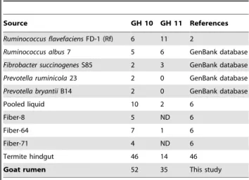
Application of degenerate primers designed based on CODE-HOP principles [20] resulted in the discovery of 52 distinct GH 10 and 35 GH 11 xylanase gene fragments from goat rumen contents. Most sequences had low identities with known xylanases in GenBank, implying that these xylanases may be as yet undiscov-ered. Moreover, these amplified partial sequences had low similarities and were distantly related based on phylogenetic analysis (Figures 1 and 2). These results suggest that there are a large number of unidentified xylanase genes in rumen, corre-sponding to the large number of uncultured microorganisms in the rumen. Compared with previous culture-independent methods such as metagenomic library construction and screening [18], pyrosequenced rumen metagenome [3], and termite hindgut metagenomics [29], far more xylanase genes were documented in our present study, and they were more diverse (Table 3), suggesting that the PCR-based culture-independent method is an efficient way to identify xylanase genes in the rumen.
Rumen bacteria and fungi are the key agents to degrade plant fiber [2], [30]. It has been proposed that rumen fungi play an important role in the initiation of plant fiber degradation during fermentative digestion in ruminants [2], [12]. Pyrosequenced rumen metagenome analysis by Brulcet al.[3] showed that all the xylanases they examined were bacterial, and no sequence was
closely related to those of fungi. In our present study, however, seven GH 11 xylanase gene fragments were retrieved directly from the rumen and showed a close relationship with rumen fungi based on phylogenetic analysis (Figure 2). Among these seven xylanase sequences, five had the highest identity to xylanases from N. patriciarumandOrpinomycessp. PC-2, and two were closely related to those fromR. flavefaciensFD-1 (Table S2). This result suggests that horizontal gene transfer from rumen bacteria to fungi may have occurred [31], [32]. In addition, the distribution of GH 11 xylanases in rumen differed from that in both tundra soil (in which more fungal than bacterial xylanase genes were detected) [20] and termite hindgut (in which GH 11 xylanases were mainly from the phyla Fibrobacteres and Spirochaetes) [29]. This result suggests that the rumen microenvironment differs from other ecosystems and has specific xylan-degrading microbial community.
Firmicutes is one of the two most abundant bacteria in rumen based on 16S rDNA library-based analysis [2], [17]. The representative genus of xylan-degrading Firmicutes in rumen is Ruminococcus [14]. Our current study showed that 33 distinct sequences (30 of GH 11 and 3 of GH 10) are closely related to the xylanases from Firmicutes, which comprises 38% of the total distinct sequences. Among them, 25 sequences (24 of GH 11 and 1 of GH 10) are related toRuminococcus. Abundance analysis also showed that xylanase genes related toRuminococcusare dominant in goat rumen (Table S2). The OTU representative of GH 11 xylanase gene fragments, R8, was subjected to full-length gene cloning and heterogeneous expression. The recombinant enzyme showed high activity to xylan. All these results suggest that the genusRuminococcusplays an important role in xylan degradation in the goat rumen.
The Cytophaga-Flexibacter-Bacteroides group represents an-other abundant group of bacteria in rumen [2], [17]. Based on phylogenetic analysis, 46 GH 10 xylanase sequences were closely related to the xylanases from Bacteroides, comprising 53% of the total distinct sequences. Abundance analysis also showed that xylanase genes related to Bacteroides were dominant in goat rumen (Table S1). More than half of them (26/46) were closely related to the xylanases fromPrevotella, which is one of the most important xylan-degrading bacterial genera in rumen [33], [34]. Many new clades were formed despite a lack of relatives, suggesting that there are other unidentified xylanase-producing Bacteroides in goat rumen. In addition to xylanases from Bacteroides, xylanase fragments from other phyla were also detected in the goat rumen. Three sequences (GR112, GR126, and GR146) in the GH 10 clone library had the highest similarity with xylanases fromX. axonopodisand Verrucomicrobiae bacteri-um. This is the first report of xylanase genes obtained from rumen that is homologous to Verrucomicrobia and Proteobacteria xylanase.
In general, rumen microorganisms can be classified into two major groups: cellulolytic and noncellulolytic [2]. The cellulolytic microorganisms include cellulolytic rumen bacteria, fungi, and some protozoa that can utilize both cellulose and xylan as a carbon source. These microorganisms, such asFibrobacter succinogenes, R. flavefaciens, andNeocallimastix frontalis, express both GH 10 and GH 11 xylanases [12], [15, [16]. The noncellulolytic microorganisms
Figure 1. Phylogenetic analysis based on the partial amino acid sequences of GH 10 xylanase genes detected in the goat rumen contents and their relationship with the reference sequences retrieved from GenBank.This tree was constructed using the neighbor-joining method (MEGA 4.0). The lengths of the branches indicate the relative divergence among the amino acid sequences. The reference sequences are marked with a closed diamond (¤) with source strains and GenBank accession numbers in parentheses. The gene fragments (GR67) used for
full-length cloning were marked with a solid square (&). The numbers at the nodes indicate bootstrap values based on 1,000 bootstrap replications and bootstrap values (.50) are displayed. The scale bar represents 0.1 amino acid substitutions per position.
doi:10.1371/journal.pone.0016731.g001
Xylanases Diversity in Goat Rumen
include members of the dominant Gram-negative Cytophaga-Flexibacter-Bacteroides phylum and Gram-positive Firmicutes, such asButyrivibrio fibrisolvens,Roseburiasp., andEubacterium rectale, which can use soluble products resulting from plant cell wall breakdown [2]. These rumen microorganisms produce only GH 10 xylanases. In our study, about 88.5% (46/52) of the GH 10 xylanase sequences were closely related to Bacteroides xylanases, whereas 85% (30/35) of the GH 11 xylanase sequences were related to xylanases from cellulolytic Firmicutes, confirming the primary distribution of GH 10 xylanase genes in noncellulolytic microorganisms and GH 11 xylanase genes in cellulolytic-xylanolytic microorganisms. GH 10 and GH 11 xylanases are distinct from each other in both three-dimensional structure [35] and mechanism of action [25]. The products of GH 11 xylanases can be further hydrolyzed by GH 10 enzymes [7]. Therefore, the different distributions of these two families imply their different roles in xylan degradation in the rumen.
To date, only a few xylanase genes have been obtained from uncultured microorganisms in rumen by direct cloning [36] or screening from the metagenomic library of rumen [18]. In our current study, two full-length xylanase genes were directly obtained from the metagenomic DNA of goat rumen contents. Both enzymes showed high activity towards different natural xylans and displayed different characteristics (Tables 1 and 2). XynGR67 had low identity (45% at maximum) with known xylanases in GenBank and similarity to a characterized cold-active xylanase [37]. Although the optimum temperature of XynGR67 was 40uC, substantial activity remained at low temperatures (22.5% at 10uC and 8.2% at 0uC). The enzyme was also thermolabile, showing a half-life of 15 min at 40uC. Compared with the cold-active xylanase XynA fromGlaciecola mesophila[37], XynGR67 displayed a much higherkcat/Kmvalue (178.5 vs. 15.4) at 4uC. These data suggest that XynGR67 has some cold-active characteristics and is distinct from other known ruminal xylanases. Using molecular methods, bacterial composition in the gastrointestinal tract are found to be dependent on host, diet, age and so on [38], [39], [40]. Of these factors, host species is the most important one [41], [42]. To expand our assessment of the xylanase gene diversity in rumen microevironment, the rumen contents of a Small Tail Han sheep that grazed under the same conditions as the Boer goat under study were subjected to xylanase gene analysis (Tables S3 and S4). Both rumens harbor similar xylan-degrading communities based on the sequence comparison analysis (Table S5, Figures S1 and S2). Similar or identical sequences were found in both rumens, and the vast majority of the amplified xylanase fragments belonged to the two major phyla of Firmicutes and Bacteroides. Based on UniFrac analysis [43], the GH 10 xylanase gene fragments in both rumens were marginally different (P = 0.02) while there was no significant difference in that of GH 11 (P = 0.35). Although the abundant sequences in each rumen are different (Table S5), the predominant OTUs of different rumens shared high identities (68% for GH 10 and 89% for GH 11, respectively). A small part of sequences are unique in each rumen (Figure S2), implying that some xylan-degrading microorganisms might be host specific [41]. The strategy reported here can be applied to explore other
gastroin-testinal tract ecosystems known to be highly specialized for raw biomass degradation (i.e., human and insect gut microbiomes) for novel xylanases.
In conclusion, an efficient culture-independent molecular method was used to explore the diversity of xylanases in the rumen ecosystem. Sequence analysis revealed a large number of unidentified and potential new xylanases in the goat rumen. Full-length cloning and heterologous expression of some genes further confirmed their function as active xylanases. The majority of GH 10 xylanase sequences were from noncellulolytic microorganisms, whereas most of the GH 11 sequences were from cellulolytic microorganisms, implying their different roles in xylan degrada-tion in the rumen ecosystem. Moreover, xylanase distribudegrada-tion in different environments is variable, suggesting that xylan-degrading microorganisms are environment specific.
Materials and Methods
Ethics statement
All animal studies were followed the regulation for the review committee of laboratory animal welfare and ethics and protocol for the review on laboratory animal welfare and ethics, Beijing Administration Office of Laboratory Animal. The animal experimentation was approved by the Committee of Laboratory Animal Welfare and Ethics, Beijing Administration Office of Laboratory Animal with the approval No. SYXK2008-0007.
Sample collection and DNA extraction
A three-year-old male Boer goat that had grazed on late-fall pasture in Inner Mongolia, China was selected. Total rumen contents were collected immediately after slaughtering and stored at270uC. The biomass was collected by centrifuging at 17,0006g, 4uC for 30 min. The sample used for DNA extraction were frozen in liquid nitrogen and ground to a fine powder with a mortar. Total genomic DNA was extracted following a protocol specific for high molecular weight DNA from environmental samples [44] and purified with an Agarose Gel DNA Purification kit (TaKaRa, Japan). The final DNA concentration was,80 ngml–1.
PCR amplification of xylanase gene fragments
Two degenerate primer sets (X10-F: 59 -CTACGACTGG-GAYGTNIBSAAYGA-39and X10-R: 59 -GTGACTCTGGAWR-CCIABNCCRT-39; and X11-F: 59 -AACTGCTACCTGKC-NITNTAYGGNTGG-39and X11-R: 59 -CCGCACGGACCAG-TAYTGNKIRAANGT-39) specific for GH 10 and GH 11 xylanases, respectively, were used to amplify xylanase gene fragments with the purified DNA as a template following the reaction system and PCR conditions as reported [20]. PCR products were visualized on an agarose gel, and bands of the expected size (,260 bp for GH 10 and 210 bp for GH 11
xylanases) were excised and purified with the TaKaRa Agarose Gel DNA Purification kit.
Cloning and sequencing of PCR products
To construct the clone library for each xylanase family, the purified PCR products were ligated into vector pGEM-T Easy
Figure 2. Phylogenetic analysis based on the partial amino acid sequences of GH 11 xylanase genes detected in the goat rumen contents and their relationship with the reference sequences retrieved from GenBank.This tree was constructed using the neighbor-joining method (MEGA 4.0). The lengths of the branches indicate the relative divergence among the amino acid sequences. The reference sequences are marked with a closed diamond (¤) with source strains and GenBank accession numbers in parentheses. The gene fragment (R8) used for
full-length retrieving is marked with a solid square (&). The numbers at the nodes indicate bootstrap values based on 1,000 bootstrap replications and bootstrap values (.50) are displayed. The scale bar represents 0.05 amino acid substitutions per position.
doi:10.1371/journal.pone.0016731.g002
Xylanases Diversity in Goat Rumen
(Promega, USA) and electroporated intoE. coliDH5acompetent cells (TaKaRa). Cells were grown on Luria-Bertani agar plates containing 100mg ml21 ampicillin, 80mg ml21 X-Gal, and 0.5mM isopropyl-b-D-1-thiogalactopyranoside (IPTG) at 37uC for 15 h. The positive transformants (white clones) in each library were randomly picked, amplified with primers M13F (59 -GTAAAACGACGGCCAGT-39) and M13R (59 -GGATAA-CAATTTCACACAGGA-39), and sequenced by Sunbiotech (China) for confirmation.
Phylogenetic analysis
Nucleotide sequences of the xylanase gene fragments were translated into amino acid sequences with EMBOSS Transeq (http://www.ebi.ac.uk/emboss/transeq) and aligned at the protein level with known sequences in the GenBank database with ClustalW. Redundant amino acid sequences were removed using CD-hit [22] with a 95% sequence identity cut-off. The protein sequence similarities were assessed by using the BLASTp programs (http://www.ncbi.nlm.nih.gov/BLAST/; until August 15, 2010). Phylogenetic trees were constructed with MEGA 4.0 [45] using the neighbor-joining method [46]. Confidence for tree topologies was estimated by bootstrap values based on 1,000 replicates. Twenty-five representative sequences originating from the rumens or human guts identified by BLASTp analysis were selected and used as references for tree construction.
Abundance analysis
Gene abundance of each GH family was estimated using distance-based operational taxonomic unit and richness determi-nation (DOTUR) software [23]. Distance matrices of the fragment sequences were calculated at the protein level with the default parameters of protdist in PHYLIP (http://evolution.genetics. washington.edu/phylip.html). Sequences were then assigned to OTUs based on UPGMA (average linkage clustering) implement-ed in DOTUR with default parameters of precision (0.01) and 1,000 bootstrap replicates.
Cloning of full-length xylanase genes
Fragments GR67, which showed the phylogenetic novelty (Figure 1) and only appeared once in the GH 10 library (see Table S1), and R8, which represented the most abundant OTU in the GH 11 library (Table S2), were subjected to full-length gene cloning. The flanking regions of these xylanase gene fragments were cloned with eight specific primers for each fragment (Table S6) following the protocol for modified TAIL-PCR [47]. PCR products were electrophoresed on 1.3% agarose gels, cloned into pGEM-T Easy vectors, sequenced, and assembled with the known fragment sequences.
Assembly of the xylanase gene sequences and identification of the open reading frames were performed with programs in Vector NTI 10.3 (InforMax, USA). The signal peptide sequence was predicted with SignalP (http://www.cbs.dtu.dk/services/SignalP/). The
Figure 3. pH and temperature activity profiles of purified recombinant XynGR67 and XynR8. AEffect of pH on XynGR67 activity. Activities at various pHs were assayed at 30uC.BEffect of temperature on XynGR67 activity in McIlvaine buffer (pH 6.0).CEffect of pH on XynR8 activity. Activities at various pHs were assayed at 30uC.DEffect of temperature on XynR8 activity in McIlvaine buffer (pH 5.5). The error bars represent the mean6SD (n = 3).
DNA and protein sequence identities/similarities were assessed with the BLASTn and BLASTp programs (http://www.ncbi.nlm.nih. gov/BLAST/), respectively.
Xylanase expression and activity assay
The coding sequences of two representative xylanase genes without the signal peptide were amplified using two primer sets (Table S6), cloned into plasmid pET-22b(+), and transformed into E. coli BL21 (DE3) competent cells for recombinant expression. Positive transformants were grown in Luria-Bertani medium containing 100mg ml21ampicillin at 37uC to an A600 of,0.6.
Expression was induced with 0.8 mM IPTG at 25uC for 12 h. Xylanase activity was determined by measuring the release of reducing sugar from substrate with the 3, 5-dinitrosalicylic acid method [48]. Reactions containing 0.1 ml of enzyme preparation and 0.9 ml of 1% (w/v) substrate in McIlvaine buffer (pH 6.0) were incubated at 30uC for 10 min, followed by addition of 1.5 ml of 3, 5-dinitrosalicylic acid reagent. The mixture was boiled for 5 min, cooled to room temperature, and the absorbance at 540 nm (A540) was measured. Using a standard curve generated with D-xylose, the absorbance was converted into moles of
reducing sugars produced.
Purification and characterization of recombinant XynGR67 and XynR8
To purify the His-tagged recombinant proteins (XynGR67 and XynR8), culture supernatant was collected after centrifugation (12,0006g, 4uC for 15 min) and further concentrated with an
ultrafiltration membrane (PES5000; Sartorius Stedim Biotech, Germany). The concentrated supernatant was loaded onto a Ni2+
-NTA agarose gel column (Qiagen, Germany) with a linear imidazole gradient of 20–200 mM in Tris-HCl buffer (20 mM Tris-HCl, 500 mM NaCl, 10% glycerol, pH 7.6). SDS-PAGE was used to determine the purity and apparent molecular mass of recombinant XynGR67. The protein concentration was
deter-mined with the Bradford method [49] with bovine serum albumin as a standard.
The optimal pH for xylanase activity of the purified recombinant XynGR67 and XynR8 were determined at 30uC in buffers of pH 4.0 to 9.0. The enzyme stability at different pHs were estimated by measuring the residual activity after incubating the enzyme solution in buffers at pH 3.0–10.0 at 25uC for 1 h. The buffers used were McIlvaine buffer (0.2 M Na2HPO4/0.1 M citric acid) for pH 3.0–7.5, and 0.1 M Tris-HCl for pH 7.5–9.0.
The optimal temperature for purified recombinant XynGR67 and XynR8 were determined over the range of 0–80uC in McIlvaine buffer (pH 5.5 for XynR8 and pH 6.0 for XynGR67). Thermostability of XynGR67 was determined after pre-incubating the enzyme in McIlvaine buffer (pH 6.0) at 30uC or 40uC without substrate for various periods while XynR8 was assayed at 55uC and 60uC in McIlvaine buffer (pH 5.5).
TheKm, Vmax, andkcatvalues for both recombinant xylanases were determined in McIlvaine buffer (pH 5.5 for XynR8 and pH 6.0 for XynGR67) containing 1–10 mg ml21birchwood xylan at 40uC and 55uC, respectively.Km and Vmax were determined from a Lineweaver-Burk plot with the non-linear regression computer program GraFit (Erithacus Software, UK). Three independent experiments were averaged, and each experiment included three replicates.
The substrate specificity of purified recombinant enzymes were assayed by incubating the enzyme solution with 1% (w/v) substrates including birchwood xylan, beechwood xylan, wheat arabinoxylan or wheat arabinoxylan (insoluble) under standard conditions. Enzymatic activities againstp-nitrophenyl cellobioside
Table 3.Amount of GH 10 and 11 xylanases in the genomes
ofRuminococcus flavefaciensFD-1,Prevotella ruminicola23,
pyrosequenced rumen, termite hindgut and goat rumen in this study.
Source GH 10 GH 11 References
Ruminococcus flavefaciensFD-1 (Rf) 6 11 2
Ruminococcus albus7 5 6 GenBank database
Fibrobacter succinogenesS85 2 3 GenBank database
Prevotella ruminicola23 2 0 GenBank database
Prevotella bryantiiB14 2 0 GenBank database
Pooled liquid 10 2 6
Fiber-8 5 ND 6
Fiber-64 7 1 6
Fiber-71 4 ND 6
Termite hindgut 46 14 46
Goat rumen 52 35 This study
doi:10.1371/journal.pone.0016731.t003
Table 1.Kinetic parameters of XynGR67 and XynR8 towards birchwood xylan.
Enzyme Temperature (6C)
Vmax
(mmol min–1mg–1)
Km
(mg ml–1)
kcat (s–1)
kcat/Km
(ml s–1mg–1)
XynGR67 40 2,150.0672.4 1.4360.16 1,505.0650.4 1,052.5
4 500.4614.3 1.9660.17 350.0610.1 178.6
XynR8 55 2,647.9660.9 3.2760.22 1,306.3630.1 399.5
doi:10.1371/journal.pone.0016731.t001
Table 2.Substrate specificity of the purified recombinant
XynGR67 and XynR8.
Substrate Relative activity (%)
XynGR67 XynR8
Birchwood xylan 100 100
Beechwood xylan 150.763.1 196.862.7
Wheat arabinoxylan 53.462.5 384.463.4
Wheat arabinoxylan (insoluble) 63.161.9 54.662.2
pNP-cellobioside NA NA
pNP-xyloside NA NA
doi:10.1371/journal.pone.0016731.t002
Xylanases Diversity in Goat Rumen
and p-nitrophenyl xyloside were examined under standard conditions at a final concentration of 2 mM.
Nucleotide sequence accession numbers
The nucleotide sequences of GH 10 and 11 xylanase gene fragments were deposited into the GenBank database under accession numbers FJ919156–FJ919224, HM773534–HM773543, and HM773544–HM773578. Accession numbers HQ219689 and HQ219690 were assigned to the full-length xylanase genesxynR8 andxynGR67of the goat rumen contents, respectively.
Supporting Information
Figure S1 Phylogenetic analysis based on the partial amino acid sequences of GH 10 xylanase genes detected in the goat and sheep rumen contents and their relationship with the reference sequences retrieved from GenBank.This tree was constructed using the neighbor-joining method (MEGA 4.0). Sequences from goat rumen were colored in red and those from sheep were in green. Sequence clusters that unique in each rumen were marked with Sheep rumen I–IV or Goat rumen A–C. The lengths of the branches indicate the relative divergence among the amino acid sequences. The reference sequences are marked with a closed diamond (¤) with source strains and GenBank accession numbers in parenthe-ses. The numbers at the nodes indicate bootstrap values based on 1,000 bootstrap replications and bootstrap values (.50) are displayed. The scale bar represents 0.1 amino acid substitutions per position.
(PDF)
Figure S2 Phylogenetic analysis based on the partial amino acid sequences of GH 11 xylanase genes detected in the goat and sheep rumen contents and their relationship with the reference sequences retrieved from GenBank.This tree was constructed using the neighbor-joining method (MEGA 4.0). Sequences from goat rumen were colored in red and those from sheep were colored in green. The lengths of the branches indicate the relative divergence among the amino acid sequences. The reference sequences are marked with a closed diamond (¤) with source strains and GenBank accession numbers in parentheses. The numbers at the nodes indicate bootstrap values based on 1,000 bootstrap replications and
bootstrap values (.50) are displayed. The scale bar represents 0.1 amino acid substitutions per position.
(PDF)
Table S1 The GH 10 xylanase gene fragments detected in the goat rumen contents and their closest relative based on amino acid sequence identity and similarity.
(DOC)
Table S2 The GH 11 xylanase gene fragments detected in the goat rumen contents and their closest relative based on amino acid sequence identity and similarity.
(DOC)
Table S3 The GH 10 xylanase gene fragments detected in the sheep rumen contents and their closest relatives based on amino acid sequence identity and similarity.
(DOC)
Table S4 The GH 11 xylanase gene fragments detected in the sheep rumen contents and their closest relatives based on amino acid sequence identity and similarity.
(DOC)
Table S5 Summary of the GH 10 and GH 11 xylanase fragment sequences obtained from the goat and sheep rumen contents.
(DOC)
Table S6 Primers used for xylanase genes cloning and expression.
(DOC)
Acknowledgments
We thank Dr. Chenggang Jiang for his assistance with sample collection.
Author Contributions
Conceived and designed the experiments: GW BY. Performed the experiments: GW XP HL YW. Analyzed the data: GW HH PS. Contributed reagents/materials/analysis tools: QD HZ. Wrote the paper: GW KM PY.
References
1. Bunzel M, Steinhart H (2003) Dietary fibre from plant cell walls: chemical and structural characteristics. Ernahrungs Umschau 50: 469–475.
2. Flint HJ, Bayer EA (2008) Plant cell wall breakdown by anaerobic microorganisms from the Mammalian digestive tract. Ann NY Acad Sci 1125: 280–288. 3. Brulc JM, Antonopoulos DA, Miller ME, Wilson MK, Yannarell AC, et al. (2009)
Gene-centric metagenomics of the fiber-adherent bovine rumen microbiome reveals forage specific glycoside hydrolases. Proc Natl Acad Sci USA 106: 1948–1953. 4. Kopecny J, Williams AG (1988) Synergism of rumen microbial hydrolases
during degradation of plant polymers. Folia Microbiol (Praha) 33: 208–212. 5. Ohgren K, Bura R, Saddler J, Zacchi G (2007) Effect of hemicellulose and lignin
removal on enzymatic hydrolysis of steam pretreated corn stover. Bioresour Technol 98: 2503–2510.
6. Yang B, Wyman CE (2004) Effect of xylan and lignin removal by batch and flowthrough pretreatment on the enzymatic digestibility of corn stover cellulose. Bioresour Technol 86: 88–98.
7. Collins T, Gerday C, Feller G (2005) Xylanases, xylanase families and extremophilic xylanases. FEMS Microbiol Rev 29: 3–23.
8. Kulkarni N, Shendye A, Rao M (1999) Molecular and biotechnological aspects of xylanases. FEMS Microbiol Rev 23: 411–456.
9. Flint, HJ, Whitehead TR, Martin JC, Gasparic A (1997) Interrupted catalytic domain structures in xylanases from two distantly related strains ofPrevotella ruminicola.Biochim Biophys Acta 1337: 161–165.
10. Dalrymple BP, Swadling Y, Layton I, Gobius KS, Xue GP (1999) Distribution and evolution of the xylanase genes xynA and xynB and their homologues in strains ofButyrivibrio fibrisolvens.Appl Environ Microbiol 65: 3660–3667.
11. Flint HJ, Zhang JX, Martin J (1994) Multiplicity and expression of xylanases in the rumen cellulolytic bacteriumRuminococcus flavefaciens.Curr Microbiol 29: 139–143. 12. Gomez de Segura B, Durand R, Fevre M (1998) Multiplicity and expression of xylanases in the rumen fungusNeocallimastix frontalis.FEMS Microbiol Lett 164: 47–53.
13. Morrison M, Daugherty SC, Nelson WC, Davidsen T, Nelson KE, et al. (2010) The FibRumBa database: a resource for biologists with interests in gastrointes-tinal microbial ecology, plant biomass degradation, and anaerobic microbiology. Microb Ecol 59: 212–213.
14. Berg Miller ME, Antonopoulos DA, Rincon MT, Band M, Bari A, et al. (2009) Diversity and strain specificity of plant cell wall degrading enzymes revealed by the draft genome ofRuminococcus flavefaciensFD-1. PLoS ONE 4: e6650. 15. Flint HJ, Bayer EA, Rincon MT, Lamed R, White BA (2008) Polysaccharide
utilization by gut bacteria: potential for new insights from genomic analysis. Nat Rev Micro 6: 121–131.
16. Qi M, Nelson KE, Daugherty SC, Nelson WC, Hance IR, et al. (2008) Genomic differences betweenFibrobacter succinogenes S85 andFibrobacter intestinalis DR7, identified by suppression subtractive hybridization. Appl Environ Microbiol 74: 987–993.
17. Edwards JE, McEwan NR, Travis AJ, John Wallace R (2004) 16S rDNA library-based analysis of ruminal bacterial diversity. Antonie Van Leeuwenhoek 86: 263–281.
19. Singh B, Gautam SK, Verma V, Kumar M, Singh B (2008) Metagenomics in animal gastrointestinal ecosystem: Potential biotechnological prospects. Anaer-obe 14: 138–144.
20. Wang G, Wang Y, Yang P, Luo H, Huang H, et al. (2010) Molecular detection and diversity of xylanase genes in alpine tundra soil. Appl Microbiol Biotechnol 87: 1383–1393.
21. Solomon V, Teplitsky A, Shulami S, Zolotnitsky G, Shoham Y, et al. (2007) Structure-specificity relationships of an intracellular xylanase fromGeobacillus stearothermophilus.Acta Crystallogr D Biol Crystallogr 63: 845–859.
22. Li W, Godzik A (2006) Cd-hit: a fast program for clustering and comparing large sets of protein or nucleotide sequences. Bioinformatics 22: 1658–1659. 23. Schloss PD, Handelsman J (2005) Introducing DOTUR, a computer program
for defining operational taxonomic units and estimating species richness. Appl Environ Microbiol 71: 1501–1506.
24. Hayashi H, Abe T, Sakamoto M, Ohara H, Ikemura T, et al. (2005) Direct cloning of genes encoding novel xylanases from the human gut. Can J Microbiol 51: 251–259.
25. Jeffries TW (1996) Biochemistry and genetics of microbial xylanases. Curr Opin Biotechnol 7: 337–342.
26. Wouters J, Georis J, Engher D, Vandenhaute J, Dusart J, et al. (2001) Crystallographic analysis of family 11 endo-b-1,4-xylanase Xyl1 fromStreptomyces
sp. S38. Acta Crystallogr D Biol Crystallogr 57: 1813–1819.
27. Raymond JA, Christner BC, Schuster SC (2008) A bacterial ice-binding protein from the Vostok ice core. Extremophiles 12: 713–717.
28. Beg QK, Kapoor M, Mahajan L, Hoondal GS (2001) Microbial xylanases and their industrial applications: a review. Appl Microbiol Biotechnol 56: 326–338. 29. Warnecke F, Luginbuhl P, Ivanova N, Ghassemian M, Richardson TH, et al. (2007) Metagenomic and functional analysis of hindgut microbiota of a wood-feeding higher termite. Nature 450: 560–565.
30. Dehority BA, Tirabasso PA (2000) Antibiosis between ruminal bacteria and ruminal fungi. Appl Environ Microbiol 66: 2921–2927.
31. Garcia-Vallve S, Romeu A, Palau J (2000) Horizontal gene transfer of glycosyl hydrolases of the rumen fungi. Mol Biol Evol 17: 352–361.
32. Gilbert HJ, Hazlewood GP, Laurie JI, Orpin CG, Xue GP (1992) Homologous catalytic domains in a rumen fungal xylanase: evidence for gene duplication and prokaryotic origin. Mol Microbiol 6: 2065–2072.
33. Dodd D, Kocherginskaya SA, Spies MA, Beery KE, Abbas CA, et al. (2009) Biochemical analysis of ab-D-xylosidase and a bifunctional xylanase-ferulic acid esterase from a xylanolytic gene cluster inPrevotella ruminicola23. J Bacteriol 191: 3328–3338.
34. Gasparic A, Martin J, Daniel A, Flint HJ (1995) A xylan hydrolase gene cluster inPrevotella ruminicolaB(1)4: sequence relationships, synergistic interactions, and oxygen sensitivity of a novel enzyme with exoxylanase andb-(1,4)-xylosidase activities. Appl Environ Microbiol 61: 2958–2964.
35. Biely P, Vrsanska M, Tenkanen M, Kluepfel D (1997) Endo-b-1,4-xylanase families: differences in catalytic properties. J Biotechnol 57: 151–166. 36. Liu JR, Yu B, Lin SH, Cheng KJ, Chen YC (2005) Direct cloning of a xylanase
gene from the mixed genomic DNA of rumen fungi and its expression in intestinalLactobacillus reuteri.FEMS Microbiol Lett 251: 233–241.
37. Guo B, Chen XL, Sun CY, Zhou BC, Zhang YZ (2009) Gene cloning, expression and characterization of a new cold-active and salt-tolerant endo-b -1,4-xylanase from marine Glaciecola mesophila KMM 241. Appl Microbiol Biotechnol 84: 1107–1115.
38. Hopkins MJ, Harp RS, Macfarlane GT (2001) Age and disease related changes in intestinal bacterial populations assessed by cell culture, 16S rRNA abundance, and community cellular fatty acid profiles. Gut 48: 198–205.
39. Leser TD, Lindecrona RH, Jensen TK, Jensen BB, Moller K (2000) Changes in bacterial community structure in the colon of pigs fed different experimental diets and after infection withBrachyspira hyodysenteriae. Appl Environ Microbiol 66: 3290–3296.
40. Shi PJ, Meng K, Zhou ZG, Wang YR, Diao QY, Yao B (2008) The host species affects the microbial community in the goat rumen. Lett Appl Microbiol 46: 132–135.
41. Dick LK, Bernhard AE, Brodeur TJ, Santo Domingo JW, Simpson JM, et al. (2005) Host distributions of uncultivated fecal Bacteroidales bacteria reveal genetic markers for fecal source identification. Appl Environ Microbiol 71: 3184–3191.
42. Weimer PJ, Stevenson DM, Mantovani HC, Man SL (2010) Host specificity of the ruminal bacterial community in the dairy cow following near-total exchange of ruminal contents. J Dairy Sci 93: 5902–5912.
43. Lozupone C, Knight R (2005) UniFrac: a new phylogenetic method for comparing microbial communities. Appl Environ Microbiol 71: 8228–8235. 44. Brady SF (2007) Construction of soil environmental DNA cosmid libraries and
screening for clones that produce biologically active small molecules. Nat Protoc 2: 1297–1305.
45. Tamura K, Dudley J, Nei M, Kumar S (2007) MEGA4: Molecular Evolutionary Genetics Analysis (MEGA) Software Version 4.0. Mol Biol Evol 24: 1596–1599. 46. Saitou N, Nei M (1987) The neighbor-joining method: a new method for
reconstructing phylogenetic trees. Mol Biol Evol 4: 406–425.
47. Huang H, Wang G, Zhao Y, Shi P, Luo H, et al. (2010) Direct and efficient cloning of full-length genes from environmental DNA by RT-qPCR and modified TAIL-PCR. Appl Microbiol Biotechnol 87: 1141–1149.
48. Miller GL, Blum R, Glennon WE, Burton AL (1960) Measurement of carboxymethylcellulase activity. Anal Biochem 1: 127–132.
49. Bradford MM (1976) A rapid and sensitive method for the quantitation of microgram quantities of protein utilizing the principle of protein-dye binding. Anal Biochem 72: 248–254.
Xylanases Diversity in Goat Rumen