Inhibition of Calcium Influx Reduces
Dysfunction and Apoptosis in Lipotoxic
Pancreatic
β
-Cells via Regulation of
Endoplasmic Reticulum Stress
Yuren Zhou1☯, Peng Sun1,2☯, Ting Wang1, Kaixian Chen1,2, Weiliang Zhu1, Heyao Wang1 *
1Shanghai Institute of Materia Medica, Chinese Academy of Sciences, 555 Zuchongzhi Road, Shanghai, 201203, China,2School of Pharmacy, Shanghai University of Traditional Chinese Medicine, 1200 Cailun Road, Shanghai, 201203, China
☯These authors contributed equally to this work. *[email protected]
Abstract
Lipotoxicity plays an important role in pancreaticβ-cell failure during the development of type 2 diabetes. Prolonged exposure ofβ-cells to elevated free fatty acids level could cause deterioration ofβ-cell function and induce cell apoptosis. Therefore, inhibition of fatty acids-inducedβ-cell dysfunction and apoptosis might provide benefit for the therapy of type 2 dia-betes. The present study examined whether regulation of fatty acids-triggered calcium influx could protect pancreaticβ-cells from lipotoxicity. Two small molecule compounds, L-type calcium channel blocker nifedipine and potassium channel activator diazoxide were used to inhibit palmitic acid-induced calcium influx. And whether the compounds could reduce pal-mitic acid-inducedβ-cell failure and the underlying mechanism were also investigated. It was found that both nifedipine and diazoxide protected MIN6 pancreaticβ-cells and primary cultured murine islets from palmitic acid-induced apoptosis. Meanwhile, the impaired insulin secretion was also recovered to varying degrees by these two compounds. Our results veri-fied that nifedipine and diazoxide could reduce palmitic acid-induced endoplasmic reticulum stress to generate protective effects on pancreaticβ-cells. More importantly, it suggested that regulation of calcium influx by small molecule compounds might provide benefits for the prevention and therapy of type 2 diabetes.
Introduction
During the development of type 2 diabetes (T2D), obesity induced elevation level of free fatty acids (FFAs) causes both insulin resistance and pancreaticβ-cell failure [1,2]. And early appearance ofβ-cell failure could subsequently lead to insufficient insulin secretion, thereby breaking normal glycemic control [1]. It is known that FFAs play an important role in the nor-mal function of pancreaticβ-cells. However, pleiotropic effects of FFAs have also been verified
OPEN ACCESS
Citation:Zhou Y, Sun P, Wang T, Chen K, Zhu W, Wang H (2015) Inhibition of Calcium Influx Reduces Dysfunction and Apoptosis in Lipotoxic Pancreaticβ -Cells via Regulation of Endoplasmic Reticulum Stress. PLoS ONE 10(7): e0132411. doi:10.1371/ journal.pone.0132411
Editor:Tohru Minamino, Niigata University Graduate School of Medical and Dental Sciences, JAPAN
Received:March 11, 2015
Accepted:June 12, 2015
Published:July 6, 2015
Copyright:© 2015 Zhou et al. This is an open access article distributed under the terms of the
Creative Commons Attribution License, which permits unrestricted use, distribution, and reproduction in any medium, provided the original author and source are credited.
Data Availability Statement:All relevant data are within the paper and its Supporting Information files.
Funding:This work was supported by grant from Shanghai Natural Science Foundation (14ZR1447700), National Natural Science Foundation of China (81473262, 81072681), National Science and Technology Major Project“Key New Drug Creation and Manufacturing Program”
(2012ZX09301001-001, 2013ZX09103001-001).
[3]. FFAs supply could augment glucose-stimulated insulin secretion, while chronically in excess, FFAs can impair insulin biosynthesis, secretion and induceβ-cell apoptosis [2,3]. Nonetheless, the molecular mechanisms of FFAs-inducedβ-cell failure are complex and not fully understood.
Under physiological conditions, acute stimulation of FFAs could activate receptors in pan-creaticβ-cells, such as G-protein coupled receptor 40 (GPR40), to amplify insulin secretion pathway via increasing intracellular calcium concentration [4,5]. Medium- and long-chain FFAs like palmitic acid (PA) could stimulate voltage-sensitive Ca2+influx and directly mobilize Ca2+from intracellular endoplasmic reticulum (ER) Ca2+pools in pancreaticβ-cells [6,7]. Therefore, chronic elevate FFAs could persistently augment Ca2+metabolism in mitochondria, which might be related to cell apoptosis [8]. More importantly, sustained elevation of intracel-lular Ca2+concentration ([Ca2+]i) could induce ER-stress response, asβ-cells have a
well-devel-oped ER and are highly susceptible to ER-stress [9,10]. Together factors indicate that Ca2+ signal is strongly involved in FFAs-inducedβ-cell dysfunction and apoptosis. It has been reported that some Ca2+chelators or Ca2+signal blockers had a protective effect on FFAs-inducedβ-cell apoptosis [11,12]. Meanwhile, our previous study revealed that using a small molecule antagonist of GPR40 to block Ca2+release also reduced PA-induced apoptosis in pan-creaticβ-cells [13]. Thus, regulation of Ca2+release might provide benefit forβ-cell protection during the development of T2D.
The aim of this study was to investigate the possible effect of inhibition of sustained Ca2+ influx on lipotoxicβ-cells. Using an classic L-type Ca2+channel blocker nifedipine, which has been reported to inhibit Ca2+influx and mediate insulin secretion in pancreaticβ-cells [14] and diazoxide, a potassium channel activator which could also block Ca2+influx during GSIS [15], the effects of regulation of Ca2+influx on chronic PA-treated pancreaticβ-cells were studied.
Materials and Methods
Cell culture and murine pancreatic islets isolation
Mouse insulinoma cell line MIN6 cells were kindly provided by Prof. S. Seino [16]. The cells were cultured in Dulbecco’s modified Eagle’s medium (DMEM) supplemented with 10% fetal bovine serum (FBS), 25 mM glucose and 50μMβ-mercaptoethanol at 37°C under 5% CO2. All
cell culture reagents were purchased from GIBCO (Carlsbad, CA, USA).
Pancreatic islets were isolated as described in our previous work [17]. Briefly, 6-week-old male C57BL/6J mice (Slac, Shanghai, China) were used to isolate islets by collagenase V (Sigma-Aldrich) digestion, then the islets were cultured in RPMI-1640 medium with 10% FBS, 10000 units/mL penicillin, and 10000μg/ mL streptomycin with 11.1 mM glucose. For islets experiments, islets were isolated from single animal and at least three parallel preparations were performed for each experiments. All animal care and experiments were permitted by Institutional Animal Care and Use Committees of Shanghai Institute of Materia Medica (No. 2013-10-WHY-12).
Cell viability assay and Hoechst33342 staining
(MTT). Briefly, MIN6 cells were supplemented with MTT (0.5mg/ml) and incubated for 4 h. Then dimethyl sulfoxide (DMSO) solution was used to dissolve formazan crystals. Cell viability was calculated by the absorbance at 490 nm wavelength.
Hoechst33342 staining was performed as described in detail in our previous work [17]. Briefly, cells were planted into a six-well plate. After treatment with 0.5 mM PA and different concentration of compounds, the cells were fixed with 4% paraformaldehyde and then stained with Hoechst33342 (10μg/ml) for 15 min. The apoptotic cells were observed and photo-graphed under a fluorescence microscope (DP70, Olympus, Tokyo, Japan), and the apoptotic ratio in each group was calculated according to 10 random insights.
Western blot analysis
After treatment as above described, MIN6 cells were lysed by RIPA buffer (Beyotime Bio, Shanghai, China). Then proteins were collected and separated by SDS–PAGE and transferred to membranes. After blocking, membranes were incubated with following primary antibodies with different dilutions: rabbit anti-phospho-eukaryotic translation initiation factor 2 (eIF2α, 1:1000, Cell Signaling, Beverly, MA, USA), rabbit anti-eIF2α(1:1000, Cell Signaling), mouse-anti-C/EBP-homologous protein (CHOP, F-168, 1:500, Santa Cruz, CA, USA), rabbit-anti-cleaved caspase-3 (Asp 175, 1:1000, Cell Signaling), rabbit-anti-phospho-c-Jun N-terminal protein kinase (JNK, 1:1000, Cell Signaling), anti-JNK (1:1000, Cell Signaling), rabbit-anti-β-actin (1:5000, Sigma-Aldrich). Then horseradish peroxidase (HRP)-conjugated second-ary antibodies (1:10000; Jackson Laboratories, PA, USA) and SuperSignal West Pico Chemilu-minescence kit (Pierce, IL, USA) was used to visualize resulting immunocomplex. All blotting were performed at least three times, the optical density of each bands were analyzed by Image J software (NIH, MD, USA).
Cell immunofluorescence
For cell immunofluorescence, MIN6 cells were grown on glass coverslips in a six-well plate and incubated with 0.5% BSA alone (control), PA/BSA or PA/BSA mixed with different com-pounds for 48 h. Then the cells were fixed in 4% paraformaldehyde at 4°C overnight. After fixa-tion, the MIN6 cells were incubated with primary antibodies (mouse anti-CHOP, dilution 1:100; rabbit anti-insulin antibody, dilution 1:100, Cell Signaling) and followed by secondary antibodies (AlexaFluor 546-conjugated goat anti-mouse IgG and AlexaFluor 488-conjugated goat anti-rabbit IgG, dilution 1:400, Invitrogen, CA, USA). The nuclei of cells were stained by 4’-6-diamidino-2-phenylindole (DAPI), then coverslips were mounted and the image were taken by fluorescence microscopy (DP70, Olympus). For islet immunofluorescence, the islets were incubated with rabbit-anti-pancreatic/duodenal homeobox-1 (PDX1, dilution 1:100, Upstate, VA, USA) primary antibody and followed by Alexafluor 546-conjugated goat anti-rabbit IgG (Invitrogen). The PDX1-positive cell rate was calculated as PDX1-positive cell num-ber divided by total islet cell numnum-ber.
Calcium mobilization assay
The calcium mobilization assay was described in our previous work [19]. Briefly, MIN6 Cells were cultured in 96-well plates for 24 h, and then the medium was replaced with 3μM Fluo-4/ AM (Invitrogen) and 2.5 mM probenecid (Sigma-Aldrich) in Hanks' balanced salt solution (HBSS, Beyotime). After 90 min incubation, the plates were washed with HBSS, and pre-incu-bated with different concentration of nifedipine and diazoxide for 10 min. Then 0.5 mM PA was used to stimulate calcium release and the fluorescence absorption of Fluo-4 was examined to calculate calcium release by Flexstation III (Molecular Devices, CA, USA).
Glucose-stimulated insulin secretion
MIN6 cells or islets of approximately the same size (150μm in diameter) were plated in 24-well plates (250,000 cells or 10 islets per well). Cells/islets were incubated with 0.5 mM PA in the presence/absence of different compound for 48 h. Then the medium was removed and pre-incubated by HBSS without glucose for 1 h at 37°C. Thereafter, the cells/islets were incu-bated in HBSS containing 5 mM glucose for 1 h and then in 25mM glucose for another 1 h. The supernatant was collected and the insulin concentration was measured by enzyme-linked immunosorbent assay (ELISA) (Millipore, MA, USA) according to its protocol.
Data analysis
All data are expressed as mean ± SE. The comparison of different groups was assessed by two-tailed unpaired Student’s t-test or one-way-ANOVA followed by Dunnet’s post hoc test. P<0.05 indicated statistically significant difference.
Results
Nifedipine and diazoxide protects MIN6 cells from PA-induced apoptosis
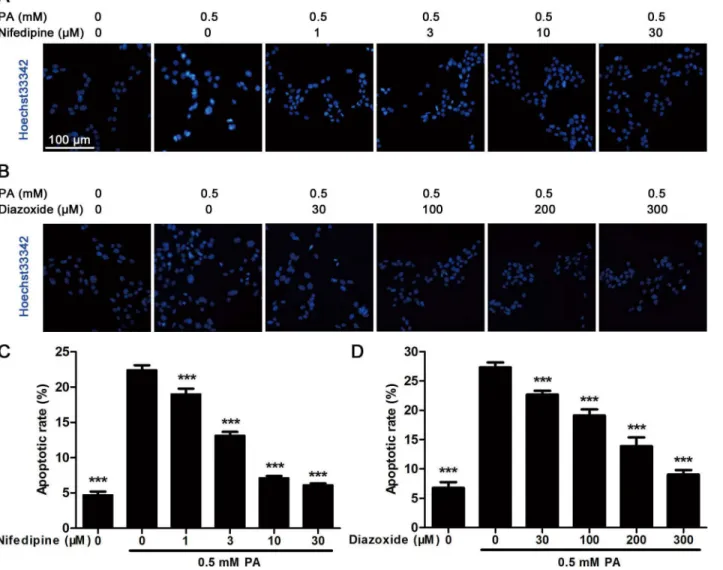
There was less effect of both nifedipine and diazoxide on MIN6 cell viability after 48 h incuba-tion (Fig 1A and 1B). However, 48 h treatment of 0.5 mM PA significantly reduced cell viability to approximately 60% compared to control cells (Fig 1A and 1B). Co-incubated with nifedipine and diazoxide dose-dependently inhibited PA-induced decreasing in cell viability (Fig 1A and 1B). Meanwhile, western blot analysis showed that the expression of cleaved caspase-3, an apoptotic marker, was obviously increased in PA-treated cells. Similarly, nifedipine reduced cleaved caspase-3 expression since 1μM concentration (Fig 1C and 1E). Although not as effec-tive as nifedipine, co-incubation of 200μM diazoxide with PA still decreased cell apoptosis (Fig 1D and 1F).To further evaluate the protective effect of nifedipine and diazoxide on MIN6 cells, Hoechst33342 staining was performed to visualize apoptotic cells. Nuclei of apoptotic cells would display a high condensed chromatin compared with normal cells after Hoechst staining. As shown inFig 2A and 2B, 0.5 mM PA-treated cells showed brighter nuclei with nuclei shrinkage and highly condensed DNA. However, an obviously reduced apoptotic rate was observed in co-incubation of both nifedipine and diazoxide with PA compared to PA-treated alone group in a dose-dependent manner (Fig 2A–2D).
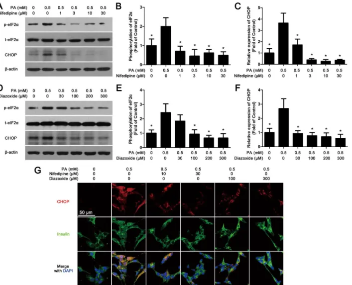
eIF2α. Meanwhile, both western blot (Fig 3A, 3C, 3D and 3F) and immunofluorescence (Fig 3G) experiments showed that the expression of CHOP in nifedipine or diazoxide co-treated with PA cells was obviously decreased in comparison with PA-treated alone group.
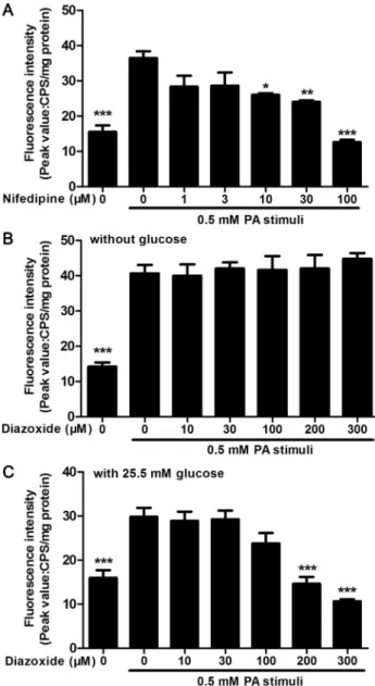
PA-stimulated calcium release in MIN6 cells was inhibited by nifedipine
and diazoxide under different conditions
To observe the effect of nifedipine and diazoxide on PA-stimulated Ca2+release in MIN6
β-cells, the Fluo-4 dye was used to determine intracellular Ca2+concentrations. It was found that nifedipine, a L-type Ca2+channel blocker, produced a direct concentration dependent inhibition of PA-stimulated Ca2+release in MIN6 cells (Fig 4A). And diazoxide, an Fig 1. Nifedipine and diazoxide protects MIN6 cells from PA-induced apoptosis.(A) After 48 h incubation of nifedipine in the presence/absence of PA, the cell viability was measured by MTT assay. The cell viability was shown as inhibitory ratio (% of control),***p<0.001 denote significant difference versus the PA-treated alone group, n = 6. (B) After 48 h incubation of diazoxide in the presence/absence of PA, the cell viability was measured by MTT assay.***p<0.001 denote significant difference versus the PA-treated alone group, n = 6. (C) After the cells were treated with 0.5 mM PA in the presence/absence of nifedipine for 48 h, the expression of cleaved caspase-3 was detected by western blot. (D) After the cells were treated with 0.5 mM PA in the presence/absence of diazoxide for 48 h, the expression of cleaved caspase-3 was detected. (E)(F) Quantitative analysis of western blot. The optical density of each blot band was determined and adjusted by the optical density ofβ-actin.*p<0.05;**p<0.01;***p<0.001 denote significant difference versus the PA-treated alone group, n = 3.
ATP-sensitive K+channel opener, could inhibit glucose-induced increasing of ATP concentra-tion, thereby reducing membrane hyperpolarization and activation of voltage-dependent Ca2+ channel [20]. Thus, we also detected the effect of diazoxide on PA-stimulated Ca2+release in MIN6 cells. However, diazoxide did not alter Ca2+response to PA in MIN6 without glucose during monitoring time (Fig 4B). As diazoxide exerted protective effects on PA-impaired MIN6 cells, we then detected the PA-stimulated Ca2+release in the presence of 25.5 mM glu-cose, which was the concentration of glucose used in common culture medium of MIN6 cells. It was found that diazoxide at 100μM could slightly suppress PA-induced Ca2+release in MIN6 cells, while 200 and 300μM diazoxide significantly inhibited Ca2+release in the presence of glucose.
Fig 2. Hoechst33342 staining analysis in MIN6 cells.(A) After the cells were treated with 0.5 mM PA in the presence/absence of nifedipine at indicated concentrations for 48 h, Hoechst33342 staining was performed to detect apoptotic cells. Blue fluorescence indicated all nucleus, the lighter and shrinkage dots were apoptotic cells. Scale bar = 100μm and referred to all panels. (B) After the cells were treated with 0.5 mM PA in the presence/absence of diazoxide for 48 h, Hoechst33342 staining was performed. (C)(D) The apoptotic rate was calculated as apoptotic cell number divided by total cell number. 10 random sights in each well were selected to count apoptosis, and the data from six duplicated wells were analyzed (n = 6).***p<0.001 denote significant difference
versus the PA-treated alone group.
Nifedipine and diazoxide suppressed PA-induced phosphorylation of
JNK
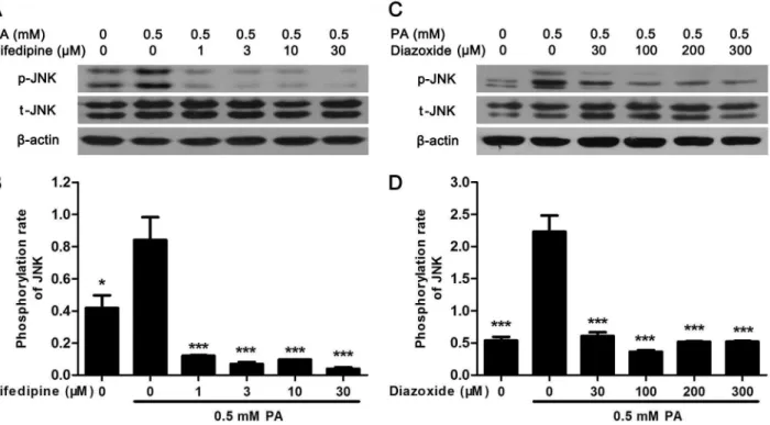
To further explore the signal pathway between calcium signal and apoptosis, the mitogen-acti-vated protein kinase (MAPK) signal was examined. It was found that PA-actimitogen-acti-vated JNK phos-phorylation was significantly suppressed in MIN6 cell by nifedipine or diazoxide (Fig 5A–5C). However, no alteration was found in PA-activated extracellular signal-regulated kinase (ERK) and p38 MAPK (Data not shown), suggested that inhibition of JNK phosphorylation but not ERK or p38 MAPK might be involved in the cytoprotective effect of nifedipine and diazoxide on PA-impairedβ-cells.
Fig 3. Nifedipine and diazoxide attenuate PA-activated ER-stress.(A) After MIN6 cells were co-treated with nifedipine and 0.5 mM PA for 48 h, the phosphorylation of eIF2αand the expression of CHOP were detected by western blot.β-actin was used for normalization. (B) The phosphorylation rate of eIF2αwas calculated as optical density of phosphorylated-eIF2α(p-eIF2α) divide by total eIF2α(t-eIF2α).*p<0.05 denote significant difference versus the PA-treated alone group, n = 3. (C) The expression of CHOP was calculated by optical density.*p<0.05 denote significant difference versus the PA-treated alone group, n = 3. (D) After MIN6 cells were co-treated with diazoxide and 0.5 mM PA for 48 h, the phosphorylation of eIF2α, the expression of CHOP were detected by western blot. (E) The phosphorylation rate of eIF2αwas calculated by optical density.*p<0.05 denote significant difference versus the
PA-treated alone group, n = 3. (F) The expression of CHOP was calculated by optical density.*p<0.05 denote significant difference versus the PA-treated alone group, n = 3. (G) After treatment, the cells were fixed and stained with CHOP and insulin antibodies. Red fluorescence indicated CHOP expression while green marked insulin. The nuclei were stained with DAPI dye. Scale bar = 50μm and referred to all panels.
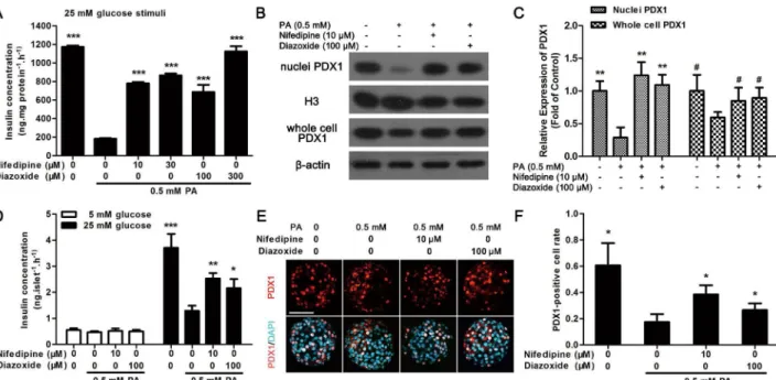
Nifedipine and diazoxide improved GSIS function in PA-impaired
pancreatic
β
-cells
As nifedipine and diazoxide generated protective effect on PA-impairedβ-cells, whether these two compounds could resist PA-induced impairment of insulin secretion in MIN6β-cells and primary cultured islets was investigated. It was found that nifedipine at a concentration of 10 and 30μM, diazoxide at 100 and 300μM could partly restore PA-impaired GSIS in MIN6 cells (Fig 6A). We next detected the expression of PDX1 in nuclei and whole cell to investigate the translocation of PDX1. It was found that the nuclei PDX1 was significantly reduced after 48 h PA treatment in MIN6 cells (Fig 6B and 6C). However, both 10μM nifedipine and 100μM Fig 4. Nifedipine and diazoxide inhibited PA-stimulated Ca2+release.(A) Pre-incubated of nifedipine dose-dependently inhibited 0.5 mM PA-stimulated Ca2+release in MIN6 cells. (B) There was no significant change in PA-induced Ca2+release between diazoxide co-treated and PA-treated alone group. The Ca2+ mobilization buffer did not contain glucose. (C) In the presence of 25.5 mM glucose, pre-incubated of diazoxide dose-dependently inhibited 0.5 mM PA-stimulated Ca2+release in MIN6 cells.
*p<0.05;**p<0.01; ***p<0.001 denote significant difference versus the PA-treated alone group, n = 6.
diazoxide increased the expression of nuclei PDX1 (Fig 6B and 6C). And PA-induced decreas-ing of PDX1 expression in whole cell was also significantly recovered by nifedipine and diazox-ide (Fig 6B and 6C).
Moreover, as the results in MIN6 cells, after 48 h incubation, 10μM nifedipine and 100μM diazoxide could both increase 25 mM glucose-induced insulin secretion in PA-treated isolated islets in comparison with PA-treated alone islets (Fig 6D). And nifedipine or diazoxide co-treated with PA could slightly improve 5 mM glucose-stimulated insulin secretion, but this effect was not statistically significant (Fig 6D). Meanwhile, translocation of PDX1 to the nucleus was detected by immunofluorescence to further evaluate the protective effect of nifedi-pine or diazoxide on PA-treated islets. As shown inFig 6E and 6F, PDX1 transported to nucleus was significantly reduced under PA-stimulation. Nonetheless, 10μM nifedipine and
100μM diazoxide partly restored the transport function of PDX1 to the nucleus (Fig 6E
and 6F).
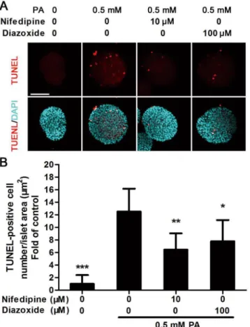
Nifedipine and diazoxide decreased PA-induced apoptosis in cultured
islets
To further evaluate whether the two Ca2+influx inhibitors, nifedipine and diazoxide, could reduce PA-induced apoptosis in primary cultured islets, TUNEL staining was used to indicate apoptosis by detecting DNA fragmentation. After the islets were treated with 0.5 mM PA for 48 h, the number of TUNEL-positive cells increased significantly compared to untreated islets (Fig 7A and 7B). As the experiments performed in MIN6 cells, nifedipine and diazoxide reduced PA-induced apoptosis in islets significantly (Fig 7A and 7B).
Fig 5. Nifedipine and diazoxide suppressed PA-induced phosphorylation of JNK.(A) After MIN6 cells were treated in 0.5 mM PA with/without different concentration of nifedipine for 48 h, the phosphorylation of JNK was detected by western blot. (B) The phosphorylation rate of JNK was calculated as optical density of phosphorylated-JNK (p-JNK) divide by total JNK (t-JNK).*p<0.05;***p<0.001 denote significant difference versus the PA-treated alone group, n = 3. (C) After the cells were treated in 0.5 mM PA with/without different concentration of diazoxide, the phosphorylation of JNK was detected by western blot. (D) The phosphorylation rate of JNK was calculated.***p<0.001 denote significant difference versus the PA-treated alone group, n = 3.
Discussion
During the development of T2D, western diets rich in saturated fats increase plasma lipid lev-els, which leads to obesity and insulin resistance, thereby aggravating compensatory insulin secretion in pancreaticβ-cells [21,22]. Meanwhile, elevated FFAs could not be oxidized in mitochondria but shunted into the esterification, caused accumulation of long-chain acyl-CoA ester in cytoplasm, consequently induced lipotoxicity and directly affectβ-cells [23]. Together factors could induceβ-cell dysfunction and apoptosis in T2D. Thus, reducing lipotoxicity to improve pancreaticβ-cell survival and function could provide benefit for improvement of T2D.
Chronic exposure to high levels of FFAs, especially saturated FFAs like PA, could activate Ca2+signal and induce abnormal cytosolic Ca2+homeostasis inβ-cells, thereby causing stress reaction and cell apoptosis [11,24]. Moreover, other stress factors, such as high level glucose and inflammatory cytokine induced apoptosis could be also associated with Ca2+signal [25,
26]. Therefore, inhibition of Ca2+release might provide benefit forβ-cell protection. It was reported that using some Ca2+influx blocker, including nifedipine and pituitary adenylate cyclase-activating polypeptide (PACAP), could reduce Ca2+toxicity in isletβ-cells [6,25,27]. Meanwhile, inhibition of GPR40-mediated PA-induced Ca2+release could also reduceβ-cell dysfunction [28]. Our previous study showed that using a small molecule GPR40 antagonist, DC260126, could reduce chronic FFAs-induced ER-stress and apoptosis inβ-cells [13]. And Fig 6. Nifedipine and diazoxide protected against PA-induced impairment of GSIS in pancreatic beta cells.(A) After MIN6 cells were treated with 0.5 mM PA in the presence/absence of nifedipine or diazoxide for 48 h, the GSIS induced by 25 mM glucose in MIN6 cells were determined.***p<0.001 denote
significant difference versus the PA-treated alone group, n = 6. (B) After MIN6 cells were treated with 0.5 mM PA in the presence/absence of nifedipine or diazoxide for 48 h, the expression of PDX1 in nuclei and whole cell was determined by western blot. (C) The expression of PDX1 was calculated by optical density.**p<0.01 compared to PA-treated alone group in nuclei PDX1 detection; ## p<0.01 compared to PA-treated alone group in whole cell PDX1 detection, n = 3. (D) After cultured islets were treated with PA in the presence/absence of different compounds for 48 h, the GSIS induced by 5 mM and 25 mM glucose were detected.*p<0.05;**p<0.01;***p<0.001 denote significant difference versus the PA-treated alone group, n = 6. (E) Cultured islets were
treated with PA in the presence/absence of different compounds for 48 h, then the PDX1 location was marked with red fluorescence, nuclei were dyed with DAPI to show blue fluorescence. Scale bar = 50μm and referred to all panels. (F) The PDX1-positive cell rate was calculated as PDX1-postive nuclei number divided by total nuclei number in each islet. 10 islets were analyzed from six duplicated wells.*p<0.05 denote significant difference versus the PA-treated alone group, n = 6.
inhibition of Ca2+release by DC260126 also decreased hyperinsulinemia and protectedβ-cells in diabetic animals [18,29]. These data were in according with results in the present study that using nifedipine or diazoxide, PA-inducedβ-cell apoptosis could be obviously reduced. How-ever, Tan et al. reported that 10μM diazoxide could not protect MIN6 cells from high glucose and FFAs-induced apoptosis [30], as other studies stated that 100μM diazoxide generate pro-tective effect onβ-cells under Ca2+signal-mediated impairment [15,31]. The different dose used in these experiment might contribute to the diverse results. Interestingly, we confirmed that unlike nifedipine, diazoxide could not inhibit Ca2+influx in MIN6β-cells in the absence of glucose. Thus, the protective efficacy of diazoxide might be less than L-type Ca2+channel blocker nifedipine. It might partially explain the controversial about the protective effect of diazoxide on lipotoxicβ-cells. Anyhow, it could still inferred that the different inhibitory pat-tern of the two compounds, nifedipine and diazoxide, on Ca2+influx was still associated with its protective effect onβ-cells. On the contrary, some compounds, like etoposide [32],
2,3,7,8-Tetrachlorodibenzo-p-Dioxin (TCDD) [26] and human islet amyloid polypeptide [33] could activate Ca2+influx, thereby causingβ-cell death. Together results suggested that inter-vening Ca2+release by compounds could provide benefits forβ-cell protection.
Fig 7. Nifedipine and diazoxide reduced cell apoptosis in PA-impaired islets.(A) Cultured islets were treated with PA in the presence/absence of different compounds for 48 h, then TUNEL staining was performed. Red fluorescence nuclei indicate apoptotic cells. Blue fluorescence showed all nuclei. Scale bar = 100μm and referred to all panels. (B) Apoptotic rate was calculated as TUNEL-positive cell number modified by islet area. 10 islets were analyzed from six duplicated wells.*p<0.05;**p<0.01 denote significant difference versus the PA-treated alone group, n = 6.
It was reported that Ca2+release from ER ofβ-cells could be observed in response to PA, thereafter this rapid depletion of ER Ca2+could contribute to PA-induced ER-stress and apo-ptosis inβ-cells [24,34]. Meanwhile, sustained Ca2+release increased demands on the ER for synthesis of insulin, subsequently increased unfolded protein response to initiate cell apoptosis [1]. During this process, one important ER-stress marker, CHOP induction in response to PA was also partially mediated by Ca2+influx [10]. Another ER-stress marker, PRKR-like endo-plasmic reticulum kinase (PERK) was also rapidly activated by PA inβ-cells, thereby activating apoptotic proteins like caspase-3 [35]. We found that PA-activated CHOP expression and PERK-mediated eIF2αphosphorylation in ER-stress were significantly increased after chronic PA stimulation, which corroborated the results in our previous study [13]. Oppositely, using Ca2+influx blockers nifedipine and diazoxide, PA-activated elevation of CHOP and eIF2α
phosphorylation were obviously suppressed, suggested that inhibition of Ca2+signal could be beneficial for reducing ER-stress and apoptosis in pancreaticβ-cells. In addition, we also tested the effect of nifedipine and diazoxide on cytokines- and H2O2-treated MIN6β-cell models. It
was found that there was less effect of these two compounds on neither cytokines- nor H2O2
-induced apoptosis in MIN6 cells (S1 Fig). Thus, the protective effect of nifedipine and diazox-ide might be mainly ascribed to ER-stress but not oxidative stress or inflammation.
During the development of T2D, insulin resistance could increase insulin secretion to main-tain normoglycemia, which termedβ-cell compensation [36]. However, sustained elevation of insulin secretion finally causesβ-cell exhaustion, subsequently induces apoptosis [36,37]. Thus, inhibition of Ca2+influx might not only suppress ER-stress to protect againstβ-cell apoptosis, but also reduce insulin secretion to decreaseβ-cell compensation. In the process of T2D, hyper-glycemia and hyperlipidemia could activation JNK phosphorylation to augment Forkhead box protein O1 (FOXO1), which is a transcription factor to regulate insulin synthesis inβ-cells [38]. FOXO1 could promote PDX1 translocation into nucleus, consequently augment insulin secretion [38,39]. On the contrary, chronic exposure to PA inhibits insulin gene transcription via altering PDX1 nuclear localization [39]. It was showed that nifedipine and diazoxide could both partially increase PA-impaired PDX1 expression and translocation to nuclei, thereby restor-ing GSIS ofβ-cells. Moreover, JNK activation could also directly induced ER-stress and apoptosis inβ-cells, whereas suppression of JNK phosphorylation by specific inhibitor, such as SP600125, could improveβ-cell survival both in vitro and in vivo [38,40]. In according with these results, we found that PA-induced JNK activation could be approximately abolished by nifedipine and diazoxide. It was reported that Ca2+could activate JNK via calmodulin-dependent protein kinase II (CaMK II) [41,42]. And JNK phosphorylation could directly promote ER-stress andβ-cell apoptosis [11,38]. Therefore, inhibition of JNK phosphorylation might be associated with the protective effect of the two Ca2+influx blockers on pancreaticβ-cells. However, the detailed rela-tionship between Ca2+influx and lipotoxicity in pancreaticβ-cells still need further investigation.
Conclusions
It was found that both nifedipine and diazoxide could inhibit PA-stimulated Ca2+release, thereby reducing PA-mediated lipotoxicity in pancreaticβ-cells. And inhibition of Ca2+release also suppressed chronic sustained insulin secretion to reduceβ-cell decompensation. These present data provided one possibility that inhibition of chronic FFAs-stimulated insulin secre-tion by regulasecre-tion Ca2+release might provide benefit forβ-cell protection in T2D.
Supporting Information
S1 Fig. Nifedipine and diazoxide had less effect on H2O2and cytokine-treated MIN6 cells.
Acknowledgments
We gratefully acknowledge the kind provision of MIN6 cells by Professor S. Seino.
Author Contributions
Conceived and designed the experiments: HW PS. Performed the experiments: YZ PS TW. Analyzed the data: YZ PS. Contributed reagents/materials/analysis tools: KC WZ. Wrote the paper: PS HW.
References
1. Prentki M, Nolan CJ. Islet beta cell failure in type 2 diabetes. J Clin Invest. 2006; 116(7):1802–12. doi: 10.1172/JCI29103PMID:16823478; PubMed Central PMCID: PMC1483155.
2. Cernea S, Dobreanu M. Diabetes and beta cell function: from mechanisms to evaluation and clinical implications. Biochem Med (Zagreb). 2013; 23(3):266–80. PMID:24266296; PubMed Central PMCID:
PMC3900074.
3. Giacca A, Xiao C, Oprescu AI, Carpentier AC, Lewis GF. Lipid-induced pancreatic beta-cell dysfunc-tion: focus on in vivo studies. Am J Physiol Endocrinol Metab. 2011; 300(2):E255–62. doi:10.1152/ ajpendo.00416.2010PMID:21119027.
4. Gromada J. The free fatty acid receptor GPR40 generates excitement in pancreatic beta-cells. Endocri-nology. 2006; 147(2):672–3. doi:10.1210/en.2005-1388PMID:16418431.
5. Itoh Y, Kawamata Y, Harada M, Kobayashi M, Fujii R, Fukusumi S, et al. Free fatty acids regulate insu-lin secretion from pancreatic beta cells through GPR40. Nature. 2003; 422(6928):173–6. doi:10.1038/ nature01478PMID:12629551.
6. Remizov O, Jakubov R, Dufer M, Krippeit Drews P, Drews G, Waring M, et al. Palmitate-induced Ca2 +-signaling in pancreatic beta-cells. Mol Cell Endocrinol. 2003; 212(1–2):1–9. PMID:14654245.
7. Nolan CJ, Madiraju MS, Delghingaro-Augusto V, Peyot ML, Prentki M. Fatty acid signaling in the beta-cell and insulin secretion. Diabetes. 2006; 55 Suppl 2:S16–23. doi:10.2337/diabetesPMID:17130640.
8. Cnop M, Hannaert JC, Hoorens A, Eizirik DL, Pipeleers DG. Inverse relationship between cytotoxicity of free fatty acids in pancreatic islet cells and cellular triglyceride accumulation. Diabetes. 2001; 50 (8):1771–7. PMID:11473037.
9. Cnop M, Ladriere L, Igoillo-Esteve M, Moura RF, Cunha DA. Causes and cures for endoplasmic reticu-lum stress in lipotoxic beta-cell dysfunction. Diabetes Obes Metab. 2010; 12 Suppl 2:76–82. doi:10. 1111/j.1463-1326.2010.01279.xPMID:21029303.
10. Cunha DA, Hekerman P, Ladriere L, Bazarra-Castro A, Ortis F, Wakeham MC, et al. Initiation and exe-cution of lipotoxic ER stress in pancreatic beta-cells. J Cell Sci. 2008; 121(Pt 14):2308–18. doi:10. 1242/jcs.026062PMID:18559892; PubMed Central PMCID: PMC3675788.
11. Choi SE, Kim HE, Shin HC, Jang HJ, Lee KW, Kim Y, et al. Involvement of Ca2+-mediated apoptotic signals in palmitate-induced MIN6N8a beta cell death. Mol Cell Endocrinol. 2007; 272(1–2):50–62. doi: 10.1016/j.mce.2007.04.004PMID:17507155.
12. Rorsman P, Arkhammar P, Bokvist K, Hellerstrom C, Nilsson T, Welsh M, et al. Failure of glucose to elicit a normal secretory response in fetal pancreatic beta cells results from glucose insensitivity of the ATP-regulated K+ channels. Proc Natl Acad Sci U S A. 1989; 86(12):4505–9. PMID:2543980; PubMed
Central PMCID: PMC287299.
13. Wu J, Sun P, Zhang X, Liu H, Jiang H, Zhu W, et al. Inhibition of GPR40 protects MIN6 beta cells from palmitate-induced ER stress and apoptosis. J Cell Biochem. 2012; 113(4):1152–8. doi:10.1002/jcb. 23450PMID:22275065.
14. Trus M, Corkey RF, Nesher R, Richard AM, Deeney JT, Corkey BE, et al. The L-type voltage-gated Ca2+ channel is the Ca2+ sensor protein of stimulus-secretion coupling in pancreatic beta cells. Bio-chemistry. 2007; 46(50):14461–7. doi:10.1021/bi7016816PMID:18027971.
15. Sargsyan E, Ortsater H, Thorn K, Bergsten P. Diazoxide-induced beta-cell rest reduces endoplasmic reticulum stress in lipotoxic beta-cells. J Endocrinol. 2008; 199(1):41–50. doi:10.1677/JOE-08-0251
PMID:18644846.
17. Wang T, Sun P, Chen L, Huang Q, Chen K, Jia Q, et al. Cinnamtannin D-1 Protects Pancreatic beta-Cells from Palmitic Acid-Induced Apoptosis by Attenuating Oxidative Stress. J Agric Food Chem. 2014; 62(22):5038–45. doi:10.1021/jf500387dPMID:24815044.
18. Sun P, Wang T, Zhou Y, Liu H, Jiang H, Zhu W, et al. DC260126: a small-molecule antagonist of GPR40 that protects against pancreatic beta-Cells dysfunction in db/db mice. PLoS One. 2013; 8(6): e66744. doi:10.1371/journal.pone.0066744PMID:23776696; PubMed Central PMCID: PMC3679087. 19. Hu H, He LY, Gong Z, Li N, Lu YN, Zhai QW, et al. A novel class of antagonists for the FFAs receptor
GPR40. Biochem Biophys Res Commun. 2009; 390(3):557–63. doi:10.1016/j.bbrc.2009.10.004
PMID:19818732.
20. Iglesias P, Diez JJ. Management of endocrine disease: a clinical update on tumor-induced hypoglyce-mia. Eur J Endocrinol. 2014; 170(4):R147–57. doi:10.1530/EJE-13-1012PMID:24459236.
21. Cnop M, Welsh N, Jonas JC, Jorns A, Lenzen S, Eizirik DL. Mechanisms of pancreatic beta-cell death in type 1 and type 2 diabetes: many differences, few similarities. Diabetes. 2005; 54 Suppl 2:S97–107.
PMID:16306347.
22. Kahn BB. Type 2 diabetes: when insulin secretion fails to compensate for insulin resistance. Cell. 1998; 92(5):593–6. PMID:9506512.
23. Prentki M, Joly E, El-Assaad W, Roduit R. Malonyl-CoA signaling, lipid partitioning, and glucolipotoxi-city: role in beta-cell adaptation and failure in the etiology of diabetes. Diabetes. 2002; 51 Suppl 3: S405–13. PMID:12475783.
24. Gwiazda KS, Yang TL, Lin Y, Johnson JD. Effects of palmitate on ER and cytosolic Ca2+ homeostasis in beta-cells. Am J Physiol Endocrinol Metab. 2009; 296(4):E690–701. doi:10.1152/ajpendo.90525. 2008PMID:19141690.
25. Yanagida K, Yaekura K, Arima T, Yada T. Glucose-insensitivity induced by Ca(2+) toxicity in islet beta-cells and its prevention by PACAP. Peptides. 2002; 23(1):135–42. PMID:11814628.
26. Kim YH, Shim YJ, Shin YJ, Sul D, Lee E, Min BH. 2,3,7,8-tetrachlorodibenzo-p-dioxin (TCDD) induces calcium influx through T-type calcium channel and enhances lysosomal exocytosis and insulin secre-tion in INS-1 cells. Int J Toxicol. 2009; 28(3):151–61. doi:10.1177/1091581809336885PMID: 19546254.
27. Wang Y, Gao L, Li Y, Chen H, Sun Z. Nifedipine protects INS-1 beta-cell from high glucose-induced ER stress and apoptosis. Int J Mol Sci. 2011; 12(11):7569–80. doi:10.3390/ijms12117569PMID: 22174617; PubMed Central PMCID: PMC3233423.
28. Kristinsson H, Smith DM, Bergsten P, Sargsyan E. FFAR1 is involved in both the acute and chronic effects of palmitate on insulin secretion. Endocrinology. 2013; 154(11):4078–88. doi: 10.1210/en.2013-1352PMID:24035997.
29. Zhang X, Yan G, Li Y, Zhu W, Wang H. DC260126, a small-molecule antagonist of GPR40, improves insulin tolerance but not glucose tolerance in obese Zucker rats. Biomed Pharmacother. 2010; 64 (9):647–51. doi:10.1016/j.biopha.2010.06.008PMID:20888730.
30. Tan C, Voss U, Svensson S, Erlinge D, Olde B. High glucose and free fatty acids induce beta cell apo-ptosis via autocrine effects of ADP acting on the P2Y(13) receptor. Purinergic Signal. 2013; 9(1):67–79.
doi:10.1007/s11302-012-9331-6PMID:22941026; PubMed Central PMCID: PMC3568432. 31. Chang I, Cho N, Kim S, Kim JY, Kim E, Woo JE, et al. Role of calcium in pancreatic islet cell death by
IFN-gamma/TNF-alpha. J Immunol. 2004; 172(11):7008–14. PMID:15153522.
32. Arora DK, Mohammed AM, Kowluru A. Nifedipine prevents etoposide-induced caspase-3 activation, prenyl transferase degradation and loss in cell viability in pancreatic beta-cells. Apoptosis. 2013; 18 (1):1–8. doi:10.1007/s10495-012-0763-9PMID:23054080; PubMed Central PMCID: PMC3545074.
33. Casas S, Novials A, Reimann F, Gomis R, Gribble FM. Calcium elevation in mouse pancreatic beta cells evoked by extracellular human islet amyloid polypeptide involves activation of the mechanosensi-tive ion channel TRPV4. Diabetologia. 2008; 51(12):2252–62. doi:10.1007/s00125-008-1111-zPMID: 18751967.
34. Kharroubi I, Ladriere L, Cardozo AK, Dogusan Z, Cnop M, Eizirik DL. Free fatty acids and cytokines induce pancreatic beta-cell apoptosis by different mechanisms: role of nuclear factor-kappaB and endoplasmic reticulum stress. Endocrinology. 2004; 145(11):5087–96. doi:10.1210/en.2004-0478
PMID:15297438.
35. Diakogiannaki E, Morgan NG. Differential regulation of the ER stress response by long-chain fatty acids in the pancreatic beta-cell. Biochem Soc Trans. 2008; 36(Pt 5):959–62. doi:10.1042/ BST0360959PMID:18793169.
36. Poitout V, Robertson RP. Minireview: Secondary beta-cell failure in type 2 diabetes—a convergence of
glucotoxicity and lipotoxicity. Endocrinology. 2002; 143(2):339–42. doi:10.1210/endo.143.2.8623
37. Kaiser N, Nesher R, Donath MY, Fraenkel M, Behar V, Magnan C, et al. Psammomys obesus, a model for environment-gene interactions in type 2 diabetes. Diabetes. 2005; 54 Suppl 2:S137–44. PMID: 16306331.
38. Martinez SC, Tanabe K, Cras-Meneur C, Abumrad NA, Bernal-Mizrachi E, Permutt MA. Inhibition of Foxo1 protects pancreatic islet beta-cells against fatty acid and endoplasmic reticulum stress-induced apoptosis. Diabetes. 2008; 57(4):846–59. doi:10.2337/db07-0595PMID:18174526.
39. Hagman DK, Hays LB, Parazzoli SD, Poitout V. Palmitate inhibits insulin gene expression by altering PDX-1 nuclear localization and reducing MafA expression in isolated rat islets of Langerhans. J Biol Chem. 2005; 280(37):32413–8. doi:10.1074/jbc.M506000200PMID:15944145; PubMed Central
PMCID: PMC1361267.
40. Hirosumi J, Tuncman G, Chang L, Gorgun CZ, Uysal KT, Maeda K, et al. A central role for JNK in obe-sity and insulin resistance. Nature. 2002; 420(6913):333–6. doi:10.1038/nature01137PMID: 12447443.
41. Enslen H, Tokumitsu H, Stork PJ, Davis RJ, Soderling TR. Regulation of mitogen-activated protein kinases by a calcium/calmodulin-dependent protein kinase cascade. Proc Natl Acad Sci U S A. 1996; 93(20):10803–8. PMID:8855261; PubMed Central PMCID: PMC38236.