Adiponectin is protective against endoplasmic
reticulum stress-induced apoptosis of endothelial cells
in sepsis
Yun Hou
1*, Xi Feng Wang
2*, Zhi Qiang Lang
3, Yin Chuan Jin
1, Jia Rong Fu
4, Xiao Min Xv
4,
Shi Tian Sun
4, Xin Xin
4and Lian Shuang Zhang
1 1Department of Histology and Embryology, Binzhou Medical University, Yan Tai, China
2Department of Critical Care Medicine, Yu Huang Ding Hospital, Qingdao University, Yan Tai, China 3Department of Pathology, Yu Huang Ding Hospital, Qingdao University, Yan Tai, China 4College of Clinical Medicine, Bin Zhou Medical University, Yan Tai, China
Abstract
Endoplasmic reticulum (ER) stress is a critical molecular mechanism involved in the pathogenesis of sepsis. Hence, strategies for alleviating this stress may be essential for preventing cardiovascular injuries under sepsis. Adiponectin is secreted by adipocytes and its levels are decreased in sepsis. The purpose of this study was to investigate the protective effects of adiponectin treatment on endothelial cells and its mechanism. Male Wistar rats underwent cecal ligation and puncture (CLP) before being treated with adiponectin (72 and 120mg/kg). The levels of malondialdehyde (MDA) in plasma,
histological structure, and apoptosis of endothelial cells were evaluated. In vitro, human umbilical vein endothelial cells (HUVECs) were treated with adiponectin at 10 and 20mg/mL for 24 h after stimulation by lipopolysaccharide (LPS). The
levels of reactive oxygen species (ROS), ultrastructure, rate of apoptosis, the expression of inositol-requiring enzyme 1a(IRE1a) protein, and its downstream molecules (78 kDa glucose-regulated protein (GRP78), C/EBP homologous protein (CHOP), and caspase-12) were detected. The results showed that the levels of MDA and ROS induced by CLP or LPS stimulation were increased. Furthermore, endothelial cell apoptosis was increased under sepsis. The IRE1apathway was
initiated, as evidenced by activated IRE1a, increased GRP78, and up-regulated CHOP and caspase-12 in HUVECs. Following treatment with adiponectin, the number of apoptotic endothelial cells was markedly decreased. These findings demonstrated that treatment with adiponectin decreased apoptosis of endothelial cells caused by sepsis by attenuating the ER stress IRE1apathway activated by oxidative stress.
Key words: Adiponectin; Apoptosis; Endoplasmic reticulum stress; Endothelial cell; Sepsis
Introduction
Sepsis is a life-threatening clinical condition character-ized by organ dysfunction and shock (1). Organ dysfunc-tion is closely related to blood circuladysfunc-tion disorders. Septic patients commonly suffer from a dysregulated and acti-vated coagulation system. The occurrence of coagulation dysfunction is related to endothelial cells that are located in the innermost layer of blood vessels, making them one of the primary targets of the inflammatory response during sepsis (2). Lipopolysaccharide (LPS) and other inflamma-tory factors in blood stimulate endothelial cells (3) and result in endothelial cell injury (4). Previous studies have shown that some proinflammatory cytokines, especially tumor necrosis factor-a, interleukin (IL)-1, and procoagulants
such as tissue factor, intercellular adhesion molecule-1, and vascular cell adhesion molecule-1 are released from damaged endothelium (5,6) and further promote coagu-lation disorders. Together, these can induce disseminated intravascular coagulation and eventually lead to multiple organ failure (7,8).
Endoplasmic reticulum (ER) is an important organelle for cellular protein synthesis and processing, maintaining stability of the intracellular environment. ER stress plays an important role in the pathological states of sepsis (9,10). Thus, it is crucial to investigate novel interventions to improve the endothelial injury induced by ER stress, and prevent coagulation disorders in severely septic patients.
Correspondence: Lian Shuang Zhang:<[email protected]>
*These authors contributed equally to this work.
Sepsis also accompanies oxidative stress, which induces the formation of reactive oxygen species (ROS) and malondialdehyde (MDA), a marker of lipid peroxidation (11). The acceleration of ER malfunction is associated with oxidative stress (12,13).
Adiponectin is a serum adipokine secreted primarily by adipocytes. It is associated with obesity, atherosclerosis, and coronary artery disease (14). Apart from its antiox-idant and anti-inflammatory activities in some disease states (15), previous studies have shown that adiponectin can improve the function of vascular endothelium (16). Moreover, recent research showed that adiponectin pro-moted the proliferation of bovine mammary epithelial cells via inhibiting ER stress responses (17).
Adiponectin has different oligomeric isoforms, includ-ing trimeric, hexameric, and the high molecular weight (HMW) oligomeric complex (18). Septic patients exhib-it low levels of total adiponectin (19) and a significant increase in total and HMW adiponectin, which is related to clinical recovery from sepsis (20) (Supplementary Table S1). However, few reports focus on whether adiponectin can protect the endothelium and is involved in the mechanisms of sepsis. Hence, the purpose of this study was to investigate the protective effects of adipo-nectin treatment on alleviating apoptosis of endothelial cells and reveal its related mechanism about oxidative stress and ER stress in rat and human umbilical vein endothelial cell (HUVECs) models of sepsis.
Material and Methods
Design ofin vivoexperiments
All the animal experiments were carried out in accord-ance with the Guide for the Care and Use of Laboratory Animals by the National Institutes of Health and the Ethical Committee of Binzhou Medical University. Eight-week-old Wistar rats weighing 250 to 300 g were main-tained on a 12-h light/dark cycle at 22±3°C. The animals were allowed free access to food and water. After one week of acclimation, the animals were randomly divided into 4 groups of 12 rats each: sham, cecal liga-tion and puncture (CLP), CLP + adiponectin (72mg/kg) treatment, and CLP+adiponectin (120mg/kg) treatment. The CLP model was generated using the method described in our previous experiments (21). The dose of adiponectin was selected based on the results of pre-experiments.
The rats in the sham group received 120 mg/kg of saline intravenously (iv), 12 h after undergoing a sham operation consisting of laparotomy and bowel manipula-tion but no CLP; the rats in the CLP group received 120mg/kg salineiv, 12 h after undergoing CLP; and the rats in the adiponectin treatment groups 72 or 120mg/kg of adiponectinivafter undergoing CLP. After 24 h, the rats were sacrificed and the abdominal aorta was removed and fixed until subsequent analysis.
Measurement of plasma MDA
The concentration of MDA was determined using an MDA kit (Jiancheng Biotechnology Company, China). All the reagents and samples were prepared according to the manufacturer’s protocol. Absorbance was measured at 532 nm with a microplate reader (DNM-9602G, Perlong Medical Equipment, China).
TdT-mediated dUTP nick end labeling (TUNEL) The apoptosis of endothelial cells in the tissue sections was detected by the terminal deoxyribonucleotide transfer-ase-mediated nick-end labeling (TUNEL) assay kit (Key-gen, China), according to the manufacturer’s instructions. The slides were placed in DAB for 5 min and stained with hematoxylin. These analyses were performed under a light microscope at 400 magnification with 15 differentfields using computer-aided software (Olympus X71-F22PH, Japan). The gray values of the apoptotic cells were quantified by computer-assisted image analysis (Leica LAS Image Analysis V4.0, Germany).
Cell culture and design ofin vitroexperiments Human umbilical vein endothelial cells (HUVECs) were purchased from ATCC Manassas (No. CRL-1730, USA). The cells were cultured in a complete medium consisting of DMEM (HyClone, USA), supplemented with 10% fetal bovine serum (Gibco, USA), 100 U/mL of penicillin, and 100mg/mL of streptomycin.
The cells were divided into control, LPS stimulation, and LPS stimulation+adiponectin treatment groups. LPS (1 mg/mL of LPS for 12 h) was added to the culture medium of the LPS stimulation group; the LPS stimulation+ adiponectin treatment groups were treated with 10mg/mL
or 20 mg/mL of adiponectin for 24 h after exposure to 1mg/mL of LPS for 12 h. To explore the role of oxidative stress and the inositol-requiring enzyme 1a (IRE1a) pathways at the cellular level, the HUVECs were pre-treated with a ROS inhibitor (NAC, 1 mM) and an IRE1a inhibitor (STF-083010, 10mM) for 4 h prior to LPS exposure. An equivalent volume of DMEM was added to the control group. Five replicate wells were used for each treatment group.
Laser confocal microscopy
The cells were incubated with 2,7-dichlorofluorescein-diacetate (DCFH-DA, 50 ng/mL, Jiancheng, China) at 37°C for 30 min in the dark. The cells were subsequently washed with PBS and observed under a confocal laser scanning microscope (Leica), and the images were captured. The mean fluorescence intensity of dichlorofluorescein (DCF) was analyzed.
Western blotting
The cell protein lysates were electrophoretically sepa-rated and transferred to polyvinylidene fluoride (PVDF) membranes. After blocking, the PVDF membranes were incubated overnight at 4°C with IRE1a(1:800), p-IRE1a (1:600), caspase 12 (1:800), C/EBP homologous protein (CHOP, 1:1000), and 78 kDa glucose-regulated protein (GRP78, 1:1000), all rabbit antibodies (CST, USA). The membranes were then incubated with anti-rabbit IgG secondary antibody (CST). After washing, the proteins bound to the antibody were measured by a chemilumi-nescent reagent (Thermo Fisher Scientific, USA). Den-sitometric analyses were performed using the Image LabtSoftware (Bio-Rad Laboratories Inc., USA).
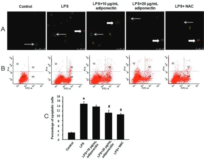
Laser confocal microscopy andflow cytometry The LPS-induced apoptosis of cells was observed by an apoptosis detection kit (Keygen), according to the
Figure 2.Effects of adiponectin on the expres-sions of TUNEL positive cells in each group.Top panel, The arrows indicate TUNEL positive cells. Bottom panel, Gray values of the TUNEL positive cells in the four groups. Scale bar: 30mm. *Po0.05 compared to the sham group,#P
manufacturer’s protocols. The cells were stained with FITC-conjugated annexin V and propidium iodide (PI) and observed under a confocal laser scanning microscope (Leica).
To evaluate the rate of apoptosis, the HUVECs were stained with both FITC-conjugated annexin V and PI, and analyzed by an apoptosis detection kit (KeyGen). The cells incubated with FITC-conjugated annexin V and PI were analyzed by aflow cytometer (Millipore, USA). The different populations of cells were identified by the different labeling patterns in the Annexin V-PI analysis.
Transmission electron microscopy
Transmission electron microscopy (TEM) was per-formed according to experimental procedures. The cells were fixed by immersing in Karnovsky’s fixative at 4°C after washing and dehydrating in increasing concentra-tions of ethanol, after which the cells were embedded in Epon using Beem capsules (Germany). Ultrathin 60-nm thick sections were cut and stained with lead citrate and uranyl acetate and viewed under a transmission electron microscope (JEM 2100, Japan).
Statistical analysis
Data were analyzed using the SPSS software, version 17.0 (USA). The in vivo differences among groups were analyzed by the Kruskal-Wallis test, while the in vitro
differences were analyzed by a one-way analysis of variance (ANOVA) followed by the Fisher’s least significant difference (LSD) test. The data are reported as means±SE. The results were considered significant at Po0.05.
Results
Treatment with adiponectin downregulated plasma levels of MDA
The plasma levels of MDA were significantly higher in the CLP group than those of the sham group; how-ever, there was no difference in the plasma levels of MDA between the adiponectin (72 mg/kg) treatment group and the CLP group (P40.05). Compared to the plasma levels of MDA in the CLP group, those of the adiponectin (120 mg/kg) treatment group were signifi-cantly lower (Po0.05) (Figure 1).
Adiponectin treatment alleviated the apoptosis of endothelial cells in vascular tissue
The results showed that the immunopositive TUNEL staining was observed in the endothelial cells of all groups (Figure 2). The number of TUNEL-positive cells was signif-icantly higher in the CLP group compared to that of the sham group (Po0.05). However, the number of TUNEL-positive cells decreased significantly in the adiponectin (120mg/kg) treatment group compared to the CLP group (Figure 2).
Adiponectin treatment downregulated the levels of ROS in HUVECs
The intracellular levels of ROS were determined using the DCFH-DA probe (Figure 3A). As shown in Figure 3B, ROS significantly increased in the LPS group compared to the control group. The reduction in the expression of ROS was significant in the LPS+20mg/mL adiponectin group compared to the LPS group (Po0.05). Additionally, NAC decreased the production of ROS in the LPS-stimulated HUVECs (Po0.05).
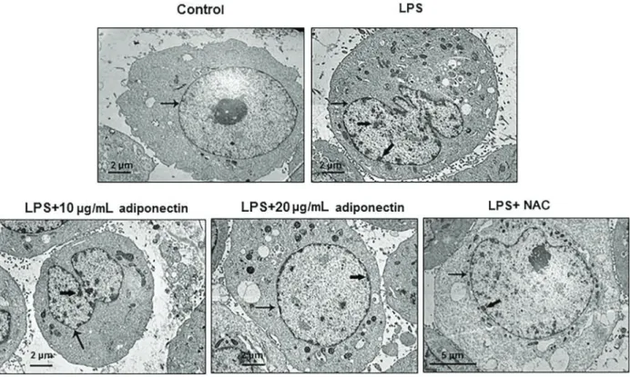
Adiponectin treatment altered the ultrastructure of HUVECs
To gain more information on an ultrastructural level, TEM was used to observe the ultrastructure of the HUVECs.
As shown in Figure 4, the cell membrane and nuclei appeared clear in the control group. However, in the LPS group and adiponectin (10mg/mL) treatment group, the chromatin appeared fragmented, as indicated by the thick arrow (Figure 4). Chromatin aggregation improved following treatment with adiponectin, and was only aggregated in the inner side of the nuclear membrane in the adiponectin (20 mg/mL) treatment group and the STF-083010 (IRE1ainhibitor)-treated group.
Treatment with adiponectin reduced apoptosis by inhibiting the activation of IRE1ain the HUVECs
The early apoptotic cells were stained with green fluorescent dye (indicated by thin arrows) and the late apoptotic cells were stained with green and red fluores-cent dyes (indicated by thick arrows) (Figure 5A). The results of flow cytometry indicated that there was a significant increase in the percentage of apoptotic cells in the LPS group compared to the control group, and a significant decrease in the adiponectin (20 mg/mL) treatment group (Po0.05) compared to the LPS group. Moreover, treatment with the IRE1a inhibitor (STF-083010, 10mM) significantly decreased the percentage of LPS-induced apoptotic cells (Po0.05) (Figure 5B and C).
Treatment with adiponectin altered the expression of proteins involved in the IRE1apathway in the HUVECs
The expression of IRE1a remained unaltered in all groups (P40.05). The expression of p-IRE1asignificantly increased in the LPS group compared to the control group (Po0.05), but decreased significantly in the adiponectin (20 mg/mL) treatment group (Po0.05), compared to the LPS group. Moreover, treatment with NAC, the inhibitor of oxidative stress, significantly decreased the expres-sion of p-IRE1a in the LPS-treated HUVECs (Po0.05, Figure 6A, B, and C). The expression of GRP78, CHOP, and caspase-12 increased significantly in the LPS group compared to the control group (Po0.05). However, treatment with adiponectin and STF-083010 signifi-cantly decreased the expressions of GRP78, CHOP,
and caspase-12 compared to those after LPS treatment (Po0.05, Figure 6D, E, F, and G).
Discussion
Coagulation dysfunction under sepsis has become the major factor affecting the prognosis of critically ill patients in intensive care units. Endothelial cells are related to coagulation because they produce numerous factors, such as tissue factor, that affect this process (8,22). Coagulation cascades triggered by endothelial damage can induce microcirculatory dysfunction that is the major mechanism in sepsis (8). As such, it is important to understand the endothelial cell injury occurring under sepsis and investigate potential therapies.
Adiponectin is secreted from adipose tissue, and endogenous adiponectin exerts protective effects against hypertensive vascular injury (23). Previous studies reported that the adiponectin concentration was lower in some cardiovascular diseases (24), and was reduced in mice subjected to CLP (25). One recent study showed that mortality was increased in adiponectin-null mice follow-ing CLP (26), (Table S1), and clinical recovery from sepsis, which is related to an increase in total and HMW adipo-nectin, was investigated (20). Thus, together, these studies indicate that exogenous supplementation with adiponectin may have protective effects on sepsis.
In our study, sepsis was induced in rats by CLP, which closely resembles the clinical pathophysiologic course of sepsis (27). Our results showed that endothelial cell injury and apoptosis were induced following CLP. This injury was characterized by nuclear pyknosis and exfoliation from the vessel wall. In addition, the number of cells stained positive with the TUNEL assay was increased. Exogenous adiponectin at a dose of 120mg/kg attenuated endothelial cell apoptosis in the septic rats.
In vitro, HUVECs apoptosis was induced by LPS stimulation. The specific manifestations were the appear-ance of chromatin fragments under TEM and an increased amount of apoptosis. Adiponectin at a concentration of 20 mg/mL decreased the extent of HUVECs apoptosis. These results suggest that adiponectin may play a protective role in endothelial cells and alleviate their injury in sepsis. Thesefindings are consistent with previous reports that adiponectin could exert vasculo-protective effects by inhibit-ing endothelial cell activation (28,29).
To gain insight into the protective mechanisms of adiponectin for endothelial cell injury, oxidative and ER stress of HUVECs were examined. The ER is the key organelle for cellular protein synthesis and processing, maintaining stability of the intracellular environment (30). Various extracellular (deprivation of calcium ions, hypoxia, and hyper-/hypo-osmosis) and intracellular (DNA damage or DNA replication stress) stresses can influence cell homeostasis (31). To adapt to such persistent unresolved stresses, misfolded proteins in the ER accumulate, and ER stress signals, such as IRE1a, are activated (32,33). Under physiological conditions, the luminal domains of IRE1aare bound to the ER molecular chaperone GRP78, which keeps it inactive. When unfolded (or misfolded), proteins accumulate in the lumen of the ER and IRE1ais activat-ed through dissociation from GRP78. Upon activation, IRE1ainduces signal transduction events that alleviate the
accumulation of unfolded/misfolded proteins in the ER by increasing expression of GRP78 (34). Moreover, IRE1a activation can simultaneously activate the CHOP signaling pathway (35,36), resulting in cell apoptosis (37).
LPS, a constituent of the cell wall of gram-negative bacteria, is an activator of oxidative stress in sepsis, inducing the formation of ROS and MDA (11,12). ROS are associated with accelerated ER malfunction (13,14). The present results demonstrated that treating HUVECs with NAC down-regulated the phosphorylation of IRE1a, and the IRE1ainhibitor (STF-083010) reduced apoptosis of HUVECs as detected by TEM andflow cytometry.
Previous studies have shown that adiponectin alle-viated ER stress in various cells and tissues, including human liver cells (38), mouse adipocytes (39), and rat smooth muscle cells (40). The current results showed that treatment with either adiponectin at a concentration of 20 mg/mL, or STF-083010, significantly decreased LPS-induced GRP78, CHOP, and cleaved caspase-12 expression, and apoptosis of HUVECs. This demon-strated the protective effects of adiponectin on endothelial cells.
In summary, we have confirmed for thefirst time that the administration of adiponectin may alleviate endothelial cell apoptosis by suppressing the ER stress IRE1a path-way. Therefore, endothelium protection with adiponectin may be a potential therapeutic strategy for coagulation dysfunction in sepsis. This will provide an early treatment reference strategy for clinical prevention of coagulopathy caused by sepsis. Although we demonstrated the protective effect of adiponectin on endothelial injury induced by sepsis, further mechanisms still need to be investigated, including finding new cell signaling pathways and new effective factors. In addition, clinical trials are needed to verify its effects on sepsis therapy.
Supplementary Material
Click here to view [pdf].
Acknowledgments
This study was supported by Nature Science Founda-tion from Shandong Province (No. ZR2016HL24, ZR2016 HP05 and ZR2017BH039), the National Natural Science Foundation of China (No.81601049), and the Project of Shandong Province Higher Educational Science and Technology Program (No. J16LK01)
References
1. Singer M. The new sepsis consensus definitions (Sepsis-3): the good, the not-so-bad, and the actually-quite-pretty. Intensive Care Med2016; 42: 2027–2029, doi: 10.1007/ s00134-016-4600-4.
2. Deutschman CS, Tracey KJ. Sepsis: current dogma and new perspectives.Immunity2014; 40: 463–475, doi: 10.1016/ j.immuni.2014.04.001.
impairment, and mortality in rats submitted to sepsis by cecal ligation and puncture.J Surg Res2012; 178: 390–400, doi: 10.1016/j.jss.2012.01.041.
4. Xu S, Zhou Z, Li H, Liu Z, Pan X, Wang F, et al. BMSCs ameliorate septic coagulopathy by suppressing infl amma-tion in cecal ligaamma-tion and puncture-induced sepsis.J Cell Sci 2018; 131: jcs211151, doi: 10.1242/jcs.211151.
5. Pruitt JH, Copeland EM, Moldawer LL. Interleukin-1 and interleukin-1 antagonism in sepsis, systemic inflammatory response syndrome, and septic shock.Shock1995; 3: 235– 251, doi: 10.1097/00024382-199504000-00001.
6. Barriere SL, Lowry SF. An overview of mortality risk predic-tion in sepsis.Crit Care Med1995; 23: 376–393, doi: 10.1097/ 00003246-199502000-00026.
7. Vincent JL, De Backer D. Microvascular dysfunction as a cause of organ dysfunction in severe sepsis.Crit Care2005; 9 (Suppl 4): S9–S12, doi: 10.1186/cc3748.
8. Schouten M, Wiersinga WJ, Levi M, van der Poll T. Infl am-mation, endothelium, and coagulation in sepsis.J Leukoc Biol2008; 83: 536–545, doi: 10.1189/jlb.0607373. 9. Schildberg FA, Schulz S, Dombrowski F, Minor T. Cyclic
AMP alleviates endoplasmic stress and programmed cell death induced by lipopolysaccharides in human endothelial cells. Cell Tissue Res 2005; 320: 91–98, doi: 10.1007/ s00441-004-1066-4.
10. Kozlov AV, Duvigneau JC, Miller I, Nürnberger S, Gessl-bauer B, Kungl A, et al. Endotoxin causes functional endoplasmic reticulum failure, possibly mediated by mito-chondria.Biochim Biophys Acta2009; 1792: 521–530, doi: 10.1016/j.bbadis.2009.03.004.
11. Novelli GP. Role of free radicals in septic shock.J Physiol Pharmacol1997; 48: 517–527.
12. Chaudhari N, Talwar P, Parimisetty A, Lefebvre d’ Hellen-court C, Ravanan P. A molecular web: endoplasmic reticulum stress, inflammation, and oxidative stress. Front Cell Neurosci2014; 8: 213, doi: 10.3389/fncel.2014.00213. 13. Lin CL, Lee CH, Chen CM, Cheng CW, Chen PN, Ying TH, et al. Protodioscin induces apoptosis through ROS-mediated endoplasmic reticulum stress via the JNK/p38 activation pathways in human cervical cancer cells. Cell Physiol Biochem2018; 46: 322–334, doi: 10.1159/000488433. 14. Arita Y, Kihara S, Ouchi N, Takahashi M, Maeda K,
Miyagawa J, et al. Paradoxical decrease of an adipose-specific protein, adiponectin, in obesity.1999. Biochem Biophys Res Commun2012; 425: 560–564, doi: 10.1016/ j.bbrc.2012.08.024.
15. Fasshauer M, Blüher M. Adiponectin in health and disease. Trends Pharmacol Sci 2015; 36: 461–470, doi: 10.1016/ j.tips.2015.04.014.
16. Okamoto Y, Kihara S, Ouchi N, Nishida M, Arita Y, Kumada M, et al. Adiponectin reduces atherosclerosis in apolipopro-tein E-deficient mice. Circulation 2002; 106: 2767–2770, doi: 10.1161/01.CIR.0000042707.50032.19.
17. Jeong W, Bae H, Lim W, Bazer FW, Song G. Adiponectin: A prosurvival and proproliferation signal that increases bovine mammary epithelial cell numbers and protects them from endoplasmic reticulum stress responses.J Anim Sci 2017; 95: 5278–5289, doi: 10.2527/jas2017.1885. 18. Wang Y, Lam KS, Yau MH, Xu A. Post-translational modifi
ca-tions of adiponectin: mechanisms and functional implicaca-tions. Biochem J2008; 409: 623–633, doi: 10.1042/BJ20071492.
19. Teoh H, Quan A, Bang KW, Wang G, Lovren F, Vu V, et al. Adiponectin deficiency promotes endothelial activation and profoundly exacerbates sepsis-related mortality.Am J Physiol Endocrinol Metab 2008; 295: E658–E664, doi: 10.1152/ ajpendo.90384.2008.
20. Welters ID, Bing C, Ding C, Leuwer M, Hall AM. Circulating anti-inflammatory adipokines high molecular weight adipo-nectin and Zinc-a2-glycoprotein (ZAG) are inhibited in early sepsis, but increase with clinical recovery: a pilot study. BMC Anesthesiology 2014; 14: 124, doi: 10.1186/1471-2253-14-124.
21. Zhang L, Yao J, Wang X, Li H, Liu T, Zhao W. Poly (ADP-ribose) synthetase inhibitor has a heart protective effect in a rat model of experimental sepsis.Int J Clin Exp Pathol 2015; 8: 9824–9835.
22. Aird WC. The role of the endothelium in severe sepsis and multiple organ dysfunction syndrome. Blood 2003; 101: 3765–3777, doi: 10.1182/blood-2002-06-1887.
23. Guo R, Han M, Song J, Liu J, Sun Y. Adiponectin and its receptors are involved in hypertensive vascular injury.Mol Med Rep2018; 17: 209–215, doi: 10.3892/mmr.2017.7878. 24. Iacobellis G, Di Gioia C, Petramala L, Chiappetta C, Serra V, Zinnamosca L, et al. Brown fat expresses adiponectin in humans.Int J Endocrinol2013; 2013: 126751, doi: 10.1155/ 2013/126751.
25. Tsuchihashi H, Yamamoto H, Maeda K, Ugi S, Mori T, Shimizu T, et al. Circulating concentrations of adiponectin, an endogenous lipopolysaccharide neutralizing protein, decrease in rats with polymicrobial sepsis. J Surg Res 2006; 134: 348–353, doi: 10.1016/j.jss.2006.01.001. 26. Uji Y, Yamamoto H, Tsuchihashi H, Maeda K, Funahashi T,
Shimomura I, et al. Adiponectin deficiency is associated with severe polymicrobial sepsis, high inflammatory cytokine levels, and high mortality.Surgery2009;145: 550–557, doi: 10.1016/j.surg.2009.01.010.
27. Chaudry IH, Wichterman KA, Baue AE. Effect of sepsis on tissue adenine nucleotide levels.Surgery1979; 85: 205–211. 28. Ouchi N, Kihara S, Arita Y, Okamoto Y, Maeda K, Kuriyama H, et al. Adiponectin, an adipocyte-derived plasma protein, inhibits endothelial NF-kB signaling through a cAMP-dependent pathway. Circulation 2000; 102: 1296–1301, doi: 10.1161/01.CIR.102.11.1296.
29. Ehsan M, Singh KK, Lovren F, Pan Y, Quan A, Mantella LE, et al. Adiponectin limits monocytic microparticle-induced endothelial activation by modulation of the AMPK, Akt and NFkB signaling pathways.Atherosclerosis2016; 245: 1–11, doi: 10.1016/j.atherosclerosis.2015.11.024.
30. Baumann O, Walz B. Endoplasmic reticulum of animal cells and its organization into structural and functional domains. Int Rev Cytol2001; 205: 149–214, doi: 10.1016/S0074-7696 (01)05004-5.
31. Oakes SA, Papa FR. The role of endoplasmic reticulum stress in human pathology.Annu Rev Pathol2015; 10: 173– 194, doi: 10.1146/annurev-pathol-012513-104649. 32. Park MA, Zhang G, Martin AP, Hamed H, Mitchell C,
Hylemon PB, et al. Vorinostat and sorafenib increase ER stress, autophagy and apoptosis via ceramide-dependent CD95 and PERK activation.Cancer Biol Ther2008; 7: 1648– 1662, doi: 10.4161/cbt.7.10.6623.
cardiomyocyte apoptosis via the PERK-eIF2asignaling path-way.J Geriatr Cardiol2012; 9: 258 268, doi: 10.3724/SP. J.1263.2012.02292.
34. Gardner BM, Walter P. Unfolded proteins are Ire1-activating ligands that directly induce the unfolded protein response. Science2011; 333: 1891–1894, doi: 10.1126/science.1209126. 35. Walter P, Ron D. The unfolded protein response: from stress pathway to homeostatic regulation. Science 2011; 334: 1081–1086, doi: 10.1126/science.1209038.
36. Oyadomari S, Mori M. Roles of CHOP/GADD153 in endoplasmic reticulum stress. Cell Death Differ2004; 11: 381–389, doi: 10.1038/sj.cdd.4401373.
37. Song XJ, Yang CY, Liu B, Wei Q, Korkor MT, Liu JY, et al. Atorvastatin inhibits myocardial cell apoptosis in a rat model with post-myocardial infarction heart failure by downregulating
ER tress response. Int J Med Sci 2011; 8: 564–572, doi: 10.7150/ijms.8.564.
38. Jung TW, Hong HC, Hwang HJ, Yoo HJ, Baik SH, Choi KM. C1q/TNF-related protein 9 (CTRP9) attenuates hepatic steatosis via the autophagy-mediated inhibition of endo-plasmic reticulum stress. Mol Cell Endocrinol 2015; 417: 131–40, doi: 10.1016/j.mce.2015.09.027.
39. Liu Z, Gan L, Wu T, Feng F, Luo D, Gu H, et al. Adiponectin reduces ER stress-induced apoptosis through PPARa transcriptional regulation of ATF2 in mouse adipose. Cell Death Dis2016; 7: e2487, doi: 10.1038/cddis.2016.388. 40. Lu Y, Bian Y, Wang Y, Bai R, Wang J, Xiao C. Globular