mTOR Promotes Regulatory T Cell Expansion and
Graft-Versus-Host Disease Protection by IL-2 in
Allogeneic Bone Marrow Transplantation
Atsushi Satake1,2, Amanda M. Schmidt1, Shosaku Nomura2, Taku Kambayashi1*
1Department of Pathology and Laboratory Medicine, Perelman School of Medicine at the University of Pennsylvania, Philadelphia, Pennsylvania, United States of America, 2First Department of Internal Medicine, Kansai Medical University, Osaka, Japan
Abstract
Regulatory T cells (Treg)s attenuate excessive immune responses, making their expansion beneficial in immune-mediated diseases including allogeneic bone marrow transplantation (BMT)-associated graft-versus-host disease (GVHD). We have recently reported that Treg expansion does not require phospholipase Ccactivation when IL-2 is provided. As such, the combination of IL-2 and a calcineurin inhibitor (Cyclosporine A; CsA) expands Tregs while inhibiting Tconv proliferation and protects against a mouse model of multiple sclerosis. However, CsA inhibits Treg proliferation in the presence of a TCR stimulus, suggesting that CsA may negatively impact Treg proliferation when they receive strong allogeneic MHC-mediated TCR signals. In this study, we show that CsA inhibits Treg proliferation and inducible Treg generation in allogeneic but not in syngeneic BMT when 2 is provided. In contrast to CsA, the mTOR inhibitor (Rapamycin) almost completely suppressed IL-2-mediated Treg proliferation. However, CsA and Rapamycin inhibited Treg proliferation to a similar extent when TCR stimulation was provided. Furthermore, Rapamycin promoted Treg expansion and inducible Treg generation in allogeneic BMT recipients treated with IL-2. Consistent with these observations, CsA abrogated while Rapamycin promoted the protective effect of IL-2 on allogeneic BMT-induced GVHD. These results suggest that while CsA permits IL-2-induced Treg proliferation in the syngeneic setting (absence of strong TCR signals), CsA in combination with IL-2 may be detrimental for Treg proliferation in an allogeneic setting. Thus, in allogeneic settings, an mTOR inhibitor such as Rapamycin is a better choice for adjunct therapy with IL-2 in expansion of Tregs and protection against allogeneic BMT-induced GVHD.
Citation:Satake A, Schmidt AM, Nomura S, Kambayashi T (2014) Inhibition of Calcineurin Abrogates While Inhibition of mTOR Promotes Regulatory T Cell Expansion and Graft-Versus-Host Disease Protection by IL-2 in Allogeneic Bone Marrow Transplantation. PLoS ONE 9(3): e92888. doi:10.1371/journal.pone.0092888
Editor:Nathalie Labrecque, Maisonneuve-Rosemont Hospital, Canada
ReceivedNovember 19, 2013;AcceptedFebruary 27, 2014;PublishedMarch 21, 2014
Copyright:ß2014 Satake et al. This is an open-access article distributed under the terms of the Creative Commons Attribution License, which permits
unrestricted use, distribution, and reproduction in any medium, provided the original author and source are credited.
Funding:This work was supported by grants from the National Blood Foundation, the University of Pennsylvania internal funds, and the National Institutes of Health (R01HL111501, R01HL107589). The funders had no role in study design, data collection and analysis, decision to publish, or preparation of the manuscript.
Competing Interests:The authors have declared that no competing interests exist. * E-mail: [email protected]
Introduction
To maintain immune tolerance, pathogenic self MHC-reactive T cells are excluded by negative selection in the thymic medulla. Nevertheless, some T cells that emigrate to the periphery still have an ability to mount autoimmune responses. To attenuate the response of such self-reactive T cells and to limit immunopathol-ogy in overexuberant immune responses directed against foreign antigens, several peripheral tolerance mechanisms are in place. One important process involves the inhibition of conventional T cells (Tconv)s by regulatory T cells (Treg)s, a subset of T cells with suppressive properties [1,2]. Patients and mice with mutations of the Treg lineage-determining transcription factor, Foxp3, harbor no Tregs and display Tconv hyperreactivity [3]. As such, they succumb to lethal systemic autoimmunity unless transplanted with allogeneic hematopoietic stem cells that reconstitute their immune system with functional Tregs. In addition to limiting T cell responses against self MHC/peptide complexes and to pathogens, Tregs also prevent allogeneic T cell responses observed in graft rejection and graft-versus-host disease (GVHD), a frequent and severe complication in hematopoietic stem cell transplantation [4–
6]. Therefore, the selective enrichment of Tregs is a promising strategy to regulate harmful immune responses against allogeneic antigens.
prolifera-tion in the presence of a TCR stimulus, suggesting that CsA may negatively impact Treg proliferation when they receive strong allogeneic MHC-mediated TCR signals.
To test this notion, we hereby investigated the impact of pharmacological TCR signaling inhibition and IL-2 on the expansion of Tregs in the allogeneic setting. Using a mouse bone marrow transplantation (BMT) model, we show that the combi-nation of CsA and IL-2 expands Tregs in syngeneic BMT but inhibits Treg expansion and inducible Treg (iTreg) generation in allogeneic BMT. In contrast, Rapamycin (Rapa), an mTOR inhibitor, promoted Treg expansion and inducible Treg (iTreg) generation in allogeneic BMT. Consistent with these observations, we found that CsA abrogates while Rapa promotes the protective effect of IL-2 on GVHD in allogeneic BMT. These results suggest that while CsA permits IL-2-induced Treg proliferation in the syngeneic setting (absence of strong TCR signals), CsA in combination with IL-2 may be detrimental for Treg proliferation in an allogeneic setting. Thus, in allogeneic settings, an mTOR inhibitor such as Rapa is a better choice for adjunct therapy with IL-2 in expansion of Tregs and protection against allogeneic BMT-induced GVHD.
Materials and Methods
Mice
C57BL/6 (B6), B6D2F1, and B6.SJL (CD45.1 congenic) mice were purchased from the National Cancer Institute. Foxp3 green fluorescent protein knock-in (Foxp3.GFP KI), mice were pur-chased from Jackson Laboratories. B6D2F1 (CD45.1/CD45.2 heterozygous) and CD45.1+Foxp3.GFP KI mice were created by
crossing B6.SJL mice to DBA2 mice and Foxp3.GFP KI mice, respectively. Mice were 6 to 16 weeks of age at time of sacrifice. Mice were housed in specific pathogen-free conditions and treated in strict compliance with Institutional Animal Care and Use Committee (IACUC) regulations of the University of Pennsylva-nia. All animal studies were approved by the IACUC, protocol number 804245.
Flow cytometry, cell sorting, and data analysis
Antibodies for flow cytometry were purchased from BD Pharmingen (San Diego, CA): Fc block (2.4G2), anti-CD25 (PC61), anti–CD4 (RM4-5); eBioscience (San Diego, CA): anti-Foxp3 (FJK-16s), anti-CD45.1 (A20); Biolegend (San Diego, CA): anti-CD45.1 (A20), anti-CD45.2 (104), anti-CD8a(53–6.7), anti-CD3 (17A2); Molecular Probes, Invitrogen (Carlsbad, CA): LIVE/ DEAD Fixable Aqua Dead Cell Stain Kit and CFSE. Cells were stained as previously reported and analyzed by an LSR II or a FACSCanto (BD Biosciences, San Jose, CA). For cell sorting, T cells were purified with CD4 and CD8 magnetic beads using MACS columns (Miltenyi Biotec, Auburn, CA) prior to cell surface staining. FACS sort was performed with a FACSAria cell sorter (BD Biosciences) at the University of Pennsylvania Flow Cytometry and Cell Sorting Core. FACS-sorted populations were typically of .95% purity. Data were analyzed with FlowJo software (TreeStar, Ashland, OR). Dead cells were excluded from analysis with LIVE/DEAD Fixable Aqua Dead Cell staining. Statistical analysis was performed by t test, paired t test, ANOVA, or log-rank test using Prism (GraphPad) as appropriate.
In vitroTreg proliferation assays
For Treg proliferation assays, FACS-sorted Foxp3.GFP+CD4+
Tregs were labeled with CFSE. CFSE-labeled Tregs (16104cells/
well) and MACS-sorted dendritic cells (DC)s (16105cells/well)
were plated in 200ml T cell media (MEM-awith 10% FBS, 1%
penicillin/streptomycin, 10 mM HEPES, and 161025M
2-mercaptoethanol) with mouse granulocyte macrophage colony stimulating factor (10 ng/ml; PeproTech, Rocky Hill, NJ) and human IL-2 (50 U/ml; PeproTech) in 96-well flat bottom plates. CD11c+DCs were obtained from spleens of mice subcutaneously
injected 8–10 days prior with FLT3L-expressing EL4 cells. Cells were cultured with or without the indicated factors at 37uC and analyzed by flow cytometry 4 days later.
Induction and assessment of GVHD
B6D2F1 mice were irradiated with a total of 1000 cGy in 2 equal doses separated by 12 hours. Irradiated mice were intravenously injected with 56106spleen cells and 36106 T-cell-depleted BM cells from B6 mice. For detection of Treg subsets during GVHD, irradiated B6D2F1 mice were injected with 1.56106 FACS-sorted Tconvs (CD45.1+CD4+Foxp3.GFP2 and
CD45.1+CD8+Foxp3.GFP2), 0.15
6106 FACS-sorted Tregs
(CD45.2+CD4+Foxp3.GFP+), and 3
6106 T cell–depleted BM
cells (CD45.2+). Host mice were treated with vehicle (PBS), CsA
(25 mg/kg), or Rapa (0.5 mg/kg) for 5 days (days 0–4) with or without concomitant IL-2 immune complexes (IL-2 IC)s (1.5mg/ mouse) for 3 days (days 0–2). IL-2 ICs were prepared by mixing 1.25mg of anti-IL-2 antibody (clone JES6-1D; BioXCell, West Lebanon, NH) with 0.25mg of mouse IL-2 (eBioscience) for 30 minutes on ice in 200ml PBS. PBS, CsA, Rapa and IL-2 ICs were injected intraperitoneally as indicated. Mice were monitored every day for survival. The degree of clinical GVHD was assessed 2–3 times per week by a scoring system that sums changes in 5 clinical parameters: wt loss, posture, activity, fur texture, and skin integrity [13]. Mice were euthanized when their wt dropped to,30% of their initial body wt.
Treg suppression assays
16106MACS-enriched CD4+T cells from Foxp3.GFP reporter
mice (CD45.2+) were cultured in 6-well tissue culture plates with
56106irradiated splenocytes (feeder cells) with anti-CD3/CD28 (1mg/ml each) and IL-2 in the presence or absence of Rapa (10 ng/ml) or CsA (100 ng/ml) for 5 days. The expanded GFP+
Tregs, freshly isolated GFP+ Tregs from Foxp3.GFP reporter
mice, and CD4+Foxp32 Tconvs from WT B6.SJL Foxp3.GFP reporter mice (CD45.1+) were FACS-sorted. The CD4+Foxp32 Tconvs were CFSE-labeled and 1.56104 CFSE-labeled Tconvs were cultured at various ratios with Tregs (starting at 1.56104 cells) in the presence of irradiated T cell-depleted CD45.2+feeder
cells (1.56105cells) and soluble anti-CD3 (1mg/ml) in 96-well flat bottom tissue culture plates. CFSE dilution of Tconvs (CD4+CD45.1+) was assessed by flow cytometry after 4 days in
culture and the division index of Tconvs was assessed using FlowJo software. The division index represents the average number of cell divisions occurring for the gated population.
In vitroiTreg conversion assay
56104FACS-sorted Tconvs (CD4+Foxp3.GFP2) and CD8+
T-cells (CD8+Foxp3.GFP2) from Foxp3.GFP-reporter mice were stimulated in 96-well culture plates pre-coated with anti-CD3 (2mg/mL) and anti-CD28 (2mg/mL) in the presence of IL-2 (50 U/ml) and human TGFb(0.2 ng/ml for CD4+and 1 ng/ml
for CD8+T-cell cultures). In some wells, CsA (100 ng/ml) and/or
Results
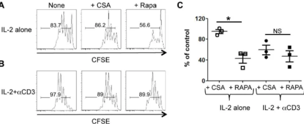
Rapa but not CsA inhibits IL-2-induced Treg proliferation Treg proliferationin vitro requires IL-2 and interactions with dendritic cells (DC)s. In this setting, co-stimulatory molecules (CD80, CD86, and OX40L) but not MHC class II expression by DCs are required for IL-2-induced Treg proliferation [10,11]. Consistent with this observation, we recently reported that CsA minimally affects Treg proliferation induced by IL-2 and DCs [12]. Since the AKT-mTOR pathway is a major signaling component downstream of co-stimulatory signals, we tested how the mTOR inhibitor (Rapa) would affect Treg proliferation induced by IL-2 and DCs. As predicted, Rapa but not CsA inhibited the proliferation of Tregs induced by IL-2 and DCs (Fig. 1A), highlighting the importance of costimulatory signals in this setting.
Rapa and CsA inhibit IL-2-induced Treg proliferation to a similar extent in the presence of overt TCR stimulation
We previously reported that CsA inhibits Treg proliferation when stimulated with anti-CD3, IL-2, and DCs, suggesting that calcineurin activation is crucial for IL-2-induced Treg prolifera-tion in the presence of TCR stimulaprolifera-tion. To test how this might compare to mTOR inhibition by Rapa, we tested the effects of Rapa and CsA on Treg proliferation induced by IL-2 and DCs in the presence of anti-CD3. Treg proliferation was suppressed by CsA in the presence of anti-CD3, such that CSA and Rapa now equally inhibited Treg proliferation in this setting (Figure 1A, B, and C). These data suggest that in the presence of TCR stimulation, both calcineurin and mTOR signaling is required for the optimal proliferation of Tregs.
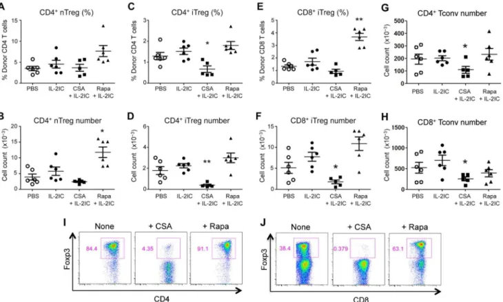
IL-2 plus CsA suppresses while IL-2 plus Rapa promotes the expansion of pre-existing donor-derived Treg expansion and inducible Treg (iTreg) generation during allogeneic BMT
To test how CsA and Rapa impact Treg proliferation in the setting where Tregs receive strong TCR signals in vivo, we employed an allogeneic BMT model and examined the Treg pool in allogeneic BMT mice administered IL-2 (in the form of immune complexes; IL-2 ICs) with or without CsA or Rapa. During allogeneic BMT, the pool of Tregs is formed by expansion of pre-existing derived Tregs and by the conversion of donor-derived Tconvs into iTregs in the recipient. We examined both
subsets of Tregs, since a large fraction of the Treg pool during GVHD consists of CD4+and CD8+iTregs that have converted
from alloreactive naı¨ve Tconvs and contribute to GVHD protection. [14–16]. The different subsets of Tregs were distin-guished by mixing FACS-sorted congenically disparate (CD45.1 vs. CD45.2) allogeneic Tregs and Tconvs (CD4+and CD8+) and
injecting them together with the BM cells into the irradiated recipients. Consistent with our in vitro results, CsA plus IL-2 treatment reduced the total number of pre-existing donor-derived Tregs compared to treatment with IL-2 alone on Day 8 post-BMT (Figure 2A and B). In contrast, Rapa augmented Treg numbers when given in conjunction with IL-2 (Figure 2A and B). Moreover, the absolute number and percentages of CD4+and CD8+iTregs
were markedly decreased in CSA plus IL-2-treated mice but enhanced in Rapa plus IL-2-treated mice (Figure 2C–F). The relative preservation of Treg percentages in CSA plus IL-2-treated mice could be due to the reduction of Tconv numbers in these mice (Figure 2G–H). Although RAPA plus IL-2 treatment slightly but not significantly decreased CD8+Tconv numbers, no effect on
CD4+Tconv numbers was observed. A similar effect of Rapa and
CsA was observed within vitroCD4+and CD8+iTreg conversion
assays, whereby Rapa augmented and CsA suppressed iTreg formation (Figure 2I–J). Notably and unexpectedly, IL-2 treatment did not significantly increase the total numbers or percentages of CD4+and CD8+Tregs at Day 8 post BMT. This was also true at
an earlier time point (Day 5 post BMT; Figure 3), suggesting that IL-2 treatment does not increase Treg numbers in the allogeneic BMT setting at these time points.
To test the impact of CsA and Rapa on Treg function, we next tested the suppressive ability of Tregs that were expanded in IL-2/ anti-CD3 in the presence or absence of CsA or Rapa. Compared to freshly isolated Tregs, IL-2/anti-CD3-expanded Tregs showed markedly increased inhibitory ability against Tconv proliferation (Fig. 4). This was true even when the Tregs were expanded in CsA or Rapa.
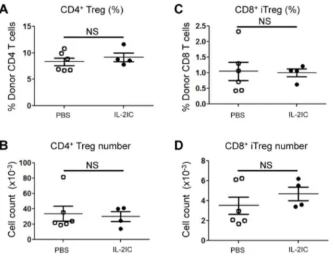
Although our data suggested that CsA suppresses and Rapa augments Treg numbers in allogeneic BMT in the presence of IL-2, we could not exclude the possibility that the inhibitory effect of CsA on Treg expansion was secondary to inflammation and lymphopenia induced by myeloablative conditioning (irradiation) during BMT. To test this possibility, IL-2 and CsA were administered to mice receiving syngeneic BMT with the same amount of irradiation. In contrast to the allogeneic BMT setting, IL-2 treatment resulted in an expansion of Tregs, which was not
Figure 1. Calcineurin signaling restores Treg proliferation in the presence of Rapa.CFSE-labeled FACS-sorted Tregs were co-cultured with B6-derived DCs and IL-2 with or without Rapa or CsA in the absence (A) or presence (B) of anti-CD3 antibody. One representative histogram showing Treg CFSE dilution in each condition is shown. (C) The division index of Tregs cultured in the presence of anti-CD3 antibody was normalized to the control and is plotted as mean6SEM ofn= 3 independent experiments.*p,0.01 by unpaired, two-tailed Student t test.
inhibited by CsA (Figure 5A–B). Together, these data suggest that CsA blocks nTreg proliferation and iTreg generation in allogeneic BMT mice treated with IL-2.
CsA blocks while Rapa augments the protective effect of IL-2 in GVHD
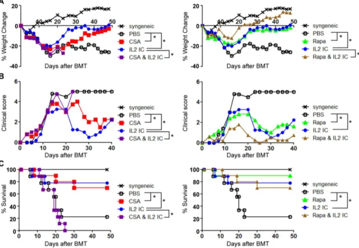
To test whether the effects of CsA and Rapa on Treg numbers correlated with GVHD protection, allogeneic BMT mice were treated with various combinations of IL-2, Rapa, and CsA and monitored for weight loss, GVHD clinical score, and mortality. The injection of IL-2 to allogeneic BMT mice significantly protected against GVHD-induced weight loss, clinical score, and mortality. Furthermore, as reported recently by others [17], Rapa and IL-2 displayed synergism in protecting against GVHD-induced weight loss (Figure 6A), although we could not observe a significant synergistic effect in clinical score or GVHD mortality (Figure 6B-C). Although CsA alone demonstrated significant benefit in protection against GVHD, the combination of CsA and IL-2 severely and significantly worsened disease outcome including weight loss, clinical score, and mortality (Figure 6A–C). These results suggest that the combination of CsA and IL-2 is detrimental in treatment of acute GVHD, which correlates with the negative effects of CsA on Tregs in the allogeneic BMT setting.
Discussion
Understanding the requirements for Treg proliferation is important to devise strategies involving the manipulation of Treg numbers in various disease settings. Previous studies from our laboratory and others demonstrated that Treg expansion does not rely heavily on TCR stimulation as long as exogenous IL-2 is provided [11,12]. As such, pharmacological inhibition of the TCR signaling pathway by CsA had little impact on IL-2-induced Treg proliferationin vitroand in vivo in a syngeneic setting. However, when additional TCR stimulation was provided in anin vitroassay, the proliferation of Tregs was markedly enhanced and now became an important signal for maximal Treg proliferation [12]. This led us to hypothesize that the requirements of Treg proliferation could be different in an allogeneic BMT setting, where a fraction of the Tregs would be expected to receive strong allogeneic MHC-driven TCR signals. To test this notion, we examined the impact of CsA on Tregs in an allogeneic BMT setting and found that CsA negatively affected Treg numbers and iTreg generation even in the presence of IL-2. This negative effect of CsA correlated with a significantly exacerbated GVHD outcome in mice treated with CsA and IL-2. In contrast, CsA did not affect Treg numbers in syngeneic BMT mice treated with IL-2.
Figure 2. CsA suppresses while Rapa promotes pre-existing donor-derived Treg expansion and iTreg generation during GVHD.
GFP2Tconvs (CD4+and CD8+) and GFP+CD4+Tregs were FACS-sorted from CD45.1+Foxp3 GFP-reporter mice and CD45.2+Foxp3 GFP-reporter mice,
respectively, combined with T cell-depleted BM cells (CD45.2+), and injected into irradiated B6D2F1 mice. Host mice were treated with vehicle (PBS),
IL-2 ICs, IL-2 ICs plus CsA (25 mg/kg), or IL-2 ICs plus Rapa (0.5 mg/kg) for 5 days. IL-2 ICs were given only for the first 3 days (Days 0, 1, and 2). Eight days after BMT, donor Tregs in the spleen were analyzed by flow cytometry. (A) The percentage and (B) absolute number of pre-existing donor nTregs, (C) the percentage and (D) absolute number of CD4+iTregs, (E) the percentage and (F) absolute number of CD8+iTregs, (G) the absolute
number of CD4+Tconvs, and (H) the absolute number of CD8+Tconvs are plotted as mean
6SEM ofn= 5–6 mice/group from two independent experiments.*p,0.05;**p,0.001 by two-tailed Student t test compared with the group treated with IL-2 ICs. (I) FACS-sorted CD4+or (J) CD8+
In addition to investigating the effects of CsA on Treg numbers, we examined the role of the mTOR signaling pathway under the same conditions. In in vitro assays, we previously found that costimulatory signals provided by DCs was critical for Treg proliferation in the absence of MHC class II [18]. Thus, we predicted that the mTOR inhibitor Rapa would suppress Treg
proliferation, as AKT-mTOR is a critical signaling pathway downstream of costimulatory molecules. Indeed, Rapa inhibited Treg proliferation induced by IL-2 and DCs. However, in contrast to CsA treatment, Rapa did not reduce the Treg proliferation further when TCR stimulation was provided. In fact, in an allogeneic BMT setting, IL-2 and Rapa synergized to increase Treg numbers and enhance iTreg generation. The increase in iTreg generation is consistent with results from ourin vitroiTreg conversion assays, where Rapa augmented IL-2/TGFb-induced conversion of Tconvs into iTregs. However, why pre-existing nTregs were also expanded by IL-2 and Rapa in allogeneic BMT is unclear, as Rapa partially inhibited Treg proliferation in vitro even in the presence of anti-CD3. One possibility is that Rapa has a strong negative effect on Tconvs [19,20], leading to an increased Treg:Tconv ratio that allowed IL-2 to expand Tregs more efficiently. However, our data suggest that only CD8+ Tconvs
and not CD4+Tconvs are affected by Rapa plus IL-2 treatment.
Thus, the discrepancy between thein vitroandin vivoeffect of Rapa on Treg numbers and the Treg:Tconv ratio in the allogeneic BMT setting cannot be explained at this time and warrants further investigation.
GVHD-induced weight loss but not survival or clinical score was improved with the combination of Rapa and IL-2 compared to either treatment alone. Compared to previous findings described by Shin et al. [17], the synergistic effects of IL-2 and Rapa on GVHD protection were not as pronounced in our study. This could potentially be due to differences in the GVHD induction protocol. While Shin et al. used purified Tconvs to induce GVHD, our study was performed with splenocytes that contain both Tconvs and Tregs. Thus, the strong effect of Rapa in augmenting the IL-2-mediated conversion of Tconvs into iTregs could have contributed more to protection against GVHD in the study by Shin et al. Nevertheless, even in the presence of nTregs, Figure 3. IL-2 ICs do not expand CD4+or CD8+Tregs on Day 5 post allogeneic BMT.Spleen cells and T-cell-depleted BM cells from WT
B6.SJL mice (CD45.1+) were injected into irradiated B6 (CD45.2+) mice. Host mice were treated with vehicle (PBS) or IL-2 ICs for 3 days (Days 0, 1, and
2). Five days after BMT, donor CD4+and CD8+Tregs in the spleen were analyzed by flow cytometry. (A) The percentage and (B) absolute number of
CD4+Tregs and (C) the percentage (E) and absolute number (F) of CD8+iTregs are plotted as mean
6SEM of n= 4–6 mice/group from two independent experiments. NS = not significant by two-tailed Student t test.
doi:10.1371/journal.pone.0092888.g003
Figure 4. IL-2 plus anti-CD3-expanded Tregs in the presence or absence of CsA or Rapa suppress Tconv proliferation. MACS-sorted CD4+ T cells from CD45.2+ Foxp3 GFP-reporter mice were
cultured with irradiated feeder cells in IL-2 plus anti-CD3/CD28 in the presence or absence of CsA or Rapa. 5 days later, GFP+ Tregs were
sorted by flow cytometry and co-cultured with CFSE-labeled FACS-sorted splenic Foxp32CD4+T cells (Tconvs) from CD45.1+Foxp3
GFP-reporter mice at various Tconv:Treg ratios for four days in the presence of irradiated T cell-depleted splenocytes and anti-CD3. FACS-sorted GFP+Tregs from freshly isolated splenocytes of CD45.2+Foxp3
GFP-reporter mice were also used for comparison. The division index of Tconvs is plotted against various Treg:Tconv ratios. One representative ofn= 2 independent experiments is shown.
Figure 5. CsA does not inhibit IL-2-induced Treg expansion in the absence of overt TCR stimulation.Spleen cells and T-cell-depleted BM cells from WT B6.SJL mice (CD45.1+) were injected into irradiated B6 (CD45.2+) mice. Host mice were treated with PBS alone, IL-2 ICs alone, or CsA
+ IL-2 ICs as well as the GVHD model. Eight days after BMT, donor CD4+Tregs (CD45.1+) in the spleen analyzed by flow cytometry. (A) The percentage and
(B) absolute number of donor CD4+Tregs are plotted as mean
6SEM ofn= 5–6 mice/group from two independent experiments.*p,0.05 by two-tailed Student t test compared with the group treated with IL-2 ICs.
doi:10.1371/journal.pone.0092888.g005
Figure 6. CsA abolishes while Rapa promotes the beneficial effect of IL-2 ICs in protection against acute GVHD.B6 splenocytes and T cell-depleted BM cells were injected into irradiated B6D2F1 mice. The mice were treated with either vehicle (PBS), CsA, Rapa, IL-2 ICs, CsA+IL-2 ICs, or
Rapa+IL-2 ICs on days 1–3. Changes in (A) wt and (B) GVHD score are plotted as the mean ofn= 5 mice/group vs. days post transplant. One representative of 2 independent experiments is shown.*p,0.05 by ANOVA with Tukey’s post hoc test. (C) Survival was monitored over a 7-week period. Two independent experiments were combined with a total ofn= 9 or 10 mice/group to generate the survival curve.*p,0.05 by log-rank test. Note that the CsA and Rapa treatment groups were separated into two graphs (left and right) for visualization purposes. The syngeneic and PBS-treated groups are identical for both plots.
our data support a beneficial effect in combining Rapa and IL-2 in the treatment of GVHD.
The combination of CsA and IL-2 is highly effective against EAE compared to treatment with either agent alone [12]. This is in striking contrast to the acute GVHD setting, where CsA suppressed the beneficial effect of IL-2 on disease outcome. We speculate that these differences arise, because strong allogeneic MHC-stimulated TCR signals drive nTreg proliferation to keep up with the expansion of alloreactive Tconvs. Thus, as opposed to the syngeneic setting, TCR signals become critical in nTreg expansion, which is inhibited by CsA. In addition, the negative effect of CsA on iTreg generation may also contribute to the detrimental effect of CsA, as both CD4+and CD8+iTregs are
important in GVHD protection [14–16]. However, other differ-ences in EAE compared to GVHD including the precursor frequency of the disease-specific T-cells (high in GVHD), the fullness of the T-cell compartment (lymphopenia in GVHD), and the degree of inflammation (high in GVHD) also need to be considered.
Another intriguing observation is that CsA negatively impacted GVHD outcome when given with IL-2 but was protective when given alone. IL-2 is a crucial factor for Treg survival and growth [21,22] and can be utilized by Tregs more effectively than Tconvs because of their constitutive expression of the high affinity IL-2 receptor CD25/CD122/CD132 complex. While this is true in the absence of inflammation, Tconvs inducibly express the high affinity IL-2 receptor complex upon activation. Thus, during massive T cell activation occurring during GVHD, excessive IL-2 may expand not only Tregs but also Tconvs. In the present study, we limited the injection of IL-2 to the first 3 days, as we observed high mortality of hosts in which IL-2 was administered continu-ously (data not shown). Mice injected continucontinu-ously with IL-2 showed an expansion of highly activated Tconvs, suggesting that extra IL-2 would augment pathogenic Tconvs harmful to the allogeneic BMT host. Thus, the negative effects of IL-2 may be brought out by the concurrent suppression of Tregs by CsA, which would allow IL-2-mediated expansion and activation of Tconvs. Alternatively, the mechanism of action of CsA could be in part mediated by suppression of IL-2 production from alloreactive Tconvs, which is necessary for their expansion. If fact, the reduction in IL-2 production by CsA treatment alone has been proposed to negatively affect Treg numbers during GVHD [23]. Although the provision of IL-2 in combination with CsA did not rescue the Treg defect during allogeneic BMT in our study, it could still potentially reverse the beneficial inhibitory effects that CsA has on suppressing alloreactive Tconv proliferation. While we do not know the exact mechanism of why CSA and IL-2 cancel out each other’s beneficial effects in the treatment of acute GVHD, we propose that this combination should be avoided in this disease setting.
The failure of IL-2 treatment to increase absolute numbers of Tregs during allogeneic BMT was unexpected. This may occur because the Tregs in the allogeneic setting are maximally proliferating such that the expansion cannot be further
augment-ed. Indeed, previous studies have shown that Treg proliferation is more dependent on TCR stimulation than on IL-2 in a lymphopenic setting [7]. This then begs the question of why IL-2 treatment attenuates GVHD in the absence of Treg expansion. It is possible that IL-2 protects against GVHD by augmenting Treg function. In our previous study [12], we found that Tregs isolated from IL-2-treated mice are significantly more suppressive than Tregs from PBS-treated mice.
Alterations in Treg function could also partially contribute to the negative effects of CsA in IL-2-mediated protection against GVHD, as Treg function was slightly reduced when Tregs were expanded with anti-CD3 and IL-2 in the presence of CsA. In addition to its effects on Treg proliferation, NFAT, the transcrip-tion factor downstream of calcineurin, plays a critical role for the ability of Tregs to exert their suppressive function [24]. Thus, many factors contribute to why a calcineurin inhibitor would not synergize with IL-2 to protect against acute GVHD. Although the combination of calcineurin inhibition and IL-2 might be detrimental in acute GVHD as shown by our current study, it could be still effective in chronic GVHD. In contrast to acute GVHD that is caused by alloreactive mature T cells, chronic GVHD is potentially caused by host thymic dysfunction leading to inadequate negative selection and/or Treg generation from the thymus [25,26]. Chronic GVHD presents with autoimmune disease manifestations including scleroderma, lupus, and Sjog-ren-like conditions. Thus, chronic GVHD may mimic more of a syngeneic autoimmune disease scenario, where CsA and IL-2 combination therapy might be beneficial. In support of this notion, IL-2 treatment was found to be effective against chronic GVHD in humans and although more than 50% of patients were concom-itantly treated with a calcineurin inhibitor in this clinical study, many of them still exhibited an elevation in Treg numbers and showed disease improvement [27].
In summary, we have demonstrated that pharmacological TCR inhibition plus IL-2 does not allow the expansion of nTregs and the generation of iTregs in the allogeneic BMT setting. Unlike the syngeneic setting, CsA and IL-2 does not suppress allogeneic immune responses, indicating that IL-2 treatment against acute GVHD would not be recommended in the clinical setting because almost all recipients are treated with a calcineurin inhibitor for GVHD prophylaxis. Thus, depending on the setting, the signaling pathways leading to expansion of the Treg pool need to be carefully considered and the immunosuppressive regimen needs to be tailored to most effectively increase the Treg:Tconv ratio.
Acknowledgments
We thank the Kambayashi, Behrens, Nichols, and Koretzky lab members for helpful discussions.
Author Contributions
Conceived and designed the experiments: AS TK. Performed the experiments: AS AMS. Analyzed the data: AS AMS TK. Contributed reagents/materials/analysis tools: SN. Wrote the paper: AS TK.
References
1. Sakaguchi S, Yamaguchi T, Nomura T, Ono M (2008) Regulatory T cells and immune tolerance. Cell 133: 775–787.
2. Josefowicz SZ, Rudensky A, Sakaguchi S, Yamaguchi T, Nomura T, et al. (2009) Control of regulatory T cell lineage commitment and maintenance Regulatory T cells and immune tolerance. Immunity 30: 616–625.
3. Fontenot JD, Gavin MA, Rudensky AY (2003) Foxp3 programs the development and function of CD4+CD25+regulatory T cells. Nat Immunol 4: 330–336.
4. Joffre O, Santolaria T, Calise D, Al Saati T, Hudrisier D, et al. (2008) Prevention of acute and chronic allograft rejection with CD4+CD25+Foxp3+ regulatory T lymphocytes. Nat Med 14: 88–92.
5. Edinger M, Hoffmann P, Ermann J, Drago K, Fathman CG, et al. (2003) CD4+CD25+ regulatory T cells preserve graft-versus-tumor activity while inhibiting graft-versus-host disease after bone marrow transplantation. Nat Med 9: 1144–1150.
7. Setoguchi R, Hori S, Takahashi T, Sakaguchi S (2005) Homeostatic maintenance of natural Foxp3(+) CD25(+) CD4(+) regulatory T cells by interleukin (IL)-2 and induction of autoimmune disease by IL-2 neutralization. J Exp Med 201: 723–735.
8. Gavin MA, Clarke SR, Negrou E, Gallegos A, Rudensky A (2002) Homeostasis and anergy of CD4(+)CD25(+) suppressor T cells in vivo. Nat Immunol 3: 33– 41.
9. Bhandoola A, Tai X, Eckhaus M, Auchincloss H, Mason K, et al. (2002) Peripheral expression of self-MHC-II influences the reactivity and self-tolerance of mature CD4(+) T cells: evidence from a lymphopenic T cell model. Immunity 17: 425–436.
10. Zou T, Caton AJ, Koretzky GA, Kambayashi T (2010) Dendritic cells induce regulatory T cell proliferation through antigen-dependent and -independent interactions. J Immunol 185: 2790–2799.
11. Swee LK, Bosco N, Malissen B, Ceredig R, Rolink A (2009) Expansion of peripheral naturally occurring T regulatory cells by Fms-like tyrosine kinase 3 ligand treatment. Blood 113: 6277–6287.
12. Satake A, Schmidt AM, Archambault A, Leichner TM, Wu GF, et al. (2013) Differential targeting of IL-2 and T cell receptor signaling pathways selectively expands regulatory T cells while inhibiting conventional T cells. J Autoimmun 44: 13–20.
13. Cooke KR, Kobzik L, Martin TR, Brewer J, Delmonte J Jr, et al. (1996) An experimental model of idiopathic pneumonia syndrome after bone marrow transplantation: I. The roles of minor H antigens and endotoxin. Blood 88: 3230–3239.
14. Sawamukai N, Satake A, Schmidt AM, Lamborn IT, Ojha P, et al. (2012) Cell-autonomous role of TGFbeta and IL-2 receptors in CD4(+) and CD8(+) inducible regulatory T-cell generation during GVHD. Blood 119: 5575–5583. 15. Robb RJ, Lineburg KE, Kuns RD, Wilson YA, Raffelt NC, et al. (2012)
Identification and expansion of highly suppressive CD8(+)FoxP3(+) regulatory T cells after experimental allogeneic bone marrow transplantation. Blood 119: 5898–5908.
16. Beres AJ, Haribhai D, Chadwick AC, Gonyo PJ, Williams CB, et al. (2012) CD8+Foxp3+regulatory T cells are induced during graft-versus-host disease and mitigate disease severity. J Immunol 189: 464–474.
17. Shin HJ, Baker J, Leveson-Gower DB, Smith AT, Sega EI, et al. (2011) Rapamycin and IL-2 reduce lethal acute graft-versus-host disease associated with
increased expansion of donor type CD4+CD25+Foxp3+regulatory T cells. Blood 118: 2342–2350.
18. Zou T, Satake A, Ojha P, Kambayashi T (2011) Cellular therapies supplement: the role of granulocyte macrophage colony-stimulating factor and dendritic cells in regulatory T-cell homeostasis and expansion. Transfusion 51 Suppl 4: 160S– 168S.
19. Wang Y, Camirand G, Lin Y, Froicu M, Deng S, et al. (2011) Regulatory T cells require mammalian target of rapamycin signaling to maintain both homeostasis and alloantigen-driven proliferation in lymphocyte-replete mice. J Immunol 186: 2809–2818.
20. Zeiser R, Leveson-Gower DB, Zambricki EA, Kambham N, Beilhack A, et al. (2008) Differential impact of mammalian target of rapamycin inhibition on CD4+CD25+Foxp3+regulatory T cells compared with conventional CD4+T cells. Blood 111: 453–462.
21. Fontenot JD, Rasmussen JP, Gavin MA, Rudensky AY (2005) A function for interleukin 2 in Foxp3-expressing regulatory T cells. Nat Immunol 6: 1142– 1151.
22. D’Cruz LM, Klein L (2005) Development and function of agonist-induced CD25+Foxp3+regulatory T cells in the absence of interleukin 2 signaling. Nat Immunol 6: 1152–1159.
23. Zeiser R, Nguyen VH, Beilhack A, Buess M, Schulz S, et al. (2006) Inhibition of CD4+CD25+regulatory T-cell function by calcineurin-dependent interleukin-2 production. Blood 108: 390–399.
24. Wu Y, Borde M, Heissmeyer V, Feuerer M, Lapan AD, et al. (2006) FOXP3 controls regulatory T cell function through cooperation with NFAT. Cell 126: 375–387.
25. Matsuoka K, Kim HT, McDonough S, Bascug G, Warshauer B, et al. (2010) Altered regulatory T cell homeostasis in patients with CD4+lymphopenia following allogeneic hematopoietic stem cell transplantation. J Clin Invest 120: 1479–1493.
26. Sakoda Y, Hashimoto D, Asakura S, Takeuchi K, Harada M, et al. (2007) Donor-derived thymic-dependent T cells cause chronic graft-versus-host disease. Blood 109: 1756–1764.