the Epithelial-to-Mesenchymal Transition in Breast
Cancer
Michael S. Hwang1, Nancy Yu1, Susanna Y. Stinson1¤, Peng Yue2¤, Robert J. Newman3, Bernard B. Allan3, David Dornan1*¤
1Department of Molecular Diagnostics and Cancer Cell Biology, Genentech, Inc., South San Francisco, California, United States of America,2Department of Bioinformatics and Computational Biology, Genentech, Inc., South San Francisco, California, United States of America,3Department of Molecular Biology, Genentech, Inc., South San Francisco, California, United States of America
Abstract
The epithelial-to-mesenchymal transition (EMT) is a highly conserved physiological program involved in development and tissue repair; however, its aberrant activation has been implicated in accelerating the progression of a variety of cancers. In breast cancer, the microRNAs (miRNAs) miR-221 and miR-222 (miR-221/222) are differentially expressed in the clinically more aggressive basal-like subtype compared to luminal subtype of breast cancer and upregulation of miR-221/222 induces the EMT by targeting the 39untranslated region (39UTR) of the GATA family transcriptional repressor TRPS1 (tricho-rhino-phalangeal syndrome type 1). The complete mechanism through which miR-221/222 promotes the EMT, however, is not fully understood. We identified adiponectin receptor 1 (ADIPOR1), a receptor for the adipocytokine adiponectin, as a direct target of miR-221/222. ADIPOR1 is expressed at higher levels in the luminal compared to the basal-like subtype of breast cancer cell lines, which can be reduced by miR-221/222 targeting of its 3’UTR. In addition, miR-221/222 were negatively correlated with ADIPOR1 expression across breast cancer cell lines and tumors. ADIPOR1 depletion by siRNA in MCF10A cells induced the EMT and increased cell invasion. Depletion of ADIPOR1 by siRNA induced activation of the canonical nuclear factor-kappaB (NF-kB) and subsequent phosphorylation of signal transducer and activator of transcription 3 (STAT3) in an interleukin 6 (IL6)-dependent manner. Finally, overexpression of ADIPOR1 in the basal-like cell line, MDA-MB-231, attenuated cell invasion and promoted the mesenchymal-to-epithelial transition (MET). We conclude that ADIPOR1 negatively regulates EMT in breast cancer and provides an additional node by which miR-221/222 induces the EMT. These results suggest that ADIPOR1 may play an important role in breast cancer progression and metastasis, and could potentially offer an alternative therapeutic strategy for basal-like breast cancer patients.
Citation:Hwang MS, Yu N, Stinson SY, Yue P, Newman RJ, et al. (2013) miR-221/222 Targets Adiponectin Receptor 1 to Promote the Epithelial-to-Mesenchymal Transition in Breast Cancer. PLoS ONE 8(6): e66502. doi:10.1371/journal.pone.0066502
Editor:Elad Katz, AMS Biotechnology, United Kingdom
ReceivedFebruary 25, 2013;AcceptedMay 6, 2013;PublishedJune 11, 2013
Copyright:ß2013 Hwang et al. This is an open-access article distributed under the terms of the Creative Commons Attribution License, which permits unrestricted use, distribution, and reproduction in any medium, provided the original author and source are credited.
Funding:The authors have no support or funding to report.
Competing Interests:All authors, except MSH, are current employees of Genentech, Inc. or Gilead Sciences, Inc. and hold stock with their respective employer. This does not alter the authors’ adherence to all the PLOS ONE policies on sharing data and materials.
* E-mail: [email protected]
¤ Current address: Gilead Sciences, Inc., Foster City, California, United States of America
Introduction
Breast cancer is a heterogeneous disease, with discrete subtypes that can—in part—be characterized by their pathogenesis, molecular markers, and phenotype [1]. Five major breast cancer subtypes have been identified through mRNA profiling efforts: luminal A, luminal B, HER2-positive, basal-like, and claudin-low [2,3]. Interestingly, the claudin-low subtype displays molecular features of EMT and cancer stem cells [4,5,6]. Consistently, the basal-like subtype, which largely overlaps with the triple-negative subtype, also exhibits aggressive clinical behavior and poor prognosis towards both targeted therapies and chemotherapy alike [7]. Compared to luminal, the basal-like subtype differen-tially expresses molecular markers such as epithelial cadherin (E-cadherin), vimentin, and fibronectin, commensurate with cells that have undergone EMT. This process gives cells a license to gain mobility and invade through the basement membrane that
Adiponectin receptor 1 (ADIPOR1) is one of several receptors for the adipocytokine adiponectin [12]. Adiponectin is made exclusively by adipocytes, and affects insulin sensitivity and proliferation in a variety of cell types [13]. Low levels of adiponectin are associated with obesity and higher risk of breast cancer [14,15], while adiponectin has also been implicated with reducing breast cancer invasion via LKB1 signaling [16]. ADIPOR1 is a member of the progestin and adipoQ receptor (PAQR) family of cell-surface receptors which have seven transmembrane domains but do not couple to G-proteins. Instead, PAQRs are thought to be enzymes with intracellular, plasma membrane localized ceramidase activity. Recently, the ceramidase activity of ADIPOR1 has been shown to be required for protection of pancreaticb-cells and cardiomyocytes from fatty acid-induced apoptosis and for improved insulin sensitivity in obese mice through removal of hepatic ceramide [17]. The intracellular signaling pathways downstream of ADIPOR1, however, have not been well characterized. In this study, we uncovered a unique relationship between miR-221/222, ADIPOR1, and EMT and provide further insight into miR-221/222-mediated EMT and breast cancer pathogenesis.
Materials and Methods
Cell Lines, Tumors, and Reagents
All breast cell lines were procured from ATCC. The MDA-MB-231 cell line was maintained in RPMI containing 10% FBS supplemented with L-gluatamine, and the MCF10A cell line was maintained in Dulbecco’s Modified Eagle Medium/Ham’s F12 (DMEM/F12) media containing 5% horse serum, EGF (20 ng/ ml), hydrocortisone (0.5mg/ml), cholera toxin (100 ng/ml), and
insulin (10mg/ml). A supporting figure shows the short tandem repeat (STR) profiles for both cell lines used (Figure S2). Primary breast tumor samples were obtained from ILSbio, SeraCare Life Sciences, and Asterand. SMARTpool and individual siRNA, in addition to microRNA mimics, were purchased from Dharmacon. Cells were seeded at 50,000–100,000 cells/ml and transfected with RNAiMAX or Lipofectamine 2000 (Invitrogen). siRNA was transfected at a concentration of 100 nM, unless otherwise indicated. Transfection efficiencies of $75% were achieved as assessed by Green Fluorescent Protein (GFP) control. Site-directed mutagenesis (Stratagene) was performed according to the manu-facturer’s protocol, and luciferase activity was measured by Dual-Glo reagents (Promega). Cucurbitacin (Sigma-Aldrich) and AG490 (Axon Medchem) were used at a concentration of 1mM, and all neutralizing antibodies (R&D Systems) were used at a concentra-tion of 20mg/ml. ADIPOR1 overexpression, tagged cDNA clone (Origene) was stably selected at 500mg/ml Geneticin (Invitrogen). Microarray and Quantitative Real Time PCR
RNA was purified using RNeasy (Qiagen) or Cells-to-CT (Ambion). For mRNA microarray, RNA was run on Affymetrix HGU133P2 chips, and mRNA from 55 basal and luminal breast cancer cell lines were profiled. For qPCR, cDNA was generated with the High Capacity RNA-to-cDNA Kit (Roche). cDNA was diluted at 1:100, and used for 20ul total reactions on the 7900HT Fast Real-Time PCR System (Applied Biosystems). Primers from TaqMan Gene Expression Arrays (Applied Biosystems) were used: GAPDH (Hs02758991_g1), ADIPOR1 (Hs01114951_m1), E-cadherin (Hs01023895_m1), Vimentin (Hs00185584_m1), and Fibronectin (Hs00415006_m1). Reactions were performed in duplicate, and delta-delta-Cycle Threshold (ddCt) values were calculated after normalization to GAPDH. All microarray gene expression data for the breast cancer cell lines can be obtained
from Gene Expression Omnibus (www.ncbi.nlm.nih.gov/geo) with accession number GSE12790.
Western Blot Analysis and Immunofluorescence
For Western Blot Analysis, all cells were lysed with RIPA buffer supplemented with cOmplete, Mini, EDTA-free Protease Inhib-itor Cocktail Tablets (Roche) and Halt Protease InhibInhib-itor Cocktail (Thermo Scientific) and subsequently clarified by centrifugation. Protein concentration was quantified with the BCA Protein Assay Kit (Pierce). Reduced lysates were resolved on NuPAGE 4–12% Bis-Tris Gels (Invitrogen) and transferred with the Trans-Blot Turbo Transfer System (Bio-Rad). Membranes were blocked and incubated in 5% bovine serum albumin (BSA). The following antibodies were used: E-cadherin (BD Bioscience), Vimentin (Santa Cruz), Actin (Cell Signaling), p-IKKa/bSer176/180 (Cell Signaling), IKKa(Cell Signaling), IKKb(Cell Signaling), p-IkBa
Ser32 (Cell Signaling), IkBa (Cell Signaling), p-STAT3 Tyr705 (Cell Signaling), STAT3 (Cell Signaling), and anti-Flag M2 (Sigma-Aldrich). For immunofluorescence, cells were transfected in an eight chamber glass slide, fixed in ice cold methanol for 5 minutes at –20uC and permeabilized in acetone for 2 minutes at – 20uC. Cells were then stained with anti-Vimentin (Sigma-Aldrich) at room temperature for 1 hour followed by anti-mouse-cy3 (Jackson Lab) and Hoechst 33258. Cells were imaged and nuclei were quantitated on the Cellomics VTI Arrayscan.
Invasion and Cell Growth Assays
For MCF10A invasion assays, 56104cells were seeded on the
upper side of 24-well BD BioCoat Matrigel Invasion Chamber inserts (BD Biosciences) 3 days post-transfection, and allowed to invade towards 10% horse serum. After 24 hours, cells on the upper side of the membrane were mechanically removed. Cells that invaded to the lower side were fixed and stained using the Diff-Quik Kit (Siemens). The membranes were photographed and the cells counted. For MDA-MB-231 cells, the high-throughput scratch CellPlayer 96-Well Kinetic Cell Invasion Assay (Essen Bioscience) was used according to the manufacturer’s protocol. 56104 cells were seeded, and the upper-layer of Matrigel was
diluted to a concentration of 6 mg/ml. Images were collected via IncuCyte every 3 hours.
To assay growth rates, cells were first seeded into 96-well plates, with each well containing 26103cells. The cells were then imaged
every 3 hours for confluency percentage on an IncuCyte to obtain growth curves.
Statistical Analysis
Spearman rank correlation coefficients or Student’s t-test
calculations were used to determined statistical significance using GraphPad Prism.
Results
ADIPOR1 is a direct target of miR-221/222 and negatively regulates EMT
chain reaction (qRT-PCR) (Figure 1A). In addition, ADIPOR1 mRNA abundance was higher in luminal cell lines compared to basal-like cell lines(Figure 1B). Furthermore, 221 and miR-222 expression was negatively correlated with ADIPOR1 mRNA levels across breast cancer cell lines as well as tumors derived from breast cancer patients (Figures 1C – F), suggesting that ADIPOR1 may be an endogenous target of miR-221/222. However, to determine if ADIPOR1 is indeed a direct target of miR-221/222, we performed site-directed mutagenesis on nucleotides 1 and 2 of the predicted seed sequence within the conserved 3’UTR of ADIPOR1 (Figure 1C). The wild-type and mutant 3’UTR of ADIPOR1 were then cloned into a luciferase reporter construct [10]. Relative to the scrambled miR mimic, miR-221/222 decreased luciferase expression of the wild-type construct in a dose-dependent manner, but was unable to perturb the luciferase expression of the mutant 3’UTR construct in MCF10A cells (Figure 1C).
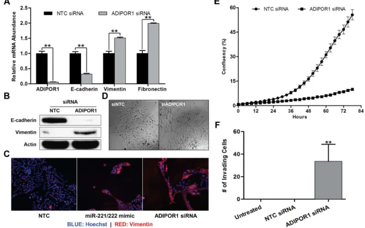
Since miR-221/222 has previously been shown to promote EMT [10], we wished to determine if ADIPOR1 silencing by small interfering ribonucleic acid (siRNA) would have a similar effect on molecular markers of EMT by qRT-PCR and western blotting. Dharmacon siRNA #2 produced the strongest knock-down of ADIPOR1 by mRNA, and this correlated with the largest decrease in E-cadherin and increase in vimentin relative to the other siRNAs in the pool; similar effects were seen with other siRNAs tested (Figure S1). All subsequent ADIPOR1 siRNA assays were therefore performed with Dharmacon siRNA #2. Indeed, ADIPOR1 siRNA downregulated E-cadherin and upre-gulated vimentin and fibronectin mRNA (Figure 2A). To confirm that mRNA levels correlated with protein expression, we performed a western blot with antibodies to E-cadherin and vimentin against samples treated with either non-targeting control (NTC) or ADIPOR1 siRNA and observed reduction in E-cadherin and elevation in vimentin protein expression (Figure 2B). Vimentin protein expression was also increased by both miR-221/222 mimic and ADIPOR1 siRNA treatment as determined by immunofluorescence (Figure 2C). Moreover, MCF10A cells transfected with ADIPOR1 siRNA adopted a more elongated and fibroblastic cellular morphology, consistent with a mesenchymal phenotype (Figure 2D). We also sought to determine whether cell invasion was enhanced by knocking down ADIPOR1. Despite attenuated growth rates, an observation that is consistent with EMT, we observed an increase in the invasion of MCF10A cells transfected with ADIPOR1 compared to NTC siRNA towards 10% serum (Figures 2E and 2F).
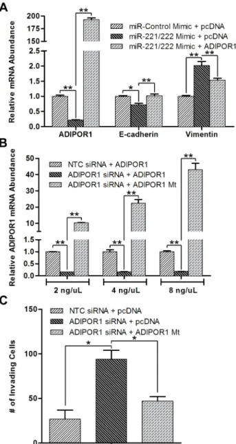
To confirm that ADIPOR1 was not only a target of miR-221/ 222, but also rescues a miR-221/222 mimic-induced EMT in MCF10A cells, we co-transfected an ADIPOR1 overexpression plasmid lacking its native 3’UTR with miR-221/222 mimic and indeed saw that increased ADIPOR1 levels rescued E-cadherin and vimentin expression (Figure 3A). To rule out the possibility ADIPOR1 siRNA has substantial off-target effects, we generated a siRNA resistant mutant of ADIPOR1 and found that not only was it able to rescue ADIPOR1 expression in a dose-dependent manner, but also blunted the number of invading MCF10A cells caused by transfection of ADIPOR1 siRNA (Figure 3B and 3C).
Collectively, these data suggest that ADIPOR1 is a target of miR-221/222, ADIPOR1 abundance negatively correlates with EMT markers and phenotypes, and depletion of ADIPOR1 can elicit molecular and phenotypic properties associated with EMT.
ADIPOR1 protects against the EMT by negatively regulating the NF-kB, IL6, and JAK2/STAT3 signaling axis.
Next we sought to determine a mechanism of action for ADIPOR1 suppression of the EMT. Transfecting ADIPOR1 or NTC siRNA into MCF10A cells in 96-well Cignal 45-Pathway Reporter Arrays revealed a number of upregulated and downreg-ulated pathways (Figure 4A). Notably, the two highest expressed reporters were STAT3 and NF-kB, both well-known drivers of the EMT in breast cancer, as well as other cancers [19,20,21,22]. To confirm the results from the Cignal 45 reporter assay, we assessed markers of canonical NF-kB and STAT3 signaling by western blot. I KappaB Kinase a/b (IKKa/b) and Inhibitor of kB a
(IkBa) phosphorylation increased 12 hours post ADIPOR1 siRNA transfection, and STAT3 phosphorylation increased after 36 hours (Figure 4B). The temporal phosphorylation of canonical NF-kB and STAT3 suggested that there might exist secondary messengers to allow for crosstalk and regulation between the two signaling nodes. To address that question, we performed a 41-plex Luminex secreted factor array on conditioned media after ADIPOR1 targeting by siRNA. Conditioned media was collected 5 days post transfection and 28 cytokines and growth factors were detectable within the linear range of the assay (Figure 4C). Noteworthy was the increased abundance of all but one analyte in response to ADIPOR1 knockdown, reminiscent of a quasi-cytokine storm or hyperquasi-cytokinemia, and consistent with classic canonical NF-kB activation [23]. Co-treatment of neutralizing antibodies against the top three upregulated cytokines in addition to IL6—a well-documented mediator of EMT via Janus Activated Kinase (JAK)/STAT3 signaling [24,25,26]—with siRNA target-ing ADIPOR1 revealed the unexpected result that only anti-IL6Ra was able to blunt an ADIPOR1 siRNA-induced EMT. This suggested that IL6 is a required, intermediate secondary messenger produced after NF-kB activation, which subsequently induces STAT3 phosphorylation in an autocrine manner (Figure 4D). Furthermore, small-molecule inhibitors against JAK2 (AG490) and STAT3 (Cucurbitacin) were able to attenuate an ADIPOR1 siRNA-mediated EMT (Figure 4E) [27,28,29]. Be-cause STAT3 has been implicated as a key driver for the EMT, we tested whether STAT3 phosphorylation could be perturbed with the neutralizing antibody anti-IL6Ra, and the small-molecule inhibitors AG490 and Cucurbitacin. We observed attenuated phospho-STAT3 levels when the inhibitors and neutralizing antibody were treated in concert with siRNA targeting ADIPOR1 in MCF10A cells (Figure 4F). Intriguingly, we also noted a reduction in cell invasion, with correlation to relative STAT3 phosphorylation (Figures 4F and 4G). Put together, we propose that ADIPOR1 protects against the EMT by suppressing the
NF-kB, IL6, and JAK2/STAT3 signaling axis.
ADIPOR1 overexpression attenuates the EMT and invasion
wished to investigate how ADIPOR1 expression would affect cell invasion. Despite similar growth curves, the pool and clone derivatives reduced invasion compared to parental MDA-MB-231 cells (Figures 5C and 5D). In addition, cell invasion rates correlated well with the degree of MET induction. Together, these data suggest that ADIPOR1 abundance in the luminal-subtype of breast cancers protects against the EMT, and this protection can be conferred by overexpression in the basal-like subtype.
Discussion
Breast cancer is the second most common cancer worldwide after lung and the leading cause of cancer deaths in women [30]. The triple negative subtype of breast cancer, with its poor prognosis and lack of approved targeted therapies, remains a
significant unmet medical need. Recently, small RNA molecules known as miRNAs have emerged as post-transcriptional regulators of mRNA stability, and have been identified to enhance several aspects of breast cancer pathogenesis including metastasis [31], invasion [32], and self-renewal [33]. Each of these phenotypes can be further modulated by the EMT program [34]. miR-221/222 has previously been reported to promote the EMT by negatively regulating TRPS1 that leads to increased ZEB2 expression [10]. Here, we found that miR-221/222 additionally targets ADIPOR1 and abrogates ADIPOR1 inhibition of the EMT. Our data show that miR-221/222 simultaneously targets two genes, ADIPOR1 and TRPS1, which likely allows for tight control of the EMT. Both the NF-kB/IL6/JAK2/STAT3 cascade, which we show here is modulated by miR-221/222-mediated downregulation of ADI-POR1, and ZEB2 expression, which we have previously shown is
Figure 1. ADIPOR1 is a direct target of miR-221/222.(A) qRT-PCR analysis of MCF10A cells, 48 hours post transfection with either miR-Control or miR-221/222 mimic. Data represent means of triplicates6SD. (B) ADIPOR1 mRNA expression data taken from microarray analysis of 32 luminal and 23 basal-like breast cancer cell lines. Data represented as boxplot, with whiskers indicating maximum and minimum values of the data set. (C) qRT-PCR analysis of ADIPOR1 mRNA expression versus miR-221 levels in 49 breast cancer cell lines. (D) qRT-qRT-PCR analysis of ADIPOR1 mRNA expression versus miR-221 levels in 27 primary breast tumors. (E) qRT-PCR analysis of ADIPOR1 mRNA expression versus miR-222 levels in 49 breast cancer cell lines. (F) qRT-PCR analysis of ADIPOR1 mRNA expression versus miR-222 levels in 27 primary breast tumors. (G) The predicted ADIPOR1 wild-type (WT) and mutant 3’UTR sequence for miR-221/222 is shown above. Below, MCF10A cells were co-transfected with the reporter constructs and an increasing titration of miR-221/222 mimic. Data represent means of triplicates6SD.
doi:10.1371/journal.pone.0066502.g001
Figure 2. ADIPOR1 negatively regulates the EMT.(A) qRT-PCR analysis of MCF10A cells, 48 hours post transfection with either non-targeting control (NTC) or ADIPOR1 siRNA. Data represent means of triplicates6SD; ** = P,0.001. (B) Western Blot analysis of E-cadherin and vimentin 5 days post siRNA transfection in MCF10A cells. (C) Immunofluorescence of vimentin and Hoechst staining in MCF10A cells 5 days post mimic or siRNA transfection. (D) Phase-contrast images of MCF10A cells 5 days post siRNA transfection. (E) Growth curve of MCF10A cells. Cells were initially transfected in a 6-well plate for 3 days prior to being transferred into a 96-well plate to assess growth rates. Data represent means of octuplets6SD. (F) Invasion of MCF10A cells was assessed by a Boyden chamber invasion assay 3 days post siRNA transfection. Data represent means of quadruplicates6SD; ** = P,0.001.
repressed by TRPS1, are well-characterized effector pathways in cancer metastasis, invasion, and EMT. It is intriguing that altering TRPS1 or ADIPOR1 alone is sufficient to modulate an EMT phenotype since there is very little evidence that the two pathways are directly related; however, there are many ways to induce EMT and the fact that miR-221/222 is targeting two independent pathways that, together, are signaling to nodes key for mediating EMT in breast cancer, underscores the importance of our findings and warrants further investigation. One potential limitation of our study is that most of our work was done in two cell lines, MCF10A and MDA-MB-231, and could limit the generalization of our
findings; however, the negative correlation between miR-221 and miR-222 expression and ADIPOR1 mRNA expression in breast tumors as well as in breast cancer cell lines suggests that our model could hold true in other systems.
Genetic variants of ADIPOR1 and its ligand adiponectin have been associated with increased risk of breast cancer [35] and circulating adiponectin levels are inversely correlated with breast cancer in post-menopausal women [15], while higher levels of adiponectin are associated with improved survival in obese, insulin-resistant breast cancer patients [36]. In mouse models of mammary gland tumors, adiponectin has been proposed to play an indirect role in tumor growth, through the regulation of non-tumor endothelial cells and angiogenesis [37]. This activity may be linked to the observed overexpression of T-cadherin, the most well-defined adiponectin receptor [38], in endothelial vascular cells of breast tumors [39,40]. Our data do not directly implicate adiponectin in the regulation of the EMT in the luminal-subtype of breast cancer cell lines. In fact, we suspect that, as ADIPOR1 is an enzyme with intrinsic activity, the loss of this activity in cells is sufficient to activate the EMT program in an adiponectin-independent manner. None of the experiments described here use exogenous, recombinant adiponectin, although the presence of adiponectin in serum means we cannot rule out some role for and contributions from adiponectin in the activities described.
Consistent with previous reports, we have demonstrated NF-kB and IL6 as drivers of the EMT phenotype. The canonical NF-kB pathway is required for induction and maintenance of EMT in a murine mammary carcinoma model; specifically, it has been shown that activation of NF-kB signaling promoted EMT in the absence of TGF-beta, whereas inhibition of NF-kB signaling prevented and reversed EMT [22]. Recombinant IL6 treatment or stable IL6 overexpression generated an EMT via the JAK/ STAT3/Snail signaling pathway, and IL6 overexpression dis-played increased metastasis in a severe combined immunodefi-ciency (SCID) mouse xenograft model [26]. In the epithelial-like T47D breast cancer cells, IL6-induced EMT generated CD44+
cells with stem-like properties [25]. Not surprisingly, IL6 is highly expressed in claudin-low tumors and is part of the claudin-low signature [6]. Along with its well-documented role in controlling the initiation and progression of human cancers [41,42], NF-kB signaling has also been implicated in drug resistance against both targeted therapies and chemotherapy alike [43,44,45]. Tumor cells of breast cancer patients that survive after conventional chemotherapy treatment often exhibit molecular markers consis-tent with a mesenchymal phenotype [4]; meanwhile, exposure of luminal-subtype breast cancer cells to chemotherapy results in shifts of traits towards that of basal-like cells [46]. In our previous study on miR-221/222, treatment of basal-like breast cancer cells with a mitogen-activated protein/extracellular signal-regulated kinase kinase (MEK) inhibitor resulted in MET and alludes to the strategy that dual treatment with MEK and NF-kB or JAK2 inhibitors may be able to dampen metastasis and sensitize previously resistant and refractory cells to chemotherapy [10]and suggest that patients with basal-like breast cancer may benefit with a MEK inhibitor combined with a/NF-kB or JAK2 inhibitor.
Collectively our data may serve to help unravel the complexities behind EMT signaling and the miR-221/222-mediated EMT mechanism of action. Further studies towards identifying the signaling intermediates of, and binding partners for, ADIPOR1 could additionally provide an avenue for therapeutic intervention by revealing novel targets for drug design for the treatment of basal-like breast cancers.
Figure 3. ADIPOR1 wildtype and siRNA mutant plasmids rescue miR-221/222 mimic and ADIPOR1 siRNA-induced EMT, respec-tively. (A) qRT-PCR analysis of MCF10A cells, 48 hours post co-transfection with plasmid (1mg/ml) and mimic. Data represent means
Figure 4. ADIPOR1 protects against the EMT by negatively regulating the NF-kB, IL6, and JAK2/STAT3 signaling axis.(A) Cignal
co-treatment of neutralizing antibody and siRNA. Data represent means of triplicates6SD; ** = P,0.001. (E) qRT-PCR analysis of MCF10A cells, 48 hours post co-treatment of drug and siRNA. Data represent means of triplicates6SD; * = P,0.05, ** = P,0.001. (F) Western Blot analysis of phosphorylated and total STAT3 in MCF10A cells upon combination drug/neutralizing antibody and siRNA treatment. Lysate collected 36 hours post treatment. (G) Invasion of MCF10A cells as assessed by a Boyden chamber invasion assay 3 days post combination drug/neutralizing antibody and siRNA treatment. Data represent means of quadruplicates6SD; * = P,0.05, ** = P,0.001..
doi:10.1371/journal.pone.0066502.g004
Figure 5. ADIPOR1 overexpression attenuates the EMT and invasion.(A) Western Blot analysis of anti-Flag and canonical NF-kB markers in MDA-MB-231 cells 2 days after passaging cells. (B) qRT-PCR analysis of MDA-MB-231 cells 2 days after passaging cells. Data represent means of triplicates6SD; * = P,0.05, ** = P,0.001. (C) Growth curve of MDA-MB-231 cells. Data represent means of octuplets6SD. (D) Invasion of MDA-MB-231 cells as assessed by the CellPlayer 96-Well Kinetic Cell Invasion Assay. Data represent means of octuplets6SD.
Supporting Information
Figure S1 Deconvolution of Dharmacon and Qiagen ADIPOR1 siRNA in MCF10A cell lines.siRNA oligos were transfected into MCF10A cells for 48 hours and ADIPOR1 mRNA levels were determined by qRT-PCR analysis.
(PDF)
Figure S2 STR profiles for the MCF10A and MDA-MB-231 cell lines used in this study.
(PDF)
Acknowledgments
We thank Ho-June Lee for experimental guidance, and thought-provoking input and discussions.
Author Contributions
Conceived and designed the experiments: MSH NY SYS PY RJN BBA DD. Performed the experiments: MSH NY SYS PY RJN. Analyzed the data: MSH NY SYS PY BBA DD. Contributed reagents/materials/ analysis tools: MSH NY SYS PY BBA DD. Wrote the paper: MSH BBA DD.
References
1. Reis-Filho JS, Tutt AN (2008) Triple negative tumours: a critical review. Histopathology 52: 108–118.
2. Perou CM, Sørlie T, Eisen MB, van de Rijn M, Jeffrey SS, et al. (2000) Molecular portraits of human breast tumours. Nature 406: 747–752. 3. Sørlie T, Perou CM, Tibshirani R, Aas T, Geisler S, et al. (2001) Gene
expression patterns of breast carcinomas distinguish tumor subclasses with clinical implications. Proc Natl Acad Sci U S A 98: 10869–10874.
4. Creighton CJ, Li X, Landis M, Dixon JM, Neumeister VM, et al. (2009) Residual breast cancers after conventional therapy display mesenchymal as well as tumor-initiating features. Proc Natl Acad Sci U S A 106: 13820–13825. 5. Hennessy BT, Gonzalez-Angulo AM, Stemke-Hale K, Gilcrease MZ,
Krishnamurthy S, et al. (2009) Characterization of a naturally occurring breast cancer subset enriched in epithelial-to-mesenchymal transition and stem cell characteristics. Cancer Res 69: 4116–4124.
6. Prat A, Parker JS, Karginova O, Fan C, Livasy C, et al. (2010) Phenotypic and molecular characterization of the claudin-low intrinsic subtype of breast cancer. Breast Cancer Res 12: R68.
7. Thike AA, Cheok PY, Jara-Lazaro AR, Tan B, Tan P, et al. (2010) Triple-negative breast cancer: clinicopathological characteristics and relationship with basal-like breast cancer. Mod Pathol 23: 123–133.
8. Thiery JP, Acloque H, Huang RY, Nieto MA (2009) Epithelial-mesenchymal transitions in development and disease. Cell 139: 871–890.
9. Yang J, Weinberg RA (2009) Epithelial-mesenchymal transition: at the crossroads of development and tumor metastasis. Dev Cell 14: 818–829. 10. Stinson S, Lackner MR, Adai AT, Yu N, Kim HJ, et al. (2011) TRPS1 targeting
by miR-221/222 promotes the epithelial-to-mesenchymal transition in breast cancer. Sci Signal 4: ra41.
11. Guo H, Ingolia NT, Weissman JS, Bartel DP (2010) Mammalian microRNAs predominantly act to decrease target mRNA levels. Nature 466: 835–840. 12. Yamauchi T, Kamon J, Ito Y, Tsuchida A, Yokomizo T, et al. (2003) Cloning of
adiponectin receptors that mediate antidiabetic metabolic effects. Nature 423: 762–769.
13. Scherer PE, Williams S, Fogliano M, Baldini G, Lodish HF (1995) A novel serum protein similar to C1q, produced exclusively in adipocytes. J Biol Chem 270: 26746–26749.
14. Miyoshi Y, Funahashi T, Kihara S, Taguchi T, Tamaki Y, et al. (2003) Association of serum adiponectin levels with breast cancer risk. Clin Cancer Res 9: 5699–5704.
15. Mantzoros C, Petridou E, Dessypris N, Chavelas C, Dalamaga M, et al. Adiponectin and breast cancer risk. J Clin Endocrinol Metab 89: 1102–1107. 16. Taliaferro-Smith L, Nagalingam A, Zhong D, Zhou W, Saxena NK, et al. (2009)
LKB1 is required for adiponectin-mediated modulation of AMPK-S6K axis and inhibition of migration and invasion of breast cancer cells. Oncogene 28: 2621– 2633.
17. Holland WL, Miller RA, Wang ZV, Sun K, Barth BM, et al. (2011) Receptor-mediated activation of ceramidase activity initiates the pleiotropic actions of adiponectin. Nat Med 17: 55–63.
18. Lewis BP, Burge CB, Bartel DP (2005) Conserved seed pairing, often flanked by adenosines, indicates that thousands of human genesare microRNA targets. Cell 120: 15–20.
19. Fang X, Cai Y, Liu J, Wang Z, Wu Q, et al. (2011) Twist2 contributes to breast cancer progression by promoting an epithelial-mesenchymaltransition and cancer stem-like cell self-renewal. Oncogene 30: 4707–4720.
20. Xiong H, Hong J, Du W, Lin YW, Ren LL, et al. (2012) Roles of STAT3 and ZEB1 proteins in E-cadherin down-regulation and human colorectal cancer epithelial-mesenchymal transition. Oncogene 287: 5819–5832.
21. Tobar N, Villar V, Santibanez JF (2010) ROS-NFkappaB mediates TGF-beta1-induced expression of urokinase-type plasminogenactivator, matrix metallopro-teinase-9 and cell invasion. Mol Cell Biochem 340: 195–202.
22. Huber MA, Azoitei N, Baumann B, Gru¨nert S, Sommer A, et al. (2004) NF-kappaB is essential for epithelial-mesenchymal transition and metastasis in in a model of breast cancer progression. J Clin Invest 114: 569–581.
23. Droebner K, Reiling SJ, Planz O (2008) Role of hypercytokinemia in NF-kappaB p50-deficient mice after H5N1influenza A virus infection. J Virol 82: 11461–11466.
24. Sullivan NJ, Sasser AK, Axel AE, Vesuna F, Raman V, et al. (2009) Interleukin-6 induces an epithelial-mesenchymal transition phenotype in human breast cancer cells. Oncogene 28: 2940–2047.
25. Xie G, Yao Q, Liu Y, Du S, Liu A, et al. (2012) IL-6-induced epithelial-mesenchymal transition promotes the generation of breast cancer stem-like cells analogous to mammosphere cultures. Int J Oncol 40: 1171–1179.
26. Yadav A, Kumar B, Datta J, Teknos TN, Kumar P (2011) IL-6 promotes head and neck tumor metastasis by inducing epithelial-mesenchymal transition via the JAK-STAT3-SNAIL signaling pathway. Mol Cancer Res 9: 1658–1667. 27. Meydan N, Grunberger T, Dadi H, Shahar M, Arpaia E, et al. (1996) Inhibition
of acute lymphoblastic leukaemia by a Jak-2 inhibitor. Nature 379: 645–648. 28. Blaskovich MA, Sun J, Cantor A, Turkson J, Jove R, et al. (2003) Discovery of
JSI-124 (cucurbitacin I), a selective Janus kinase/signal transducer and activator of transcription 3 signaling pathway inhibitor with potent antitumor activity against human and murine cancer cells in mice. Cancer Res 63: 1270–1279. 29. Nefedova Y, Nagaraj S, Rosenbauer A, Muro-Cacho C, Sebti SM, et al. (2005)
Regulation of dendritic cell differentiation and antitumor immune responsein cancer by pharmacologic-selective inhibition of the janus-activated kinase 2/ signal transducers and activators of transcription 3 pathway. Cancer Res 65: 9525–9535.
30. Hutchinson L (2010) Breast cancer: challenges, controversies, breakthroughs. Nat Rev Clin Oncol 7: 669–670.
31. Tavazoie SF, Alarco´n C, Oskarsson T, Padua D, Wang Q, et al. (2008) Endogenous human microRNAs that suppress breast cancer metastasis. Nature 451: 147–152.
32. Ma L, Teruya-Feldstein J, Weinberg RA (2007) Tumour invasion and metastasis initiated by microRNA-10b in breast cancer. Nature 449: 682–688. 33. Yu F, Yao H, Zhu P, Zhang X, Pan Q, et al. (2007) let-7 regulates self renewal
and tumorigenicity of breast cancer cells. Cell 131: 1109–1123.
34. Tomaskovic-Crook E, Thompson EW, Thiery JP (2009) Epithelial to mesenchymal transition and breast cancer. Breast Cancer Res 11: 213. 35. Kaklamani VG, Sadim M, Hsi A, Offit K, Oddoux C, et al. (2008) Variants of
the adiponectin and adiponectin receptor 1 genes and breast cancer risk. Cancer Res 68: 3178–3184.
36. Duggan C, Irwin ML, Xiao L, Henderson KD, Smith AW, et al. (2011) Associations of insulin resistance and adiponectin with mortality in women with breast cancer. J Clin Oncol 29: 32–39.
37. Landskroner-Eiger S, Qian B, Muise ES, Nawrocki AR, Berger JP, et al. (2009) Proangiogenic contribution of adiponectin toward mammary tumor growth in vivo. Clin Cancer Res 15: 3265–3276.
38. Denzel MS, Scimia MC, Zumstein PM, Walsh K, Ruiz-Lozano P, et al. (2010) T-cadherin is critical critical for adiponectin-mediated cardioprotection in mice. J Clin Invest 120: 4342–4352.
39. Hebbard LW, Garlatti M, Young LJ, Cardiff RD, Oshima RG, et al. (2008) T-cadherin supports angiogenesis and adiponectin association with the vasculature in a mouse mammary tumor model. Cancer Res 68: 1407–1416.
40. Andrews JL, Kim AC, Hens JR (2012) The role and function of cadherins in the mammary gland. Breast Cancer Res 14: 203.
41. Basse`res DS, Baldwin AS (2006) Nuclear factor-kappaB and inhibitor of kappaB kinase pathways in oncogenic initiation and progression. Oncogene 25: 6817– 6830.
42. Dutta J, Fan Y, Gupta N, Fan G, Ge´linas C (2006) Current insights into the regulation of programmed cell death by NF-kappaB. Oncogene 25: 6800–6816. 43. Wang CY, Cusack JC Jr, Liu R, Baldwin AS Jr (1999) Control of inducible chemoresistance: enhanced anti-tumor therapy through increased apoptosis by inhibition of NF-kappaB. Nat Med 5: 412–417.
44. Cilloni D, Messa F, Arruga F, Defilippi I, Morotti A, et al. (2006) The NF-kappaB pathway blockade by the IKK inhibitor PS1145 can overcome imatinib resistance. Leukemia 20: 61–67.
45. Baldwin AS (2001) Control of oncogenesis and cancer therapy resistance by the transcription factor NF-kappaB. J Clin Invest 107: 241–246.