Clinical significance of miR-140-5p and miR-193b expression in patients
with breast cancer and relationship to IGFBP5
Gökçe Güllü
1, Irem Peker
1, Aptullah Haholu
2, Fatih Eren
1, Zafer Küçükodaci
2, Bülent Güleç
3,
Hüseyin Baloglu
4, Can Erzik
1, Ayse Özer
1and Mustafa Akkiprik
11
Department of Medical Biology, School of Medicine, Marmara University, Istanbul, Turkey.
2
Department of Pathology, Haydarpasa Training Hospital, Gülhane Military Medical Academy, Istanbul,
Turkey.
3Department of General Surgery, Haydarpasa Training Hospital, Gülhane Military Medical Academy,
Istanbul, Turkey.
4
Department of Pathology, Anadolu Medical Center, Istanbul, Turkey.
Abstract
The functional role of IGFBP5 in breast cancer is complicated. Experimental and bioinformatics studies have shown that IGFBP5 is targeted by miR-140-5p and miR-193b, although this has not yet been proven in clinical samples. The aim of this study was to evaluate the expression of miR-140-5p and miR-193b in breast cancer and adjacent normal tissue and assess its correlation with IGFBP5 and the clinicopathological characteristics of the tumors. IGFBP5 pro-tein expression was analyzed immunohistochemically and IGFBP5, miR-140 and miR-193b mRNA expression lev-els were analyzed with real-time RT-PCR. Tumor tissue had higher miR-140-5p expression than adjacent normal tissue (p = 0.015). Samples with no immunohistochemical staining for IGFBP5 showed increased miR-140-5p ex-pression (p = 0.009). miR-140-5p exex-pression was elevated in invasive ductal carcinomas (p = 0.002), whereas basal-like tumors had decreased expression of miR-140-5p compared to other tumors (p = 0.008). Lymph node-positive samples showed an approximately 13-fold increase in miR-140-5p expression compared to lymph node-negative tissue (p = 0.049). These findings suggest that miR-140-5p, but not miR-193b, could be an important determinant of IGFBP5 expression and clinical phenotype in breast cancer patients. Further studies are needed to clarify the expressional regulation of IGFBP5 by miR-140-5p.
Keywords: breast cancer, ER alpha, IGFBP5, micro RNA, miR-140, miR-193b.
Received: May 26, 2014; Accepted: October 6, 2014.
Introduction
Breast cancer is the most common cancer and the leading cause of cancer-related deaths among women worldwide, according to the World Health Organization (WHO). The treatment of breast cancer remains largely in-effective, primarily because of the complex etiology of this disease, acquired drug resistance and our incomplete un-derstanding of the molecular pathways involved (Eroleset al., 2011).
The insulin-like growth factor (IGF) signaling path-way has an important role in cell growth, differentiation, apoptosis regulation (Valentinis and Baserga, 2001) and tu-mor development (Khandwalaet al., 2000). IGF signaling involves two growth factors (IGF-I, IGF-II), two IGF re-ceptors (IGF-IR, IGF-IIR), seven well-defined
IGF-bin-ding proteins (IGFBPs), a group of IGFBP-related proteins that bind IGFs with low affinity and IGFBP proteases. IGFBP5, the most conserved member of the IGFBP family, (Mohan and Baylink, 2002) is involved in carcinogenesis via IGF-dependent and independent pathways (Beattieet al., 2006; Krickeret al., 2010), and influences the rate of apoptosis, cell motility and survival (Akkipriket al., 2008; Güllüet al., 2012).
The functional role of IGFBP5 in breast cancer is complex, with numerous studies examining its involve-ment in cell survival and apoptosis in normal and cancer cells. Butt et al. (2005) showed that IGFBP5 activated caspases 8 and 9 and caused apoptosis through Bcl-2 in the intrinsic apoptotic pathway in MDA-MB-231 breast cancer cells. IGFBP5 has been reported to inhibit cell growth and cause G2/M arrest in human breast cancer and PANC-1 pancreatic cancer cells (Butt et al., 2005; Johnson and Haun, 2009). IGFBP5 protein levels in breast cancer pa-tients are related to metastasis, poor prognosis, drug
sensi-Send correspondence to Mustafa Akkiprik. Department of Medical Biology, School of Medicine, Marmara University, Tibbiye C., No. 49, 34668, Haydarpasa, Istanbul, Turkey. E-mail: makkiprik@marmara.edu.tr.
Research Article
tivity and limited response to endocrine treatment, although some studies have suggested metastatic and anti-migratory effects for this protein (Becker et al., 2012). Identification of the molecular regulators of IGFBP5 ex-pression in breast cancer patients is critical to understand-ing the developmental mechanisms of breast cancer.
Epigenetic factors, such as micro RNAs (miRNA), may regulate IGFBP5 expression level in breast cancer. miRNAs are small, non-protein coding RNA gene products 20-24 nucleotides long that mediate target mRNA degrada-tion or transladegrada-tion (Palmero et al., 2011). miRNAs can serve as oncogenes or tumor suppressors. Bioinformatics analyses have shown that miR-193b targets IGFBP5 but this association has not yet been demonstrated experimen-tally. miR-140 expression was recently shown to be re-duced in response to estrogen stimulation of ERa-positive breast cancer cells (Zhanget al., 2012). Promoter analyses revealed that ERabinds to a specific estrogen response ele-ment flanking the 140 promoter and suppresses miR-140 transcription. The stem cell self-renewal regulator SOX2 is a novel target of miR-140 and the miR-140/SOX2 pathway that critically regulates the survival of breast tu-mor cells. This finding provides a new link between ERa signaling and breast cancer stem cell maintenance (Zhang
et al., 2012). miRNA microarray analysis of breast cancer tissue samples revealed that miR-140 is down-regulated more than two-fold in primary breast cancer samples than in adjacent normal tissues (Yan et al., 2008). The transfection of chondrocytes with pre-miR-140 has shown that IGFBP5 is a direct target of miR-140 (Tardifet al., 2009). Inconsistent expression analysis results of mesen-chymal stromal cells suggest that miR-140 has tissue-spe-cific effects on IGFBP5 expression (Buechliet al., 2013).
ER-negative breast cancer patients have lower IGFBP5 mRNA levels than ER-positive patients (Liet al., 2007), although some studies have reported no association between IGFBP5 levels and ER expression (Mitaet al., 2007). ER-positive breast cancer patients with low levels of IGFBP5 mRNA have a better disease-free survival rate (Mitaet al., 2007). One of the best-known ERa-regulating miRNAs is miR-193b and ERa-negative tumors have lower miR-193b expression than ERa-positive tumors (Yoshimoto et al., 2011). Increased expression of miR-193b decreases tumor migration, invasion and prolifera-tion. Leivonenet al.(2009) reported that miR-193b directly targets ERaby binding to the 3-UTR region of ERato in-hibit estrogen-induced proliferation; consequently, higher levels of miR-193b expression led to better disease-free survival in breast cancer patients.
IGFBP5 expression, which is known to be regulated by miR-140-5p, has variable expression in breast cancer tissue. miR-140-5p can be regulated by ER expression and ER expression is known to be altered by miR-193b. Our aim in this study was to analyze the expression levels of
IGFBP5, miR-140-5p, miR-193b and ERain breast cancer tissues, and their relation to each other and to the patients clinicopathological characteristics.
Materials and Methods
Specimen collection
Human breast cancer Formalin-fixed, paraffin-embedded (FFPE) sections were provided by Gülhane Mil-itary Medical Academy Haydarpasa Training Hospital (Is-tanbul, Turkey). All patients provided written informed consent prior to participation in this study. All patients were diagnosed with breast cancer between 2005 and 2011 and had undergone mastectomy. Forty-eight tumor samples were examined by pathologists and divided into subgroups based on their expression of ER, PgR, Her2, CK5/6 and EGFR: 11 were classified as Luminal A, 11 as Luminal B, 12 as Her2 and 14 as Basal-like (www.cap.org). The Her2 status of tumor samples was determined by fluorescencein situhybridization (FISH).
This study was done with the understanding and writ-ten consent of each subject, and conformed to The Code of Ethics of the World Medical Association (Declaration of Helsinki), as published in theBritish Medical Journal(18
July 1964). Marmara University Clinical Research Ethical Committee approved this study on 03/21/2012 (protocol no: 53).
RNA, miRNA isolation and cDNA synthesis
FFPE sections of the tissue samples were depa-raffinized prior to miRNA isolation. miRNA was extracted using High Pure miRNA isolation kits (Roche, Germany), according to the manufacturer’s instructions. The concen-tration and purity of RNA were determined spectrophoto-metrically based on the absorption at 260 to 320 nm. DEPC-treated water used in all spectrophotometric analy-ses. 20 ng of miRNA was used for cDNA synthesis in a re-action volume of 20mL. cDNA was synthesized using a Universal cDNA synthesis kit (Exiqon, USA), according to the manufacturer’s instructions. cDNA samples were stored at -20 °C until used.
The isolation of total RNA from FFPE sections was done using a Roche High Pure RNA paraffin kit (Roche), according to the manufacturer’s instructions. 500 ng of to-tal RNA was used for cDNA synthesis in a reaction volume of 20mL, in conjunction with a Transcriptor High Fidelity cDNA synthesis kit (Roche).
Immunohistochemistry
retrieval, tissue sections were incubated overnight with IGFBP5 primary antibody (antibody C-18, diluted 1:50; Santa Cruz Biotechnology, Santa Cruz, CA, USA), washed with phosphate-buffered saline (PBS) and incubated with secondary antibody for 30 min. IGFBP5 protein was visual-ized using streptavidin-HRP and diaminobenzidine (DAB). Two independent pathologists performed immuno-histochemistry on 48 tumor samples and 32 samples of ad-jacent normal tissue. Sections that contained no stained cells were classified as negative (-), sections with 1-3 stained cells out of 10 cells examined were classified as low positive (+), those with 4-7 stained cells out of 10 cells were classified as medium positive (++) and those with 8-10 stained cells out of 10 cells were classified as strongly posi-tive (+++). Posiposi-tive staining was observed in 34 of the tu-mor and 22 of the normal tissue samples and all positive staining was cytoplasmic. Negative, low and strong stainings are shown in Figure 1.
Real-time quantitative PCR (Q-PCR)
Real-time quantitative PCR for 193b and miR-140 was done using miRCURY LNA Universal RT microRNA PCR SYBR Green master mix (Exiqon, Vedbaek, Denmark) and an RNU-5G PCR primer set for the reference gene, an miR140-5p primer set for miR-140 and an miR-193b primer set for miR-193b (Exiqon, Vedbaek, Denmark). Thermocycling conditions in a Biometra TPersonal system (Goettingen, Germany) were as follows: polymerase activation at 95 °C for 10 min, fol-lowed by 50 amplification cycles of 95 °C for 10 s and 60 °C for 1 min. Melting curves were obtained from 40-95°C with continuous ramps of 0.1 °C. All reactions were done in duplicate for the reference gene, 140, miR-193b and no-template samples. All analyses and quantifications were done using LightCycler 480 software (Roche, Germany). Relative expression was quantified by the delta delta Ct method subsequent to the normalization of miR-140 and miR-193b expression in relation to RNU-5G.
Real-time quantitative PCR for IGFBP5 andb-actin (reference housekeeping gene) were done using
LightCycler 480 Probes Master (Roche). An aliquot of cDNA (2.5mL) was used in a reaction volume of 10mL. All reactions were done in duplicate, and all analyses and quantifications were done using LightCycler 480 software. Relative expression was quantified by the delta delta Ct method, subsequent to the normalization of IGFBP5 ex-pression in relation tob-actin.
Statistical analysis
Statistical analysis was done using SPSS Statistics 17.0 software. miR-140 expression levels in IGFBP5-posi-tive samples were compared with IGFBP5-negaIGFBP5-posi-tive sam-ples, and tumor diameter and vascular invasion were compared between the two groups with the Mann-Whitney U-test. The correlation between miR-140-5p and miR-193b expression was calculated using Spearman’s correlation coefficient, p < 0.05 indicate significance. Table 1 summa-rizes the statistical significance of the various variables for tumor tissues.
Results
Patient demographics and tumor characteristics
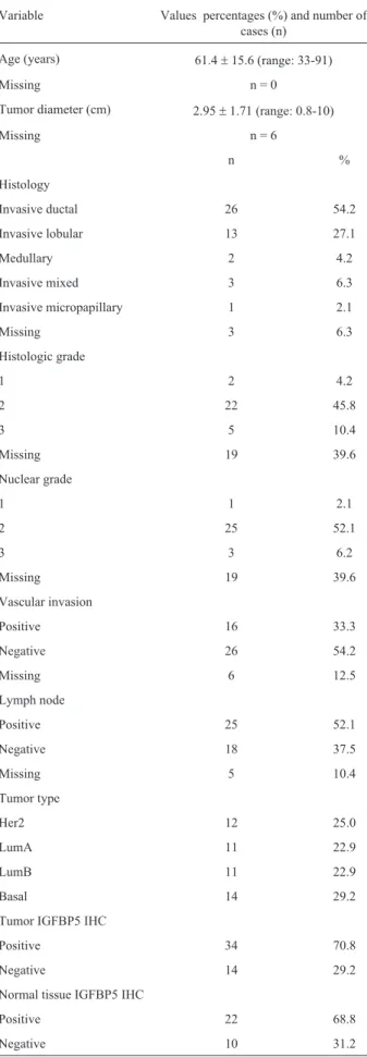
Table 2 summarizes the histology, histological grade, tumor diameter, nuclear grade, vascular invasion and lymph node status for the patients examined in this study. Some of the data are missing and these missing items were excluded from the statistical analyses. All statistical analy-ses of miRNA expression and the clinopathological charac-teristics are shown in Table 1.
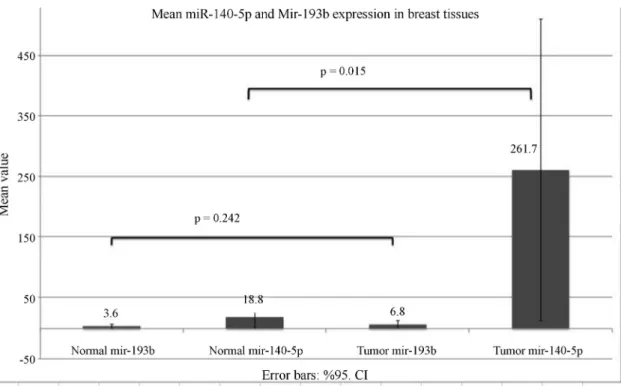
miR-140 and miR-193b expression in breast cancer tissue and adjacent non-cancerous tissue
The miRNA expression in tumor (n = 48) and adja-cent normal (n = 32) tissues was examined to determine whether miR-140 or miR-193b expression was a tumori-genic property (Figure 2). Tumor tissue samples had higher miR-140-5p expression than adjacent normal tissues (mean = 261.7 and median = 1.8 for tumor samples and mean = 18.79 and median = 0.57 for normal tissues; p = 0.015). These results indicate that enhanced miR-140
expression was characteristic of tumors and could be a po-tential biomarker for breast cancer. There was no difference in the miR-193b expression of tumors and adjacent normal tissue.
Spearman’s correlation was used to assess the corre-lation between miR-140-5p and miR-193b expression in tissue samples and both were found to be positively corre-lated in tumor samples (n = 48), in normal tissue samples (n = 32) and in all tissue samples (n = 80) (correlation
coef-ficient (CC) = 0.616, p = 0.000 for tumor tissue, CC = 0.507, p = 0.003 for normal tissue, and CC = 0.583, p = 0.0000 for all samples, respectively).
IGFBP5 protein immunohistochemistry and correlation with miR-140 and miR-193b
Tumor tissue samples and all tissue samples were classified into two groups as IGFBP5-positive (expressing the protein) and IGFBP5-negative (not expressing the
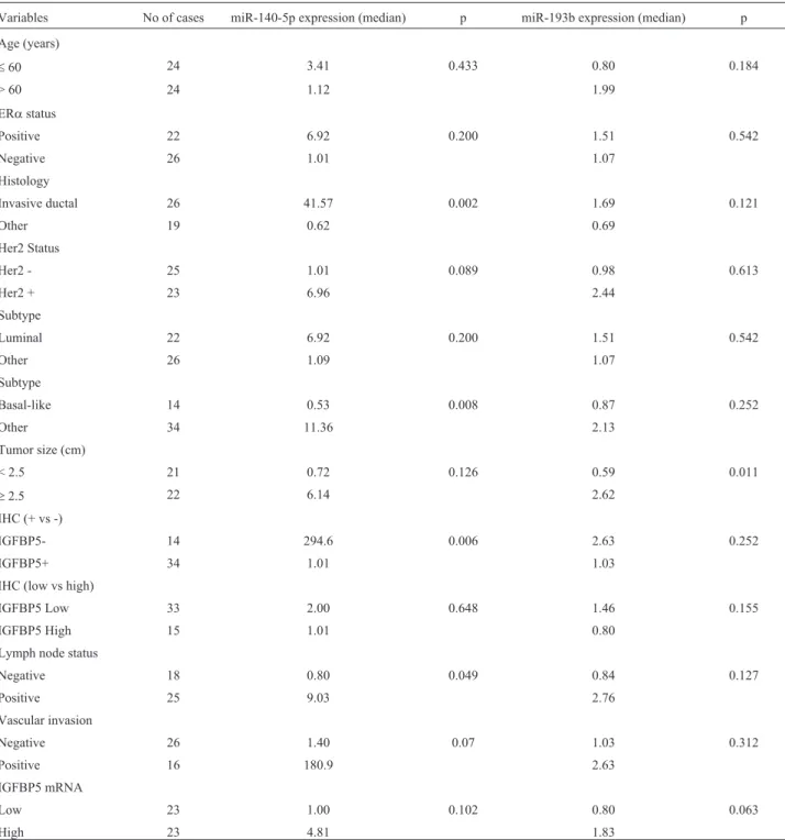
pro-Table 1- miR-140-5p and miR-193b expression in invasive breast carcinomas in relation to clinicopathological characteristics and IGFBP5 expression
Variables No of cases miR-140-5p expression (median) p miR-193b expression (median) p
Age (years)
£60 24 3.41 0.433 0.80 0.184
> 60 24 1.12 1.99
ERastatus
Positive 22 6.92 0.200 1.51 0.542
Negative 26 1.01 1.07
Histology
Invasive ductal 26 41.57 0.002 1.69 0.121
Other 19 0.62 0.69
Her2 Status
Her2 - 25 1.01 0.089 0.98 0.613
Her2 + 23 6.96 2.44
Subtype
Luminal 22 6.92 0.200 1.51 0.542
Other 26 1.09 1.07
Subtype
Basal-like 14 0.53 0.008 0.87 0.252
Other 34 11.36 2.13
Tumor size (cm)
< 2.5 21 0.72 0.126 0.59 0.011
³2.5 22 6.14 2.62
IHC (+ vs -)
IGFBP5- 14 294.6 0.006 2.63 0.252
IGFBP5+ 34 1.01 1.03
IHC (low vs high)
IGFBP5 Low 33 2.00 0.648 1.46 0.155
IGFBP5 High 15 1.01 0.80
Lymph node status
Negative 18 0.80 0.049 0.84 0.127
Positive 25 9.03 2.76
Vascular invasion
Negative 26 1.40 0.07 1.03 0.312
Positive 16 180.9 2.63
IGFBP5 mRNA
Low 23 1.00 0.102 0.80 0.063
High 23 4.81 1.83
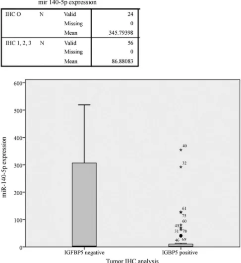
tein). Based on this classification, 29.2% of tumor samples and 31.3% of normal tissue samples were IGFBP5-negative and there was no significant difference between the two groups. Differences in miR-140-5p expression be-tween these groups were analyzed with the Mann-Whitney U-test. Tumor samples (n = 48) and all of the samples that were analyzed immunohistochemically (n = 80; regardless of whether they were normal or tumor tissue) showed greater miR-140-5p expression in IGFBP5-negative than in IGFBP5-positive samples (p = 0.006 and 0.009, respec-tively) (Figure 3). Immunohistochemically IGFBP5-negative samples showed increased expression of miR-140-5p, but this was not the case for miR-193b.
mRNA expression of IGFBP5 and correlation with miR-140 and miR-193b
Spearmans correlation coefficient was also used to examine the correlation between the protein (immunohis-tochemistry) and mRNA (RT-PCR) levels of IGFBP5. Based on this analysis, IGFBP5 protein expression in tumor samples was found to be negatively correlated with mRNA expression of this protein (CC = -0.295; p = 0.047), when protein expression [in which negative and low positive (+) staining were considered as negative results, and medium positive (++) and strong positive (+++) staining were con-sidered as positive results] was compared with IGFBP5 mRNA expression levels, calculated relative to the refer-ence gene [low (< 1) and high (> 1)] (Table 3). Although there was no significance, IGFBP5 mRNA-negative sam-ples showed a tendency to express higher levels of miR-140-5p, which suggested that miR-140-5p could post-transcriptionally regulate IGFBP5 expression.
Relationship between the clinicopathological
characteristics and miR-140, miR-193b and IGFBP5 protein levels
There was no correlation between miR-193b and miR-140-5p expression and patient age, ERastatus, Her2 status, vascular invasion and nuclear grade. However, inva-sive ductal carcinomas showed greater expression of miR-140-5p than other histological types (p = 0.002, median ex-pression = 41.57 and 0.618 for invasive ductal carcinomas and others, respectively). No such association was seen for miR-193b. On the contrary, basal-like tumors showed de-creased expression of miR-140-5p compared to other tu-mors (median miR-140 expression = 0.525 and 11.36 for basal-like tumors and other tumors, respectively; p = .008). No such relationship was observed for miR-193b. Lymph node-positive samples had an approximately 13-fold grea-ter expression of miR-140-5p than lymph node-negative samples (median expression = 0.799 and 9.031 for lymph node-negative and -positive samples, respectively; p = 0.049).
Comparison of the diameters of vascular invasive and non-invasive tumors based on the Mann-Whitney U-test
Table 2- Patient demographics and tumor characteristics.
Variable Values percentages (%) and number of cases (n)
Age (years) 61.4±15.6 (range: 33-91)
Missing n = 0
Tumor diameter (cm) 2.95±1.71 (range: 0.8-10)
Missing n = 6
n %
Histology
Invasive ductal 26 54.2
Invasive lobular 13 27.1
Medullary 2 4.2
Invasive mixed 3 6.3
Invasive micropapillary 1 2.1
Missing 3 6.3
Histologic grade
1 2 4.2
2 22 45.8
3 5 10.4
Missing 19 39.6
Nuclear grade
1 1 2.1
2 25 52.1
3 3 6.2
Missing 19 39.6
Vascular invasion
Positive 16 33.3
Negative 26 54.2
Missing 6 12.5
Lymph node
Positive 25 52.1
Negative 18 37.5
Missing 5 10.4
Tumor type
Her2 12 25.0
LumA 11 22.9
LumB 11 22.9
Basal 14 29.2
Tumor IGFBP5 IHC
Positive 34 70.8
Negative 14 29.2
Normal tissue IGFBP5 IHC
Positive 22 68.8
showed that the former were greater than the latter (n = 42, p = 0.003). The median diameter of vascular non-invasive tumors was 2.05 cm. Tumors > 2.5 cm in diameter had greater expression of miR-193b than tumors < 2.5 cm (me-dian expression = 0.59 and 2.62 for small and large tumors, respectively; p = 0.011).
All of the tumor samples and 27 of the adjacent nor-mal tissue samples were miR-193b-positive and only five of the adjacent normal tissues were negative; all samples of the latter group were immunohistochemically IGFBP5-positive and four were ER-negative. There was no signifi-cant correlation between the expression levels of IGFBP5, ER and miR-193b.
Discussion
We analyzed miRNA expression in breast cancer tu-mors (n = 48) and adjacent normal mammary (n = 32) tis-sues and found that tumor tissue had higher miR-140-5p expression than adjacent normal tissue. miR-140 has been considered a possible tumor suppressor miRNA because of its regulatory actions in the MAPK/ERK, TGF-b and SOX2 pathways (Zhanget al., 2012; Yanget al., 2013). However, as shown here, breast tumor samples had higher expression of miR-140 than normal breast tissue, although it was found to be down-regulated in some breast tumors. The role of miR-140 in breast tumorigenesis is unclear, but has been associated with drug resistance, tumor growth and metastasis in various cancers. miR-140 and SOX2, a stem
cell self-renewal regulator, are crucial components in the maintenance of breast cancer stem cells. The expression of miR-140 in ERa-positive breast cancer cells decreases sub-sequent estrogen stimulation because ERa suppresses miR-140 transcription by binding to the estrogen response element flanking the miR-140 promoter. miR-140 targets the SOX2 gene, thereby regulating breast tumor cell sur-vival (Zhanget al., 2012). However, we found no associa-tion between ERa-positive breast cancer tissue and miR-140 expression levels.
Recent studies have described anti-proliferative and anti-metastatic effects of miR-140 in Hepatocellular carci-noma (HCC), which suggests a possible role in tumor sup-pression. miR-140 targets the Tissue Growth Factor b receptor 1 (TGFBR1) and Fibroblast Growth Factor 9 (FGF9), thereby inhibiting TGF-band MAPK/ERK signal-ing (Yanget al., 2013). Our findings indicate that miR-140 expression has tumorigenic activity and could be a poten-tial biomarker for breast cancer. However, more data about patient demographics and clinical outcome are needed to evaluate whether patients with higher expression of miR-140 have any clinical advantage, such as response to treat-ment, metastasis-free survival, disease-free survival and overall survival.
Recently, Liet al.(2013) showed that miR-140 ex-pression is down-regulated in ductal carcinoma in situ
(DCIS) stem cells compared to normal mammary stem cells, and that SOX9 and ALDH1, direct targets of
miR-140, were markedly activated in DSIC stem cells. Restoration of miR-140 expression in ERa-nega-tive/basal-like DCIS cells resulted in a decrease in SOX9 and ALDH1 expression and decreased tumor growth in vivo(Liet al., 2013). As shown here,
ERa-negative/basal-like samples had the lowest miR-140 expression (2.5-fold and 4-fold lower than in other tumors and normal tissue samples, respectively). This could explain the aggressive-ness of basal-like tumors in which miR-140 is suppressed,
stem cell factors are induced and the tumors are more resis-tant to therapy. Normal tissues had about a 15-fold lower expression of miR-140 compared to tumors, excluding ERa-negative/basal-like tumors. Based on these findings, we conclude that normal breast tissues need optimal ex-pression of miR-140 for cell renewal, and that basal-like tu-mors, which have lower expression of this protein, have aggressive characteristics compared to other types of breast cancer.
Invasive ductal tumors showed a 70-fold greater miR-140 expression than other tumors (median = 42.57 and 0.618, respectively; p = 0.002). In addition, patients with positive lymph nodes had greater expression of miR-140 than the other groups (median expression = 0.80 and 9.03, respectively; p = 0.049); this finding suggests that miR-140 may be involved in lymph node invasion. To our knowl-edge, this is the first demonstration of a significant associa-tion between miR-140 and lymph node metastasis in breast cancer tissue. Previously, miR-140 had only been reported to be down-regulated in primary breast cancer tissue
com-Figure 3- miR-140-5p expression in tissues with positive and negative immunohistochemical staining for IGFBP5. Of the 80 samples analyzed immunohistochemically, those that were IGFBP5-negative showed significantly greater miR-140-5p expression than IGFBP5-positive samples (p = 0.009; Mann-Whitney U-test); miR-140 expression is 4-fold less than in tissues that do not express IGFBP5 protein. The numbers beside some of the as-terisks (*) states the sample number. The error bars indicate the 95% confidence intervals (CI).
Table 3- Correlation between IGFBP5 mRNA (RT-PCR) and protein (immunohistochemistry - IHC) expression levels.
IHC results
Negative Positive Total (n)
mRNA ex-pression
Low 13 11 24
High 18 4 22
pared to normal breast tissue, but it was not shown to be as-sociated with lymph node invasion (Yan et al., 2008). Further data are needed to shed light on the relationship be-tween miR-140 and invasiveness.
Tardifet al.(2009) showed that IGFBP5 was directly
targeted for mRNA degradation by miR-140, but expres-sion analysis of miR-140 during chondrogenic differentia-tion did not confirm that IGFBP5 was a direct target of miR-140 (Buechli et al., 2013). These authors indicated
that IGFBP5 was not directly targeted by miR-140 for deg-radation, which would be expected if miR-140 facilitated its decay; the possibility that the translation of IGFBP5 could be inhibited by miR-140 was not tested. As shown here, there was no correlation between IGFBP5 mRNA ex-pression and miR-140. In addition, regardless of the type of tissue (tumor or normal), in samples in which IGFBP5 pro-tein was expressed miR-140 expression was four-fold lower than in samples not expressing IGFBP5 protein (Fig-ure 3). These results indicate that miR-140 post- trans-criptionally regulates IGFBP5 by inhibiting its translation rather than by degrading its mRNA. We conclude that miR-140 is more effective at repressing IGFBP5 protein expression in normal tissues than in tumor samples and that the higher expression of miR-140 in IGFBP5-negative tu-mors reflects this proteins effectiveness.
Although IGFBP5 mRNA is up-regulated in cancer tissue (Liet al., 2007), we found no significant difference among our samples. IGFBP5 mRNA levels have been posi-tively correlated with ER and PR status, and negaposi-tively cor-related with HER2 overexpression, with low levels of IGFBP5 mRNA in tumors being associated with better prognosis and disease-free survival (Hunget al., 2008). In contrast to this study, we found no significant correlation between IGFBP5 mRNA levelsvs. ER and PR status or HER2 overexpression.
There was a significant positive correlation between miR-140-5p and miR-193b expression in all tissue sam-ples. However, there was no significant correlation be-tween ER status and miR-193b, in contrast to the findings of Yoshimotoet al.(2011) who reported that ERa-negative tumors had relatively lower miR-193b expression than ERa-positive tumors. Foekenset al.(2000) reported a rela-tion between miR-193b and uPA that plays a key role in in-vasion and metastasis in breast cancers. uPA expression is increased in metastatic breast cancer and is negatively cor-related with miR-193b. This may mean that miR-193b could be a biomarker for metastatic breast cancer (Nohet al., 2011). On the other hand, miR-193b acts as a tumor suppressor that limits proliferation, migration and invasion in acute myeloid leukemia by regulating c-Kit proto-onco-gene (Gaoet al., 2011), and also suppresses human hepa-tocellular carcinoma cells (Xuet al., 2010), melanoma (by regulating Mcl-1) (Chenet al., 2010) and non-small cell lung cancer cells (Huet al., 2012). In the present study, it was not possible to analyze our data based on metastatic
be-havior or tumor invasiveness since we did not have the necessary data for the patients.
In conclusion, our results show that breast tumor tis-sue had higher miR-140-5p expression than adjacent nor-mal tissues and that patients with positive lymph nodes had greater miR-140 expression than other individuals. On the other hand, basal-like breast tumors had lower expression of miR-140-5p than other types of breast tumors. Thus, miR-140 expression is characteristic of breast tumors in general and may be an important molecule for understand-ing lymph node invasion and tumor differentiation in breast cancer. There was no correlation between IGBFP5 mRNA and miR-140 expression, but miR-140 expression was lower in tumors expressing IGFBP5 protein compared to samples which not expressing this protein. There was a sig-nificant positive correlation between miR-140-5p and miR-193b expression in all tissue samples, but there was no association between miR-193b expression and any other clinical features. Further studies, including clinical fol-low-up of the patients, would be useful in clarifying the role of miR-140, miR-193b and IGFBP5 in breast cancer and in assessing the usefulness of these miRNAs and IGFBP5 as suppressors or inducers of breast carcinogenesis.
Acknowledgments
The authors thank Dr. Nadiye Pinar Ay (Department of Public Health, School of Medicine, Marmara Univer-sity) for assistance in statistical analyses. This work was partly supported by a grant (SBAG-111S161 to MA) from the Scientific and Technological Research Council of Tur-key and by grants (SAG-C-YLP-110412-0067 and SAG-C-YLP-210311-0048 to MA) from the Research Foundation of Marmara University (BAPKO).
References
Akkiprik M, Feng Y, Wang H, Chen K, Hu L, Sahin A, Krishnamurthy S, Ozer A, Hao X and Zhang W (2008) Multifunctional roles of insulin-like growth factor binding protein 5 in breast cancer. Breast Cancer Res10:212. Beattie J, Allan GJ, Lochrie JD and Flint D (2006) Insulin-like
growth factor binding protein-5 (IGFBP5): A critical mem-ber of the IGF axis. Biochem J 395:1-19.
Becker MA, Hou X, Harrington SC, Weroha SJ, Gonzalez SE, Ja-cob KA, Carboni JM, Gottardis MM and Haluska P (2012) IGFBP ratio confers resistance to IGF targeting and corre-lates with increased invasion and poor outcome in breast tu-mors. Clin Cancer Res 18:1808-1817.
Buechli ME, Lamarre J and Koch TG (2013) MicroRNA-140 ex-pression during chondrogenic differentiation of equine cord blood-derived mesenchymal stromal cells. Stem Cells Dev 22:1288-1296.
Chen J, Feilotter HE, Paré GC, Zhang X, Pemberton JG, Garady C, Lai D, Yang X and Tron VA (2010) MicroRNA-193b re-presses cell proliferation and regulates cyclin D1 in mela-noma. Pathology 176:2520-2529.
Eroles P, Bosch A, Pérez-Fidalgo JA and Lluch A (2011) Molecu-lar biology in breast cancer: Intrinsic subtypes and signaling pathways. Cancer Treat Rev 6:698-707.
Foekens J, Peters HA, Look MP, Portengen H, Schmitt M, Kra-mer MD, Brünner N, Jänicke F, Meijer-van Gelder ME, Henzen-Logmans SC,et al.(2000) The urokinase system of plasminogen activation and prognosis in 2780 breast cancer patients. Cancer Res. 60:636-643.
Gao XN, Lin J, Gao L, Li YH, Wang LL and Yu L (2011) MicroRNA-193b regulates c-Kit proto-oncogene and re-presses cell proliferation in acute myeloid leukemia. Leuk Res 35:1226-1232.
Güllü G, Karabulut S and Akkiprik M (2012) Functional roles and clinical values of insulin-like growth factor binding pro-tein-5 in different types of cancers. Chin J Cancer 6:266-280.
Hu H, LiS, Liu J and Ni B (2012) MicroRNA-193b modulates proliferation, migration, and invasion of non-small cell lung cancer cells. Acta Biochim Biophys Sin (Shanghai) 44:424-430.
Hung PS, Kao SY, Shih YH, Chiou SH, Liu CJ, Chang KW and Lin SC (2008) Insulin-like growth factor binding protein-5 (IGFBP-5) suppresses the tumourigenesis of head and neck squamous cell carcinoma. J Pathol 214:368-376.
Johnson SK and Haun RS (2009) Insulin-like growth factor bind-ing protein-5 influences pancreatic cancer cell growth. World J Gastroenterol 15:3355-3366.
Khandwala HM, McCutcheon IE, Flyvbjerg A and Friend KE (2000) The effects of insulin-like growth factors on tumori-genesis and neoplastic growth. Endocr Rev 21:215-244. Kricker JA, Hyde CE, Van Lonkhuyzen DR, Hollier BG, Shooter
GK, Leavesley DI, Herington AC and Upton Z (2010) Mechanistic investigations into interactions between IGF-I and IGFBPs and their impact on facilitating cell migration on vitronectin. Growth Factors 28:359-369.
Leivonen SK, Mäkelä R, Ostling P, Kohonen P, Haapa-Paananen S, Kleivi K, Enerly E, Aakula A, Hellström K, Sahlberg N, et al.(2009) Protein lysate microarray analysis to identify microRNAs regulating estrogen receptor signaling in breast cancer cell lines. Oncogene 28:3926-3936.
Li Q, Yao Y, Eades G, Liu Z, Zhang Y and Zhou Q (2013) Downregulation of miR-140 promotes cancer stem cell for-mation in basal-like early stage breast cancer. Oncogene 20:2589-2600.
Li X, Cao X, Li X, Zhang W and Feng Y (2007) Expression level of insulin-like growth factor binding protein 5 mRNA is a
prognostic factor for breast cancer. Cancer Sci 98:1592-1596.
Mita K, Zhang Z, Ando Y, Toyama T, Hamaguchi M, Kobayashi S, Hayashi S, Fujii Y, Iwase H, Yamashita H (2007) Prog-nostic significance of insulin-like growth factor binding pro-tein (IGFBP)-4 and IGFBP-5 expression in breast cancer. Jpn J Clin Oncol 37:575-582.
Mohan S and Baylink DJ (2002) IGF-binding proteins are multi-functional and act via IGF-dependent and independent mechanisms. J Endocrinol 175:19-31.
Noh H, Hong S, Dong Z, Pan ZK, Jing Q and Huang S (2011) Im-paired microRNA processing facilitates breast cancer cell invasion by upregulating urokinase-type plasminogen acti-vator expression. Genes Cancer 2:140-150.
Palmero EI, de Campos SG, Campos M, de Souza NC, Guerreiro ID, Carvalho AL and Marques MM (2011) Mechanisms and role of microRNA deregulation in cancer onset and progres-sion. Genet Mol Biol 34:363-370.
Tardif G, Hum D, Pelletier JP, Duval N and Martel-Pelletier J (2009) Regulation of the IGFBP-5 and MMP-13 genes by the microRNAs miR-140 and miR-27a in human osteo-arthritic chondrocytes. BMC Musculoskelet Disord 10:148. Valentinis B and Baserga R (2001) IGF-I receptor signalling in
transformation and differentiation. Mol Pathol 54:133-137. Xu C, Liu S, Fu H, Li S, Tie Y, Zhu J, Xing R, Jin Y, Sun Z, Zheng
X (2010) MicroRNA-193b regulates proliferation, migra-tion and invasion in human hepatocellular carcinoma cells. Eur J Cancer 15:2828-2836.
Yan LX, Huang XF, Shao Q, Huang MY, Deng L, Wu QL, Zeng YX and Shao JY (2008) MicroRNA miR-21 overexpression in human breast cancer is associated with advanced clinical stage, lymph node metastasis and patient poor prognosis. RNA 11:2348-2360.
Yang H, Fang F, Chang R and Yang L (2013) MicroRNA-140-5p suppresses tumor growth and metastasis by targeting trans-forming growth factor breceptor 1 and fibroblast growth factor 9 in hepatocellular carcinoma. Hepatology 58:205-217.
Yoshimoto N, Toyama T, Takahashi S, Sugiura H, Endo Y, Iwasa M, Fujii Y and Yamashita H (2011) Distinct expressions of microRNAs that directly target estrogen receptorain hu-man breast cancer. Breast Cancer Res Tr 130:331-339. Zhang Y, Eades G, Yao Y, Li Q and Zhou Q (2012) Estrogen
re-ceptorasignaling regulates breast tumor-initiating cells by down-regulating miR-140 which targets the transcription factor SOX2. J Biol Chem 287:41514-41522.
Associate Editor: Anamaria Aranha Camargo