Int J Anat Res 2015, 3(4):1685-88. ISSN 2321-4287 1685 Original Research Article
AN ANATOM ICAL STUDY OF GRACILIS M USCLE AND ITS VASCULAR
PEDICLES
M .S. Rajeshw ari * ¹, B.N. Roshan kumar ².
ABSTRACT
Address for Correspondence: Dr. M . S. Rajeshwari, Professor of Anat omy, Bangalore M edical College and Research Inst it ut e, Bangalore, Karnat aka, India.
E-M ail: rajeshw ari.roshan8@gmail.com
Background:Gracilis m uscle being easily accessible and funct ionally a w eak m uscle is suit able for m uscle graft t o replace t he dam aged m uscle in any part of t he body. The lengt h of t he m uscle, vascular pedi cles and lim it ed donor sit e m or bidit y helps t he surgeon t o plan accordingly. The m uscle receives a num ber of vascular pedicles ranging from one t o five. The source of t hese pedicles varies.
M aterial and M ethods:The st udy w as conduct ed on 36 form alin fixed low er lim bs of bot h sexes of unknow n age
from t he depart m ent of Anat omy, BM CRI, Bangalore.
Results and Discussion:In 75% of lim bs t w o vascular pedicles w ere seen penet rat ing t he m uscle at differ ent levels and in 25% accessory pedicles w ere seen in t he low er 2/3rd of t he m uscle.
Conclusion:The findings suggest t hat t he first vascular pedicle t o t he m uscle is alw ays const ant in posit ion accom panied by it s venae com it ans and branch from obt urator nerve and is placed at a dist ance of 10.5cm s±2cm s from t he pubic t ubercle.
KEY W ORDS:Gracilis M uscle, Vascular Pedicle, M uscle Transplantat ion, Flap Reconst r uct ion.
INTRODUCTION
Int ernat ional Journal of Anatomy and Research, Int J Anat Res 2015, Vol 3(4):1685-88. ISSN 2321- 4287 DOI: ht t p:/ / dx.doi.org/10.16965/ ijar.2015.316
Access this Article online
Quick Response code Web site:
Received: 17 Nov 2015 Accept ed: 14 Dec 2015 Peer Review : 17 Nov 2015 Published (O): 31 Dec 2015 Revised: None Published (P): 31 Dec 2015
Int ernat ional Journal of Anat omy and Research ISSN 2321-4287
ww w.ijmhr.org/ ijar.htm
DOI: 10.16965/ ijar.2015.316
*1 Professor, Depart ment of Anat omy, Bangalore M edical College & Research Inst it ute, Bangalore,
Karnat aka, India.
2 Professor and Head, Depart ment of Ort hopaedics, RajaRajeshw ari M edical College Bangalore,
Karnat aka, India.
funct ion in pat ients with faecal incont inence [1].
“Gracilis” t he muscle derives it s name from t he lat in w ord m eaning slender. It is t he m ost superficial of t he adduct or group of m uscles w hich is t hin, flat and broad above and t hick, narrow and tapering below. It arises by a t hin aponeuroses from t he medial m argins of t he low er half of t he body of t he pubis, t he w hole of t he inferior ramus of pubis and t he adjoining part of ischial ramus. The fibres descend and curve around t he medial condyle of t he t ibia, w here it fans out and at t aches t o t he upper part of t he Gracilis muscle is used oft enly in reconst ruct ive
Int J Anat Res 2015, 3(4):1685-88. ISSN 2321-4287 1686
M .S. Rajeshw ari, B.N. Roshan kum ar. AN ANATOM ICAL STUDY OF GRACILIS M USCLE AND ITS VASCULAR PEDICLES.
M ATERIALS AND M ETHODS
m edial sur f ace of t he t ibia j ust below t he condyle, proximal t o that of semitendinosus, and it s upper edge is overlapped by t he t endon of sart orius, w it h w hich it blends part ly. It is inner-vated by t he anterior division of obt urat or nerve and derives it s blood supply from t he obt urat or ar t er y, m ed i al ci rcu m f l ex f em or al ar t er y, d escend i n g geni cu lar ar t er y, or f ro m t h e superior and inferior medial genicular art eries or from femoral art ery [2]. Usually t w o or t hree v ascu l ar p ed i cles acco m p an ied by v en ae comit ans, ent er t he muscle from it s deep sur-face and nourish it. Of t hese the proximal pedicle is generally t he dominant blood supply t o t he m uscle and supplies 70% of t he bulk of t he muscle. The number of m inor pedicles range from one t o five w hich dist ally supply 15%-30% of t he bulk of t he muscle [3].
The st udy w as conduct ed on 36 formalin fixed low er limbs of bot h sexes,30 male and 6 female o f u nk n o w n age f r om t he dep ar t m en t o f Anat omy, BM CRI, Bangalore. The skin over t he medial compart ment of t he t high w as reflect ed and t he gracilis muscle w as exposed, the muscle was cleaned from the sorrounding st ructures and along it s ent ire lengt h t o not e t he number and ent ry point s of t he vascular pedicles.
The object ives of t he st udy is t o find t he t ot al lengt h of t he muscle, w idt h of t he muscle at t he point of ent ry of t he neurovascular pedicle, number of vascular pedicles and source of t he art eries.
OBSERVATIONS
Table 1: Show ing M ean lengt h and M ean w idt h of t he m uscle. (Fig.-1)
M ean W idth 3.9±2cm
M ean Length 42.2±2cm
Fig. 1: Show ing lengt h of t he gracilis m uscle.
Fig. 2: Show ing One Principal/ M ain pedicle and one accessory Pedicle.
Fig. 3: Sh o w in g on e pr i n ci p al/ m ain p ed i cle and 2 Accessory pedicle.
Fig. 4: Sh o w i n g On e Pr in cip al/ m ai n p ed i cl e an d 3 Accessory Pedicles.
Pr in ci pal / M ai n Pe d i cl e
1st Accessor y
Pe d i cl e
Pr in ci pal / M ai n Pe d i cl e
1st Accessor y
Pe d i cl e
2n d Accessor y
Pe d i cl e
Pr in ci pal / M ai n Pe d i cl e
1st Accessor y
Pe d i cl e
2n d Accessor y
Pe d i cl e
3r d Accesso r y Pedicle
Int J Anat Res 2015, 3(4):1685-88. ISSN 2321-4287 1687
DISCUSSION
M .S. Rajeshw ari, B.N. Roshan kum ar. AN ANATOM ICAL STUDY OF GRACILIS M USCLE AND ITS VASCULAR PEDICLES.
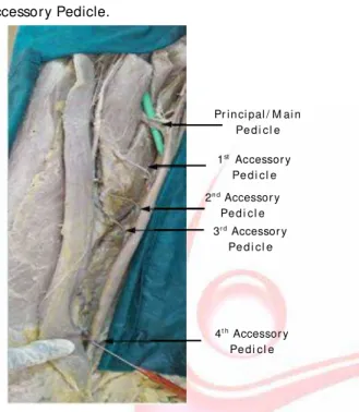
Fig. 5: Sh ow in g On e Pr inci pal / M ai n pedicl e an d 4 Accessory Pedicle.
Table 2: Show ing Num ber of Pedicles. (Fig.-2,3,4,5)
Number of Pedicles Percentage Principal (M ain) + 1 accessory pedicle 75.0% (27)
Principal + 2 accessory pedicle 16.6% (6)
Principal + 3 accessory pedicle 2.7% (1)
Principal + 4 accessory pedicle 5.5% (2)
Table 3:Show ing Ent rypoint of Pedicles.
Pedicles Distance from the pubic tubercle
Principal Pedicle 10.5±2 cms
1st accessory pedicle 22±1.5 cms
2nd accessory pedicle 28±1.5 cms
3rd accessory pedicle 30±1.5 cms
4t h accessory pedicle 36±1.5 cms
Table 4: Show ing origin of t he Pedicles.
PFA OA FA DGA SA
Principal
Pedicle 55% 6.15% 5.55%
1st accessory
pedicle - - 83.30%
2nd accessory
pedicle 11.10%
3rd accessory
pedicle 2.70%
4t h accessory
pedicle 5.50%
Pedicles Source of the pedicle M CFA
33.30%
16.70%
5.50%
w hich t he proximal pedicle is considered as t he principal or t he m ain pedicle and provides nut rit ion t o t he upper t w o t hirds of t he muscle. The accessory pedicle, if present t hey are seen mainly in t he low er t w o t hirds of t he muscle. Occasionally a small m inor accessory pedicle t hat is proximal t o t he main or principal pedicle m ay be present w hich is know n as proxim al accessory pedicle [3]. The dom inant pedicle originat es from t he art ery for adduct ors in 46% and 73% respect ively [4,5]. The vessels normally ent ered t he deep surface of t he muscle t hrough
t he anterior border.M ajorit y of t he muscle had
one principal/ main pedicle and one accessory p ed i cle and b el o n ged t o Typ e-B o f [ 6] classificat ion. The source of principal pedicle o b ser v ed i n o u r st ud y i s si m i l ar t o t h e observat ions seen in st udy [7]. The source of t hese art eries is not alw ays const ant but varies from one populat ion t o anot her.
M u scl e gr af t s ar e f r eq uen t ly n eed ed i n reconst ruct ive surgery anyw here in t he body. M an y sur geon s p r ef er gr acil i s m u scl e f o r t ransplant as it fulfills t he necessary crit eria and t he donor morbidit y is less [8].
Gracilis being a slender muscle is t he surgeons first choice for foot and ankle reconst ruct ion and in t he t reat ment of chronic ost eomyelit is as it adequat ely fill t he small defect creat ed by t he debridement [9].
Gracilis muscle derives it s vascular supply from a w ide range of art eries. The number of pedicles t o t he muscle ranges from one t o five, out of
Table 5: Show s com parison of t he M ean lengt h & w idt h.
Authors M ethodology M ean Length M ean width
Present study Dissection 42.2±2 cm 3.9±0.5 cm
AJ.Hussey et al 2007 [10]
M achii.V et al 2008 [11]
Harbans Singh 2011 [12]
38.4 cm 6.2 cm
CT Angiography 41±2.1 cm 44±1 mm
Dissection 43±2.08 cm (M ) -37.1±0.76 cm (F)
Dissection & Radiology
Table 6: Show s com parison of t he ent ry point of principal pedicle t aken from pubic t ubercle.
Authors M ethodology
Dissection & Radiology 9.4cm
CT Angiography 10±1.3cm
AJ.Hussey et al 2007 [10] M achii.V et al 2008 [11]
Entry point of Principal Pedicle
Dissection & Contrast
Radiograph 10±2cm Dissection & Contrast
Radiography 9.28 cm Present study Dissection 10.5±2cm
Iain.S.W hitaker 2012 [3]
Dorothee .C. 2006 [13] Pr in ci pal / M ai n
Pe d i cl e
1st Accessor y
Pe d i cl e
3r d Accessor y
Pe d i cl e 2n d Accessor y
Pe d i cl e
4t h Accessor y
Int J Anat Res 2015, 3(4):1685-88. ISSN 2321-4287 1688
Table 7: Show s com par ison of Presence of Pr incipal (M ain) and Accessory pedicle.
Xia ochun et al 2012 [6] Present study
M ethodology Contrast Radiograph Dissection
Single principal Pedicle 8.30%
-One principal + 1 accessory. P 41.70% 75.00%
One principal + 2 accessory. P 33.30% 16.60%
One principal + 3 accessory. P 16.70% 2.90%
One principal + 4 accessory. P - 5.50%
Table 8: Show s com parison of source of Principal (M ain) vessel/ Pedicle.
A.A FA
46%
-33.30%
73% 7.7% Dual
81.25%
Source PFA M CFA
Iain.s.whitaker et al 2012 [3] 70%
M achii.V et al 2008 [11] 45% 9%
Xiaochun et al 2012 [6] 54.20% 12.50%
5.50%
Singh et al 2011 [12] 70% 30%
19.20%
Dorothee et al 2006 [13] -
-Jurisic et al 1993 [5]
Present study 33.30% 6.15% 55.00%
CONCLUSION
Though previous and present st udies report on t h e n u m b er o f vascul ar ped i cl es, n o t w o ind ivid uals ar e sim ilar in t heir anat om i cal st ruct ures. Considering t he fact ors from t he present st udy t he average lengt h of t he gracilis m uscle is 42cm and t he num ber of vascular pedicles range from one t o four. A const ant neurovascular pedicle is present in t he proxi-mal one t hird of the muscle w hich can be ut ilised for all m uscle t ransplant at ion surgeries since bot h t he nerve and t he art ery t raverse t oget her t o ent er t he deep surface of t he muscle t hrough it s anterior border.
ABBREVIATIONS
M CFA - M edial Circumflex Femoral Artery
PFA - Profunda Femoris Artery
OA - Obt urat or Art ery
FA - Femoral Artery
AA - Adduct or Art ery
DGA - Descending Genicular Art ery
SA - Saphenous Art ery
Conflicts of Interests: None
REFERENCES
[1]. Shatari.T, Niim i.M . Fuiita.M . Kodaira.S.. Vascular Anat om y of Gracilis m uscle, ar t erial f indings t o e n h an ce Gr aci l o p l ast y. Su r g. Rad i o l . An at , 2000;22(1):21-4.
[2]. Gray’s Anat omy ,Churchill Livingst one, 38t h edit ion. 2000, pg 874.
[3]. Iain.S.Whitaker et al. The Gracilis myocutaneous free flap, A Quant it at ive Analysis of t he fasciocut aneous blood supply & im plicat ions for Analogous Breast reconst r uct ion. PLos ONE25/2012;7(5):e36367. [4]. Enr ico vigat o et al. The clinical r ole of gr acilis
m uscle; Pelviperineology 2007;26:149-151 [5]. M .Jurisic et al. Anat om ic basis for use of gracilis
m uscle f lap. Anat om ic basis of M edical, Surgical, and Radiological Anat omy. Sept 1993;15(3):163-168.
[6]. Xiaochun An et al. Art er ial Anat om y of t he Gracilis m uscle as det erm ined by Lat ex inject ion & glycerin t ransparency. Clinical Anat omy 2012;25;231-34. [7]. Di v ya. N. Up ad hyaya, Vai b av kh an n a, Su r aj i t
Bhat t achar ya, Sandeep Garg, Ram esh Kohli. The t ransversely split gr acilis t w in f ree flaps, Indian Journal of Plast ic Surgery, 2010;43(2):173-76. [8]. Huem er G.M , Dunst KM , M aurer .H and Ninkovic.
M . Area enlargem ent of t he gracilis m uscle f lap t h r o u gh m i cr o scop i cal l y ai ded i n t r am uscu l ar dissect ion, ideas and innovat ion. M icrosur ger y 2004;24(5);369-73.
[9]. P.Lorea, N. vercruysse, B.C.Coessens. Use of gracilis m u scl e f r e e f l ap Re co n st r u ct i o n o f ch r o n i c ost eom yelit is of foot and ankle, Act a Ort hopaedica Belgica, 2001;67-3.
[10]. AJ.Hussey, AJ Laing, Padriac Jam es Regan, Annals of plast ic surgery, 11/2007;59(4):404-9.
[11]. M achi.V et al. Gracilis m uscle and it s use in clinical reconst ruct ion. An Anat om ical, Em bryological and Radiological st udy. 2008;21(7):696-704.
[12]. Harbans Singh, Ram andeep Kaur, Neena Gupta. M orphom et r ic st udy of gracilis m uscle and it s role in clinical Reconst ruct ion. J.Anat . Soc. India. 2011; 60(2):202-206.
[ 13] .Dor o t h ee Coqu er el -Begh un et al. The Gr acil is m usculocut aneous f lap, vascular supply of t he m u scl e an d ski n co m p o n e n t s, su r gi cal an d Radiological Anat om y 2006:28(6):588-95.
How to cite this article
:
M .S. Rajeshw ari, B.N. Roshan kum ar. AN ANATOM ICAL STUDY OF GRACILIS M USCLE AND ITS VASCULAR PEDICLES. Int J Anat Res
2015;3(4):1685-1688. DOI: 10.16965/ ijar.2015.316