Distribuição do colágeno tipo I, colágeno tipo III e versican
na lâmina própria da prega vocal humana de fetos e
adultos : método histoquímico e imunoistoquímico
Tese apresentada à Faculdade de Medicina da
Universidade de São Paulo para obtenção do
Título de Doutor em Ciências
Área de concentração: Otorrinolaringologia
Orientador: Prof. Dr. Luiz Ubirajara Sennes
Dados Internacionais de Catalogação na Publicação (CIP)
Preparada pela Biblioteca da
Faculdade de Medicina da Universidade de São Paulo ©reprodução autorizada pelo autor
Bühler, Rogerio Borghi
Distribuição do colágeno tipo I, colágeno tipo III e versican na lâmina própria da prega vocal humana de fetos e adultos : método histoquímico e imunoistoquímico / Rogerio Borghi Bühler. -- São Paulo, 2008.
Tese(doutorado)--Faculdade de Medicina da Universidade de São Paulo. Departamento de Oftalmologia e Otorrinolaringologia.
Área de concentração: Otorrinolaringologia. Orientador: Luiz Ubirajara Sennes.
Descritores: 1.Colágneo tipo I 2.Colágeno tipo III 3.Versicanas 4.Cordas vocais 5.Laringe 6.Imunoistoquímica
DEDICATÓRIA
Aos meus pais pela oportunidade da vida.
A minha mãe pelo planejamento e investimento em minha educação.
Ao meu avô João pelo exemplo de vida.
A minha irmã Gisele pela amizade eterna.
A minha esposa Karina pela compreensão, companheirismo e apoio. Pelo exercício pleno do amor na figura de esposa, de amiga e de mãe.
AGRADECIMENTOS
Ao Prof. Dr. Luiz Ubirajara Sennes, pela oportunidade de poder participar de um grupo tão seleto da Otorrinolaringologia, pelo exemplo de dedicação e competência na área acadêmica e pelo incentivo no desenvolvimento deste estudo.
Ao Prof. Dr. Domingos Hiroshi Tsuji, pela simplicidade e objetividade na abordagem de temas diversos indispensáveis ao nosso desenvolvimento profissional e pessoal.
Ao Prof. Dr. Richard Voegels, pela confiança, apoio e incentivo que me conduziram ao curso de Pós-Graduação.
A Prof. Dra. Thais Mauad, Professora Associada do Departamento de Patologia da Faculdade de Medicina da Universidade de São Paulo, pela “co-orientação”, paciência, competência e dedicação nos ensinamentos de sua área.
Ao Prof. Dr. Ricardo Ferreira Bento, Professor Titular da Disciplina de Clínica Otorrinolaringológica da Faculdade de Medicina da Universidade de São Paulo, pela competência e dedicação no gerenciamento da Clínica proporcionando condições adequadas para o desenvolvimento acadêmico e profissional de seus alunos.
Ao Dr. Luis Fernando Ferraz, médico do Departamento de Patologia Faculdade de Medicina da Universidade de São Paulo, pela ajuda indispensável na elaboração das análises estatísticas, pelo altruísmo e amor à pesquisa.
Ao Prof. Dr. Osíris de Oliveira Camponês do Brasil, Professor Convidado da Disciplina de Otorrinolaringologia da Universidade Federal de São Paulo, pela paixão e vanguarda no exercício profissional da Laringologia e Cirurgia de Cabeça e Pescoço despertando em mim o interesse nestas áreas.
Ao Dr. Erich Christiano Madruga de Melo, ex-Doutorando da Disciplina de Clínica Otorrinolaringológica da Faculdade de Medicina da Universidade de São Paulo, pela vanguarda da pesquisa, disponibilidade e boa vontade nas sugestões e discussões que culminaram no êxito deste estudo.
Ao Dr. André Duprat, Professor do Departamento de Otorrinolaringologia da Faculdade de Ciências Médicas da Santa Casa de São Paulo, exemplo de atuação na Laringologia, pelas oportunidades e incentivo.
Ao Dr. Rui Imamura, pela disponibilidade e dedicação na orientação dos projetos de pesquisa da Clínica.
Ao Dr. Michel Cahali e Dr. Ronaldo Frizzarini pelas ponderações e sugestões no desenvolvimento deste estudo.
Ao Prof. Dr. Chao Lung Wen, Chefe da Disciplina de Telemedicina do Departamento de Patologia Faculdade de Medicina da Universidade de São Paulo, pela oportunidade da utilização dos recursos gráficos deste estudo.
Ao Prof. Dr. Carlos Augusto Pasqualucci, Diretor do SVOC-USP e seus funcionários, pelo apoio na obtenção das laringes.
Ao Dr. Flávio Sakae, colega da Pós-graduação, pela amizade e troca de informações indispensáveis para a conclusão deste estudo.
Aos colegas e residentes do Hospital do Servidor Público Estadual de São Paulo pela compreensão e apoio.
A Sandra de Moraes, pela dedicação na elaboração do material histológico.
Aos funcionários técnicos e administrativos do Departamento de Patologia pela dedicação no exercício de suas funções.
As secretárias e funcionários do Departamento de Otorrinolaringologia, em especial a Marileide, Márcia e Luci, pelo auxílio em todos os momentos necessários.
“O homem pretende ser imortal, e para isso defende princípios efêmeros. Um dia, inexoravelmente, descobrirá que para ser imortal deverá defender princípios absolutos. Neste dia morrerá para a carne, efêmera, e viverá para o espírito, eterno. Será imortal.”
NORMALIZAÇÃO
Esta tese está de acordo com as seguintes normas, em vigor no momento desta publicação:
Referências: adaptado de Internacional Committee of Medical Journals Editors
(Vancouver)
Universidade de São Paulo. Faculdade de Medicina. Serviço de Biblioteca e Documentação. Guia de apresentação de dissertações, teses e monografias. Elaborado por Anneliese Carneiro da Cunha, Maria Julia A.L. Freddi, Maria F. Crestana, Marinalva D.S. Aragão, Suely C. Cardoso, Valéria Vilhena. São Paulo: Serviço de Biblioteca e Documentação; 2005.
SUMÁRIO
Revista Laryngoscope – Instrução para autores Artigo publicado em Revista Indexada Internacional Artigo a ser submetido à Revista Indexada Internacional Lista de abreviaturas, símbolos e siglas
Lista de Figuras Lista de Tabelas Resumo
Summary
1. INTRODUÇÃO ... 1
1.1 Objetivos ... 6
2. REVISÃO DA LITERATURA... 7
2.1 Fibras colágenas ... 8
2.2 Proteoglicanos ... 9
2.3 Ultraestrutura da prega vocal humana em fetos... 11
2.4 Ultraestrutura da prega vocal humana em adultos ... 13
3. MÉTODOS ... 21
3.1 Aspecto ético... 22
3.2 Grupo fetal ... 22
3.2.1 Casuística ... 22
3.2.2 Isolamento da prega vocal... 23
3.2.3 Histologia ... 23
3.2.3.1 Fibras colágenas ... 23
3.2.3.2 Proteoglicano versican ... 24
3.2.4 Análise morfométrica... 25
3.3 Grupo adulto ... 29
3.3.1 Casuística ... 29
3.2.2 Isolamento da prega vocal... 30
3.2.3 Histologia ... 31
3.2.3.1 Fibras colágenas ... 31
3.2.3.2 Proteoglicano versican ... 32
3.2.4 Análise morfométrica... 32
3.2.5 Análise estatística... 33
4. RESULTADOS... 34
4.1 Grupo fetal ... 35
4.2 Grupo adulto ... 44
5. DISCUSSÃO ... 54
5.1 Grupo fetal ... 55
5.2 Grupo adulto ... 59
5.3 Consideraçãoes finais... 62
6. CONCLUSÕES ... 63
6.1 Grupo fetal ... 64
6.2 Grupo adulto ... 65
8. REFERÊNCIAS... 66
The Laryngoscope is an international peer-reviewed periodical dedicated to the advancement of patient care in otolaryngology–head and neck surgery. As such, The Laryngoscope publishes original articles relating to the clinical and basic science aspects of otolaryngology–head and neck surgery. The Laryngoscope reserves the right to exclusive publication of all accepted manuscripts, and will not consider any manuscript previously published or under review by another publication. Once accepted for review, the manuscript must not be submitted elsewhere. Unethical publishing such as plagiarism, undisclosed conflicts of interest, inappropriate authorship, and duplicate publication are forbidden. This includes publication in a non-otolaryngologic journal or in another language. In case of doubt, disclosure is essential and the editor is available for consultation. Transfer of copyright to The Laryngoscope is a prerequisite of publication. All authors must sign the copyright transfer form. (This does not preclude publication of abstracts in the transactions or proceedings of the various societies.)
Authors must disclose any financial relationship at the time of submission and must be updated by the authors prior to the time of publication. Information that could be perceived as potential conflict of interest must be stated, including personal relationships, interests, and affiliations over the past three years. This information includes, but is not limited to, grants or funding, employment, affiliations, patents, inventions, honoraria, consultancies, royalties, stock options/ownership, or expert testimony.
Manuscripts are subject to peer review and revision may be required as a condition of acceptance. These instructions apply to all submissions.
Manuscripts reporting original scientific investigation, both basic science and clinical reports, are encouraged to use the manuscript format described below. In addition to full-length original manuscripts, The Laryngoscope will consider for publication Contemporary Reviews, Scientific Reviews, Rapid Communications, Case Reports, Letters to the Editor, and “How I Do It” submissions.
Contemporary Review manuscripts should review topics of contemporary interest and importance, and ideally should address controversial issues by expressing both sides of the controversy. The review should emphasize the best evidence currently available. We especially invite collaborative efforts by authors representing different points of view.
Scientific Review manuscripts should address contemporary topics in otolaryngology– head and neck surgery that are controversial or in a state of rapid flux. The review should be comprehensive and authoritative as reflected by a contemporary bibliography. The manuscript format should conform to the format described below (see Manuscript Preparation) for original scientific manuscripts.
clinical situations. The key to an acceptable Case Report is the identification of a clinical pearl or clinical wisdom that could benefit future patients. Case Reports should be limited to four double-spaced typewritten pages and no more than eight references. An abbreviated abstract limited to less than 100 words that captures the essential value of the Case Report should be included.
Letters to the Editor should be directed to the Editor regarding manuscripts previously published in which significant scientific controversy exists. Letters to the Editor deemed appropriate for publication will be submitted to the author(s) of the manuscript of interest comment. Letters to the Editor should be limited to three double-spaced type written pages including references.
“How I Do It” submissions report innovative solutions to clinical problems. Originality and quality of illustrations (when appropriate) are essential ingredients. “How I Do It” manuscripts should have a clear practical value and be no more than four double-spaced typewritten pages. No abstract is required.
Authorship Criteria and Responsibility
The Laryngoscope insists that all authors are truly qualified to be listed as such. Others who have contributed to the work but are not qualified to be authors should be “acknowledged” at the end of the article.
Authorship credit is based only on having made a substantial contribution to the published work by virtue of meeting all the following three criteria:
1. Conception and design of project or analysis of the manuscript data;
2. Drafting or critically revising the content of the manuscript submitted for publication, and;
3. Giving final approval of the version to be published.
All three criteria must be met for an individual to be listed as an author or co-author on a published paper.
Please note that other criteria, which do not qualify an individual for “author status,” include the following:
1. Supplying funding or other resources; 2. Collecting data (only);
3. General supervision of the research group, and; 4. Being departmental chair or division chief.
Special Approval
“Corresponding Author,” the submission will not show up in your queue for approval.
Manuscript Submission
Until August 27th, 2008 Authors are to submit their manuscripts through the Web-based tracking system at https://lscope.edmgr.com
Starting August 27th, 2008 Authors must submit their manuscripts online through http://mc.manuscriptcentral.com/lscope
Manuscripts submitted online are received on the day of submission and quickly assigned to reviewers. Through individual Author Centers on this website, authors can view the status of their manuscripts as they progress through the review process. Notification of the disposition of each manuscript will be sent by E-mail to the corresponding author on the day of decision. To submit your manuscript online:
x Go to http://mc.manuscriptcentral.com/lscope
x Click on the "Check for Existing Account" button at the bottom of the opening page. If you do not already have an account, then create one by clicking on the "Create an Account" button. You then will be able to submit your manuscript.
x Click on “Author Center.” Follow the on-screen instructions carefully. Submit the complete manuscript with text (including references), tables, and figures as separate files. You do not need to mail paper copies of your manuscript.
x At the end of a successful submission, you will see a confirmation screen with your manuscript number, and you will receive a separate E-mail confirmation of manuscript reception by the journal. If these two messages do not appear, then go into your Author Center and make sure that you have clicked on the “Submit” button or contact technical support at support@scholarone.com .
Manuscript format: The manuscript for the body of the text should not exceed 15 double-spaced typewritten pages. (Please see above additional requirements for Rapid Communication, “How I Do It,” etc.)
The elements of a full-length article should be in the following sequence: Title Page, Structured Abstract and Key Words, Text (Introduction, Materials and Methods, Results, Discussion, Conclusion), Acknowledgment, References, Tables, and Figure Legends. Each of these elements should begin on a new page, and each page should have a short running title (see next section: Title Page).
Title pages:
A. Title page must be submitted as a separate file on the first page of the online system. This should contain: article title (not to exceed 75 characters, including spaces).
address and postal codes) and telephone and telefax numbers. If the paper was presented at a meeting, give society name, city, state, country, and exact date meeting was held.
The title page must also include disclosure of funding received for this work from any of the following organizations: National Institutes of Health (NIH); Wellcome Trust; Howard Hughes Medical Institute (HHMI); and other(s).
Structured abstract and key words: Limit the abstract to 250 words. Do not cite references in the abstract. Limit the use of abbreviations and acronyms. Use the following subheads: Objectives/Hypothesis, Study Design (randomized, prospective, etc.), Methods, Results, and Conclusions.
Text: The text is to be divided into five sections with the following headings: Introduction, Materials and Methods, Results, Discussion, and Conclusion. Define abbreviations at first mention in text and in each table and figure. If a brand name is cited, supply the manufacturer’s name and address (city and state/country). The introduction should be limited to two paragraphs of pertinent information. The discussion should not be an exhaustive review of the literature; it should be succinct and limited to conclusions that can be reached based on the results.
Abbreviations: Use generic names for drugs. List supplier of manufacturer for products and instruments; include supplier’s city and state (e.g., Glaxo Wellcome, Research Triangle Park, NC). Audiograms must be plotted according to ISO standards and must be in black and white. For commonly accepted abbreviations, consult Logan’s Medical and Scientific Abbreviations. Authors are encouraged to consult Dorland’ Illustrated Medical Dictionary (28th Edition), American Medical Association Manual of Style, and
Council of Biology Editors Style Manual (available from the Council of Biology Editors, 9650 Rockville Pike, Bethesda, MD 20814, USA). The full term for which an abbreviation stands should precede its first use unless it is a standard unit of measurement.
Style: Pattern manuscript style after the American Medical Association Manual of Style
(9th Edition), Stedman’s Medical Dictionary (27th Edition) and Merriam Webster’s Collegiate Dictionary (10th Edition) should be used as standard references. Refer to drugs and therapeutic agents by their accepted generic or chemical names, and do not abbreviate them. Use code numbers only when a generic name is not yet available. In that case, supply the chemical name and a figure giving the chemical structure of the drug. Capitalize the trade names of drugs and place them in parentheses after the generic names. To comply with trademark law, include the name and location (city and state in USA; city and country outside USA) of the manufacturer of any drug, supply, or equipment mentioned in the manuscript. Use the metric system to express the units of measure and degrees Celsius to express temperatures, and SI units rather than conventional units.
the patient. For a photograph of a minor, signed parental permission is required.
Internal Review: All authors are strongly encouraged to have their manuscripts thoroughly and critically reviewed within their institution before submitting to The Laryngoscope.
References: The authors are responsible for the accuracy of the references. The Journal complies with the reference style given in “Uniform Requirements for Manuscripts Submitted to Biomedical Journals” (available from The New England Journal of Medicine, Bulk Reprints, 1440 Main Street, Waltham, MA 02154, USA; send self-addressed stamped envelope). References are to be cited in numerical order in text and identified by Arabic numerals set in superscript type. Authors will be charged $3.00 for each reference over 15. The reference section should be typed double-spaced at the end of the text, following the sample formats given below. For abbreviations of journal names, refer to List of Journals Indexed in Index Medicus (available from the Superintendent of Documents, U.S. Government Printing Office, Washington, DC 20402, USA; DHEW Publication No. (NIH) 91-267; ISSN 0093-3821).
Provide all authors’ names when fewer than seven; when seven or more, list the first three and add et al. Provide article titles and inclusive pages. “Unpublished observations” and “personal communications” do not qualify as references and should be placed parenthetically in the text. Accuracy of reference data is the responsibility of the author. Sample references are given below:
Journal article
1. Rand NS, Dawson JM, Juliao SF, et al. In vivo macrophase recruitment by murine intervertebral disc cells. J Spinal Disord 2001; 14:339–342.
Book chapter
2. Todd VR. Visual information analysis: frame of reference for visual perception. In: Kramer P, Hinojosa J, eds. Frames of Reference for Pediatric Occupational Therapy. Philadelphia, PA: Lippincott Williams & Wilkins; 1999:205–256.
Entire book
3. Webster NR, Galley HF. Anaesthesia Science. Oxford, UK: Blackwell Publishing, Ltd.; 2006.
Software
4. Epi Info [computer program]. Version 6. Atlanta, GA: Centers for Disease Control and Prevention; 1994.
Online journals
Websites
7. Gostin LO. Drug use and HIV/AIDS [JAMA HIV/AIDS Web site]. June 1, 1996. Available at: http://www.ama-assn.org/special/hiv/ethics. Accessed June 26, 1997.
Figures
Each figure must be identified individually and within the text of the manuscript. Black and white illustrations will be published without charge. Authors will be charged for color illustrations in print. Color illustrations online are free of charge. The Publisher will provide, upon request, an estimate of the cost of color artwork.
Digital art needs to be created/scanned and saved and submitted as either a TIFF (tagged image file format), an EPS (encapsulated postscript) file. PPT (Power Point) files will also be accepted. Electronic photographs–radiographs, CT scans, and so on–and scanned images must have a resolution of at least 300 dpi (dots per inch). Line art must have a resolution of at least 1200 dpi. If fonts are used in the artwork, they must be converted to paths or outlines or they must be embedded in the files. Color images must be created/scanned and saved and submitted as CMYK files. If you do not have the capability to create CMYK files, please disregard this step. Indicate in your cover letter that you are unable to produce CMYK files. Cite figures consecutively in the text, and number them in the order in which they are discussed.
Digital Art Checklist:
x Create and submit artwork in the actual size it will appear in the journal
x Crop out any extra white or black space surrounding the image
x Text within figures should be in an acceptable font (Helvetica is preferred) and sized consistently throughout the artwork using 8–12 point type
x Text within figures should be embedded in the file or converted to an outline or path
x For black and white images: create and save in grayscale format
x For color files: create and save in CMYK format (not RGB)
x For line art: save and submit at a resolution of at least 1200 dpi
x For images/photographs: save and submit at a resolution of at least 300 dpi
x For combination halftones: save and submit TIFF or EPS files. Do not select “Save as Compressed TIFF” when saving files. PowerPoint files are also acceptable
x Save each figure as a separate file and save them separate from the
accompanying text file(s). For multipanel or composite figures only: send as one files with each part labeled the way it is to appear in print
x Name figures in the format: corresponding author’s last name_figure 1.tif, etc.
x Upload figures consecutively to the submission site.
the image for electron micrographs, and indicate the type of stain used. Explain all symbols used in the figure.
Tables:Each table must be identified individually and within the text of the manuscript. Do not include the same information in both tables and figures. Create tables using the table creating and editing feature of your word processing software (e.g., Word, WordPerfect). Do not use Excel or comparable spreadsheet programs. Group all tables in a separate file. Tables should be typed neatly, each table on a separate sheet, with the title above and any notes below. Explain all abbreviations. Tables should be numbered consecutively beginning with Roman numeral I. A table must have at least two columns. Lists are to be incorporated into the text. Each table should appear on a separate page and should include the table title, appropriate column heads, and explanatory legends (including definitions of any abbreviations used). Do not embed tables within the body of the manuscript. They should be self-explanatory and should supplement, rather than duplicate, the material in the text. Do not use patient initials in tables. Patients should be referred to by sequential Arabic numerals, not by their initials.
Supporting Information (Supplementary Materials):
Authors may publish additional article-related materials online that complements and reinforces information published in the print journal. Supplementary material posted online is intended to enhance print article content, and may include figures, tables, movies and animation.
All supplemental materials must be submitted with the original submission via Manuscript Central for peer-review and be approved by the Editor in order to be published online. Authors should reference the fact that they have supplied supplemental data with their submission in their cover letter as well as designate the files as Supplemental Files during upload.
There are no restrictions on file types of the data that you submit. Please keep in mind, however, that the more universal the file type, the more accessible to the community.
Because all supplementary materials submitted for addition online are posted exactly as provided to the Publisher, authors are advised to review materials carefully. Data will be posted as it is submitted; it will not be professionally edited or proofread. No additional work or file processing will be performed on any submission. The Publisher will not be responsible for errors or omissions.
after the original review. Authors can use the track changes feature of the Microsoft Word program to create a marked copy. Authors also should submit all tables and
figures in separate files for production purposes.
After Acceptance
Page proofs and corrections: Corresponding authors will receive will receive electronic page proofs to check the copyedited and typeset article before publication. Portable document format (PDF) files of the typeset pages and support documents (e.g., reprint order form) will be sent to the corresponding author by e-mail. Complete instructions will be provided with the e-mail for downloading and printing the files and for returning the corrected pages to the Publisher. It is the author's responsibility to ensure that there are no errors in the proofs. Changes that have been made to conform to journal style will stand if they do not alter the authors' meaning. Only the most critical changes to the accuracy of the content will be made. Changes that are stylistic or are a reworking of previously accepted material will be disallowed. The Publisher reserves the right to deny any changes that do not affect the accuracy of the content. Authors may be charged for alterations to the proofs beyond those required to correct errors or to answer queries. Proofs must be checked carefully and corrections returned within 48 hours of receipt, as
requested in the communication accompanying the page proofs.
Reprints Authors will receive a reprint order form and a price list with the page proofs. Reprint requests should be faxed to the publisher with the corrected proofs. Reprints are normally shipped 4 to 6 weeks after publication of the issue in which the item appears. Contact the Reprint Department: Email: reprints@wiley.com with any questions.
Publisher’s Contact: Email corrected page proofs and any other related materials to Production Editor, The Laryngoscope,kaccaval@wiley.com
Manuscript Checklist (before submission)
x Title page with complete mailing address and telephone, telefax and e-mail of corresponding author
x Abstract in structured format and keywords
x References double-spaced in AMA style and in proper format, and numerical order in the body of the text
x Permission to reproduce copyrighted materials or signed patient consent forms
x Acknowledgments listed for grants and technical support
x Manuscript conforming to criteria listed in Instructions to Authors
x Clear indication of approval of appropriate institutional research oversight committee
Collagen Fiber and Versican Distribution
Within the Lamina Propria of Fetal
Vocal Folds
Rogerio Borghi Buhler, MD; Luiz U. Sennes, MD, PhD; Thais Mauad, MD, PhD;
Erich Christiano M. Melo, MD; Luiz Fernando F. Silva, MD; Paulo Hila´rio N. Saldiva, MD, PhD
Objectives:To analyze the presence and distribu-tion of collagen fibers and versican in human vocal fold lamina propria of fetal larynges.
Study Design:Cross sectional analysis of cadaveric vocal folds of human fetuses.
Methods:Seven fetal larynges obtained from 28- to 36-week-old fetuses were analyzed with the Picrosirius-polarization method, immunohistochemistry, and image analysis.
Results:Collagen fibers within the lamina propria exhibited a monolaminar distribution pattern and spatial arrangement in “wicker basket.” Versican distribution was larger in the superficial and intermediate layers when compared to the deep layer.
Conclusion:Our findings suggest that collagen and versican distribution and arrangement within the lamina propria in the developing fetus are important for vocal-ization at birth.
Key Words:Versican, collagen fibers, lamina pro-pria, fetal larynx, immunohistochemical.
Laryngoscope,118:371–374, 2008
INTRODUCTION
The lamina propria of human vocal folds is a specific laminar tissue of great importance for vocal production, as explained by the cover-body theory.1 In adults, it is an
area found immediately below the epithelium and super-ficially to the vocal muscle, composed by extracellular matrix (ECM) proteins and few mesenchymal cells.2
ECM components have a major influence on organ development, affecting replication, migration,
differentia-tion, and functioning of body structures.3In vocal folds,
arrangement of the ECM also has a key impact on biome-chanics of voice production.4The fibrillar proteins
colla-gen and elastin are responsible for the lamina propria fibrous framework. Proteoglycans and glycoproteins are non-fibrillar proteins that fill the space between fibers.3
They frequently affect the viscosity, amount of fluid, thick-ness of the lamina propria and, many times, the density and size of collagen fibers.5
Proteoglycans represent a family of macromolecules composed of a central protein and lateral chains of sul-fated polysaccharides and glycosaminoglycans (GAGs) co-valently bound.6The GAGs consist of a repeated structure
of sulfated disaccharides that are classified according to their carbohydrate component.7 Proteoglycans are
in-volved in the maintenance of biophysical properties of various tissues and in biologic interactions that take part in tissue hydric balance, vascular permeability, cell mi-gration, proliferation, and differentiation.8Versican is a
large proteoglycan chondroitin sulfate that is present in the adult human vocal fold, and it is one of the main components in the extracellular matrix of the lamina propria.5,9,10
Collagen fibers have an important role on vocal phys-iology, providing adequate tension and malleability. Melo et al.11 described the distribution of collagen fibers in
three distinct layers and their “wicker-basket” arrange-ment that is involved in vibration dynamics.
The lamina propria of the human vocal folds is de-scribed since the 13th week of gestation12and specific cells
in the macula flava are involved in the production of ECM.13 In the fetus and newborn, no histologic distinct
layers and no defined vocal ligament are present.14 –17
However, there are scarce data on composition and ar-rangement of the ECM components within the fetal vocal fold. Such knowledge is important because fetal composi-tion and arrangement of the ECM within the lamina pro-pria may have an important role in the development of phonation-induced tissue changes.15
Therefore, in this study our aim was to describe the arrangement and composition within the fetal lamina
From the Department of Otolaryngology (R.B.B.,L.U.S.,E.C.M.d.M.),
and the Department of Pathology (T.M.,L.F.F.d.S.,P.H.N.S.), the University of
Sa˜o Paulo School of Medicine, Sa˜o Paulo, Brazil.
Editor’s Note: This Manuscript was accepted for publication August 28, 2007.
This work was supported by FAPESP (Sa˜o Paulo State Research Agency) and Central Hospital–University of Sa˜o Paulo School of Medicine. Send correspondence to Rogerio Borghi Bu¨hler, Rua Aliança Liberal 1015, Ap. 101/D2, 05088-000, Sa˜o Paulo, SP, Brasil. E-mail: rbbuhler@ uol.com.br
search Project Analysis of the Clinical Board of the University of Sa˜o Paulo School of Medicine.
Human larynges were obtained from stillborn fetuses au-topsied at the Sa˜o Paulo Autopsy Service, University of Sa˜o Paulo. The larynges of seven fetuses (three females) were in-cluded in the study, with a median age of 31 weeks, ranging from 28 to 36 weeks. Three fetuses were of the white race and four of the black race. Fetuses presented no malformations and no mac-roscopic abnormalities in the larynx. We have only included cases autopsied within 24 hours after death and that were not macer-ated. After those initial procedures, in bloc exeresis of the larynx was performed and the right vocal fold was obtained from each larynx and fixed in 10% formalin solution for 24 hours. After-wards, 1- to 2-mm-thick coronal sections in relation to the vocal fold were performed on the middle portion of the fold membra-nous region, dehydrated in progressive alcoholic concentrations
and embedded in paraffin. Tissue blocks were submitted to 4-
m-thick histologic sections and hematoxylin and eosin (H&E) stained for initial analysis.
For visualization of collagen fibers, slides were stained with
Sirius red as previously described.11For the analysis of versican
expression, immunohistochemical reactions were carried out us-ing monoclonal antihuman antibody large proteoglycan versican (Seikagaku America, Inc., Ijamsville, MD). Briefly, sections were incubated in 0.05 U/mL ABC chondroitinase solution (Sigma, Oakville, ON, Canada), for 1 hour, at 37°C, and then incubated with the primary antibody (1:500 dilution) in 1% BSA/PBS
be-tween 4°C and 8°C overnight. As secondary antibody, LSAB⫹Ap
(Dako, Carpinteria, CA) and Fast Red (Sigma, Steinhein, Ger-many) were employed as chromogen. The slides were counter-stained with Mayer’s hematoxilin. Incubation with PBS supple-mented with 1% BSA instead of the primary antibody served as a negative control.
For qualitative and quantitative analyses expression, we divided lamina propria compartments according to the model
proposed by Butler et al.18For collagen analysis, Sirius red was
analyzed with the polarization method.19
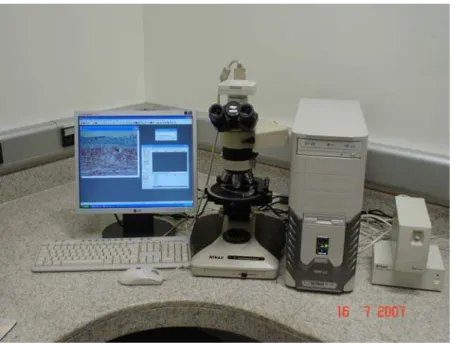
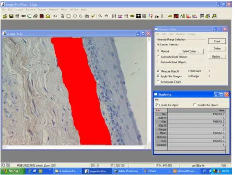
Measurement of positive stained areas were performed by image analysis using a system comprising of a light microscope Leica DMR (Leica Microsystems Wetzlar Gmb H, Wetzlar, Ger-many) connected to a computer through a video camera using the software Image Pro Plus, version 4.1 (Media Cybernetics, Silver Spring, MD). For each lamina propria compartment, three
non-overlapping areas at 400⫻magnification were analyzed,
compris-ing nine analyzed areas for each section. Results were expressed
as stained area/total area (m2/m2).
Data were expressed as medians and ranges. Comparison of stained areas within the three compartments was performed using Friedman and Wilcoxon’s non-parametric statistical tests. All tests were done in the SPSS software, version13.0 (SPSS Inc., Chicago,
IL), andPvalues lower than .05 were considered significant.
RESULTS
Collagen fibers were identified in the lamina propria of fetal vocal fold under conventional and polarized light (Fig. 1) with a “wicker basket” arrangement (Fig. 2). The distribution of collagen fibers was homogeneous on all its thicknesses, and there was no statistical difference throughout its distribution among superficial (S),
inter-mediate (I), and deep (D) layers (S⫽0.69 [0.25– 0.77]; I⫽
0.73 [0.26 – 0.91]; D⫽0.61 [0.36 – 0.81],P⫽.56). There was a diffuse positive versican staining in all laminar layers with a stronger staining in the subepithe-lial region (Fig. 3). Morphometric analysis revealed that versican density differed significantly among layers within the lamina propria, being lower in the deep layer when compared to the superficial (D⫽0.13 [0.05– 0.18]; S ⫽ 0.23 [0.05– 0.31], P ⫽ .041) and the intermediate layers (I⫽0.16 [0.09 – 0.20],P⫽.018); however, there was no difference between the superficial and intermediate layers (Fig. 4). Figure 5 shows three-dimensional graphic illustrations of the histoarchiteture of the collagen fibers.
DISCUSSION
In this study we describe the distribution of collagen fibers and versican within the lamina propria of 28- to 36-weeks-old human fetuses. Our data show that while there is a uniform distribution of collagen fibers within the lamina propria, versican density is increased in the superficial and intermediate layers. To our knowledge, we are the first to analyze versican expression within the lamina propria of human fetal vocal folds.
There are a few studies on the histologic aspects of the human vocal folds. Progressive alterations in the com-position of the lamina propria are related during fetal development. In 13-week-old fetuses it is basically com-prised of mesenchymal cells. From 16 weeks onwards, slender fibers distributed parallel to the free border begin to appear. At 23 weeks of gestation, collagen fibers are distributed in a single layer over all thicknesses of the vocal fold.12,20Similarly, previous ultrastructural studies
on the fetus vocal fold described an uniform distribution of collagen fibers without a clear layer distinction.13–16Our
quantitative analysis of collagen fibers confirms these findings.
findings on the adult population. Melo et al.11described a
“wicker basket” configuration of collagen fibers in adults, and hypothesized that this configuration might have im-portant implications for vocal production. However, there were no data available as to whether this collagen fiber configuration would be already present in fetuses, or if it would be part of the maturation process of the ECM, originating from the vibratory stimulus during phonation after birth. We now demonstrate that the “wicker basket” arrangement of these fibers is already present in 28- to
36-week-old-fetuses. Although we have not examined other important components of the vocal fold ECM and larynx innervation, this finding suggests that despite no existing
Fig. 3. Transversal histologic section of fetal vocal fold stained by specific immunohistochemical technique for the versican proteogly-can (brown coloration), showing its distribution on superficial (S), intermediate (I), and deep (D) layers of the lamina propria. Note the predominance on the subepithelial region (arrow) (200⫻ magnifica-tion); E⫽epithelium.
diffusely present in the ECM, with a significant increased density in the superficial and intermediate layers when compared to the deep layers, with a stronger staining observed in the subepithelial region. Interestingly, a sim-ilar distribution of versican in the upper dermis in mid-age fetuses (of a similar mid-age range as the present study) was described by Sorrell et al.21In adult vocal folds, Hahn
et al.12described a most intense staining in the superior
portion of the intermediate layer.
The role of versican in the fetal vocal folds is not clear. Versican forms aggregates with hyaluronic acid, and these aggregates have an importance role in tissue viscosity, osmosis, and resistance.9 In addition, these
structures form complex interactions with fibronectin, type I collagen21and elastic networks.10Versican
predom-inance within the superficial and intermediate layers might be important in determining collagen deposition and distribution in the developing vocal fold, since proteo-glycans have an important role in regulating collagen fibrillogenesis.6 The increased versican presence in the
superficial and intermediate layers may contribute to the “wicker-basket” arrangement of the developing collagen fibers at this level. On the other hand, less versican in the deeper layer of the lamina propria may provide structure for a more dense collagen arrangement, adequate for the physiologic function of transmitting tension from vocal musculature to the rest of the lamina propria during voice modulation. In addition, predominance of versican in su-perficial and intermediate layers could be important in absorbing impact during vocalization at birth.
CONCLUSION
In this study, novel information is presented about the ECM composition in the fetus vocal folds. Collagen fibers are uniformly distributed within the lamina pro-pria, and present the “wicker basket” arrangement as described in adults. In contrast, versican density is larger in the superficial and intermediate layers when compared to the deep layer.
Acknowledgments
The authors thank Prof. Dr. Chao Lung (Discipline of Telemedicine–the University of Sa˜o Paulo School of Med-icine) for assistance in three-dimensional graphic repre-sentation. The authors would also like to acknowledge the autopsy assistants of the SVOC-USP who helped gather the study material, and Sandra de Moraes for support in the immunohistochemical procedures.
fold lamina propria. Otolaryngol Head Neck Surg1998;
118:663– 667.
3. Labat-Robert J, Bihari-Varga M, Robert L. Extracelular
ma-trix.FEBS Lett1990;268:386 –393.
4. Chan RW, Gray SD, Titze IR. The importance of hyaluronic
acid in vocal fold biomechanics. Otolaryngol Head Neck
Surg2001;124:607– 614.
5. Pawlak AS, Hammond E, Hammond T, Gray SD.
Immuno-histochemical study of proteglycans in vocal folds. Ann
Otol Rhinol Laryngol1996;105:6 –11.
6. Iozzo RV. Matrix proteoglycans: from molecular design to
cellular function.Annu Rev Biochem1998;67:609 – 652.
7. Hardinghan TE, Fosang AJ. Proteoglycans: many forms and
many functions.FASEB J1992;12:137–147.
8. Zhang Y, Cao L, Yang BL, Yang BB. The G3 domain of versican enhances cell proliferation via epidermal growth
factor-like motifs.J Biol Chem1998;273:21342–21351.
9. Gray SD, Titze IR, Chan Roger, Hammond T. Vocal fold
proteoglycan and their influence on biomechanics.
Laryn-goscope1999;109:845– 854.
10. Hahn MS, Kobler JB, Zeitels SM, Langer R. Midmembranous vocal fold lamina propria proteoglycans across selected
species.Ann Otol Rhinol Laryngol2005;114:451– 462.
11. Melo ECM, Lemos M, Ximenes Filho JA, Sennes LU, Saldiva PHN, Tsuji DH. Distribution of collagen in the lamina
propria of the human vocal fold.Laryngoscope2003;113:
2187–2191.
12. Subotic R, Vecerina S, Krajina Z, Hirano M, Kurita S. Histo-logical structure of vocal fold lamina propria in foetal lar-ynx.Acta Otolaryngol (Stockh)1984;97:403– 406. 13. Sato K, Nakashima T. Vitamin A – storing stellate cells in the
human newborn vocal folds. Ann Otol Rhinol Laryngol
2005;114:517–524.
14. Sato K, Hirano M, Nakashima T. Fine structure of the human
newborn and infant vocal fold.Ann Otol Rhinol Laryngol
2001;110:417– 424.
15. Hirano M, Kurita S, Nakashima T. Growth, development and aging of human vocal folds. In: Bless DM, Abbs JH, eds.
Vocal Fold Physiology. San Diego, CA: College Hill Press; 1983:22– 43.
16. Hartnick CJ, Rehbar R, Prasad V. Development and matu-ration of the pediatric human vocal fold lamina propria.
Laryngoscope2005;115:4 –15.
17. Ishii K, Yamashita K, Akita M, Hirose H. Age related devel-opment of the arrangement of connective tissue fibers in
the lamina propria of the human vocal fold. Ann Otol
Rhinol Laryngol2000;109:1055–1064.
18. Butler JE, Hammond TH, Gray SD. Gender-related differ-ences of hyaluronic acid distribution in the human vocal
fold.Laryngoscope2001;111:907–911.
19. Junqueira LCU, Bignolas G, Brentani RR. Picrosirius stain-ing plus polarization microscopy: a specific method for
collagen detection in tissue sections.Histochem1979;11:
447– 455.
20. Kuritas S. Layer structure of the human vocal fold:
morpho-logical investigation.Otologia (Fukuoka)1980;26:973–997.
21. Sorrell JM, Carrino DA, Baber MA, Caplan AI. Versican in
human fetal skin development. Anat Embryol1999;199:
Collagen Type I, Type III and Versican Distribution within the Lamina Propria of
Human Vocal Fold
This work was supported by FAPESP (São Paulo State Research Agency), and
HC-FMUSP.
From the Department of Otolaryngology (Rogerio Borghi Bühler, MD; Luiz Ubirajara
Sennes, MD, PhD; Domingos Hiroshi Tsuji, MD, PhD) and Department of Pathology
(Thais Mauad, MD, PhD; Luiz Fernando Ferraz da Silva, MD; Paulo Hilário
Nascimento Saldiva, MD, PhD), The University of São Paulo School of Medicine, São
Paulo, Brazil.
Send correspondence to Rogerio Borghi Bühler, Rua Tenente Negrão 140, Cj. 91,
SUMMARY
Objectives: Extracellular matrix distribution within the lamina propria may be related
to phonation mechanics. In this study, we analyzed the distribution of collagen type I,
collagen type III and versican in human vocal fold lamina propria. Study design: Cross
sectional analysis of cadaveric vocal folds of adult human larynges. Methods: Twenty
larynges obtained from autopsy specimens were analyzed with immunohistochemistry
and image analysis. Results: There was a lower collagen type I density in the
intermediate layer (IL) when compared to the superficial (SL) and the deep (DL) layers.
Collagen type III presented a lower density in the IL when compared to the DL layer.
There was a lower versican density in the SL when compared to the IL and the DL.
There was a lower versican density in the lamina propria of females when compared
with males. The difference was noted in the superficial layer only. There was a positive
correlation between collagen III and versican within the lamina propria. Conclusion:
Collagen type I, type III and versican are differently distributed within the adult vocal
fold lamina propria layers.
Key Words: versican, collagen type I, collagen type III, lamina propria, vocal fold,
INTRODUCTION
Human vocal folds (VF) are histologically comprised by the epithelium, the
three layers of lamina propria (LP) and the vocalis muscle. The cover-body theory of
phonation explains how the intricate relation among the layered structures of the LP
contributes to voice production by allowing the VF to vibrate with consistency and
control.1
The extracellular matrix (ECM) proteins of the vocal fold lamina propria consist
of fibrillar and interstitial proteins. The main fibrillar proteins in the VF lamina propria
comprise the collagen and the elastic fibers. The composition of fibrillar and interstitial
proteins within the VF determine many of the oscillatory characteristics of this
structure. 2,3
Collagens represent a family of proteins that serve primarily as the supporting
elements in tissue structure and contribute to tissue tensile strength and stability but also
regulate cellular migration and tissue remodeling during growth, differentiation,
morphogenesis and wound healing.4 Collagens are a major component of the human
VF, representing 43% of the total tissue protein.5 The relative density of collagen fibers
varies within the human lamina propria layers. Some studies have previously shown that
the density of total collagen is higher in the superficial and deep layers.5,6,7 It’s
structural arrangement within the VF lamina propria is believed to have an important
impact in phonation.8
There are various subtypes of fibrillar collagens and each one is uniquely suited
appear as the fibrillar bundles providing a structure of high tensile strength. Collagen
type III is present in most tissues that require flexibility and elasticity. Collagen type I
and type III have been immunohistochemically identified as the major collagens in the
VF lamina propria5,10 but there are few quantitative data on the distribution of collagens
type I and III within the lamina propria layers of human adult vocal folds.
Proteoglycans (PG) represent a family of complex molecules composed of core
proteins to which glycosaminoglycans (GAG) side chains, such as dermatan sulfate,
chondroitan sulfate, keratan sulfate, and heparan sulfate are attached. They provide
growth supportive or suppressive function, modulate wound repair, bind and deliver
growth factors. One of these proteins, the large proteoglycan versican, has the ability to
regulate water content in tissues, thereby affecting resiliency.11,12 Versican is known to
be present in fetal and adult human vocal folds; and it interacts with collagen deposition
by regulating its fibrillogenesis.2,10,11,13 No quantitative studies analyzed versican
distribution within the 3 lamina propria layers.
It is plausible to assume that the distribution of collagen types and versican may
vary within the LP of the vocal folds, since each layer may be submitted to different
mechanical stresses during phonation. To better understand this subject, we analyzed
the distribution of collagen type I, type III and versican within the lamina propria of
METHODS
The study was approved by the Ethics Committee for Research Project Analysis
of the Clinical Board of the University of São Paulo School of Medicine.
Human larynges were obtained from cadavers autopsied in the São Paulo
Autopsy Service within 24 hours after death. Larynges were obtained from twenty
adults (10 males and 10 females). Patient’s mean (±SD) age was 67 (±9) ranging from
50 to 85 years. Mean age of males was 66 and 70 from females, without significant
statistical difference between groups. Thirteen adults were from the white race and
seven from the black. Cadavers with a medical history of neck manipulation as oral or
nasal intubation, tracheotomy, laryngeal surgery and head and neck irradiation were
excluded. Only larynges from non-smoking subjects were included in the study.
In bloc exeresis of the larynx was performed, all specimens presented no
macroscopic lesions. The left vocal fold from each larynx was obtained and fixed in
10% formalin solution for 24 hours. Afterwards, 5-mm-thick coronal sections in relation
to the vocal fold were performed on the middle portion of the fold membranous region,
dehydrated in progressive alcoholic concentrations and embedded in paraffin. Tissue
blocks were submitted to 4-µm-thick histological sections and hematoxylin-eosin
(H&E) stained for initial analysis.
For the analysis of collagen type I and type III expression, immunohistochemical
reactions were carried out using monoclonal anti-human antibody (C7510 12A, United
anti-human antibody (CP19L, Calbiochem-Novabiochem, San Diego, CA, USA) with 1:500
dilution. To analyze versican expression a specific monoclonal anti-human antibody
large proteoglycan versican (Seikagaku America, Inc; Rockville, MD, USA) was used.
For the antibody versican, sections were pretreated with a 0.05 U/ml ABC
chondroitinase solution (Sigma-Aldrich, Oakville, ON, Canada), for one hour, at 370C.
Subsequently, sections were incubated with the primary antibody in 1% BSA/PBS
between 40C and 80C overnight. As secondary antibody, LSAB+Ap (Dako, Carpinteria,
CA, USA) and Fast Red (Sigma, Steinheim, NRW, Germany) were employed as
chromogens. The slides were counter-stained with Mayer´s Hematoxylin. Incubation
with PBS supplemented with 1% BSA instead of the primary antibody served as a
negative control.
For quantitative analyses of collagens and versican expression, we divided the
lamina propria compartments according to the model proposed by Butler et al.14 in three
layers: superficial, intermediate and deep. Measurement of positive stained areas were
performed by image analysis using a system comprising of a light microscope Leica
DMR (Leica Microsystems, Wetzlar, Hesse, Germany) connected to a computer
through a video camera using the software Image Pro Plus® 4.1 version (Media
Cybernetics, Silver Spring, MD, USA). For each lamina propria compartment three
non-overlapping areas at 400x magnification were analyzed, comprising 9 analyzed
areas for each section. Results were expressed as stained area/total area (µm2/µm2).
Data were expressed as medians and ranges. Comparison of stained areas within the 3
compartments was performed using ANOVA or Kruskall-Wallis followed by post-hoc
Tukey’s b or Bonferroni respectively (depending on data distribution). Data were
Inc., Chicago, IL, USA). Correlation among ECM elements was performed using the
RESULTS
Collagens and versican stained as fibrillar structures in the lamina propria and
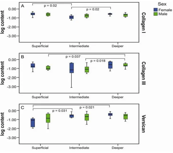
among the vocal muscle cells. There was a lower collagen type I density in the
intermediate layer when compared to the superficial (p<0.001) and the deep layers
(p=0.005) (Figure 1a).Collagen type III had a more homogenous distribution within the
VF layers, with a statistically lower density of collagen type III in the intermediate layer
when compared to the deep layer (p=0.001), but without differences in the superficial
layer (Figure 1b). There was a lower versican density in the superficial layer when
compared to the intermediate (p=0.036) and the deep layer (p=0.013) (Figure 1c).
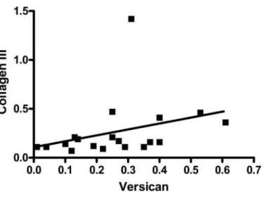
When all layers were considered together, there was a positive correlation
between versican and collagen III densities (r = 0.57, p=0.010) (Figure 2).
Morphometrical analysis categorized by gender revealed that collagen type I,
collagen type III and versican density differed significantly among layers within the
lamina propria. Females had a lower density of collagen type I in the intermediate when
compared to superficial (p=0.02) and deep layers (D=0.28 [0.10-0.51], p=0.02). For
males, no differences were observed (Figure 3a). There was no difference in collagen
type III distribution in the female group within layers. On the other hand, there was a
higher collagen type III density in the deep layer when compared to the superficial
(p=0.037) and the intermediate layer (p=0.018) in males (Figure 3b). There was lower
versican female density in the superficial layer when compared to the intermediate
(p=0.031) and the deep layer (p=0.021). There was no statistical difference in versican
There was no difference between total density of collagen type I and collagen
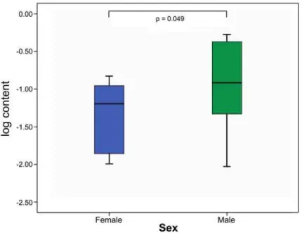
type III in male and female lamina propria vocal folds. There was a higher versican
density in the lamina propria of females when compared with males. The difference was
noted in the superficial layer only (p=0.049) (Figure 4). There were no correlations
between collagens and versican density in the lamina propria and patient´s age in male
and females. Figure 5 shows three dimensional graphic illustrations of histoarchiteture
DISCUSSION
In this study we described the distribution of collagens type I, type III and
versican within the lamina propria of age-matched female and male adult vocal folds.
Our data show that collagens type I and III and versican vary its distribution within the
lamina propria of vocal folds, with gender variation.
The distribution of total collagen (as detected with Sirius Red staining) in human
vocal folds has been described to be similar in all age groups, but with varying
concentrations according to age and gender.8 In adult normal vocal folds, most collagen
is present in a superficial band immediately below the epithelium as well as in deep
lamina propria adjacent to the vocalis muscle,6,8 with a higher concentration in males
and geriatric males.6 We confirmed this distribution for collagen type I, i.e.,
predominance in superficial and deep layers. When categorized by gender, our data
show that these differences were more accentuated within the female group, but without
differences between genders.
Collagen type III analysis also revealed differences in distribution within the
lamina propria, with a higher content in the deep than in the superficial and intermediate
layers. Our data show that these differences were more accentuated within the male
group, but again without significant differences between genders. Since collagen type
III tends to be more highly concentrated in dynamic regions of elastic tissues,
differences in the distribution of collagen types I and III can potentially give insight into
the stresses sustained by different LP regions.15 As suggested by Tateya et al,7 collagen
ligament to maintain the vocal fold shape during vibration, while the more homogenous
distribution of collagen type III may be important to maintain tissue elasticity.
Versican is a large aggregating PG that binds to hyaluronic acid (HA)11 and that
frequently interacts with elastic networks.16 The highly hydrated versican-hyaluronic
acid complex play a significant role in inhibiting cell-matrix interactions, affecting
turgor, dissipating impact and compressive stresses in the VF lamina propria.17 The
presence of versican in adult and fetal vocal folds has been previously described10,18,19,
but no studies have assessed its distribution within the LP layers in adults. Our data
show that versican density is higher in the intermediate and deeper layers, this
difference being more accentuated in females; with an increased content of versican in
the superficial layer of males. Our data are in disagreement with Hahn et al.10 that could
not find differences in versican distribution between males and females. However, those
authors examined a smaller population than ours (5 patients) using only semi
quantitative analysis.
Our previous and present data show that the distribution of versican within the
human LP varies according to age. In the fetus, versican density was higher in the
superficial layer.13 On the other hand versican density was lower in the superficial layer
of adults when compared to the other layers. The reasons for such differences are not
clear. It has been shown that, in the presence of some proteoglycans, collagen synthesis
yield thinner fibrils. Such theory could explain the higher density of collagen type I
(thick fibrils) and the smaller density of versican in the superficial layer in adults.
Further, this is also in line with our findings of a positive correlation between collagen
Understanding gender differences in ECM composition in the vocal fold LP is of
interest since it may provide insights into the greater vulnerability of women to LP
scarring and certain vocal fold disorders.5 Although adult men may have higher total
concentration of collagens in the lamina propria, our and Hahn´s data show that the
distribution of the collagens type I and type III between genders is similar. On the other
hand, the density of versican was higher in the superficial layer of males. This data may
help explain the higher predisposition in women to the occurrence of benign laryngeal
lesions like vocal nodules. Versican has likely an important role in absorbing impacts
between VF during phonation. With a smaller density of versican in the SL, it is
possible that the vocal fold is more prone to mechanical damage during phonation.
Others have detected correlation between age and collagen expression, with
increased collagen content in the elderly.6 In our study we could not find such
correlations, since our study group was comprised by an adult population of a limited
age interval, which is a limitation of the present work.
CONCLUSION
Our data show, in adults, collagen type I, type III and versican are differently
distributed within the adult vocal fold lamina propria layers, with gender variations.
Better knowledge about ECM distribution within the LP is important to understand the
ACKNOWLEDGMENTS
We thank Prof. Dr. Chao Lung (Discipline of Telemedicine - The University of São
Paulo School of Medicine, São Paulo, Brazil) for assistance in three-dimensional
graphic representation. We would also like to acknowledge the autopsy assistants of the
BIBLIOGRAPHY
1. Gray SD, Hirano M, Sato K. Molecular and cellular structure of vocal fold tissue.
In: Titze IR, ed. Vocal Fold Physiology. San Diego: Singular Publishing Group;
1993.
2. Gray SD, Titze IR, Chan R, Hammond T. Vocal fold proteoglycans an their
influence on biomechanics. Laryngoscope 1999;109:845-854.
3. Gray SD. Celular physiology of the vocal fold. Otolaryngologic Clin North Am
2000;33:679-697.
4. Myllyharju J, Kivirikko KI. Collagens modifying enzymes and their mutations in
humans, flies and worms. Trends Genet 2004;20:33-43.
5. Hahn MS, Kobler JB, Zeitels SM, Langer R. Quantitative and comparative studies
of the vocal fold extracellular matrix II: collagen. Ann Otol Rhinol Laryngol 2006;
115: 225-232.
6. Hammond TH, Gray SD, Butler JE. Age and gender-related collagen distribution
in human vocal folds. Ann Otol Rhinol Laryngol 2000; 109: 913-920.
7. Tateya T, Tateya I, Bless DM. Collagen subtypes in human vocal folds. Ann Otol
Rhinol Laryngol 2006; 115: 469-476.
8. Melo ECM, Lemos M, Ximenes Filho JA, Sennes LU, Saldiva PHN, Tsuji DH.
Distribution of collagen in the lamina propria of the human vocal fold.
Laryngoscope 2003;113:2187-2191.
9. Tateya T, Tateya I, Bless DM. Immuno-scanning electron microscopy of collagen
types I and III in human vocal fold lamina propria. Ann Otol Rhinol Laryngol
10.Hahn MS, Kobler JB, Zeitels SM, Langer R. Midmembranous vocal fold lamina
propria proteoglycans across selected species. Ann Otol Rhinol Laryngol 2005;
114: 451-462.
11.Iozzo R V. Matrix proteoglycans: from molecular design to cellular function. Annu
Rev Biochem 1998;67:609-652.
12.Hardinghan TE, Fosang AJ. Proteoglycans: many forms and many functions.
FASEB J 1992;12:137-147. 12
13.Buhler RB, Sennes LU, Mauad T, Melo ECM, Silva LFF, Saldiva PHN. Collagen
fiber and versican distribution within the lamina propria of fetal vocal folds.
Laryngoscope 2008;113:2187-2191.
14.Butler JE, Hammond TH, Gray SD. Gender-related differences of hyaluronic acid
distribution in the human vocal fold. Laryngoscope 2001;111:907-1. 14
15.Culav EM, Clark CH, Merrilees MJ. Connective tissues: matrix composition and
its relevance to physical therapy. Phys Ther 1999;79:308-319. 15
16.Kielty CM, Sherratt MJ, Shuttleworth CA. Elastic fibers. J Cell Sci
2002;115:2817-2828. 16
17.Laurent TC, Fraser JRE. Hyaluronan. FASEB J 1992;6:2397-2404. 17
18.Pawlak AS, Hammond E, Hammond T, Gray SD. Immunohistochemical study of
proteglycans in vocal folds. Ann Otol Rhinol Laryngol 1996;105:6-11.
19.Skandalis SS, Theocharis AD, Papageorgakopoulou N, Vynios DH, Theocharis
DA. The increased accumulation of structurally modified versican and decorin is
Figure 1. Expression of extracellular matrix components (ECM) in coronal histological
section of the middle membranous portion of vocal folds lamina propria in males and
females (50x magnification) and the distribution within the vocal fold layers (horizontal
line represents the median). A. Immunohistochemical expression of collagen type I B.
Lower collagen type I density in the intermediate layer (IL) when compared to the
superficial layer (SL) and the deep layer (DL). C. Immunohistochemical expression of
collagen type III. D. Lower density of collagen type III in the IL when compared to the
DL E. Immunohistochemical expression of versican. F. Lower versican density in the
Figure 2. Positive correlation between versican and collagen type III density (r = 0.57,
Figure 3. Distribution of collagen type I, type III and versican in lamina propria vocal
fold of male and females. The horizontal line represents the median. A. Females had a
lower density of collagen type I in intermediate layer (IL) when compared to the
superficial layer (SL) and the deep layer (DL). For males, no differences were observed.
B. No difference in collagen type III distribution in the females. In males there was a
higher collagen III density in DL when compared to the SL and the IL. C. Lower
versican female density in the SL when compared to the IL and DL. No difference in
Figure 4. Distribution of versican in lamina propria vocal fold showing a higher density
in the superficial layer of females when compared with males (p=0.049). The horizontal
Figure 5. Histoarchiteture of the collagen type I, collagen type III and versican
distributed within the adult lamina propria vocal fold; brown fiber = collagen type I;
LISTA DE ABREVIATURAS, SÍMBOLOS E SIGLAS
ABC do inglês avidin biotin complex, complexo avidina-biotina BSA do inglês bovine serum albumin, albumina bovina
CA estado da Califórnia, Estados Unidos da América CAPPesq Comissão de Ética para Projetos de Pesquisa 0
C grau Celsus, medida de temperatura
E epitélio
ed edição
et al. e outros
EUA Estados Unidos da América
FMUSP Faculdade de Medicina da Universidade de São Paulo I camada intermediária da lâmina própria
IL estado de Illinois, Estados Unidos da América Inc do inglês Incorporation, empresa
LIM Laboratório de Investigação Médica
LCD do inglês liquid crystal display, tela de cristal líquido Log content conteúdo (objeto do estudo) “logado”
LP lâmina própria
µm micrômetro, a milionésima parte do metro ml mililitro, um milionésimo de litro
MD estado de Maryland, Estados Unidos da América ON província de Ontário, Canadá
p proporção, representação da significância estatística
p página
% porcentagem, por cento
PBS do inglês phosphate-buffered saline, solução de fosfato tamponada
® marca registrada
r valor calculado do coeficiente de correlação de Pearson S camada superficial da lâmina própria
SPSS Statistical Package for the Social Sciences SVOC Serviço de Verificação de Óbitos da Capital U unidade internacional, medida padronizada v volume
LISTA DE FIGURAS
Figura 1. Representação gráfica esquemática do proteoglicano versican.
Proteína central (laranja) com glicosaminoglicanas ligadas; cadeias laterais com diversos sítios de ligação (vermelho: ácido hialurônico; azul: fator de crescimento epidérmico; verde: lecitina; amarelo: complemento regulador de proteína)... 5
Figura 2. Analisador digital de imagens composto por microscópio óptico
equipado com câmera de vídeo acoplado a um monitor e a computador com programa de análise digital de imagens. ... 25
Figura 3. Interface do programa de análise digital de imagem mostrando a
fotomicrografia de toda a extensão da prega vocal. (aumento de 50x). ... 27
Figura 4. Interface do programa de análise digital de imagem mostrando
fotomicrografia com delimitação da área a ser estudada marcada em verde. (aumento de 400x)... 27
Figura 5. Interface do programa de análise digital de imagem mostrando
fotomicrografia com delimitação da área a ser estudada e aplicação do padrão de cores estabelecido (área positiva) marcado em vermelho. (aumento de 400x). ... 28
Figura 6. Interface do programa de análise digital de imagem mostrando
fotomicrografia com delimitação da área total a ser medida marcada em vermelho. (aumento de 400x). ... 28
Figura 7. Histograma mostrando a distribuição pareada (curvas similares) das
idades nos grupos masculino e feminino permitindo sua comparação. ... 30
Figura 8. Representação gráfica esquemática da região interna da laringe
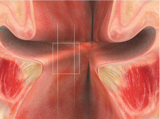
após incisão longitudinal entre as cartilagens aritenóides (visão posterior). Retângulo representando o local do corte coronal da região média da porção membranosa da prega vocal esquerda. ... 31
Figura 9. Corte histológico coronal da região média da porção membranosa de
prega vocal fetal corada com Picrosirius e visualizada sob luz convencional. (aumento de 50x) - barra = 400 μm ... 36
Figura 10. Corte histológico coronal da região média da porção membranosa de