III
Editorial
Fetal cardiac evaluation by 3D/4D
ultrasonography (STIC): what is its real
applicability in the diagnosis of congenital heart
disease?
Edward Araujo Júnior
1, Liliam Cristine Rolo
2, Luciano Marcondes Machado Nardozza
3, Antonio
Fernandes Moron
4DOI: 10.5935/1678-9741.20130002
The congenital heart diseases (CHD) are the most common major malformations at birth [1], with prevalence ranging from 0.6% to 5% of live births [2]. Despite great efforts and technological advancement of two-dimensional echocardiography (2D) in the past two decades, the accuracy in the detection of CHD at prenatal is between 31% and 96% [3].
In 2003, with the development of Spatio-Temporal Image Correlation (STIC), scientists started the use of third and fourth dimension ultrasonography (3D/4D) in fetal cardiac evaluation [4]. The STIC is a software that enables acquiring volumetric fetal heart with its vascular connections, whose images can be evaluated in either multiplanar or rendering modes, or even surface mode, in a static or moving ways (4D) by means of a sequence of cineloop, which simulates a complete cardiac cycle [4]. This software provides an innovation absent in 2D ultrasound, which is the storage volume of the heart for
an ofline analysis, in other words, in the absence of the
patient. Thus, a detailed assessment of the anatomy and the functioning fetal heart is possible without the need to
cause major discomfort to pregnant women, a relatively frequent situation when more prolonged ultrasound studies are used. Moreover, the storage allows the sending of volumes to specialized centers through an internet link, strengthening the telemedicine and improving the prenatal period tracking [5].
Standardization of volume storage is already a reality,
so the investigator responsible for ofline analysis has
knowledge of the actual position of the heart chambers with respect to the right and left fetal axis to evaluate the presence of possible cardiac isomerisms. Therefore, when the fetus is in cephalic presentation, it should be considered that the heart side corresponds to the fetal side, unlike the pelvic fetuses which stay in opposite sides [6].
The gray scale and color Doppler applications are also present in the STIC, used to improve the evaluation
of the ventricular outlow tracts, aortic and ductal arches,
besides assisting in the location of septal defects [7]. The 3D technology has allowed the development of new techniques known as inversion mode (analysis technique of liquid structures which reverses voxels of gray scale,
1. Adjunct Professor at the Federal University of São Paulo (UNIFESP), São Paulo, SP, Brazil - Main Author.
2. Doctor of Science, Department of Obstetrics, UNIFESP, São Paulo, SP, Brazil - Contribution to the article.
3. Associate Professor, Department of Obstetrics, São Paulo, SP, Brazil - Article Review.
4. Professor, Department of Obstetrics, UNIFESP, São Paulo, SP, Brazil - Article Review.
Correspondence Address:
Division of Fetal Cardiology, Department of Obstetrics, Federal University of São Paulo (UNIFESP), São Paulo, SP, Brazil.
Edward Araujo Júnior
956 Carlos Weber Street, Apto 113 - Visage - Alto da Lapa - São Paulo, SP, Brazil – Zip code: 05303-000
E-mail: [email protected]
IV
so anechoic structures such as the heart chambers, lumen vessels, stomach, bladder and renal pelvis, with inversion mode they become echogenic, whereas normally echogenic structures, such as bone, become anechoic) [8]
(Figure 1) and B-low imaging (technique that improves the weak signal relected from the blood, and suppresses
the strong signals of the surrounding structures) [9]. The inversion mode allows the reconstruction of the cardiac chambers, aortic and ductal arches, and abnormalities of
venous connections [8]. The B-low imaging shows high
sensitivity and angle independence, then it is potentially advantageous over color Doppler for the visualization of
large vessels and venous return, allowing the identiication of small vessel with low-velocity lows, such as pulmonary
veins, enhancing the detection of anomalies in pulmonary venous return [9].
Abbreviations, acronyms and symbols
2D 3D 4D CHD STIC TUI
Second Dimension Third Dimension Fourth Dimension Congenital Heart Diseases Spatio-Temporal Image Correlation Tomographic Ultrasound Imaging
Fig. 1 – Visualization of four heart chambers plane using the inversion mode
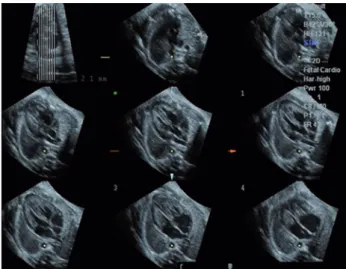
Fig. 2 - Use of TUI with simultaneous visualization of sequential axial planes of the fetal chest at heart level (3VT, 5C, 4C). TUI: tomographic ultrasound imaging. 3VT: three vessels and trachea;
5C: ive chambers; 4C: four chambers
Another technique also addressed by STIC Tomographic Ultrasound Imaging's (TUI), which enables the achievement of all the axial planes of the heart from the abdomen to the apex of the chest, increasing the fetal heart tracking and analysis [10] (Figure 2) . STIC also allows the measurement of volumes of cardiac chambers, as well as the calculation of systolic volume, ejection fraction and cardiac output. Thus, to obtain relevant information about
cardiac function is possible due to the CHD [11]. More recently, a new approach to STIC rendering mode analysis obtained measurements of mitral and tricuspid valves areas, and also and the interventricular septum, determining reference values for these parameters, making it feasible to apply in suspected or pathological cases[12,13].
Recently, some studies have attempted to correlate the technical 3D/4D (STIC) and 2D (echocardiography) with the diagnosis of CHD, however, these studies have shown
conlicting results [14,15]. In one of these studies, with the
STIC being conducted by the general obstetricians, without
speciic knowledge of the methodology, the STIC proved
itself to be inferior to 2D echocardiography, performed by a specialist in the diagnosis of CHD [14]. In another study, where the STIC was performed by experienced examiners in this methodology, the accuracy in the diagnosis of CHD was similar to that obtained by echocardiography 2D [15].
Based on recent studies, it is still early to say whether the 3D/4D ultrasound (STIC) will surpass 2D echocardiography as a gold standard in prenatal diagnosis of CHD. In our reality, the biggest obstacles are the need for sophisticated and expensive equipment and people
with speciic training in 3D/4D ultrasound. However, some beneits are already envisioned, as less operator
dependence and the possibility of sending volumes to reference centers for fetal and pediatric cardiology, which is of fundamental importance in a country like ours, where these centers are located in major cities.
In summary, only in multicenter studies, with
V
REFERENCES
1. Hoffmann JI. Incidence of congenital heart disease: I. Postnatal incidence. Pediatr Cardiol. 1995;16(3):103-13.
2. Grandjean H, Larroque D, Levi S. The performance of routine ultrasonographic screening of pregnancies in the Eurofetus Study. Am J Obstet Gynecol. 1999;181(2):446-54.
3. Stümplen I, Stümplen A, Wimmer M, Bernaschek G. Effect
of detailed fetal echocardiography as part of routine prenatal ultrasonographic screening on detection of congenital heart disease. Lancet. 1996;348(9031):854-7.
4. DeVore GR, Falkensammer P, Sklansky MS, Platt LD. Spatio-temporal image correlation (STIC): new technology for evaluation of the fetal heart. Ultrasound Obstet Gynecol. 2003;22(4):380-7.
5. Gonçalves LF, Nien JK, Espinoza J, Kusanovic JP, Lee W, Swope B, et al. What does 2-dimensional imaging add to 3- and 4-dimensional obstetrics ultrasonography? J Ultrasound Med. 2006;25(6):691-9.
6. Paladini D. Standardization of on-screen fetal heart orientation prior to storage of spatio-temporal image correlation (STIC) volume datasets. Ultrasound Obstet Gynecol. 2007;29(6):605-11.
7. Gonçalves LF, Lee W, Chaiworapongsa T, Espinoza J, Schoen ML, Falkensammer P. Four-dimensional ultrasonography of the fetal heart with spatiotemporal image correlation. Am J Obstet Gynecol. 2003;189:1792-802.
8. Lee W, Gonçalves LF, Espinoza J, Romero R. Inversion mode: a new volume analysis tool for 3-dimensional ultrasonography. J Ultrasound Med. 2005;24(2):201-7.
9. Pooh PK, Korai A. B-low and B-low spatio-temporal image
correlation in visualizing fetal cardiac blood low. Croat Med
J. 2005;46(5):808-11.
10. Yagel S, Cohen SM, Rosenak D, Messing B, Lipschuetz M, Shen O, et al. Added value of three-/four-dimensional
ultrasound in ofline analysis and diagnosis of congenital heart
disease. Ultrasound Obstet Gynecol. 2011;37(4):432-7.
11. Simioni C, Nardozza LM, Araujo Júnior E, Rolo LC, Zamith M, Caetano AC, et al. Heart stroke volume, cardiac output, and ejection fraction in 265 normal fetus in the second half of gestation assessed by 4D ultrasound using spatio-temporal image correlation. J Matern Fetal Neonatal Med. 2011;24(9):1159-67.
12. Rolo LC, Nardozza LM, Araujo Júnior E, Hatanaka AR, Rocha LA, Simioni C, et al. Reference ranges of atrioventricular valve areas by means of four-dimensional ultrasonography using spatiotemporal image correlation in the rendering mode. Prenat Diagn. 2013;33(1):50-5.
13. Nardozza LM, Rolo LC, Araujo Júnior E, Hatanaka AR, Rocha LA, Simioni C, et al. Reference range for fetal interventricular septum area by means of four-dimensional ultrasonography using spatiotemporal image correlation. Fetal Diagn Ther. 2013 [ahead of print].
14. Wanitpongpan P, Kanagawa T, Kinugasa Y, Kimura T. Spatio-temporal image correlation (STIC) used by general obstetricians is marginally clinically effective compared to 2D fetal echocardiography scanning by experts. Prenat Diagn. 2008;28(10):923-8.